UNIVERSITÉ DE MONTRÉAL
EFFECT 0F INTERFERON-TAU PROTEIN SECRETION IN
BOVINE ENDOMETRIAL CELLS AND ITS MODULATION BY
STEROID HORMONES
par Bïngtuan Wang
Centre de recherche en reproduction animale (CRRA) Département de biomédecine
Faculté de médecine vétérinaire
Thèse présentée à la Faculté des études supérieures En vue de l’obtention du grade de
Philosophiae Doctor (Ph.D) en sciences vétérinaires
091
ô
arnq
L 018
Université
Cll’
de Montré al
Direction des bibliothèques
AVIS
L’auteur a autorisé l’Université de Montréal à reproduire et diffuser, en totalité ou en partie, par quelque moyen que ce soit et sur quelque support que ce soit, et exclusivement à des fins non lucratives d’enseignement et de recherche, des copies de ce mémoire ou de cette thèse.
L’auteur et les coauteurs le cas échéant conservent la propriété du droit d’auteur et des droits moraux qui protègent ce document. Ni la thèse ou le mémoire, ni des extraits substantiels de ce document, ne doivent être imprimés ou autrement reproduits sans l’autorisation de l’auteur.
Afin de se conformer à la Loi canadienne sur la protection des renseignements personnels, quelques formulaires secondaires, coordonnées ou signatures intégrées au texte ont pu être enlevés de ce document. Bien que cela ait pu affecter la pagination, il n’y a aucun contenu manquant.
NOTICE
The author of this thesis or dissertation has gtanted a nonexclusive license allowing Université de Montréal to reproduce and publish the document, in part or in whole, and in any format, solely for noncommercial educational and research purposes.
The author and co-authors if applicable retain copyright ownership and moral rights in this document. Neither the whole thesis or dissertation, nor substantial extracts from it, may be printed or otherwise reproduced without the author’s permission.
In compliance with the Canadian Privacy Act some supporting forms, contact information or signatures may have been removed from the document. While this may affect the document page count, it does not represent any loss of content from the document.
ç
UNIVERSITÉ DE MONTRÉAL
FACULTÉ DES ÉTUDES SUPÉRIEURES
Cette thèse intitulée
EFFECT 0f INTERFERON-TAU ON PROTEIN SECRETION IN
BOVINE ENDOMETRIAL CELLS AND ITS MODULATION BY
STEROID HORMONES
Présentée par
Bingtuan Wang
A été évaluée par un jury composé des personnes suivantes Président du jury: Dr. Bruce D. Murphy
Directeur de recherche: Dr. Alan K. Goff Membre du jury: Dr. Christopher A. Price Examinateur exterme: Dr. Leslie MacLaren Représentant du doyen: Dr. Sirois Jean
n
In presenting the thesis in partial fulfiilmentofthe requirement ofa postgraduate degree from University of Montreal, I agree that the Lïbraries of [lis University may make it fteely available for inspection. I further agree that permission for copying this thesis in any mariner, in whole or in part, for scholarly purposes may be granted by
tue
professor or professors who supervised my thesis work or. in their absence, by the beaU
of the Department or the Dean of the facuhy in which rny thesis work was donc.
It is
understood that any copying or publication or use of this thesis or parts thereof for
financial gain shah not be altowed without my written permission. it is also understood that due recognition shah be given to me and to the University of Montreal
in any
scholarly use which may he made of any material in my thesis.
Request for permission to copy or to make other use of material in this thesis in whole or in part should be addressed to:
Dean of Faculté des études supérieures Faculté de médecine vétérinaire
Univeristé de Montréat 3200 rue Sicotte, C. P. 5000
SOMMAIRE DE LA THÈSE DE PHD
Dès le début de la gestation chez les ruminants, l’embryon n’a pas comme seul rôle de prévenir la sécrétion de la prostaglandine f2a mais module également la
sécrétion des protéines endométriales. L’interféron--t (IFN-t) est produit par le trophoblaste avant l’implantation chez les ruminants. Ce fateur est impliqué dans la reconnaissance maternelle de la gestation, et peut altérer la synthèse de protéines endométriales et inhiber la prolifération de certaines cellules, étant une molécule pléïotropique. Toutefois, les mécanismes impliqués dans la reconnaissance maternelle de la gestation, et dans l’implantation et le développement embryonnaires ne sont pas bien connus aux niveaux cellulaire et moléculaire. Pour améliorernotre compréhension, un système de culture primaire de cellules endométriales a été utilisé dans la présente étude. Nous proposons comme hypothèse que WN--r peut modifier la sécrétion des protéines endométriales qui sont impliquées dans l’établissement de la gestation. Les Objectifs de cette étude d’étaient d’étabir un système de culture de cellules endométriales bovines pouvant répondre aux hormones stéroïdiennes et IFN-T, puis d’examiner les effets de WN-r sur la sécrétion de protéines endométriales, et la modulation possible de cette sécrétion par les hormones stéroïdiennes.
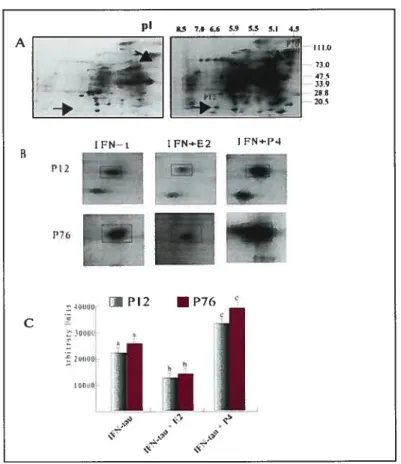
Dans la première expérience, l’effet de WN-t sur la sécrétion de protéines par les cellules épithéliales ou stromales de l’endomètre bovin a été examiné par gel SDS PAGE bi-dimensionnel (2D SDS-PAGE) et par analyse HPLC. Les résultats ont démontrés que LFN-t induit la sécrétion de plusieurs protéines par les cellules épithéliales de l’enomètre bovin et ces effets sont modulé par les hormones
progestérone régule la sécrétion de protéines positivement. L’analyse subséquente des séquences protéiques a montré que ces protéines contenaient des séquences partielles d’acides aminés correspondant au facteur inhibiteur de migration des macrophages (MIF).
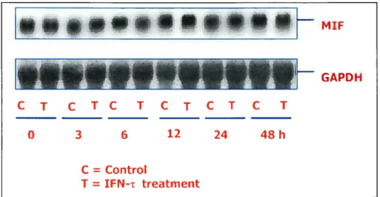
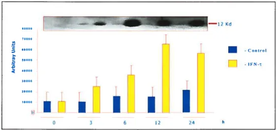
Dans la deuxième expérience, l’expression de MIF dans les cellules épithéliales de l’endomètre bovin a été examinée par buvardage de type Nothem, de type Western et par analyse immunohistochimique. La sécrétion de MIE par les cellules épithéliales bovines a été évaluée par buvardage de type Western. Les résultats ont démontrés qu’une grande expression basale de la protéine et de l’ARN messager (ARNm) de MIE était observée dans toutes les cellules, mais que les traitements hormonaux et les contrôles n’ont pas d’effet sur cette expression. Toutefois, IEN-r stimule la sécrétion de la protéine MIE par les cellules. La caractérisation de la régulation de l’expression de la protéine MW et de son ARNm par WN-t contribuerait à améliorer de façon significative notre compréhension des intéractions embryo-utérines du début de la gestion chez les ruminants.
Pour la troisième expérience, l’effet de IEN-t sur l’apoptose dans les cellules épithéliales de l’endomètre bovin en culture a été examiné par analyse TUNEL, par fragmentation d’ADN, et par buvardage de type Western. Les résultats indiquent que IEN-r et CHX augmente significativement le pourcentage de cellules à noyau apoptotique (34.3 et 46.5% respectivement), comparativement au contrôle (11.2%) (P<O.05). Le traitement des cellules avec la progestérone inhibe significativement l’habileté de IEN-t à induire l’apoptose (13.7%), comparativement à IFN-r seul (34.3%) (P<O.O5). L’analyse de fragmentation d’ADN a démontré que le traitement avec 1NF-r
résulte en une augmentation de la séparation de l’ADN relativement aux cultures contrôles non-traitées. Le buvardage de type Western a démontré que les traitements avec IFN-r et la cycloheximide résultent en une augmentation d’expression de la protéine pré-apoptotique Bax, comparativement aux cultures contrôles.
En conclusion, ces données expérimentales démontrent que WN-r est capable d’induire la sécrétion de protéines par les cellules épithéliales de l’endomètre bovin, de réguler positivement la sécrétion de MW, et d’induire l’apoptose dans ces cellules. De plus, ces effets sont modulés par les hormones stéroïdiennes. La présente étude est d’importance car elle démontre pour la première fois que (1) l’ARNm et la protéineMW sont hautement exprimés dans les cellules épithéliales de l’endomètre bovin en culture, et que la sécrétion de MW est stimulée en réponse WN-’r; (2) WN-t inhibe la prolifération des cellules épithéliales et induit l’apoptose dans les cellules épithélialesde l’endomètre bovin en culture. L’implication de IFN-t dans la reconnaissancematernelle de la gestation est connue depuis plusieurs années, mais l’étude présente rapporte pour la première fois l’implication directe de cette cytokine dans l’induction de l’expression d’un membre de la famille bcl2, Bax-Œ, et de l’apoptose dans les cellules épithélialesde l’endomètre bovin. Une autre découverte importante est que l’apoptose induitepar WN
‘ est inhibée par la progestérone, ce qui suggère que les concentrations plasmatiques
de progestérone au début de la gestation sont importantes pour la modulation des effets de
WN-r.
Ces résultats rassemblés suggèrent que MW est vraisemblablement un facteur contribuant à l’établissement du début de la gestation, mais la signification fonctionnelle de MW reste à être déterminée. Comprendre la régulation de la sécrétion de MW et son
site d’action dans le tractus reproducteur ajoutera significativement à notre compréhension des intéractions embryo-utérines du début de la gestation. Nous spéculons que WN--t pourrait jouer un rôle dans le développement de l’endomètre autour de la période d’implantation. Incidemment, la compréhension de la régulation de la sécrétion de la protéine MiE par WN-t ajoutera significativement à notre compréhension des intéractions embryo-utérines du début de la gestation.
C
Summary
During early pregnancy in ruminants, the embryo not only prevents prostaglandin F2 release, but it also modifies protein secretion ftom the endometrium. Tnterferon-t (1FN-t) is produced by the trophoblast prior to implantation in ruminants. It is involved in maternai recognition of pregnancy and is aiso a pleiotropic molecule that can alter the synthesis of endometrial proteins and inhibit proliferation of some ceils. However, the mechanisms involved in maternai recognition of pregnancy, embryo implantation and development are not weli understood at the cellular and molecular leveL To further our understanding, a primary culture system of endometrial ceils was used in these studies. We hypothesize that ffN-r modifies protein secretion from endometrium that is involved in the establishment of pregnancy. The objectives of this study were to establish an appropriate primary endometrial ceil culture system that can respond to steroid hormones and TFN-r and to examine the effect of TFN-t on protein secretion and ceil division and the possible modulation of its effects by steroid hormones.
In the first experiment, the effect of WN-r on protein secretion from bovine endometrial epithelial ceils and stromal ceils was examined by 2D SDS PAGE and HPLC analysis. The resuits showed that IFN-t induced the secretion ofseveral proteins from bovine uterine epithelial celis and this was modulated by steroid hormones. Estradiol down-regulated protein secretion and progesterone up-regulated protein secretion. Protein sequence analysis of proteins induced by IFN--r. showed partial amino acid sequences that corresponded to macrophage migration inhibitory factor (Mif).
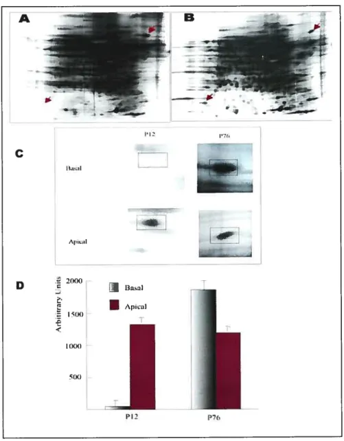
Ri the second experiment, expression of MW in isolated epithelial and stromal ceils of bovine endometrium was examined by Northern blotting, Western blotting and immunohistochemistry analysis. MW secretion from cultured bovine epithelial ceils was examined by Western blotting analysis. Results showed that MW protein and MW rnRNA were expressed in epithelial, but not in stromal ceils. There was no effect of WN-t on MW expression in the epithelial celis. However WN-r did stimulate the secretion of MW protein from the celis. The characterization and understanding of the MW protein and mRNA expression and regulation of the secretion of the MW protein by WN-i will add significantly to our understanding of early embryo-uterine interactions.
In the third experiment, the effect of WN-r on celi growth was studied. ResuÏts showed that WN-t at the 100 ng/ml dose, either alone or in the presense of P4, significantly decreased the DNA content of epithelial celis (p < 0.00 1), indicating that a decrease in proliferation or an increase in ceil death, or both, occurred. To fiirther examine the reason for this effect, apoptosis in cuÏtured epithelial celis of bovine endometrium was examined by TUNEL, DNA fragmentation and western blotting analysis. The resuits showed that WN-r and CHX significantly increased the percentage ofcells with apoptotic nuclei (34.3 and 46.5% respectively) when compared with control (11.2%) (P<0.05). Progesterone treatment of the celis significantÏy inhibited the ability
of LFN-T to induce apoptosis (13.7%) when compared with WN-r alone (34.3%)
(P<0.05). DNA fragmentation analysis showed that TNF-r treatment resulted in an increase in the appearance of DNA laddering compared with that in untreated control cultures. Western blotting analysis showed that WN-t and cycloheximide treatment
resulted in an increase in the expression of the proapoptotic protein Bax-x compared with that in control cultures.
In conclusion, these data demonstrate that WN-t can induce protein secretion from epithelial celis of bovine endometrium, stimulate MW secretion and induce apoptosis in bovine uterine epithelial celis. These effects were modulated by steroid hormones. The most significant findings in those studies are that: (1) it demonstrates for the first time that MW mRNA and protein are highly expressed in cultured bovine endometrial epithelial ceils and that the secretion of MW is stimulated in response to WN-r. (2) WN-t inhibits epithelial celi proliferation and induces apoptosis in cultured bovine endometrial epithelial ceils. Although the involvement of WN-’ç in maternai recognition of pregnancy lias been known for a number of years, this is the first report that this cytokine directly induces a bcl2 gene family member, Bax-a, and apoptosis in bovine endometrial epitheliai ceils. The other important finding is that the WN-’t induced apoptosis is inhibited by progesterone, which suggests that plasma progesterone concentration during early pregnancy is important for modulating the effect of WN-r. Taken together, our resuits suggest that MW is likely a factor contributing to the establishment of early pregnancy, however, the functional significance of MW remains to be determined. Understanding the regulation of MiT secretion and its site of action in the reproductive tract will add significantly to our understanding of early embryo uterine interactions.
TABLE 0F CONTENTS
Sommaire iv
Summary viii
Table of Contents xi
List of figures XV
List of Abbrevïations xvii
Acknowledgements xx
AVANT-PROPOS (PREFACE) Xxi
1.0. Introduction 1
2.0. Literature Review 4
2.1. MaternaI Recognition of Pregnancy 5
2.1.1. Hormonal Influences on the Outcome of Early Pregnancy 5 2.1.2. The embryonic signal—Production of Interferon-t 6 2.1.3. The maternai signai—Development ofthe luteolytic
mechanism 8
2.1.4. Uterine hormone receptors during early pregnancy 9
2.1.5. MaternaI Recognition ofPregnancy 10
2.2. Structural and Biocliemical Remodelling of Endometrium in the Estrous
Cycle and Eariy Gestation 11
2.2.1. Stuctural remodelling 12
2.2.2. Biochemical remodelling 14
2.2.2.1. Steroid hormone receptors 14
2.2.2.2. IFN-t 16
2.3.1 Implantation .17
2.3.2 Placentation. 1$
2.3.3 Embryo development 20
2.3.4 Differentiation 21
2.4. Macrophage migration inhibitory factor (MIF) 22
2.4.1 Cytokines 22
2.4.2 Discovery, cloning and structure of MW 24 2.4.3 Inhibition of macrophage migration by MW 24 2.4.4 Main characterization and biological activities of MW 25 2.4.5 Role of MW in ceil proliferation, angiogenesis
and tumorigenesis 27
2.4.6 Role of MW as a T cell cytokine 29 2.4.7 MW as a counter-regulator of glucocorticoid action 31
2.4.8 Distinct feature of MW 35
2.4.9 Molecular mechanism of MW function 38
2.4.10 Role of MW in reproduction 41
Male reproduction 41
Female reproduction 41
2.5. The role ofapoptosis in early pregnancy maintenance 43
2.5.1 Apoptosis and implantation 43
2.5.2 Clearance ofapoptotic celis 44
2.6. Conclusion 47
3.0. Hypothesis and Objectives 50
3.2. Objectives. 50
4.0. Article One 51
INFLUENCE 0F ESTRADIOL AND PROGESTERONE ON PROTEIN SECRETION INDUCED BY INTERFERON-TAU IN
CULTURED BOVINE EPITHELIAL CELLS 52
4.1. Abstract 53
4.2. Introduction 54
4.3. Materials and methods 57
4.4. Resuits 64
4.5. Discussion 66
4.6. Acknowledgements 6$
4.7. References 69
5.0. Article Two 75
INTERFERON-TAU STIMULATES SECRETION 0F
MACROPHAGE MIGRATION INHIBITORY FACTOR FROM
BOVINE ENDOMETRIAL EPITHELIAL CELLS 77
5.1. Abstract 78
5.2. Introduction 79
5.3. Materials and methods 80
5.4. Resuits $8
5.5. Discussion 90
5.6. Acknowledgements 95
5.7. References 95
6.0. Article Tree.104 PROGESTERON-MODULATED INDUCTION 0F APOPTOSIS
BY INTERFERON-TAU IN CULTURED EPITHELIAL CELLS
0F BOVINE ENDOMETRIUM 106
6.1. Abstract 107
6.2. Introduction 108
6.3. Materials and Methods 109
6.4. Resuits 115
6.5. Discussion 117
6.6. Acknowledgements 121
6.7. References 121
7.0. General Discussion 131
7.1. Proliferation and apoptosis of bovine endometrial epithelial ceil
and the establishment of early pregnancy 131
7.2. Biological function of MIE protein and the establishment
ofearly pregnancy 133
7.3. Interaction ofIFN-t, E2 and P4 and the establishment of early
pregnancy 136
8.0. General Conclusion 140
C
LIST 0F FIGURES Literature reviewFigure 1. Schematic drawing of bovine fetus in utero 19 Figure 2. Glucocorticoid-induced MTF secretion by macrophages 33 Figure 3. Effect of clearance of apoptotic ceils by macrophages 46
Article One
Figure 1. Effect ofEmbryo, conditioned medium and WN-r on proteins secreted by
endometrial epithelial ceils 71
Figure 2. Effect ofIFN-r on proteins secreted by endometrial stromal ceils 72 Figure 3. Influence ofE2 and P4 on protein secretion induced by WN-r in bovine
epithelial celis 73
Figure 4. The epithelial celis were grown on a Matrigel matrix Milliceil insert 74
Article Two
Figure 1. Effect of WN-T on protein secretion by endometrial epithelial celis 99 Figure 2. Effect ofIFN-T on MIF mRNA expression in bovine endometnal
epithelial celis 100
Figure 3. Effect of WN-T on MW protein expression in bovine endometnal
epithelial celis 101
Figure 4. Effect of WN-t on MW secretion from bovine endometrial epithelial
celis 102
Figure5. Immunolocalization of MW in bovine endometrium 103
Article Three
Figure 1. Effects of P4 and WN-r on DNA and protein content in bovine
endometrial ceils 125
Figure 2. Representative images of endometrial epithelial celis afier TUNEL
staining 126
Figure 3. The effect of IFN-t and P4 on the percentage of dead ceils in primary culture of bovine endometrial epithelial cells 127 Figure 4. The effect of 10 ng/ml P4 on the onset ofWN-t-induced apoptosis 128 Figure 5. Western blotting analysis ofproapoptotic protein Bax-Πin epithelial
celis 129
Figure 6. Effect ofIFN-r on expression ofproapoptotic protein Bax-Œ
in epithelial ceils 130
General Discussion
Figure 1. Effects ofgrowth factors on the expression ofMIF mRNA in
MC3T3-Elcells 135
C
LIST 0F ACTH Adrenocortropic hormoneBGCP-2 Bovine granulocyte chemotactic protein-2
bIFN-t Bovine interferon-t BSA Bovine serum albumin
bIP-1 Bovine trophoblast protein- 1
CL Corpus luteum
COX Cyclooxygenase
CTB Inner cytotrophoblast
CIIMI 5-Carboxymethyl-2-Hydroxymuconate isomerase DEPC Diethyl Pyrocarbonate
DR-6 Death receptor-6
DTH Delayed-type hypersensitivity E2 Estradiol-17f3
EGF Epidermal growth factor ER Estradiol receptor FCS fetal calfserum
FITC Fluorescein isothiocyanate FSH Follicle-stimulating hormone
GM-CSF Granulocyte-macrophage colony-stimulating factor HBSS Hank’s buffered saline solution
HPA Hypothalamus-pituitary-adrenal axis
IgG Immunoglobulin G
IGF-2 Insulin-like growth factor-2
IFN-t Jnterferon-t IL-1 Interleukin-1
IRE Interferon response element IRF Interferon regulatory factor ISGF-3 IFN-stimulated gene factor-3 LPS Lipopolysaccliaride
NBCS New-bom calfserum
MIF Macrophage migration inhibitory factor
NO Nitric oxide
NOS Nitric oxide synthase ODF Outer dense fibres
01 Oxytocin
oTP-1 Ovine trophoblast protein-1 OTR Oxytocin receptor
P4 Progesterone PGF-2ΠProstaglandin F-2ΠPGFS Prostaglandin F synthase PGE2 Prostaglandin E2 PGH2 Prostaglandin H2
C
PGHS Prostablandin G/H synthasePR Progesterone receptor RA Rheumatoid arthritis
rbIFN-r Recombinant bovine interferon-r SDS Sodium dodecyl sulfate
STB Outer syncytiotrophoblast TGFŒ Transforming growth factor-Œ
TIMPs Tissue inhibitors ofmetalloproteinases TNF Tumor necrosis factor
TNFR Tumor necrosis factor receptor
TUNEL Terminal deoxynucleotidyl transferase (TdT)-mediated d-UTP nick end labelling technique
TNf Tumor necrosis factor
TRML TNF-related apoptosis-inducing ligand
TRAIL-R TNFR family and through binding to its receptor TRADD TNFR1 -associated death domain
TSST-1 Toxic shock syndrome toxin-l UCRP Uniquitin cross-reactive protein VSV Vesicular stomatis virus
Acknowledgements
First of ail I would like to sincerely thank my supervisor Dr Alan K. Goff for his invaluable guidance, maximum patience, and constant encouragement, unselfish help and very thoughtffil arrangement for my study while I was studying at the Centre de recherche en reproduction animale (CRRA), University of Montreal. In addition, there are many people I need to thank for their help in the completion of this thesis and my Ph.D. studies. I ai-n greatly indebted to the members of my graduate student advisory committee, Drs Bruce D. Murphy, Christopher A. Price and Denis Vaillancourt.
I also would like to thank Dr R. M. Roberts for lis generous gifi of the
recombinant bovine interferon-’t and Dr A. Meinhardt for his MW antibody and probe which allowed me to begin the study of MW expression in bovine endometrial epithelial ceils without delay.
This study was carried out at CRRA, Faculté de médecine vétérinaire. I would like to thank ail members of the CRRA, for their fi-iendship and kindness during the course of my study. Particularly, I am grateful to Mrs Dobias-Goff, Danieli Rannou for their tecimical assistance. I am grateful to Miss Jol1e Desmarais for her heip in French translation ofmy thesis summary. I an-i also gratefiul to Drs Derek Boerboom, Angelika Stock, Khampoune Sayasith, Nicolas Gévry and Minglu Cao not only for their help invaluable for the completion ofthis work but also for their friendship.
I am indebted to express my sincere thanks to my wife Wenya, who supported
and encouraged me in spirits, to my son Kaiyang and daughters Anna and Gina for their love. Finally, I am indebted to my parents-in-law and my mother for their encouragement
Ç
AVANT-PROPOS (PREFACE)Cette thése comprend une introduction générale qui renferme l’hypothése de départ, une revue de littérature générale; trois articles comprenant chacun une introduction, une section Matériel et méthodes, des résultats, une discussion et des références; une discussion générale ainsi qu’une conclusion générale.
This thesis composes a general introduction which indicates the hypothesis of the study, a general literature review; three articles, each of which contains specific introduction, materials and methods, resuits, discussion and references; a general discussion and general conclusion.
C
1. 0. Introduction
Maternai recognition of pregnancy resuits from biochemical signaling between the conceptus (embryo and its associated membrances) and the maternai system. Pregnancy recognition signais ensure maintenance of structural and functional integrity of the corpus luteum (CL), which would otherwise regress at the end of the estrous cycle. The CL produces progesterone, the hormone of pregnancy, which is responsible for maintaining endometrial functions that permit early embryonic development, implantation, placentation, and successful fetal/placental development.
The estrous cycle of cows is uterine dependent, and the luteolytic signal responsible for structural and functional demise of the CL, or luteolysis, is prostaglandin f2 (PGf2). The uterine endometrium, primarily luminal epithelium and perhaps superficial glandular epithelium, is influenced by progesterone, estrogen, and oxytocin, through their cognate receptors, to release pulses of PGf2 required for luteolysis. The antiluteolytic signal for pregnancy recognition in cows is a novel Type I interferon named interferon-t tIEN r)(Roberts, Farin et al. 1990). WN-r is secreted by trophectoderm of cow conceptus between Day 12 and Day 25 of pregnancy and exerts a paracrine effect on the uterine endometrium to abrogate the luteolytic mechanism (Mann and Lamming 2001).
Pregnancy starts with fertilization and ends with parturition (the birth process). After fertilization, the conceptus develops through periods of cleavage,
(E
called an embryo, afler differentiation, it is cailed a fetus. During the early part of gestation, the embryo remains ftee, first in the oviduct and then the uterus. The establishment and maintenance of pregnancy involves a series of molecular signais exchanged by conceptus (embryo/foetus and associated membranes) and endometrium. During the peri-implantation period the ruminant conceptus is bathed in endometriai secretions (histotroph) which nourish and sustain it. Histotroph contains a complex mixture of proteins, carbohydrates, sugars, lipids and ions (Bazer 1989). Changes in the quantitative and qualitative pattem of endometrial protein in cyclic and pregnant ruminants have provided dues to the identity and function of the protein components of histotroph and their roles in establishment ofpregnancy.During early pregnancy the uterus, under the influence of ovarian steroids progesterone and estrogen, undergoes cellular and molecular changes to achieve a receptive state for the onset of implantation. In rats and mice, progesterone and estrogen sequentially program the uterus into pre-receptive, receptive and non receptive phases during pregnancy or pseudopregnancy (Psychoyos 1973). Blastocysts implant only in the receptive uterus. The progesterone-primed pregnant utems becomes receptive on day 4 afler it is superimposed with preimplantation ovarian estrogen secretion. Subsequently, the receptive uterus proceeds to a non-receptive phase when blastocysts can no longer implant. Although many molecular signaiing pathways have been identified associated with uterine receptivity and embryo—uterine interactions during implantation, the
definitive roies of these pathways or their interactions in these processes remain elusive.
In this respect, scientists have recentiy discovered that the mouse uterus, which was thought to 5e receptive primarily on day 4, is stiil receptive on day 5 of pseudopregnancy after biastocyst transfers. for example, mice receiving
blastocyst transfers on day 5 of pseudopregnancy show implantation when examined 48 h later. In contrast, simiiariy transferred blastocysts compietely faiied to implant in day 6 pseudopregnant recipient uteri (Song, Guan et al. 2002). Before implantation (Day 21) the embryo depends on uterine secretion for normal development and survival. Only afler piacentation, can the embryo derive nutrients and transfer waste products through maternai blood. In the cow between 30 and 35 days afier fertilization, there wiil be 3 or 4 fragile cotyledonary attachments in the pregnant hom (Song, Guan et al. 2002).
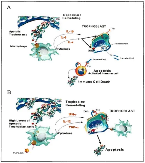
Cytokines play a pivotai role in reguiating the host inflammatory and immune responses to infection and tissue invasion. They regulate the first non specific phase of the host response by orchestrating a local inflammatory reaction and then serve to controi the subsequent specific immune response. Cytokines appear to play a critical role in the establishment of pregnancy. MIF is a pro-inflammatory cytokine involved in reproduction (letta, Todros et al. 2002).
During pregnancy, the uterus and the placenta are immunologicaily privileged sites in which immune activity is effectively diminished (Streilein 1995). Apoptosis of immune celis has been proposed as a mechanism for
C
maintaining immune privilege (Griffith and ferguson 1997). Induction of apoptotic celi death can also be a factor that limits lymphocyte proliferation following activation. Apoptosis is a complex process that removes aging or injured celis from the body and occurs in a wide variety of organisms. Regulation of apoptosis is complex and involves a family of related proteins that can promote or inhibit this process. Apoptosis may serve as a previously unsuspected mechanism that induces tolerance of the foetal allografi against matemal immune system and plays a role in early pregnancy maintenance (Jerzak and Bischof 2002).Based on the above considerations, we hypothesize that WN-r modifies protein secretion from endometrium that are involved in the establishment of pregnancy. The specific objectives ofthis project were (1) to establish an in vitro mode! to explore the effect of IFN--c on protein secretion from cultured bovine endometria! epithelial and stromal celis, (2) to identify proteins secreted into culture medium and effected by IFN-t in bovine endometrial epithelial ceils, and (3) to examine its steroid hormone modulation on protein secretion induced by
IfN-r.
2.0. LITERATURE REVIEW
This literature review will focus on (1) the events that occur during early gestation (the first three weeks) in the cow, including the hormonal influences on the outcome of early pregnancy, production of 1FN-t (embryo signal), and the
C
receptors during early pregnancy, uterine remodelling and protein synthesis, cleavage, implantation and embryo development; (2) role of MW in inflammatory and reproduction and highlight the functional and mechanistic properties of MW, and (3) the role of apoptosis in early pregnancy maintenance.2.1. Maternai Recognition of Pregnancy
2.1.1 Hormonal Influences on the Outcome of Early Pregnancy
Many of the mechanisms involved in early pregnancy are influenced by the ovarian steroid hormones, progesterone and estradiol, and many studies have investigated their roles. It has been established for many years that the concentration of pro gesterone during early pregnancy lias a marked effect on the potential outcome. Lower concentrations of plasma progesterone from about day 12 afier mating have been reported in animals in which early pregnancy fails in a number of studies (Lukaszewska and Hansel 1980; Lamming, Darwash et al. 1989; Mann and Lamming 1995). These studies clearly demonstrate that both a late post-ovulatory rise in progesterone and low luteal phase concentrations of progesterone have a detrimental effect on the outcome of early pregnancy.
Although estradiol concentrations have not been studied as comprehensively as concentrations of progesterone during early pregnancy, most studies indicate that concentrations of estradiol do flot differ between mated cows in which pregnancy is successful or fails (Lukaszewska and Hansel 1980; Gyawu and Pope 1992; Maun and Lamming 1995). In one study in beef cows, a lower
estradiol between day 14 and day 17. However, in this study, luteolysis had begun in some cows and 80 it is flot clear whether the higher concentration of
estradiol was the cause of pregnancy failure or the result of failed embryonic inhibition of luteolysis (Pritchard, Schrich et al. 1994). Thus current evidence supports the idea that estradiol does not exert the same degree of influence as progesterone over the outcome of early pregnancy. It has now been demonstrated that the concentration of luteal phase progesterone in the cow lias a profound influence on the strength of development of the luteolytic signal (Mann and Lamming 1995). Estradiol concentrations do flot differ between pregnant and non-pregnant cows, but have an important influence in controlling the strength ofthe luteolytic signal (Mann and Lamming 1995).
2.1.2. The Embryonic signai--Production oflnterferon-r
In ruminants, IFN-’t is well characterized as an important embryonic pregnancy recognition signal. LFN-r is encoded by multiple genes (Ealy, Larson et al. 2001) and is expressed and secreted by trophectoderm cells of blastocysts (Roberts, Farin et al. 1990). Secretion of WN-t by bovine blastocysts in vivo is highest between days 15 and 17, whereas in a specific culture system for bovine trophoblastic vesicles increasing IFN-t secretion was observed for a longer period (up to day 23 afier fertilization) (Stojkovic, Wolf et al. 1995).
In cows, removal of the embryo from the uterus on day 15 does not result in a delay in luteolysis, whereas removal on day 17 resuits in a significant delay.
C”
in luteo]ysis (Nortliey and french 1980). Thus it was established that the embryo exerted an anti-luteolytic effect on the cow between day 15 and day 17. Highly purified bTP- 1 obtained from culture of day 17-12 conceptuses (Helmer, Hansen et al. 1989) and recombinant bovine interferon-r (Meyer, Hansen et aI. 1995) have been shown to reduce luteolytic secretion of PGF2Πand extend luteal function in the cow. Expression of mRNA for interferon-r has been detected as early as day 12 in the cow, is maximal on days 15-16 and continues at least until day 25. This expression appears to be limited to the trophectoderm and expression is flot apparent in the endoderm or yolk sac (Farin, Imakawa et al.1990).
During the early stages of pregnancy, it is well established that progesterone stimulates the production of the endometrial secretions necessary for embryo development. The effects of this progesterone include increased endometrial protein secretion (Garrett, Geisert et al. 1988) and increased production of PGE2 (Vincent, Meredith et ai. 1986). In cows, maternai concentrations of progesterone have a marked influence on the development of the embryo (Mann, Mann et al. 1996) and its ability to produce interferon-t (Mann, Lamming et al. 1998). Cows with a late post-ovulatory increase in progesterone or lower luteal phase concentrations had embryos that, on day 16, exhibited littie or no elongation and produce littie or no interferon-’u. Conversely day 16 embryos of cows with an earlier rise in progesterone to higher luteal phase concentrations were well elongated (>4 cm) and produced large quantities of interferon-t. These findings suggest that an early increase in progesterone is
more important in stimulating embryo development and interferon-r synthesis than are later progesterone concentrations (Mann, Lamming et al. 1999).
2.1.3 The Maternai Signal—Development of the luteo]ytic Mechanism In cattie, concentrations of endometrial oxytocin receptors are low or undetectable from about day 6-8 of the luteal phase to immediately before luteolysis, about day 15-17, when concentrations begin to increase (Meyer, Mittermeier et al. 1988; Fuchs, Bebrens et al. 1990; Mann and Lamming 1994). The pulsatile secretion of luteolytic PGF2 begins on about day 17 and is associated with a small rise in endometrial oxytocin receptor concentration (Mann and Lamming 1993). By collection ofrepeated biopsy samples ofuterine endometrium, Mann et al (1999) found that oxytocin receptors, which are undetectable through much of the luteal phase (<20 fmol/mg protein), rise to a concentration of 121 ± 16 fmol /mg protein when large luteolytic episodes of PGF2 secretion are first observed. The onset of PGF2 secretion is followed by luteolysis within 48 h and oxytocin receptor concentrations continue to increase to maximum concentrations of 500-1000 fmol /mg protein at estms. furthermore, a marked increase in oxytocin-induced PGf2Œ production occurs in heifers between day 13 and day 16, despite only a slight increase in concentration of endometrial oxytocin receptors; a large increase in oxytocin receptor concentration did not occur until day 19. Thus luteolytic PGf2 release requires only a modest increase in uterine oxytocin receptors. The peak concentration of oxytocin receptor obtained at estrus are associated with the fail
in progesterone and increase in oestradiol secretion that occur as a resuit of luteolysis and are not, therefore, the cause of luteolysis (Mirando, Becker et al.
1993).
In cows oxytocin receptors first appear on the luminal epithelium of the uterine endometrium (Robinson, Mann et al. 1999). This occurs at the same time as the uterus develops the ability to release PGF2a in response to OXytOcifl (Maflfl and Lamming, 1994). This finding demonstrates that, in cows, it is the development of oxytocin receptors on the luminal epithelium that is the key event in the development of the luteolytic mechanism. The initiation of the luteolytic mechanism requires only a relatively small increase in endometrial oxytocin receptors and it is this initial increase in oxytocin receptor development within the luminal epithelium which the embryo must counteract if it is to prevent luteolysis.
2.1.4. Uterine oxytoxïn receptors during early pregnancy
The initial small rise in oxytocin receptors, localized to the luminal epithelium of the endometrium, is the event in the initiation of luteolysis. Thus the inhibition ofthis initial rise in oxytocin receptors appears to be the key event in the establishment ofpregnancy. Fuchs et al (1990) found that the presence of a viable conceptus in the uterus completely prevented both the small rise in oxytocin receptors in the uterus between day 14 and day 17, and the much larger rise seen between day 17 and day 21 in cyclic cows. On day 16, a time at which the luteolytic mechanism is beginning to develop, oxytocin receptor mRNA was
Ç
detectable in the luminai epithelium of over 40% of non-pregnant cows but was undetectabie in ail cows with an embryo (Robinson etaÏ., 1999).2.1.5. Maternai Recognïtion ofPregnancy
Antiluteolytic effects of IFN--t are responsible for maternai recognition of pregnancy, which is the term used to describe how a mother responds (physiologicaiiy) to the presence of a conceptus in her reproductive tract. In domestic ruminants, the developing embryo does not implant until relatively late in development, although the conceptus is ciearly capable of communicating with the mother weil before implantation occurs, and before the conceptus has access to the maternai circulation. Failure of the conceptus to signal its presence at the appropriate time ieads to pregnancy ioss (Demmers, Derecka et ai. 2001).
The principal roie of the embryo during the maternai recognition of pregnancy is to inhibit the development of oxytocin receptors on the endometrium and hence the release of PGf2Πthat is responsible for the demise of the corpus luteum. In sheep, it has been postulated that interferontprevents the
rise in endometrial estrogen receptors that is thought to precede the rise in oxytocin receptors necessary for the induction of luteolytic release of PGf2. This contention is supported by several studies (Spencer and Bazer 1995). However, studies in cows indicate that the initiai inhibition of oxytocin receptor development on the iuminal epitheiium and luteolytic PGF2a release occur in the absence of an effect on estrogen receptor concentrations (Robinson et al., 1999).
It is important to consider the control of PGf2Πrelease. One such mechanism in cows involves the stimulation, by the embryo, of an endometrial inhibitor of PGF2Πsynthesis (Thatcher, Meyer et al. 1995). hi sheep, pregnancy
is associated with a reduction in pulsatile release of PGf2 coupled with an increased basal secretion, i.e. a change in the pattem and not the quantity of PGF2Πsecretion. However, in cattie the attenuation of luteolytic episodes occurs in the absence of an increase in basal secretion of PGF2a (Thatcher et al., 1995). This species difference is clearly supportive of the presence of a direct inhibitory effect of the embryo on PGF2 secretion in cows.
During the early stage of pregnancy, progesterone induces differentiation and maturation by acting on the estrogen-primed endometrium through progesterone receptors. This resuits in switching on of several progesterone dependent genes. Genes, negatively regulated in the estrogen dominant phase of estrous cycle, may also be expressed. This results in synthesis of a number of endometrial proteins and other factors and consequently alters the structural and molecular profiles of endometrium (Hegele-Hartung, Mootz et al. 1992; Beier Hellwig, Bonn et al. 1994). Thus, structural and biochemical remodelling of endometrium will be discussed in the following sections.
2.2. Structural and biochemical remodelling of endometrium in
the estrous cycle and early gestation
i.e. estrous cycle, implantation and maintenance of pregnancy. Afler pregnancy recognition, maintenance of pregnancy requires reciprocal communication between the conceptus and endometrium during implantation and synepitheliochorial placentation (Wimsatt 1950). The endometrium undergoes several structural and biochemical changes in every estrous cycle and pregnancy to facilitate embryo implantation.
2.2.1. Structural remodelling
The utenis of the domestic mammal consists of a corpus (body), a cervix (neck), and two homs. The mucous membrane lining the utems is a highly glandular structrure, the endometrium. It varies in thickness and vascularity with hormonal changes in the ovary and with pregnancy (Frandson and Spurgeon 1992). Cattle have a 21 day estrous cycle govemed by follicular estrogen and luteal progesterone. These hormonal shifis allow for the development and ovulation of an egg, receptivity to mating and preparation of the uterus for pregnancy. In the uterus, intercellular communication requires integration of hormonal signals ftom the ovaries and fetus, which in tum depends on tissue and ceil specific expression of steroid receptors (Kimmins and MacLaren 2001). The mechanisms which govem such interactions in the uterus include celi-cell contact, paracrine transmission of hormones, cytokines and signaliing molecules, and extracellular matrix and celi adhesion molecule interactiona at tissue boundaries (Bai-toi, Wiley et al. 1999; Roberts, Eaiy et al. 1999). The tissue
distribution of steroid receptors is regulated during the estrous cycle and early pregnancy in a celi specific manner (Kimmins and MacLaren 2001).
The ruminant placenta shows a considerable uniformity of gross structure across the genus, a flat apposition of trophoblast (chorionic epithelium) to uterine epithelium with a variable number of placetomes. Implantation is a dynamic process requiring intricate signalling interactions between maternai endometrium and fetal trophoblast celis, and remodelling of the endometrium to accommodate placental development. Cattie and other rumimants undergo a relatively non invasive placentation process that gives rise to a synepitheliochoriai placenta. About Day 19-20 of pregnancy, trophoblast attachment begins in the region of the embryonic disk, and binucleate ceil migration begins (King 1980; Wathes 1980). These cells arise from the trophoblast, migrate across the microvillar junction, and fuse with maternai epitheliai cells to form a hybrid epithelium (Wooding 1992). Changes in the endometrial stroma have been reported during ruminant implantation, inciuding structural changes and angiogenesis as a prerequisite to cotyledon formation (King 1980; Renolds 1992). In sheep, superficial implantation and placentation is a lengthy process that begins on Days 15-16 and is not completed until Days 50-60 of pregnancy (Guillomot 1995). During this period, the ovine uterus grows substantially in order to accommodate rapid conceptus development and growth in the latter half of pregnancy. In addition to placental development in the caruncuiar areas ofthe endometrium and changes in vascularity, the intercaruncular endometrial glands grow substantially
in iength (four-ford) and width (ten-ford) during pregnancy in ewes (Stewart 2000).
Histomorphologicai analysis of non-receptive endometrium from antiprogestin treated animais clearly indicated that biocking the progesterone action induces retardation in endometriai development (Rosario, Sachdeva et al. 2003). Thus, the expression of the moiecular markers couid piay a significant roie in endometrial growth, deveiopment and thereby receptivity.
2.2.2. Biochemical remodelling 2.2.2.1 Steroid hormone receptors
It lias been now unequivocally estabiished that ovarian steroids— progesterone (P4) and estrogen (E2) are the major determinants ofmorphological and functional maturation of endometrium. Both E2 and P4 are known to paiy critical roies in reguiating the endometriai growth and deveiopment in cyclic manner. While E2 induces growth and proiiferation of endometrium, P4 induces endometrium to undergo differentiation and maturation (Rosario, Sachdeva et al. 2003). Ovarian hormones mediate their activity via specific receptors on endometrium. E2 is known in inducing the synthesis of both estrogen tER) and progesterone receptors (PR) while P4 down-reguiates the expression of ER and PR (fujishita 1997). P4 once bound to PR, initiates a series of events, which leads to synthesis of various cytokines, growth factors etc (Rosario, Sachdeva et al. 2003).
A number of studies in both cattie and sheep have demonstrated the general ability of the embryo itself, embryo-derived interferon-’r or recombinant interferon-r to inhibit the development of oxytocin receptors on the endometrium. It is now generally accepted that this is the event in the maintenance ofpregnancy in both species.
As discussed previously, induction of OTR is dependant on the action of E2 and P4. The effects of pregnancy on endometrial oxytocin, estradiol and pregesterone receptors on day 16 in non-pregnant cows and in cows with an embryo present in the uterus have been investigated (Robinson et al., 1999). In pregnant cows, there was a significant inhibition of both endometrial oxytocin receptor mRNA concentrations and oxytocin-induced secretion of PGF2a. However, despite the inhibitory effect of the embryo on the initiation of a luteolytic mechanism, measurement of both estradiol receptor mRNA and estradiol receptor protein revealed no differences between pregnant and non pregnant cows. Thus it would appear that in cows, the embryo can inhibit both the initiation of oxytocin receptor and oxytocin-induced secretion of PGf2 without affecting estradiol receptor concentrations. As with the estrogen receptor, progesterone receptors were also present at similar concentrations in both pregnant and non-pregnant cows in ail regions of the uterus studied, supporting the idea that changes in endometrial progesterone receptor concentrations are not involved in the inhibition of luteolysis during pregnancy (Robinson, Mann et al. 2001).
2.2.2.2. IFN-t
WN-t induces the expression of a number of genes, such as STAT (signal transducer and activator of transcription) 1 and 2 (Stewart, Johnson et al. 2001), WN-regulatory factor 1 (IRF-1) (Spencer, Ott et al. 1998), ubiquitin crossreactive protein (Johnson, Austin et al. 1998; Johnson, Spencer et al. 1999), Mx protein (Ott, Yin et al. 1998), granulocyte-chemotactic protein-2 (Teixeira, Austin et al. 1997), 2’, 5’-oligoadenylate synthetase (Johnson, Stewart et al. 2001). Further, IFN-r stimulates the expression of granulocyte-macrophage colony-stimulating factor, a cytokine with putative positive effects on the conceptus, in stromal celis of the endometrium (Emond, Asselin et al. 2000). Other effects of IFN-t in endometrium ceils include a reduction of oxytocin induced cyclooxygenase-2 and prostaglandin F synthetase expresstion (Xiao, Murphy et al. 1999). The regulation of those proteins by IFN-t may have important implications for cytokine networking in the uterus during pregnancy. Also, the regulation of inflammation and angiogenesis by those proteins with other cytokines may be integral to establishing early pregnancy and implantation in the cow (Teixeira, Austin et al. 1997) and to the maintenance of early pregnancy in ruminants (Johnson, Austin et al. 1998).
2.3. Early Gestation
Gestation is achieved through an array of events that include fertilization, attachment, implantation and placentation. Implantation and consequent placentation are important processes for success of gestation as remarkable
(N
changes occur between the conceptus and uterine endometrium (Wooding 1992; Weilauf 1994). In the cow, the elongation of the conceptus starts around two weeks afier fertilization and it first attaches to the endometrium at about three weeks of gestation (Wooding 1992).
2.3.1 Implantation
Implantation refers a series of highiy coordinated interactions that begin with intimate contact between apical plasma membranes of the conceptus trophectoderm and the uterine luminal epithelium and conclude with the formation of placenta as a means to support embryonic/fetal development throughout pregnancy. Before any intercellular contacts are established, secretions from the embryo and uterine endometrium exert a mutual influence to support flirther deveiopment of the conceptus. Embryonic secretory signais sustain the function of the corpus luteum during the early stages of pregnancy (Hafez 1993)
Implantation begins with an uncommon union between apical plasma membranes ofthe two genetically distinct tissues (i.e. the embryo and the uterine epithelium). The process of implantation in domestic animals (i.e. caffle, sheep) differs in a number of important ways from implantation in rodents and primate species. In contrast to rodents and primates, where the embryo attaches almost immediately to the uterine epithelium upon entering the uterus, domestic animais have a prolonged pre-implantation period upon arrivai of the embryo in the utems. Rather, the pre-implantation period is characterized by endometrial gland
secretion, and the generation of the conceptus signal for maternai recognition of pregnancy. Further, the trophoblast with its supporting layer of extra-embryonic mesoderm is simply apposed to the uterine epithelium in domestic animais: i.e. there is littie invasion of the maternai tissue by fetal tissue and the conceptus remains within the uterine lumen throughout gestation (epitheliochorial implantation) (Hafez 1993).
In the cow, there is evidence that the expression of carbohydrate determinants and ceil surface antigens change in the pre-implantation conceptuses (Dowsing, Gougoulidis et ai. 199$) and it is anticipated that protein and glycoprotein expression at the surface of the uterine epithelium is also modulated at this time (Skinner, MacLaren et ai. 1999).
2.3.2. Placentation
The endometrium provides a mechanism for attachment of the extraembryonic membranes. This union forms the placenta, and the process is called piacentation. As illustrated in Figure 1, the bovine placenta is multiplex, villous and epitheliochorial. It forms 80 — 120 placentomes {Figure 1(B)], areas
in which tufis of chorionic villi (cotyledons) attach to crypts deveioped from preformed endometrial prominences (caruncles). With formation ofthe placenta, nutrients from maternal blood can be transferred to embryonic and fetal blood and waste products from embryonic and fetal blood can be eliminated through the maternai systems (Bearden and Fuquay 2000). Cows have cotyledonary
o
Figure 1. (A) Schematic drawing of bovine fetus in utero. The grey part represents the maternai compartment composed of the endometrium (E) and the myometrium (M). Allantoamniotic membrane (A); placentome (P); intercotyledonary region (1CR). (B) Detailed schematic drawing of a placentome. The aliantochorion tAC) is smooth in the 1CR, which apposes likewise smooth E. In the piacentome, the AC is cailed cotyledon and forms fetai viilous (FV) trees that fit in maternai crypts (MC). In the AC, a loose network of mesenchymal ceils (MES) is covered with aiiantoic endoderm (AL) toward the aiiantoic cavity and with an intact trophobiast celi (TR) layer opposing the uterine epitheiium (UE). Adapted from (Schauser, Nielsen et ai. 2001)
penetrate into carunles, which aïe button-like projections on the endometrium. This union, chorionic villi and carunles, forms the placentome (also called the cotyledon) (Bearden and fuquay 2000).
Binucleate cells are found in the fetal trophectodermal epithelium of ail ruminant placentas. They are present in fairly constant proportions from implantation to parturition. The placental lactogen system in the sheep placenta is restricted to the binucleate celis. Counts of the ftequency of binucleate ceils and their migration indicated that most, if not ail, binucleate ceils migrated to the lumenal epithelia in glandular areas of the uterus (caruncles). The resuit of the migration is fusion of a binucleate celi with a uterine epitheiial ceil or a syncytial layer. This fusion delivers the characteristic binucleate ceil granules close to the matemal circulation while maintaining the trophectodermal barrier to other feto matemal exchange. The ruminant binucleate ceil therefore seems to play a central role in forming the structures and secretions at the feto-matemal interface which may be crucial in establishing and maintaining pregnancy (Wooding
1982).
2.3.3. Embryo Development
After fertilization, the zygote wilI divide many times without any increase in cytoplasm. The overail size may increase due to absorption of water, but the total ceilular material will decrease. This process of ceil division without growth is cleavage. The first cleavage will resuit in a 2-ceil embryo. This is foilowed by additional cleavages resulting in
4-ecu,
8 cell, 16-celi, 32-celi embryos, andso on. With each cleavage, ceils become smaller. In cows and ewes, an 8 to 16-ceil embryo will enter the uterus 3 to 4 days after ovulation. The 8 to 16-celi stage embryo is called a morula. By the 32 to 64 ccli stage, the morula will compact with gap junctions forming between interior celis and tight junctions forming between celis on the outside of the embryo, a necessary step in blastocyst formation. The blastocyst stage will be reached by day 7 in cattie. The development that occurs in the oviduct is critical to survival of the embryo (Killian 2004).
2.3.4. Differentiation
It is a period when the cells are in the process of forming specific organs in the body of the embryo. Notable events during differentiation include the formation of the germ layers, extraembryonic membranes, and organs. In addition, rapid changes in relative size occur during differentiation. This will occur by day 12 in cows (Peters 1987). Afler differentiation is completed, the product of conception is called a fetus rather than an embryo. This portion of gestation, between the completion of differentiation and parturition, has been termed the “the period of the fetus.” The principal development feature of this period is growth (Hafez 1993; Bearden and Fuquay 2000).
As reviewed previously, the uterus undergoes biochemical remodelling during pregnancy. Macrophage migration inhibitory factor (MW) expression and secretion are regulated by endocrinological changes during pregnancy in some
Ç
(Suzuki, Kanagawa et al. 1996). The features and functions of MIF in reproduction will be discussed in next section.2.4 Macrophage migration inhibitory factor (MIF)
2.4.1 Cytokines
Cytokines are a group of regulatory proteins with a MW of less than 100 kDa secreted by ceils of the immune system and act nonenzymaticaily in picomolar to nanomolar concentrations (Abbas, Lichtman et al. 1994). Cytokines play a pivotai role in regulating the host inflammatory and immune responses to infection and tissue invasion (Kauma 2000). They regulate the first nonspecific phase of the host response by orchestrating a local inflammatory reaction and then serve to control the subsequent specific immune response. Structurally cytokines are small Œ—, u/13—, or 3—proteins with a molecular weight
of 8-30 kDa that can ofien be grouped into subfamilies according to their structure or the structure of their con-e sponding receptors. Ex amples include the interleukin (IL) 6 family of cytokines and receptors, the chemokine family, and the tumor necrosis factor (TNF)-Fas ligand-CD3O ligand-CD28 ligand family. Cytokine biological activities are both pleiotropic and redundant, indicating that the molecular interplay leading to the balanced functioning of immune system is very complex. Novel members of the various cytokine families continue to be discovered, further adding to the complexity of the cytokine network (Durum and Oppenheim 1993; Thompson 1993).
Cytokine production and action are largely restricted to ceils of the immune system. However, it also has become clear recently that specific cytokine production and cytokine effects can occur in other celi types. An immune-neuroendocrine network of cytokine action lias been shown to participate in the regulation of inflammatory reactions and in the general host stress response. Researcli programs aimed at elucidating the pathways of immune-neuroendocrine regulation on a molecular level have demonstrated that systemic release of endocrine hormones, particularly glucocorticoids, may act to modulate immune system reactivity, and in tum that mediators of the immune response, i.e., cytokines, may serve to regulate neuroendocrine functions (Goldstein, Bowen et al. 1992; Spangelo and Gorospe 1995).
The maintenance of a state of physiological equilibrium within the host requires the interplay of various processes that have both complementary and opposing functions. Bemhagen et al (1998) initiated an exploratory research program to identify novel mediators that might be released systemically, and that can modulate inflammatory and immune responses. These studies have led to the identification of migration inhibitory factor (MIF), a previously known T ceil cytokine of largely unknown flmction, as a critical component of the immune system and counter-regulator of glucocorticoid action with unusual structural and functional features (Bernhagen, Calandra et al. 1998).
C
2.4.2 Discovery, cloning and structure ofMIFMW was identified nearly four decades ago as one of the first cytokines discovered (Bloom and Bennett 1966). MW was initially described as an immune activity isolated from the supematants of T lymphocytes and was found to inhibit the random migration of macrophages and subsequently to activate macrophage function. Over the years, MW activity was associated with macrophage phagocytosis and delayed-type hypersensitivity (Nathan, Kamovsky et al. 1971). Research on MW has been hampered because the entity responsible for the observed immune activities was not defined at a molecular level for almost three decades.
Today, MW is cloned (Bemhagen, Calandra et al. 1993) and its structure has been well characterized by crystallization, nuclear magnetic resonance spectroscopy and various biochemical methods. The structural properties of MW have recently been reviewed (Bemhagen, Calandra et al. 199$) and shah thus not be discussed in detail here.
2.4.3 Inhibition of macrophage migration by MIF
In 1966 historical experiments by Bloom and Bennett (Bloom and Bennett 1966) and David (David 1966) first identified MW as a nondialyzable protein factor produced by sensitized lymphocytes and which was associated with delayed-type hypersensitivity (DTH). MW was characterized by the activity of crude extracts to inhibit the random migration of guinea pig pentoneal exudates macrophages in vitro (Bloom and Bennett 1966; David 1966) and
subsequently to activate macrophage function (Nathan, Kamovsky et al. 1971; Nathan, Remold et al. 1973). In spite of these observations that described a cytokine activity more than 30 years ago, a detailed view ofthe biological role of MIF has remained elusive until very recently. This was due in large part to the failure to identify the molecular entity that is associated with the observed inhibition of macrophage migration. It is now clear that other immune factors such as interferon-y and IL-4 also exhibit migration inhibitory effects, and that these cytokines were also present in the complex cellular supematants that had been used in the initial MIF studies (Thurman, Braude et al. 1985; Mclnnes and Rennick 1988).
The cloning in 1989 of a human T celi protein with a molecular weight of 12.5 kDa unraveled the identity of MIF as this molecule was distinct from previously discovered cytokines with MIF activity (Weiser, Temple et al. 1989). However, the lack of biologically active, purified recombinant protein and the identification of a mitogenic contaminant within the original recombinant M1F preparations slowed further research in the field. As a resuit it was unclear at that time what the precise relationship is between the 12.5-kDa MIF protein and the various migration inhibitory assays that have been applied over the years.
2.4.4 Main characteristics and biological activities of MIF
MIF exhibits a number of unusual properties that distinguish this factor from other cytokines. The most important of these features are summarized here. MIF is considered a pleiotropic lymphocyte and macrophage cytokine, but
C
numerous reports (Waeber, Calandra et al. 1999; Fingerle-Rowson and Bucala 2001) also suggest that MW is an endocrine factor. Intriguingly, MW has been demonstrated to have at least two distinct catalytic activities, i.e. a tautomerase and an oxidoreductase activity. Accordingly, MIF has been termed “cytokine with enzyrmatic properties or cytozyme” and “secreted enzyme”. As one of the catalytic activities found is reminiscent of the oxidoreductase activities of the thioredoxin family of proteins, MW has recently been coined “redoxkine” (Ghezzi P. et al., 2000). The physiological relevance of the reported enzymatic activities of MW is flot yet resolved.MW is ubiquitously expressed in both immune and non-immune cells including various peripheral tissues. Its most critical functions encompass the regulation of macrophage function (Calandra, Bemhagen et al. 1994; Onodera, Suzuki et al. 1997), lymphocyte immunity (Bacher, Metz et al. 1996; Abe, Peng et al. 2001) and endocrine functions (Calandra, Bemhagen et al. 1995; Meinhardt, Bacher et al. 1996; Waeber, Calandra et al. 1997; Bacher, Meinhardt et al. 1998). MW is a unique counter-regulator of the immunosuppressive and anti-inflammatory activities of glucocorticoids (Calandra, Bemhagen et al. 1995; Daun and Cannon 2000).
One other non-typical property is that MW is effectively secreted from a variety of immune and some non-immune celis without having an N-terminal leader sequence or an apparent internai signal sequence for import into the endoplasmic reticulum. It has thus been concluded that MW is secreted by a
C
non-conventional Ieaderless patliway (Bembagen, Calandra et al. 1998). However, the precise mechanism of secretion has flot yet been elucidated.2.4.5 Role of MIF in celi proilferation, angiogenesis and tumorigenesis
A number of recent studies imply that MIF could be centrally involved in processes regulating celi proliferation and tumor angiogenesis (Takahashi, Nishihira et al. 1998); (Yang, Degranpre et aI. 2000). Moreover, Hudson et aï. suggest that celi cycle regulation by MW could be related to MIPs inflammatory activity (Hudson, Shoaibi et al. 1999).
In an attempt to explain the increased expression of cytosolic MW in murine colon carcinoma cells in response to growth factors, Takahashi et al. (1998) investigated the correlation between the expression of MIF and celi proliferation and found that MW expression was associated with enhanced proliferation of these ceils. Chesney et al. demonstrated that neutralizing anti MW-antibodies dramatically reduced the initial outgrowth of 38C13 B celi lymphoma celis in C3H/HeN mice (Chesney, Metz et al. 1999). As immune neutralization of MW did flot significantly affect the growth of estabïished tumors, Lue and Kleemann suggested an early primary effect for MW. They subsequently showed that neutralization of MW by anti-MW antibodies inhibited endothelial celi growth and led to a reduced number of tumor capillaries, but did not affect the proliferation of the lymphoma cells (Lue, Kleemann et al. 2002). In une with these observations, inhibition of tumor angiogenesis by anti-MW
al. 1999) and in murine colon carcinoma celis (Takahashi, Nishihira et al. 1992) and (Ogawa, Nishihira et al. 2000). In addition, Yang and coworkers identified MW as an angiogenic factor released by ectopic human endometrial ceils promoting human coronary artery endothelial ceil growth (Yang, Degranpre et al. 2000). Recombinant MW enhanced the effect of a cocktail of growth stimulating factors but not alone, and MW antibody reduced the stimulatory effect of the growth factors, suggesting together that MW lias an indirect effect on cell proliferation induced by growth factors, but may flot act in a proliferative manner itself. The studies also imply that increased cytosolic MW expression in tumors is linked to the proliferative properties of tumor ceils, a notion that is confirmed by the finding that over expression of antisense MW constructs led to an inhibition of ceil proliferation (Takahashi, Nishihira et al. 1998). However, del Vecchio and colleagues studied MW levels in different stages of prostatic adenocarcinomas (del Vecchio, Tripodi et al. 2000) and found that MW expression was stronger in low-grade than in high-grade adenocarcinomas, indicating that with histological dedifferentiation, prostate adenocarcinoma cells show a reduced MW expression and that MIF expression, while generally elevated in tumors could be inversely related to tumor development. In fact, the observation by del Vecchio and colleagues is consistent with the proposed early primary effect of MW on tumor ceil proliferation indicated by the study of Chesney et al. (1999).
Together, the recent studies suggest that modulation of ceil proliferation by MW could involve a complex regulatory system in which the proteins
JAB1/CSN5 and p53 and possibly other signalosome proteins may be involved (Bacher, Meinhardt et al. 1998; Hudson, Shoaibi et al. 1999; Kleemann, Hausser et al. 2000; Bech-Otschir, Kraft et al. 2001). Although this evidence is circumstantial at this point, detailed future studies will have to focus on these proteins to clarify their importance in MIF-mediated regulation of apoptosis, ceil proliferation and tumorigenesis.
2.4.6 Role of MIF as a T ccli cytokine
For almost 30 years MIF was considered to be a T ceil cytoldne released upon lymphocyte-specific stimulus. The macrophage was considered to be its main cellular target. Accordingly, the naming of MW referred to this activity (Bloom and Bennett 1966; David 1966). Despite the recent discovery of pituitary and macrophage MW serving crucial functions as proinflammatory mediators of sepsis and other inflammatory diseases, work performed over the past 10 years also has demonstrated that T ce!! MW plays an important role in the regu!ation of immunity.
MW is an abundant preformed constituent of resting primary mouse T cells and human and mouse T cell unes. Resting T cells a!so contained measurab!e amounts of MW mRNA. By enzyme-linked immunosorbent assay the MW protein content in these ce!ls was determined to be 4 fglcell and 170 fg/ce!! in primary T cells and T cell !ines, respectively (Nishino, Bemhagen et al. 1995). This is similar to the values observed in primary monocytes (2 fg/cell) and macrophage ce!l !ines (120 fglcell) (Bacher, Metz et al. 1996).
T ceil activation by specific antigen, mitogens, or anti-CD3 antibodies lias been found to lead to increased M1F mRNA expression and secretion of MW protein, indicating that MW production is also an important feature of T ceil activation (Bacher, Metz et al. 1996). From a historical perspective, T celis serve as a source of MW, the macrophage being its main target. Nevertheless, very littie had been described conceming the effect of purified recombinant MW on lymphocytes. It has now shown that MW is a critical component of both the T celi and B ceil activation pathways by inhibiting T ceil proliferation and B ceil antibody production in vivo (Bacher, Metz et al. 1996).
An interesting aspect of T ccli MW biology also lias came from work on so-called T suppressor factors. A molecule termed glycosylation inhibition factor that had been found to be expressed in certain T suppressor ceils and to participate in the inhibition ofthe glycosylation of an IgE binding protein is identical in primary structure with MW (Liu, Nakano et ai. 1994).
DTH reactions are mediated by pathways central to cellular immunity. T cells and monocytes/macrophages are the predominant infiltrating cell populations found in DTH sites. Cytokines play a pivotai role by regulating cellular infiltration into DTH sites and immune ceil activation. MW was the first cytokine demonstrated to be associated with DTH (Bloom and Bennett 1966; David 1966). As mentioned above, MW was thought to be released by T ceils that are iocalized at DTH sites and to act to contain infiltrating monocytes/macrophages at the inflammatory locus. Some studies on the role of MW in the classic tuberculin-induced DTH response in the mouse indicate that
the macrophage rather than the T ceil is the predorninant source of MW protein and mRNA expression within DTH inflammatory sites (Bemhagen, Bacher et al. 1996). The data showing that MW is an important component in the pathway leading to T celi activation suggest that, once released by DTH macrophages, MW acts to activate the T ceils that colocalize at the DTH site. Thus despite the involvement of the macrophage as an important source of MW these studies confirm overali the important historical observation of MW as a mediator of the DTH response
2.4.7. MIF as a counter regulator of glucocorticoid action
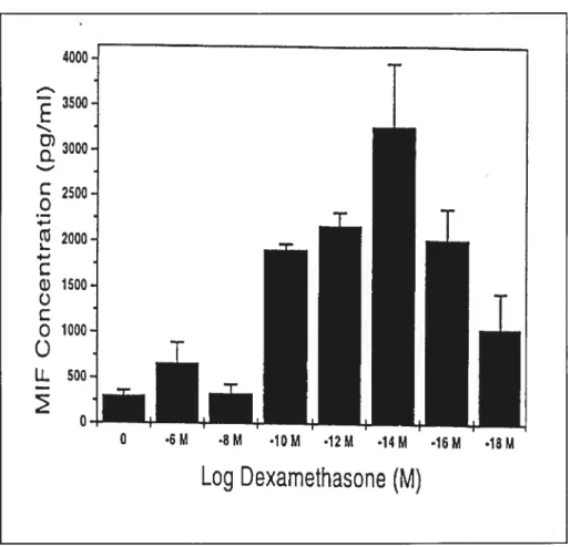
The information on the biological role of MW has corne from studies on MW and glucorticoid hormones. As rnentioned above, a key feature of the central stress response to infection and inflammation is the induction of endogenous glucocorticoid steroids. Once reÏeased, glucocorticoids are potent anti-inflammatory agents and immunosuppressants. As a mie cytokine expression is inhibited by steroids. By contrast, MW expression by monocytes/macrophages and T celis is induced rather than suppressed by glucocoticoids (Calandra, Bemhagen et al. 1995). MW release from pituitary ceils is aiso stimulated by glucocorticoid hormone, raising the intruiging possibility that steroid feedback regulation within the HPA axis acts both negatively, by inhibiting further ACTH release (via hypothalamic corticotropic releasing hormone), and positively, by stimulating MW secretion. Although flot