Three-dimensional cultured liver-on-a-Chip with mature
hepatocyte-like cells derived from human pluripotent stem cells
Ken-ichiro Kamei1 &Momoko Yoshioka1&Shiho Terada1&Yumie Tokunaga1&Yong Chen1,2Published online: 15 July 2019
# Springer Science+Business Media, LLC, part of Springer Nature 2019
Abstract
Liver-on-a-Chip technology holds considerable potential for applications in drug screening and chemical-safety testing. To establish such platforms, functional hepatocytes are required; however, primary hepatocytes are commonly used, despite prob-lems involving donor limitations, lot-to-lot variation, and unsatisfactory two-dimensional culture methods. Although human pluripotent stem cells (hPSCs) may represent a strong alternative contender to address the aforementioned issues, remaining technological challenges include the robust, highly efficient production of high-purity hepatic clusters. In addition, current Liver-on-a-Chip platforms are relatively complicated and not applicable for high-throughput experiments. Here, we develop a very simple Liver-on-a-Chip platform with mature and functional hepatocyte-like cells derived from hPSCs. To establish a method for hepatic differentiation of hPSCs, cells were first treated by inhibiting the phosphoinositide 3-kinase- and Rho-associated protein kinase-signaling pathways to stop self-renewal and improve survival, respectively, which enabled the formation of a well-defined endoderm and facilitated hepatocyte commitment. Next, a simple microfluidic device was used to create a three-dimensional (3D) culture environment that enhanced the maturation and function of hepatocyte-like cells by increasing the expression of both hepatic maturation markers and cytochrome P450. Finally, we confirmed improvements in hepatic functions, such as drug uptake/excretion capabilities, in >90% of 3D-matured hepatocyte-like cells by indocyanin green assay. These results indicated that the incorporation of hPSC-derived hepatocytes on our Liver-on-a-Chip platform may serve to enhance the processes involved in drug screening and chemical-safety testing.
Keywords Organ on a Chip . Pluripotent stem cells . Hepatocytes . Three-dimensional cell culture . Microfluidic device . Polydimethylsiloxane
1 Introduction
Organs-on-a-Chip (OoC) platforms hold considerable poten-tial for pre-clinical trials of drug development (Bhatia and Ingber 2014; Kimura et al. 2018; Marx et al. 2016; Ronaldson-Bouchard and Vunjak-Novakovic2018) as well
as toxicological tests (Ishida2018). To date, such testing has relied upon animal models; however, large challenges remain with regard to predicting the safety and efficacy of drug can-didates for human clinical use. Therefore, new in vitro models need to be developed as an alternative to animal models to predict treatment effects on humans with high predictability. OoCs represent the most suitable platforms to fulfill these requirements, as these platforms were developed to recapitu-late human physiological conditions in vitro. Currently, nu-merous OoC platforms have been reported as representative of organs, such as the brain (Park et al.2015), lung (Huh et al. 2010), heart (Zhang et al.2016), gut (Kim et al.2012), intes-tine (Trietsch et al.2017), liver (Beckwitt et al.2018; Gröger et al. 2018; Khetani and Bhatia 2008) or multiple tissues (Edington et al. 2018; Kamei et al.2017). Considering that the liver constitutes the largest organ in the human body and has many critical roles for body maintenance as well as drug metabolism and detoxification, we focused on optimizing the Liver-on-a-Chip (LC) platform, as the reported platform still
Electronic supplementary material The online version of this article (https://doi.org/10.1007/s10544-019-0423-8) contains supplementary material, which is available to authorized users.
* Ken-ichiro Kamei
1
Institute for Integrated Cell-Material Sciences (WPI-iCeMS), Kyoto University, Yoshida-Ushinomiya-cho, Sakyo-ku, Kyoto 606-8501, Japan
2
Ecole Normale Supérieure, CNRS-ENS-UPMC UMR 8640, 24 Rue Lhomond, 75005 Paris, France
retains two critical issues involving cell sources and cell cul-ture methods.
In terms of cell source, hepatocytes, which constitute the majority of cells in the liver, are mainly used for current drug screening and toxicological testing as well as LC platforms. Primary hepatocytes can be obtained from healthy donors; however, these cells cannot proliferate under standard cell-culture conditions and often experience loss of function. As an alternative, established cell lines are used for cell culture and in vitro drug testing, although these cells were often orig-inally harvested from tumors and do not represent healthy liver functions. Therefore, obtaining sufficient amounts of proper and functional hepatocytes remains challenging.
To meet this requirement, human pluripotent stem cells (hPSCs), such as embryonic (hESCs (Thomson1998)) and induced pluripotent stem cells (hiPSCs (Takahashi et al. 2007)) hold considerable potential as sources to generate he-patocytes. hPSCs exhibit two distinctive capabilities: unlimit-ed self-renewal without karyotypical abnormality and the abil-ity to differentiate into almost any cell type. These capabilities allow for the generation of hepatocyte quantities sufficient for subsequent applications such as LC platforms.
However, there remain issues with current methods related to the induction of hepatic differentiation from hPSCs to result in functional and mature hepatocytes. These methods involve either 1) formation of embryoid bodies or cell aggregates (Greenhough et al.2013), or 2) introduction of exogenous genes (e.g., HHEX (Inamura et al. 2011) and SOX17 (Takayama et al.2011)). Such methods have high potential to cause quality control difficulties involving contamination with other differentiated cells; moreover, the hepatic function-ality of the resultant cells still has considerable room for im-provement. Therefore, it is necessary to establish a novel method enabling the efficient and robust differentiation of hPSCs into functional hepatocytes.
In addition to cell sources, cell culture methods also require new innovation. Although two-dimensional (2D) culture of hepatocytes has been carried out for many years, this method is also problematic; for example, primary hepatocytes often lose their function under 2D culture. Therefore, a number of reports have suggested the use of three-dimensional (3D) cul-ture methods to render hepatocytes more functional (Schepers et al. 2016). Moreover, recent studies have shown that organoids (Asai et al.2017; Nantasanti et al.2015) hold prom-ise to obtain micro live tissues with greater functionality than those of 2D culture. However, the described cell aggregates and organoids exhibited varying size and structure as they are reliant upon only the capacity of the stem cells for tissue for-mation, resulting in low reproducibility. Therefore, these methods have yet to reach an acceptable stage for use in drug screening. Similarly, the majority of conventional LC plat-forms were based on 2D cell culture. Moreover, as most pre-viously developed LC platforms are both sophisticated and complicated and require special instruments to control the procedures, they are not user-friendly, limiting their use by general biology laboratories and their applicability for drug screening owing to the lack of high-throughput screening capability.
Here, we developed a novel LC platform by 1) establishing a protocol enabling the efficient differentiation of hepatocyte-like cells from hPSCs, resulting in high degrees of purity and reproducibility, and 2) applying a simple microfluidic 3D cell-culture platform (Kamei et al. 2016; Kamei et al. 2015), named 3D-LC platform (Fig.1). To facilitate efficient hepatic differentiation and cell survival, our method utilizes a cocktail of chemicals and growth factors. Conversely, our method does not require the use of exogenous genes, thereby eliminating concerns of genetic integration into host cells and variations in gene-delivery efficiency. After hepatic differentiation from hPSCs to hepatic progenitor cells, the cells are introduced into
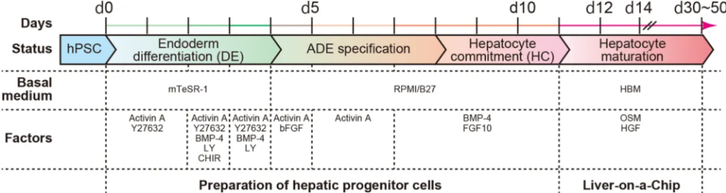
Fig. 1 The hepatic differentiation process from human pluripotent stem cells. A variety of solutions containing basal medium, growth factors, and chemicals were administered to hPSCs to efficiently induce differentiation to hepatocyte-like cells. Initially, hPSCs were treated with mTeSR-1 medium supplemented with combinations of Activin A, BMP-4, CHIR99021 (CHIR), LY294002 (LY), and Y27632 (Y) to induce definitive endoderm (DE) differentiation until Day 4 (d4). Then,
cells were treated with Activin A in RPMI medium supplemented with B27 for anterior definitive endoderm (ADE) specification until Day 8. As the next step, cells were treated with BMP-4 and FGF-10 for inducing hepatocyte commitment in RPMI medium supplemented with B27 until Day 11. Finally, cells were treated with oncostatin M (OSM) and hepatocyte growth factor (HGF) in hepatocyte basal medium for hepatocyte maturation
our simple microfluidic 3D cell culture device to prepare our 3D-LC platform with hPSC-derived hepatocyte-like cells of high-maturation status. Notably, this platform does not require any special instruments, with standard pipettes able to be used to carry out the experiments.
2 Experimental
2.1 hPSC culture
hESCs were used according to the guidelines of the ethnical committee of Kyoto University. H9 hESCs were purchased from WiCell Research Institute (Madison, WI, USA). Prior to culturing, hESC-certified Matrigel (Corning, Corning, NY, USA) was diluted with Dulbecco’s modified Eagle me-dium (DMEM)/F12 meme-dium (Sigma-Aldrich, St. Louis, MO, USA) at a 1:75 (v/v) ratio and coated onto a culture dish. Matrigel was incubated in a dish for 24 h at 4 °C. Then, excess Matrigel was removed and the coated dish was washed with fresh DMEM/F12 medium.
mTeSR-1 defined medium (Stem Cell Technologies, Vancouver, Canada) was used for daily culturing of hPSCs. For passaging, cells were dissociated with TrypLE Express (Life Technologies, Carlsbad, CA, USA) for 3 min at 37 °C and harvested. A cell strainer was used to remove undesired cell aggregates from the cell suspension, and cells were cen-trifuged at 200×g for 3 min and resuspended in mTeSR-1 medium. Cells were counted using a NucleoCounter NC-200 (Chemetec, Baton Rouge, LA, USA). mTeSR-1 medium containing 10μM of the ROCK inhibitor Y-27632 (Wako, Osaka, Japan) was used to prevent apoptosis of dissociated hPSCs on day 1. mTeSR-1 medium without the ROCK inhib-itor was used on subsequent days, with daily medium changes in a humidified incubator at 37 °C with 5% CO2for 4 to
5 days.
2.2 Hepatic differentiation from hPSCs
Prior to inducing differentiation, a cell-culture dish was coated with 0.1% gelatin in phosphate-buffered saline (PBS) at 25 °C for 30 min. The gelatin solution was then aspirated and DMEM/F12 medium (Sigma-Aldrich) supplemented with 10% (v/v) fetal bovine serum (FBS), penicillin/streptomycin, and 100μM β-mercaptoethanol (Sigma-Aldrich) was intro-duced onto the culture dish for serum coating at 37 °C for 24 h. The coated dish was then rinsed with fresh medium.
To induce endoderm differentiation, cultured hPSCs were washed with PBS and treated with TryPLE Express at 37 °C for 5 min, followed by the addition of basal medium and transfer of the cell suspension into a 15-mL tube. Cells were centrifuged at 200×g for 3 min and the supernatant was re-moved. Cells were resuspended in mTeSR-1 medium
supplemented with 10μM Y27632 and 100 ng mL−1activin A, plated on a serum-coated culture dish, and cultured in a humidified incubator at 37 °C with 5% CO2for 24 h. At the
end of day 1, the medium was replaced with fresh mTeSR-1 medium supplemented with 10μM Y27632 and 100 ng mL−1 activin A and cultured for another 24 h. On day 2, the medium was replaced with mTeSR-1 medium supplemented with 10μM Y27632, 100 ng mL−1activin A, 10 ng mL−1 BMP-4, 10μM LY294002, and 3 μM CHIR99021, and cells were incubated for 24 h. On day 3, medium was replaced with mTeSR-1 medium supplemented with 10 μM Y27632, 100 ng mL−1activin A, 10 ng mL−1BMP-4, and 10 μM LY294002, and cells were incubated for 24 h. On day 4, me-dium was replaced with Roswell Park Memorial Institute ( R P M I ) m e d i u m s u p p l e m e n t e d w i t h B - 2 7 ( L i f e Technologies), 100 ng mL−1 activin A, and 100 ng mL−1 bFGF, and cells were incubated for 24 h.
To induce ADE specification, cells were treated with RPMI medium supplemented with 50 ng mL−1activin A, with daily medium changes for 3 days. Cells were then treated with RPMI medium supplemented with 20 ng mL−1BMP-4 and 10 ng mL−1FGF-10, with daily medium changes for 4 days. On day 12, the medium was replaced with hepatocyte basal medium (Lonza, Basel, Switzerland) supplemented with 30 ng mL−1oncostatin M and 50 ng mL−1hepatocyte growth factor to induce maturation of the differentiated hepatocytes. Cells were incubated at 37 °C, and medium was changed every 2 days.
2.3 HepG2 cell culture
HepG2 cells were provided by American Type Culture Collection (Manassas, VA, USA). HepG2 cells were cultured with DMEM supplemented with 10% (v/v) FBS, 1% penicillin/ streptomycin, and 1 mM nonessential amino acids, and the me-dium was changed every 2 to 3 days. The cells were passaged with trypsin-EDTA solutions at a 1:10 to 1:20 subculture ratio.
2.4 Flow cytometry
Cells were harvested with TrypLE Express and rinsed with PBS twice prior to cell counting. For staining with antibodies, cells were diluted to a final concentration of 1 × 10⁷cells mL−1in PBS supplemented with 2% fetal calf serum (FCS). Fluorescence-labeled antibodies were added and incubated at room temperature for 30 min. As a negative control, specific isotype controls were used. After removing excess antibodies by centrifugation at 300×g for 5 min, cells were washed with PBS containing 2% FCS, and cell suspensions were applied to a FACS Canto II (BD Biosciences, Franklin Lakes, NJ, USA) for flow cytometric analysis. Data analysis was performed using FlowJo software (v9; FlowJo, LLC, Ashland, OR, USA).
2.5 RT-PCR
Total RNA was purified using an RNeasy Mini Kit (Qiagen, Hilden, Germany), and 1μg of total RNA was reverse tran-scribed to generate cDNA using PrimeScript RT master mix (Perfect Real Time; TaKaRa Bio, Shiga, Japan). A reaction mixture (25 μL) containing 20 ng cDNA, 0.2 μM PCR primers (Supplementary Table S1), and 5 U of Taq DNA polymerase (TaKaRa Bio) was subjected to PCR using a ther-mal cycler (Applied Biosystems 7300 real-time PCR system; Applied Biosystems, Foster City, CA, USA). PCR was per-formed with 30 to 35 cycles (94 °C for 30 s, 58 °C for 30 s, and 72 °C for 60 s). PCR products (10μL) were electropho-resed on 1.2% agarose gels and visualized by GelRed nucleic acid staining (Biotium, Fremont, CA, USA).
2.6 Immunocytochemistry
Cells were fixed with 4% paraformaldehyde in PBS for 20 min at 25 °C and then permeabilized with 0.5% Triton X-100 in PBS for 16 h at 25 °C. Subsequently, cells were blocked in PBS (5% normal goat serum, 5% normal donkey serum, 3% bovine serum albumin, 0.1% Tween-20) at 4 °C for 16 h and then incubated at 4 °C for 16 h with the primary antibody [anti-human A1AT rabbit IgG, 1:800; Dako, Glostrup, Denmark] in PBS with 0.5% Triton X-100. Cells were then incubated at 37 °C for 60 min with a secondary antibody (AlexaFluor 488 Donkey anti-rabbit IgG, 1:1000; Jackson ImmunoResearch, West Grove, PA, USA) in blocking buffer prior to a final incubation with 4′,6-diamidino-2-phenylindole (DAPI) at 25 °C for 30 min.
2.7 Image acquisition
The sample containing cells was placed on the stage of a Nikon ECLIPSE Ti inverted fluorescence microscope equipped with a CFI plan fluor 10×/0.30 N.A. objective lens (Nikon, Tokyo, Japan), CCD camera (ORCA-R2; Hamamatsu Photonics, Hamamatsu City, Japan), mercury lamp (Intensilight; Nikon), XYZ automated stage (Ti-S-ER motor-ized stage with encoders; Nikon), and filter cubes for fluores-cence channels (DAPI and GFP HYQ; Nikon). For image acquisition, the exposure times were set at 500 ms for DAPI and 500 ms for GFP HYQ (for A1AT).
2.8 Indocyanin green uptake/excretion assay
Briefly, 1 mg mL−1Indocyanin green (ICG) was dissolved in hepatocyte-maturation medium and cells were treated with ICG solution for 1 h, rinsed with hepatocyte-maturation me-dium, and then observed using a bright-field microscope (Olympus, Tokyo, Japan). After 24 h, cells were observed again to visualize excretion capability.
2.9 Microfluidic device fabrication
A microfluidic device was fabricated using stereolithographic 3D-printing techniques and solution cast-molding processes (Kamei et al.2015). The mold for the microfluidic channels was produced using a 3D printer (Keyence Corporation, Osaka, Japan). Sylgard 184 PDMS two-part elastomer (10:1 ratio of pre-polymer to curing agent; Dow Corning Corporation, Midland, MI, USA) was mixed, poured into a 3D-printed mold to produce a 5-mm-thick PDMS layer, and de-gassed by using a vacuum desiccator. The PDMS material was then cured in an oven at 65 °C for 48 h. After curing, the PDMS form was removed from the mold, trimmed, and cleaned. The PDMS form and a glass dish or plastic plate were corona-plasma-treated (Kasuga Denki, Inc., Kawasaki, Japan) and bonded together by baking in an oven at 80 °C.
2.10 Preparation of the liver-on-a-Chip platform
Prior to use, a microfluidic 3D cell-culture device was steril-ized in 70% ethanol and placed under ultraviolet light in a biosafety cabinet for 30 min. A microfluidic cell-culture chamber was then coated with Corning Matrigel hESC-qualified matrix (Corning) diluted to 1:75 (v/v) with DMEM/F12 (Sigma-Aldrich). After a 1-day incubation at 4 °C, excess Matrigel was removed, and the coated dish was washed with fresh DMEM/F12. Cells were harvested using trypsin and collected in a 15-mL tube. Following centrifuga-tion, cells were suspended in hepatocyte-maturation medium and introduced into a microfluidic device via a cell inlet. The microfluidic device was placed in a humidified incubator at 37 °C with 5% CO2 atmosphere, and the medium was
changed daily.
3 Results and discussion
3.1 Cell survival and robustness
of definitive-endoderm (DE) differentiation is
enhanced by phosphoinositide 3-kinase (PI3K)
and rho-associated protein kinase (ROCK) inhibition
Induction of DE differentiation is the most critical step re-quired to promote further differentiation (Fig.2). In this pro-cess, transforming growth factor (TGF)-β (Basma et al.2009; Hay et al.2008), bone morphogenetic protein (BMP)-4 (Rossi 2001), β-catenin (Bone et al. 2011) and basic fibroblast growth factor (bFGF) (Basma et al.2009)-signaling pathways play critical roles. Moreover, the PI3K-signaling pathway is critical for the maintenance of hPSC self-renewal (Huang et al. 2015), with inhibition of this pathway resulting in hPSC differentiation. Previously, Hannan et al. established a method to effectively achieve hPSC differentiation to DE
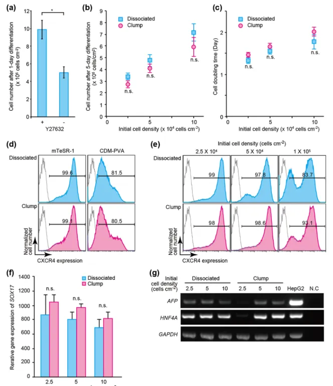
Fig. 2 Combination of ROCK (10μM Y27632) and PI3K (10 μM LY294002) inhibitors facilitates survival and inhibits self-renewal of hPSCs during definitive-endoderm (DE) differentiation. (a) 1-day treatment with mTeSR-1 basal medium in combination with Y27632 increases cell survival of H9 human embryonic stem cells (hESCs). Data represent the means ± standard deviation (n = 3). (b, c) Both dissociated and clumped H9 hESCs treated with mTeSR-1/Y27632 were able to proliferate during DE differentiation (b) with similar doubling times (c). Data represent the means ± standard deviation (n = 3).“n.s.” represents “not significant”. (d) Selection of mTeSR-1 or CDM-PVA medium to induce differentiation of dissociated or clumped H9 hESCs into DE by monitoring the expression levels of the CXCR4 DE marker. Cells treated with mTeSR-1 medium supplemented with activin
A, BMP-4, LY294002, and CHIR99021, along with Y27632 showed a more homogeneous cell population with higher CXCR4 expression. (e) Initial cell density influences the cellular heterogeneity during DE differentiation, as determined by CXCR4 expression. (f) Initial cell density is inversely correlated with expression levels of the SOX17 DE marker. At low initial cell density, SOX17 expression levels varied among experiments. Data represent the means ± standard deviation (n = 3).“n.s.” represents“not significant”. (g) Representative gel electrophoresis of RT-PCR samples to ascertain expression of α-fetoprotein (AFP) and hepatocyte nuclear factor 4α (HNF4A) differentiation marker expression on day 12. GAPDH, a housekeeping gene, was used as an internal control. Data were obtained from three independent experiments. N.C. represents a negative control (water)
using a cocktail of activin A, BMP-4, bFGF, LY294002, and CHIR99021, but without the use of exogenous genes (Hannan et al.2013). However, this method results in massive cell death and inefficient and irreproducible outcomes. Furthermore, the use of hPSC aggregates is unlikely to allow improved robustness, owing to the challenge associated with size control, which is a critical factor for lineage determination during hPSC differentiation. Although the use of dissociated hPSCs may improve robustness, the related processes contin-ue to result in large amounts of cell death.
Therefore, we used a ROCK inhibitor (Y27632) that has previously been used to facilitate the survival of dissociated self-renewing hPSCs (Watanabe et al.2007). We hypothe-sized that administration of a ROCK inhibitor would also facilitate cell survival during the early stage of DE differenti-ation. For the medium, we modified mTeSR-1 defined medi-um (Ludwig et al.2006) containing BMP-4 and bFGF (Fig.1 and Supplementary TableS1), which are also commonly used as key factors to introduce DE differentiation. We observed that hPSCs showed unstable behavior following introduction of the new culture conditions; therefore, for establishing ro-bust differentiation protocols, the initial use of self-renewing culture medium was beneficial, followed by gradually chang-ing to the differentiation medium. Toward this end, activin A, BMP-4, LY294002, and CHIR99021 were subsequently added into the mTeSR-1 medium for DE differentiation.
To confirm increases in cell survival during DE differenti-ation, we counted H9 hESCs following 1 day of treatment (Fig.2a). Cells treated with both 10μM Y27632 and 10 μM LY294002 showed significant increases in cell number, as compared with levels observed in cells treated with only LY294002. This result indicated that inhibition of the ROCK-signaling pathway improved cell survival to a degree greater than that observed following inhibition of the PI3K-signaling pathway.
We then investigated differences in outcomes based on initiating differentiation from either dissociated or clumped hPSCs according to cell number and doubling time (Fig. 2b, c). We observed that following a 5-day incubation with 10μM Y27632 and 10 μM LY294002 and DE differentiation, both dissociated and clumped hESCs increased cell numbers with similar doubling times. These results suggested that Y27632 improves cell survival even during DE differentia-tion, but not cell proliferation.
To confirm cell status following DE differentiation, the expression of C-X-C-motif chemokine receptor-4 (CXCR4), a cell surface DE-differentiation marker, was quantitatively analyzed by flow cytometry (Fig. 2d) and compared with levels measured in previously-reported chemically defined medium (CDM-PVA) (Hannan et al.2013) (Supplementary TableS2). H9 hESCs treated with mTeSR-based medium showed higher degrees of purity and enhanced CXCR4 ex-pression (99.6%) compared to those of cells cultured in
CDM-PVA medium (81.5%). Then, we investigated the effects of initial cell-seeding density on the induction of DE differentia-tion (Fig.2e). During 5-day DE differentiation, cell numbers in all cell densities increased approximately 10 folds (Fig.2b); however, the increase in initial cell number revealed bimodal cell populations with regard to CXCR4 expression (Fig.2e). These results indicated that the use of a smaller initial cell-seeding density resulted in a more homogeneous cell popula-tion with higher CXCR4 expression.
We also measured expression of the sex-determining re-gion Y box-17 (SOX17) transcription factor and endoderm-lineage marker by quantitative reverse transcription-polymerase chain reaction (RT-PCR) (Fig.2f). The use of cell clumps resulted in higher SOX17 expression as compared with using dissociated cells, with SOX17 expression subsequently decreasing along with increasing cell numbers, although low-er initial cell-seeding density (2.5 × 10⁴cells cm−2) resulted in large variations in SOX17 expression. We also confirmed he-patocyte differentiation by monitoring the expression of α-fetoprotein (AFP) and hepatocyte nuclear factor 4α (HNF4A differentiation marker mRNA on day 12 (Fig.2g). The use of cell aggregates with lower initial cell-seeding density (2.5 × 10⁴cells cm−2) did not result in AFP and HNF4A expression. Based on these results, we used 5 × 10⁴cells cm−2for further differentiation protocols consequent to our observation of higher purity and improved robustness at higher initial cell densities.
3.2 Matured hPSC-derived hepatocyte-like cells
in the LC platfrom
To facilitate maturation levels of hPSC-derived hepatocytes, we developed a simple 3D-culture method using a microfluidic device. We hypothesized that 3D-culture condi-tions would provide a more suitable environment for hepato-cytes as compared with conventional 2D-culture methods (Kruitwagen et al.2017) toward mimicking 3D micro liver tissues (Fig.3a). Previously, we reported a microfluidic device enabling 3D culture of hPSCs (Kamei et al.2016; Kamei et al. 2015) (Fig.3b), which was utilized in the present study to perform 3D culturing of hepatocyte-like cells from day 12 to enhance the maturation process. The microfluidic device was made of polydimethylsiloxane (PDMS), which exhibits good biocompatibility and gas permeability (Kamei et al.2016), and contained two microfluidic 3D cell-culture chambers [10 mm (L) × 1.5 mm (W) × 150 μm (H)] with a cell inlet (0.75-mm diameter) and a medium reservoir (3-mm diame-ter). To confirm the maturity of hPSC-derived hepatocyte-like cells at the protein level, we conducted immunocytochemistry for one of the maturation markers, α1 anti-trypsin (A1AT). Our results indicated that the A1AT hepatocyte-maturation marker was more strongly expressed in 3D-cultured hepato-cyte-like cells derived from H9 hESCs over 33 days using our
method as compared with levels observed in 2D-cultured cells (Fig.3c). Although undifferentiated H9 hESCs did not press hepatic genes, we also confirmed increased mRNA ex-pression of other hepatocyte-marker genes, such as UGT1A1, CYP3A4, CYP2A9, CYC2C19, and CYP2D6, in the 3D-cultured H9 hESC-derived hepatocyte-like cells relative to levels observed in 2D-cultured cells (Fig. 3d and Supplementary TableS3). In contrast, HepG2 cells did not express UGT1A1, TDO1, MDR/TAP, CYP3A4, CYP2A9, CYP2C19, or CYP2D6. As expected, the liver extract from the healthy donor strongly expressed all tested genes at levels exceeding those of hepatocyte-like cells derived from H9
hESCs. These results indicated that the hepatocyte-like cells derived from H9 hESCs exhibited improved gene expression relative to a HepG2 cell line and 2D-cultured hepatocyte-like cells, but were unable to match levels observed in liver ex-tracts from a healthy donor.
To investigate differences in functionality, an ICG uptake/ excretion assay, which is a non-invasive marker of drug uptake/excretion in the liver (Takayama et al.2012), was per-formed on cells from both 3D- and 2D cultures (Fig. 3e). Notably, 3D-cultured cells showed greater ICG uptake within 1 h compared to that of 2D-cultured cells, with most cells also effectively excreting the ICG after 24 h. These results
Fig. 3 Simple 3D-LC platform with matured hPSC-derived hepatocyte-like cells. (a) Illustration of 2D and 3D-LC platform for hepatocyte maturation. (b) Photograph of a microfluidic 3D-culture device (Kamei et al.2016; Kamei et al.2015) for the 3D-LC platform and the structure of a microfluidic cell-culture chamber [10 mm (L) × 1.5 mm (W) × 150μm (H)] with a cell inlet (0.75-mm diameter) and medium reservoir (3-mm diameter). (c) Immunocytochemistry to visualize the hepatocyte-maturation markerα1 anti-trypsin (A1AT) in H9 hESC-derived hepatocyte-like cells.“2D” and “3D” represent hepatocyte-like cells derived from H9 hESCs cultured in a conventional 35-mm culture dish and a microfluidic 3D cell-culture device, respectively. Scale bar represents 20μm. (d) Representative gel electrophoresis of RT-PCR
products for mRNAs associated with the cytochrome P450 family (e.g., CYP3A4, CYP1A1, CYP2A9, CYP2C19, and CYP2D6), ATP-binding cassette (ABC) transporters (MRP2 and MDR/TAP), hepatocyte-maturation markers (Albumin, UGT1A1, A1AT, and TDO1), and endoderm markers (AFP and HNF4A), obtained from independently triplicated experiments. HepG2 hepatocellular carcinoma cells were used as a control, and a liver extract from a healthy donor was used as a positive control. H9_Hep and Undif_H9 represent hepatocyte-like cells derived from H9 hESCs and undifferentiated H9 hESCs, respectively. (e) Microphotographs of 2D- and microfluidic 3D-hepatocyte-like cells treated with indocyanine green (ICG) for 1 h to visualize ICG uptake and then excretion after 24 h. Scale bar represents 500μm
indicated that hPSC-derived hepatocyte-like cells in the 3D-LC appeared more mature and functional, compared with those of 2D-cultured cells.
4 Conclusions
In this study, we developed a 3D-LC platform with matured hPSC-derived hepatocyte-like cells. In particular, we firstly established an efficient and robust method to induce hPSC differentiation into functional hepatocyte-like cells. By opti-mizing the basal medium, combination of chemicals, and ini-tial cell-seeding density for endoderm differentiation, we ob-tained a homogeneous population of endoderm-differentiated cells highly expressing CXCR4 from dissociated hPSCs. Additionally, we optimized the periods required for ADE specification and HC according to quantitative PCR analysis. To our knowledge, this represents the first demonstration of application of a microfluidic 3D cell-culture platform for the maturation of hepatocyte-like cells.
LC platforms offer critical opportunities for drug screening and chemical-safety testing in a variety of industries including cosmetics and agriculture. However, there are limited num-bers of cell sources (e.g., primary hepatocytes) available, with the number and quality varying by donor. Although current hepatocyte cell lines, such as HepG2, have been used as alter-natives, they often show different characteristics from primary hepatocytes. In addition, current 2D-culture conditions in a chip do not allow the expression of proper hepatic functions. In this study, we successfully developed a LC platform by establishing a method to induce hPSC differentiation into ma-ture hepatocytes with microfluidic 3D culma-ture to prepare func-tional hepatocyte-like cells on a chip.
LC platforms may be used for either“High-content analy-sis (HCA)” or “High-throughput screening (HTS)”. Although a number of LC platforms have been previously reported, these often provided less information than that of current HCA, are exceedingly complicated for general users, and not applicable for HTS. For drug screening and chemical-safety testing platforms, it is necessary to decide which appli-cation would be most suitable at the beginning of device de-velopment. Our device and cell culture chamber are very sim-ple and offer easy handling without requiring any special in-struments. They can be applicable for increased throughput by means of straightforward device format design, such as the use of microplates, following the recommendations of the rec-ommendation of the Society for Biomolecular Screening (SBS). Accordingly, we consider that our LC platform will fulfill current requirements and serve as a useful tool in the field of drug discovery.
Acknowledgements Funding was generously provided by the Japan Society for the Promotion of Science (JSPS; 24656502, 26560209,
16K14660, and 17H02083). Funding was also provided by the Terumo Life Science Foundation and Japan Agency for Medical Research and Development. The WPI-iCeMS is supported by the World Premier International Research Centre Initiative (WPI), MEXT, Japan.
Compliance with ethical standards
Competing financial interests Kyoto University (K.K. and M.Y.) filed a provisional Japanese patent application based on the research presented herein. The other authors have no conflict of interest.
References
A. Asai, E. Aihara, C. Watson, R. Mourya, T. Mizuochi, P. Shivakumar, K. Phelan, C. Mayhew, M. Helmrath, T. Takebe, J. Wells, J.A. Bezerra, Development 144, 1056 (2017)
H. Basma, A. Soto–Gutiérrez, G.R. Yannam, L. Liu, R. Ito, T. Yamamoto, E. Ellis, S.D. Carson, S. Sato, Y. Chen, D. Muirhead, N. Navarro– Álvarez, R.J. Wong, J. Roy–Chowdhury, J.L. Platt, D.F. Mercer, J.D. Miller, S.C. Strom, N. Kobayashi, I.J. Fox, Gastroenterology 136, 990 (2009)
C.H. Beckwitt, A.M. Clark, S. Wheeler, D.L. Taylor, D.B. Stolz, L. Griffith, A. Wells, Exp. Cell Res. 363, 15 (2018)
S.N. Bhatia, D.E. Ingber, Nat. Biotechnol. 32, 760 (2014)
H.K. Bone, A.S. Nelson, C.E. Goldring, D. Tosh, M.J. Welham, J. Cell Sci. 124, 1992 (2011)
C.D. Edington, W.L.K. Chen, E. Geishecker, T. Kassis, L.R. Soenksen, B.M. Bhushan, D. Freake, J. Kirschner, C. Maass, N. Tsamandouras, J. Valdez, C.D. Cook, T. Parent, S. Snyder, J. Yu, E. Suter, M. Shockley, J. Velazquez, J.J. Velazquez, L. Stockdale, J.P. Papps, I. Lee, N. Vann, M. Gamboa, M.E. LaBarge, Z. Zhong, X. Wang, L.A. Boyer, D.A. Lauffenburger, R.L. Carrier, C. Communal, S.R. Tannenbaum, C.L. Stokes, D.J. Hughes, G. Rohatgi, D.L. Trumper, M. Cirit, L.G. Griffith, Sci. Rep. 8, 4530 (2018)
S. Greenhough, H. Bradburn, J. Gardner, D.C. Hay, Cell. Reprogram. 15, 9 (2013)
M. Gröger, J. Dinger, M. Kiehntopf, F.T. Peters, U. Rauen, A.S. Mosig, Adv. Healthc. Mater. 7, 1700616 (2018)
N.R.F. Hannan, C.-P. Segeritz, T. Touboul, L. Vallier, Nat. Protoc. 8, 430 (2013)
D.C. Hay, J. Fletcher, C. Payne, J.D. Terrace, R.C.J. Gallagher, J. Snoeys, J.R. Black, D. Wojtacha, K. Samuel, Z. Hannoun, A. Pryde, C. Filippi, I.S. Currie, S.J. Forbes, J.A. Ross, P.N. Newsome, J.P. Iredale, Proc. Natl. Acad. Sci. 105, 12301 (2008)
T.-S. Huang, L. Li, L. Moalim-Nour, D. Jia, J. Bai, Z. Yao, S.A.L. Bennett, D. Figeys, L. Wang, Stem Cells 33, 1419 (2015) D. Huh, B.D. Matthews, A. Mammoto, M. Montoya-Zavala, H.Y. Hsin,
D.E. Ingber, Science 328, 1662 (2010)
M. Inamura, K. Kawabata, K. Takayama, K. Tashiro, F. Sakurai, K. Katayama, M. Toyoda, H. Akutsu, Y. Miyagawa, H. Okita, N. Kiyokawa, A. Umezawa, T. Hayakawa, M.K. Furue, H. Mizuguchi, Mol. Ther. 19, 400 (2011)
S. Ishida, Drug Metab. Pharmacokinet. 33, 49 (2018)
K. Kamei, Y. Mashimo, Y. Koyama, C. Fockenberg, M. Nakashima, M. Nakajima, J. Li, Y. Chen, Biomed. Microdevices 17, 36 (2015) K. Kamei, Y. Koyama, Y. Tokunaga, Y. Mashimo, M. Yoshioka, C.
Fockenberg, R. Mosbergen, O. Korn, C. Wells, Y. Chen, Adv. Healthc. Mater. 5, 2951 (2016)
K. Kamei, Y. Kato, Y. Hirai, S. Ito, J. Satoh, A. Oka, T. Tsuchiya, Y. Chen, O. Tabata, RSC Adv. 7, 36777 (2017)
S.R. Khetani, S.N. Bhatia, Nat. Biotechnol. 26, 120 (2008)
H. Kimura, Y. Sakai, T. Fujii, Drug Metab. Pharmacokinet. 33, 43 (2018) H.S. Kruitwagen, L.A. Oosterhoff, I.G.W.H. Vernooij, I.M. Schrall, M.E. van Wolferen, F. Bannink, C. Roesch, L. van Uden, M.R. Molenaar, J.B. Helms, G.C.M. Grinwis, M.M.A. Verstegen, L.J.W. van der Laan, M. Huch, N. Geijsen, R.G. Vries, H. Clevers, J. Rothuizen, B.A. Schotanus, L.C. Penning, B. Spee, Stem Cell Reports 8, 822 (2017)
T.E. Ludwig, V. Bergendahl, M.E. Levenstein, J. Yu, M.D. Probasco, J.A. Thomson, Nat. Methods 3, 637 (2006)
U. Marx, T.B. Andersson, A. Bahinski, M. Beilmann, P. Vulto, J. Wang, J. Wiest, M. Rodenburg, A. Roth, J.A.M. Adrienne, Sips 10 (2016) S. Nantasanti, A. De Bruin, J. Rothuizen, L.C. Penning, B.A. Schotanus,
Stem Cells Transl. Med. 5, 325 (2015)
J. Park, B.K. Lee, G.S. Jeong, J.K. Hyun, C.J. Lee, S.-H. Lee, Lab Chip 15, 141 (2015)
K. Ronaldson-Bouchard, G. Vunjak-Novakovic, Cell Stem Cell 22, 310 (2018)
J.M. Rossi, Genes Dev. 15, 1998 (2001)
A. Schepers, C. Li, A. Chhabra, B.T. Seney, S. Bhatia, Lab Chip 16, 2644 (2016)
K. Takahashi, K. Tanabe, M. Ohnuki, M. Narita, T. Ichisaka, K. Tomoda, S. Yamanaka, Cell 131, 861 (2007)
K. Takayama, M. Inamura, K. Kawabata, K. Tashiro, K. Katayama, F. Sakurai, T. Hayakawa, M.K. Furue, H. Mizuguchi, PLoS One 6, e21780 (2011)
K. Takayama, M. Inamura, K. Kawabata, M. Sugawara, K. Kikuchi, M. Higuchi, Y. Nagamoto, H. Watanabe, K. Tashiro, F. Sakurai, T. Hayakawa, M.K. Furue, H. Mizuguchi, J. Hepatol. 57, 628 (2012) J.A. Thomson, Science 282, 1145 (1998)
S.J. Trietsch, E. Naumovska, D. Kurek, M.C. Setyawati, M.K. Vormann, K.J. Wilschut, H.L. Lanz, A. Nicolas, C.P. Ng, J. Joore, S. Kustermann, A. Roth, T. Hankemeier, A. Moisan, P. Vulto, Nat. Commun. 8, 262 (2017)
K. Watanabe, M. Ueno, D. Kamiya, A. Nishiyama, M. Matsumura, T. Wataya, J.B. Takahashi, S. Nishikawa, S. Nishikawa, K. Muguruma, Y. Sasai, Nat. Biotechnol. 25, 681 (2007)
Y.S. Zhang, A. Arneri, S. Bersini, S.-R. Shin, K. Zhu, Z. Goli-Malekabadi, J. Aleman, C. Colosi, F. Busignani, V. Dell’Erba, C. Bishop, T. Shupe, D. Demarchi, M. Moretti, M. Rasponi, M.R. Dokmeci, A. Atala, A. Khademhosseini, Biomaterials 110, 45 (2016)
Publisher’s note Springer Nature remains neutral with regard to jurisdictional claims in published maps and institutional affiliations.