THÈSE
En vue de l’obtention du
DOCTORAT DE L’UNIVERSITÉ DE TOULOUSE
Délivré par l'Université Toulouse 3 - Paul Sabatier
Présentée et soutenue par
Thomas VIAL
Le 29 Juin 2020
Le virus de la dengue détourne le métabolisme des
phospholipides du moustique pour sa réplication
Ecole doctorale : BSB - Biologie, Santé, Biotechnologies Spécialité : MICROBIOLOGIE
Unité de recherche :
PHARMA-DEV -Laboratoire Pharmacochimie et Pharmacologie pour le Développement
Thèse dirigée par
Eric DEHARO
Jury
M. Louis Lambrechts, Rapporteur M. Jean-Luc Imler, Rapporteur Mme Isabelle Morlais, Examinatrice M. Jean-Charles Portais, Examinateur
M. Eric Deharo, Directeur de thèse M. Julien Pompon, Co-directeur de thèse
2
Dengue virus diverts the mosquito phospholipid
metabolism for effective infection
Thomas Vial
École Doctorale BSB – Biologie, Santé, Biotechnologies Université Toulouse 3 - Paul Sabatier
3
ACKNOWLEDGMENTS
These four years spent on this project have been intense, first by being based in Laos and travelling to Singapore and Toulouse to initiate the project, and then full time in Singapore. I would like to thank here all the people who contributed in any way to the realization of this thesis work in the best conditions.
First of all, I would like to thank Eric Deharo, my thesis director, who allowed me to be part of this incredible experience in Laos four years ago, by opening the doors of the IRD to me and get back to research work. Thank you for your confidence on all these different projects and especially for proposing me this research project by sending me to Singapore. You pushed me to enroll a thesis, which was not necessarily in my plans at the beginning.
I would also like to thank the person who supported and accompanied me tirelessly throughout this research project, my thesis co-director Julien Pompon. Thank you welcoming me at Duke-NUS and for allowing me to turn those valuable preliminary data into a thesis project. I do not count the precious advices, rereading, Skype meeting to lead to the big picture and to get the right message. Your availability and your support on the analysis and writing of the papers and the manuscript were essential.
I particularly thank Mariano Garcia-Blanco for having done me the honor of joining his laboratory and for having taken advantage of his always brilliant remarks and advice. Thanks also to Linfa Wang for his welcome to the EID department.
Of course, I warmly thank the members of the MGB lab team. Thanks to Shi-Chia, Mayra and KC for bringing me their experience and advices. Thanks to Vanessa and Ben for their technical support and especially to Wei Lian for helping me a lot, especially at the end of the thesis for many experiments. Thanks to Menchie for training me in insectary and for providing me with a lot of mosquitoes. I can't count the hours spent sorting female mosquitoes, dissecting midgut or testicles in the heat and humidity.
I would like to thank Guillaume Marti for training and coaching me on the metabolomics part and for welcoming me during my stays in Toulouse. These numerous samples and long hours of mass spectrometry provided a considerable amount of data. I would like to thank Nicolas Fabre for his welcome at Pharmadev and the members of UMR-152 whom I was able to meet briefly. Thanks to Franck for helped me during my stays in the lab and for received my numerous shipments of samples from Singapore.
Thanks to the members of the thesis committee, Mariano, Eng Eong Ooi and Jean-Paul Kovalic, for helping me to share my results at important moments and to oriented me on the project.
Finally, I would like to thank my family and friends who gave me their support despite the distance. First of all, my parents and my sister, who encouraged me from the beginning, when I left France. My friends and the many discussions on WhatsApp that kept me connected. Finally, I would like to thank my wife, Melanie, the first to support me, without whom it would have been difficult to move forward in the same way and with whom I look forward to sharing the next adventures.
4
ABSTRACT
More than half of the world population is at risk of dengue virus (DENV) infection because of the global distribution of its mosquito vectors. There is neither effective vaccine nor therapeutics. The only available strategy relies on insecticides, against which mosquitoes are developing resistance. Viruses utilize the host metabolome for replication and dissemination. This is particularly true for envelope viruses like DENV that relies on host lipid membranes to complete their life-cycle. To reach an optimal metabolic environment, viruses subvert the host metabolome. Understanding DENV-mosquito metabolic interactions will reveal novel strategies to stop DENV transmission. Here, we characterized how DENV hijacks the Aedes aegypti mosquito lipidome to identify targets for novel transmission-blocking interventions. To describe metabolic changes throughout the mosquito DENV cycle, we deployed a Liquid chromatography–high resolution mass spectrometry (LC-HRMS) workflow at different stages of vector infection. We revealed a major phospholipid reconfiguration throughout the DENV mosquito cycle, in cells, midguts, and whole mosquitoes. To decipher how DENV reconfigures phospholipids, we phylogenetically characterized acylglycerolphosphate acyltransferase (AGPAT) enzyme isoforms and identified those (i.e., AGPAT1) that catalyze a central rate-limiting step in phospholipid biogenesis. We found that DENV infection decreased AGPAT1 expression, which depletion enhances infection by maintaining high aminophospholipid (aminoPL) concentrations, especially phosphatidylcholine (PC) and phosphatidylethanolamine (PE), during DENV mosquito cycle. By demonstrating that DENV-mediated AGPAT1 downregulation provides a proviral environment, these results reveal the first metabolic host factor in mosquitoes and emphasize the role of aminophospholipids in DENV cellular cycle. We then undertook to precise how DENV influences aminoPL biosynthesis and what stage of DENV cellular cycle requires aminoPL reconfiguration. De novo biosynthesis of PC and PE is known as the Kennedy pathway, where a diacylglycerol (DAG) incorporates either a choline or an ethanolamine group. AminoPL remodeling by deacylation/reacylation then ensures membrane dynamism that participates in membrane rearrangements. Using isotopic labelling through ethanolamine or choline supplementation, we showed that DENV modulates PC and PE biosynthesis by interacting with membrane remodeling. Further supporting the importance of the Kennedy pathway in DENV infection, ethanolamine supplementation reduced virus titer in mosquito cells by altering composition of specific PC and PE. While ethanolamine-mediated aminoPL disruption did not alter attachment, internalization or translation, it reduced replication and resulted in a lower ratio of infectious particles, likely because of deficient replication. These results strongly support the importance of aminoPLs in DENV infection of mosquitoes and reveal the importance of aminoPL composition in replication. PC and PE are the most abundant phospholipid species in eukaryotic cells and contribute to cell membrane architecture, especially in the endoplasmic reticulum, where replication takes place. Disruption of aminoPL reconfiguration may represent a novel strategy to interfere with DENV subversion of mosquito metabolome.
5
RESUME
Plus de la moitié de la population mondiale est exposée au risque d'infection par le virus de la dengue (DENV) en raison de la distribution mondiale de ses moustiques vecteurs. Il n'existe ni vaccin ni traitement efficace. La seule stratégie disponible repose sur les insecticides, contre lesquels les moustiques développent une résistance. Les virus utilisent le métabolome de l'hôte pour la réplication et la dissémination. C'est particulièrement vrai pour les virus enveloppés comme le DENV qui dépend des membranes lipidiques de l'hôte pour compléter son cycle de vie. Pour atteindre un environnement métabolique optimal, les virus perturbent le métabolome de l'hôte. La compréhension de ces altérations chez les moustiques vecteurs pourrait révéler de nouvelles stratégies pour bloquer la transmission du DENV. Ici, nous avons caractérisé comment le DENV détourne le lipidome du moustique Aedes aegypti. Pour décrire les changements métaboliques tout au long du cycle du DENV chez le moustique, nous avons débeloppé une méthode de chromatographie liquide et de spectrométrie de masse à haute résolution (LC-HRMS) à différents stades de l'infection chez le vecteur. Nous avons révélé une reconfiguration majeure des phospholipides tout au long du cycle du DENV chez le moustique, dans les cellules, l’intestin moyen et le moustique entier. Pour déchiffrer la façon dont le virus reconfigure les phospholipides, nous avons caractérisé phylogénétiquement les isoformes de l'enzyme acylglycerol-phosphate acyltransférase (AGPAT) et identifié celles qui catalysent une étape limitante dans la biogenèse des phospholipides. Nous avons constaté que l'infection par le DENV diminuait l'expression de AGPAT1, dont la déplétion renforce l'infection en maintenant des concentrations élevées d'aminophospholipides (aminoPL), en particulier la phosphatidylcholine (PC) et la phosphatidyléthanolamine (PE), pendant le cycle du DENV chez le moustique. En démontrant que la sous-régulation de AGPAT1, causé par le virus, fournit un environnement proviral, nous révèlons le premier facteur métabolique hôte chez les moustiques et soulignent le rôle des aminophospholipides dans le cycle cellulaire viral. Nous avons ensuite cherché à confirmer que le virus influence la biosynthèse des aminoPL et déterminer à quel stade du cycle viral la reconfiguration des aminoPL est nécessaire. La biosynthèse de novo de PC et de PE est connue sous le nom de voie de Kennedy, où un diacylglycérol (DAG) incorpore soit un groupe choline, soit un groupe éthanolamine. Le remodelage des AminoPL par déacylation/réacylation assure ensuite un dynamisme des membranes qui participe aux réarrangements membranaires. En utilisant un marquage isotopique avec une supplémentation en éthanolamine ou en choline, nous avons montré que le virus module la biosynthèse des PC et des PE en interagissant avec le remodelage membranaire. Soulignant l'importance de la voie de Kennedy dans l'infection par le DENV, la supplémentation en éthanolamine a réduit le titre du virus dans les cellules de moustiques en modifiant la composition de PC et PE. Bien que la supplémentation en éthanolamine n'ait pas modifié l'attachement, l'internalisation ou la traduction, elle réduit la réplication et entraîne un ratio plus faible de particules infectieuses, probablement en raison d'une réplication déficiente. Ces résultats confirment l'importance des aminoPL dans l'infection des moustiques par le DENV et révèlent l'importance de la composition des aminoPL dans la réplication. Les PC et PE sont les espèces de phospholipides les plus abondantes dans les cellules eucaryotes et contribuent à l'architecture de la membrane cellulaire, en particulier dans le réticulum endoplasmique, où la réplication a lieu. L'inhibition de la reconfiguration des aminoPL par la supplémentation en éthanolamine pourrait représenter une nouvelle stratégie pour interférer avec la perturbation du métabolome des moustiques par le virus de la dengue.
6
Popular thesis summary
Dengue is endemic in tropical and subtropical regions, and has now encroached onto temperate regions because of the geographic expansion of its vector, Aedes aegypti. In the absence of effective treatment and vaccine, the only intervention is vector mosquito containment. Here, we explore the changes induced by dengue virus (DENV) in mosquito metabolite content, to uncover targets for blocking transmission. DENV relies on host metabolism to proliferate, particularly the membrane lipids that compose the architecture of the host cell. We described metabolic changes incurred by DENV throughout the mosquito cycle. Membrane lipids, called phospholipids, were highly reconfigured through reduction of an enzyme involved in their biogenesis to produce a pro-viral environment. Furthermore, we showed that the production chain of the two main phospholipid species is altered by DENV to promote viral replication. Our work comprehensively describes metabolic changes associated with DENV infection, reveal how DENV subdues the host membrane, emphasize the importance of phospholipids and identify their role in replication in mosquitoes.
Résumé de thèse vulgarisé
La dengue est endémique dans les régions tropicales, et empiète désormais sur les régions tempérées en raison de l'expansion géographique de son vecteur, Aedes aegypti. En l'absence de traitement et de vaccin efficaces, le confinement des moustiques est le seul moyen de contrôle. Ici, nous explorons les changements induits par le virus de la dengue au niveau du métabolisme des moustiques tout au long de leur cycle de vie, afin de découvrir des cibles pour bloquer la transmission. Le virus s'appuie sur le métabolisme de l'hôte pour proliférer, en particulier les lipides membranaires qui composent l'architecture de la cellule hôte. Les lipides membranaires, appelés phospholipides, sont fortement reconfigurés, à travers la réduction d'une enzyme impliquée dans leur biogenèse pour produire un environnement pro-viral. Nous avons montré que la chaîne de production des deux principales espèces de phospholipides est modifiée par le virus pour favoriser sa réplication. Nos travaux décrivent les changements métaboliques membranaires causés par le virus de la dengue et identifient leur rôle dans la réplication chez les moustiques.
7
TABLE OF CONTENTS
ACKNOWLEDGMENTS ... 3
ABSTRACT ... 4
LIST OF FIGURES AND TABLES ... 11
LIST OF SYMBOLS AND ABBREVIATIONS ... 13
CHAPTER 1 – INTRODUCTION TO HOST-VIRUS METABOLIC INTERACTIONS IN DENGUE VIRUS ... 17
1. Dengue: a disease in expansion without efficient control ... 17
1.1. Dengue ... 17
1.1.1. A global burden ... 17
1.1.2. From asymptomatic to severe dengue ... 18
1.1.2.1 Clinical phase ... 18
1.1.2.2 Symptom Classification ... 21
1.1.2.3 Secondary dengue infection ... 21
1.1.2.4 Diagnosis ... 21
1.2. DENV vectors ... 22
1.2.1. Aedes aegypti and Aedes albopictus ... 22
1.2.2. DENV cycle in mosquito ... 24
1.3. Dengue virus (DENV) ... 26
1.3.1. DENV biology ... 26
1.3.2. DENV cellular life cycle ... 29
1.3.2.1 Virus entry ... 29
1.3.2.2 Polyprotein translation ... 31
1.3.2.3 Replication ... 31
1.3.2.4 Assembly ... 33
1.4. Prevention and treatment ... 33
1.4.1. Antiviral developments ... 33
1.4.2. Vaccines ... 34
1.4.2.1. Dengvaxia, a partially efficacious vaccine ... 34
1.4.2.2. Other vaccine in clinical trial ... 35
1.4.3. Vector control ... 36
1.4.3.1. Environmental methods ... 36
8
Wolbachia-infected mosquitoes ... 37
Genetically modified mosquitoes ... 38
Larvicide organisms ... 38
1.4.3.3. Chemical methods ... 39
Insecticides ... 39
Insecticide resistance ... 40
Vector competence and tolerance to infection ... 41
2. The metabolome ... 42 2.1. Overview ... 42 2.2. Energy pathways ... 44 2.3. Lipid metabolism ... 45 2.3.1. Overview ... 45 2.3.2. Lipid biogenesis ... 45 2.3.3. Phospholipid metabolism ... 47 2.3.3.1. Phospholipid (PL) biogenesis ... 50 2.3.3.2. Phospholipid remodeling ... 54
2.3.3.3. Phospholipid structure and biochemical property ... 56
Structure and membrane curvature ... 56
Membrane asymmetry ... 57
Electrostatics ... 58
Packing defects ... 58
Lipid phase ... 59
Protein insertion ... 60
2.3.4. Lipid droplet: a lipid storage constrained by phospholipids ... 62
2.3.5. Specificity of mosquito lipid metabolism ... 62
2.3.6. PL mediated signaling and innate immunity ... 63
2.4. Metabolomics ... 65
3. Metabolic alterations upon DENV infection ... 68
3.1. Alteration of energy pathways ... 68
3.2. Lipids as biomarker of dengue infection ... 70
3.3. Lipid regulations in DENV-infected mosquito ... 71
3.4. DENV cellular cycle is intricately linked to membrane lipids ... 72
3.4.1. Lipid virus structure ... 72
3.4.2. Attachment and entry ... 72
9
3.4.4. Replication and assembly ... 73
3.5. Lipids as targets for DENV antivirals ... 75
4. Aim of the thesis ... 78
CHAPTER 2 – DENGUE VIRUS REDUCES AGPAT1 EXPRESSION TO ALTER PHOSPHOLIPIDS AND ENHANCE INFECTION IN AEDES AEGYPTI ... 80
1. Presentation of the publication ... 80
2. Publication ... 81
2.1. Abstract ... 81
2.2. Introduction ... 82
2.3. Results ... 83
2.3.1. Mosquito phospholipidome is reconfigured throughout DENV infection 83 2.3.2. DENV infection modulates expression of AGPAT1 that is involved in PL biogenesis ... 86
2.3.3. Depletion of AGPAT1 but not AGPAT2 promotes DENV infection by increasing aminoPL concentrations in cells ... 86
2.3.4. AGPAT1 depletion promotes DENV infection by amplifying aminoPL reconfiguration in mosquitoes ... 89
2.4. Discussion ... 91
2.5. Materials and methods ... 92
3. Supplement information ... 104
CHAPTER 3 – DENV ACTIVATES PHOSPHOLIPID REMODELING FOR REPLICATION ... 119 1. Presentation ... 119 2. Advanced results ... 119 2.1. Abstract ... 119 2.2. Introduction ... 120 2.3. Results ... 122
2.3.1. Inhibition of the Kennedy pathway favors DENV multiplication ... 122
2.3.2. Activation of the CDP:Ethanolamine branch of the Kennedy pathway reduces DENV multiplication ... 127
2.3.3. DENV reconfigures PLs through remodeling first and then de novo synthesis ... 130
2.3.4. Kennedy pathway activation hampers replication ... 133
2.4. Discussion ... 136
2.5. Materials and methods ... 138
10 CHAPTER 4 – DISCUSSION ... 153
1. DENV alters the phospholipid metabolism for its benefits in mosquito ... 154 1.1. Mosquito phospholipid species reconfiguration during DENV infection .. 154 1.2. DENV reduces the phospholipid biogenesis AGPAT1 gene ... 156 1.3. Phospholipid remodeling contributes to DENV infection ... 159 1.4. DENV NS proteins recruit metabolic host protein ... 160 2. The DENV viral life cycle is intimately associated with the phospholipids .... 162 2.1. Alteration of membrane PL composition for the replication step ... 162 2.2. Model of phospholipid needs in DENV life cycle ... 163 3. Host lipid metabolism alteration confirms potential antiviral target strategy .. 164 4. Exogenous factor linked to PL in vector transmission ... 166 5. A metabolomics approach to vector-pathogen interaction ... 168
11
LIST OF FIGURES AND TABLES
CHAPTER 1 – Introduction to host-virus metabolic interactions in dengue virus
Figure 1. The global distribution of dengue…………...20
Figure 2. DENV cycle in humans and mosquitoes...26
Figure 3. Phylogenetic tree of mosquito borne flavivirus...27
Figure 4. Overview of the metabolism...43
Figure 5. Phospholipid composition of cell membranes...48
Figure 6: Biosynthetic pathway of phospholipids......53
Figure 7. Structures of common fatty acids in phospholipids...54
Figure 8. Phospholipid remodeling...56
Figure 9. Phospholipid structure and biochemical properties...61
Figure 10. DENV life cycle...77
CHAPTER 2 – Dengue virus reduces AGPAT1 expression to alter phospholipids and enhance infection in Aedes aegypti Figure 1. Aminophospholipid composition is altered throughout DENV infection in mosquitoes...84
Figure 2. DENV infection decreases AGPAT1 but not AGPAT2 expression...87
Figure 3. AGPAT1 but not AGPAT2 depletion increases DENV multiplication and aminoPL in cells...89
Figure 4. AGPAT1 depletion increases DENV multiplication and consumption of aminoPLs in mosquitoes...90
Figure S1. Quantification of DENV infection in Aag2 cells, A. aegypti mosquito and midgut...104
Figure S2. LC-HMRS analytical pipeline...105
Figure S3. Spectral similarity network from mosquito MS features...106
Figure S4. Ion intensity of regulated metabolites in cells, midguts and mosquitoes infected with DENV and mock...107
Figure S5. AGPAT genes are regulated by DENV infection, from Colpitts transcriptomic data...108
Figure S6. UV-inactivated DENV does not regulate AGPAT1 expression...109
Figure S7. Metabolomic impact of AGPAT1 and AGPAT2 depletion and infection in cells as measured by ion intensity...109
Figure S8. AGPAT1 and 2 expression in cells after the other AGPAT depletion...110
Figure S9. Ethanolamine supplementation partially rescued infection increase upon AGPAT1 depletion...110
12 Figure S10. Metabolomic impact of AGPAT1 and AGPAT2 depletion in uninfected mosquitoes as
measured by ion intensity...111
Figure S11. Impact of AGPAT1-depletion in mosquitoes on DENV infection rate and gRNA copies...112
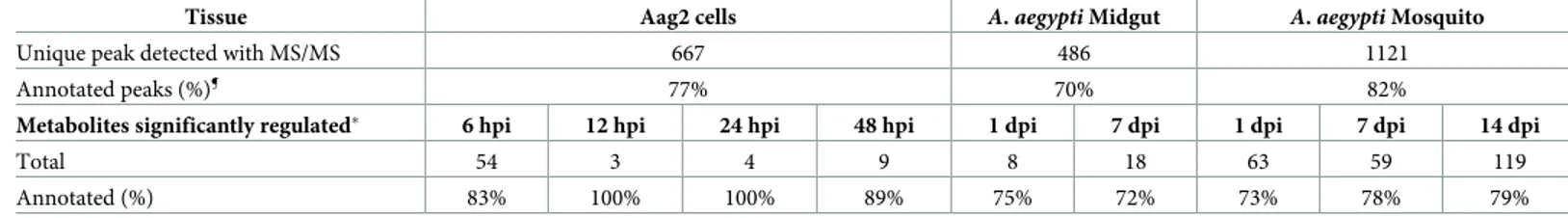
Table 1. Summary of metabolites detected across mosquito tissues...85
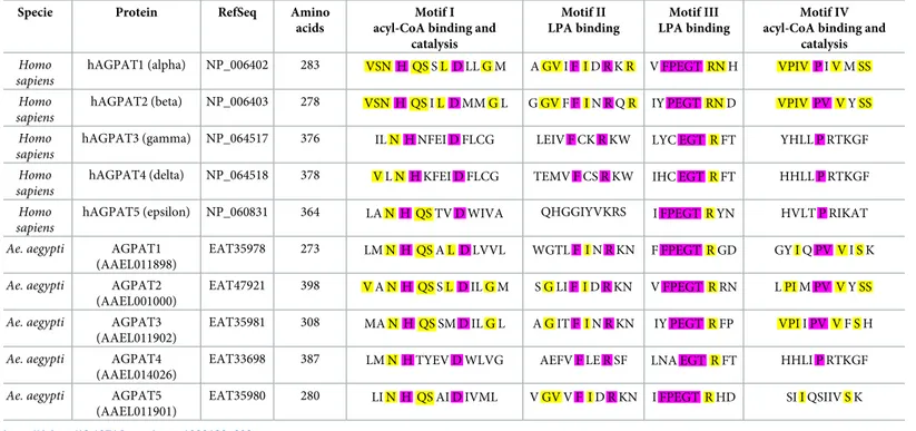
Table 2. Acyltransferase motif comparison between human and Ae. aegypti AGPAT homologues...88
Table S1. Identification of mosquito specific metabolites by spectral similarity...113
Table S3. Primers for dsRNA...116
Table S4. Primers for Real-Time qPCR...116
Table S5. AGPAT FASTA protein sequences...116
Chapter 3 – DENV activates phospholipid remodeling for replication Figure 1. Impact of Kennedy pathway inhibition on DENV and phospholipid reconfiguration...125
Figure 2. Impact of Kennedy pathway activation on DENV and phospholipid reconfiguration...129
Figure 3. Impact of DENV on each of the branches of Kennedy pathway ...132
Figure 4. Impact of Kennedy pathway activation on DENV cellular cycle...135
Figure S1. Kennedy pathway gene expression after silencing...145
Figure S2. Ion intensity of regulated metabolites in DENV-infected cells after Kennedy pathway gene depletion...146
Figure S3. Impact of ethanolamine and choline supplementation on DENV replication at MOI 5...147
Figure S4. Ion intensity of regulated metabolites in choline or ethanolamine supplemented cells infected with DENV...148
Figure S5. Isotope labeled 13C ethanolamine (Etn) or choline (Cho) incorporation on phospholipid metabolites on mock-infected cells...149
Figure S6. Impact of ethanolamine and choline pre-treated virus on attachment and internalization... ...150
Table S1. Isotope labeled 13C ethanolamine (Etn) or choline (Cho) incorporation on phospholipid metabolites in mock-infected cells...150
Table S2. Isotope labeled 13C ethanolamine (Etn) or choline (Cho) incorporation on phospholipid metabolites in DENV and mock-infected cells between 25 and 72 hours after supplementation...151
Table S3. Primers for dsRNA synthesis...151
13
LIST OF SYMBOLS AND ABBREVIATIONS
AA Amino acid
ABC ATP-binding cassette
AC Acylcarnitine
ACC Acetyl-CoA carboxylase
ADE Antibody-dependent enhancement
AGPAT Acyl-sn-glycerol-3-phosphate O-acyltransferases
AminoPL Aminophospholipids
AMPK Adenosine monophosphate-activated protein kinase
APLS Amphipathic lipid packing sensor
ARA Acid arachidonic
ATP Adenosine triphosphate
AUP1 Ancient ubiquitous protein 1
BMP Bis(monoacylglycerol)phosphate
Bti Bacillus thuringiensis israelensis
C Capsid protein
CDC Center for disease control and prevention
CDP Cytidine diphosphate
CDS CDP-diacylglycerol synthase
Cho Choline
CI Cytoplasmic Incompatibility
CK/EK Choline kinase/Ethanolamine kinase
CL Cardiolipin CLS Cardiolipin synthase CM Convoluted membranes CoA Coenzyme A CPT DAG:CDP-choline cholinephosphotranferase CT CTP:phosphocholine cytidyltransferase CTP Cytidine triphosphate
CYD Chimeric yellow fever dengue
DAG Diacylglycerol
DC Dendritic cells
DENV Dengue virus
DF Dengue fever
14
DHA Docosahexaenoic acid
DSS Dengue shock syndrome
dsRNA Double-stranded RNA
E Envelope protein
ECDC European centre for disease prevention and control
EK Ethanolamine kinase
EPA Ecosapentaenoic acid
EPT DAG:CDP-ethanolamine ethanolaminephosphotranferase
ER Endoplasmic reticulum
ET CTP:ethanolamine cytidyltransferase
Etn Ethanolamine
FA Fatty acid
FAS Fatty acid synthase
G3P Glycerol-3-phosphate
GABA Gamma-Aminobutyric acid
GAG Glycosaminoglycans
GAPDH Glyceraldehyde-3-phosphate dehydrogenase
GC Gas chromatography
GL Glycerolipid
GPAT Glycerol-3-phosphate acyltransferase
GST Glutathione transferases
HCV Hepatitis C virus
HILIC Hydrophilic interaction liquid chromatography
HSP Heat-shock protein
IFN Interferon
IL Interleukin
ITM Insecticide-treated materials
ITN Insecticide-treated bed nets
JEV Japanese encephalitis virus
LA Linoleic acid
LC Liquid chromatography
LD Lipid droplet
LLIN Long-lasting insecticides-nets
LPLAT Lysophospholipid acyltransferase
LPCATs Lysophosphatidylcholine acyltransferase
15 LysoPC Lysophosphatidylcholine LysoPE Lysophosphatidylethanolamine LysoPI Lysophosphatidylinositol LysoPG Lysophosphatidylglycerol LysoPL Lysophospholipid M/prM Membrane/premembrane protein
MAM Mitochondria-associated membranes
MS Mass spectrometry
MBOAT Membrane-bound O-acyltransferase
MR Mannose receptor
MSI Metabolomics standards initiative
MUFA Monounsaturated fatty acids
NS Non structural
NIAID National institute of allergy and infectious diseases
NHP Non-human primate
NADH Nicotinamide adenine dinucleotide
NDGA Nordihydroguaiaretic acid
NMR Nuclear magnetic resonance
ORF Open reading frame
PA Phosphatidic acid
PAF Platelet-activating factor
PC Phosphatidylcholine
PCA Principal component analysis
PE Phosphatidylethanolamine
PE-Cer, Ceramide Phosphoethanolamine
PEMT PE methyltransferase
PFU Plaque-forming unit
PI Phosphatidylinositol PIP Phosphoinositide PIS PI synthase PL Phospholipid PLA Phospholipase A PLB Phospholipase B PLC Phospholipase C PLD Phospholipase D
16 PG Phosphatidylglycerol PGP Phosphatidylglycerol phosphate PGPS PGP synthase PS Phosphatidylserine PSD PS decarboxylase PSS PS synthase
PUFA Poly-unsaturated fatty acids
RIDL Release of insects carrying dominant lethals
RC Replication complex
RNA Ribonucleic acid
RNP Ribonucleoprotein
dsRNA Double-stranded RNA
RNAi RNA interference
SIT Sterile Insect Technique
SL Sphingolipid
SM Sphingomyelin
SMS Sphingomyelin synthase
SREBP Sterol regulatory element-binding proteins
T Tubular structures
TBEV Tick-borne encephalitis virus
TAG Triacylglycerol
TCA Tricarboxylic acid
TIC Total ion current
TLR Toll-like receptor
TNF- α Tumor necrosis factor-α
UTR Untranslated regions
Ve Double-membrane vesicle
VIP Variable influence on projection
VP Vesicle packet
WHO World Health Organization
WNV West Nile virus
YFV Yellow Fever virus
ZKV Zika virus
(+)ssRNA Positive single strand RNA
17
CHAPTER 1 – Introduction to host-virus metabolic
interactions in dengue virus
1. Dengue: a disease in expansion without efficient control
1.1. Dengue
1.1.1. A global burden
Dengue is an arthropod-borne viral disease (arbovirose) that currently infects about 400 million people every year throughout the tropical and subtropical world [1]. Dengue virus (DENV) is the most widespread arbovirus. It emerged in the second part of the 20th century [2], and ever since its incidence has increased 30-fold, according to WHO. Asia is the continent the most affected by dengue as it bears 70% of total infection. For instance, India represents 34% of the total infection. Africa is estimated to contribute 16% of global infections. However, African dengue burden is probably underestimated because of poor surveillance and presence of other diseases presenting similar symptoms. Americas represents 14% of infections, with majority of cases in Brazil and Mexico. The annual global cost incurred by dengue is estimated at US$ 8.9 billion, according to a study conducted in 2013 in 142 countries with active DENV transmission [3]. The average cost per dengue case is US$152, although actual cost varies according to clinical outcome. Estimated hospital admission for dengue costs US$70 but rises to US$84,730 for fatal cases. Dengue economic burden is higher than for other major infectious diseases, such as cholera, viral gastroenteritis, Chagas or rabies, because of the lack of available treatment and insufficient systematic diagnosis. Other factors associated to dengue outbreaks worsen the cost. The healthcare system can be congested during epidemic episode, resulting in difficulties
18
to provide accurate diagnosis. Loss of productivity, ability to work, impact on tourism also increase the economic burden [4].
DENV is transmitted by the bite of an infected female mosquito of the genus
Aedes. Because of the Aedes geographical distribution more than 40% of the global
population is at risk to contract dengue [5], encompassing 128 countries [6]. Aedes distribution now extends in all continents, including North America and Europe [5] in addition to the usual subtropical regions. Consequences are an expanding distribution of DENV (Fig 1).
1.1.2. From asymptomatic to severe dengue
About 75% of people infected with DENV remain asymptomatic (not registered in hospital). The other 25% represents around 100 million patients that experience a range of different symptoms from flu-like illness to vascular leakage, hemorrhages, organ failure and shock [1].
1.1.2.1 Clinical phase
The incubation period is usually 5-10 days and up to 14 days, followed by sudden onset of symptoms divided in 3 phases: febrile, critical and recovery.
The febrile phase starts with high fever during 3-7 days, vomiting and multiple symptoms (rash, headache, bone pain, retro-orbital pain, flush, hematuria, minor bleeding). The liver can be enlarged and sensitive. A decrease in total white cells can suggest dengue at this stage. The early febrile phase can be difficult to distinguish from other febrile diseases. Moreover, those symptoms can not differentiate an outcome into severe and non-severe dengue.
The critical phase begins when the fever decreases and complications start to develop. Most patients improve in this phase, but some can undergo vascular leak
19
syndrome. Increased vascular permeability can result in plasma leakage, intravascular volume depletion and lead to dengue shock syndrome (DSS) [7]. In the latter case, fluid overload induces respiratory distress. Vascular leak syndrome usually resolves within 1 or 3 days. Patients can also suffer from severe bleeding, especially during prolonged shock. Although uncommon, organ impairment can occur and includes hepatitis, encephalitis and myocarditis.
The recovery phase lasts 1-2 weeks with good supportive care. Fluids are reabsorbed, then hematocrit, white blood cells and platelets stabilize. Some patients may experience rash and remain tired for several days.
20
Figure 1. The global distribution of dengue. Adapted from [1,8], CDC Dengue heatmap (/www.healthmap.org/dengue), and data from the
European Centre for Disease Prevention and Control (ECDC) (www.ecdc.europa.eu/en/dengue). National and local consensus of complete presence (red) or absence (blue) reported by local transmission. This map does not take into account imported dengue cases by travelers.
21
1.1.2.2 Symptom Classification
A previous classification established by WHO in 1997 differentiated dengue fever (DF), dengue shock syndrome (DSS) and dengue hemorrhagic fever (DHF). In 2009, WHO revised the classification in dengue without complications and severe dengue. Severe dengue is defined by one of these complications: shock syndrome or respiratory distress caused by plasma leakage or fluid accumulation, severe bleeding, or organ impairment [9].
1.1.2.3 Secondary dengue infection
Following DENV infection with one serotype, the adaptive immune response will provide long-term immunity to this serotype and short-term protection (3 months to 2 years) against heterologous serotype infection [10]. However, with vanning heterologous protection, the risk of severe dengue with secondary infection increases. Cross-reactive antibodies induced by the primary infection bind the heterologous serotype and facilitate virus entry into target cells via Fc receptors. This phenomenon is called antibody-dependent enhancement (ADE). Antibody-FC receptor interaction is an alternative DENV entry mechanism during ADE in the course on secondary heterologous infection [11]. ADE also contributes to decreasing immune response and antiviral response against the infection, through FC receptor signaling which prevent lysosomal degradation of DENV [12].
1.1.2.4 Diagnosis
Diagnosis will depend on the time after disease onset [9]. Before 5 days, viral RNA is detected by nucleic acid amplification, the virus is isolated in cell culture or viral protein NS1 is detected by immunoassay. After 5 days, DENV does not persist in blood,
22
but NS1 is still detected for a few days, especially during primary infection. From about 4 days onwards, IgM antibodies can be detected, with a peak at 10-14 days, before decreasing and disappearing after 3 months. IgGs appear later, from day 10 onwards, at lower concentration and remains detectable for life. Serological assay may not be sufficient for detection due to cross-reactivity with other flaviviruses, especially in regions where flaviviruses circulate or for patients vaccinated against yellow fever or Japanese encephalitis.
1.2. DENV vectors
1.2.1. Aedes aegypti and Aedes albopictus
The main vector of DENV is Aedes aegypti mosquito. It is highly adapted to urban areas as it preferentially breeds in man-made containers with stagnant water. It mainly bites humans during the day and can take several blood meals from different people, increasing chances of transmission [13]. Aedes albopictus, the second major vector of DENV, is found in peri-urban and rural areas and feeds on humans as well as other mammals [14]. Both species are quickly colonizing new regions, spreading risk of infection to new regions [5]. Multiple factors such as urbanization, globalization, trade, population growth, travel and global warming are associated with the enlarged distribution of the two vectors [15]. Population growth in urban tropical areas closely correlates with the increase in dengue epidemics [16]. Higher population density amplifies DENV transmission dynamic, illustrated by the fact that a mosquito can bite several people during a blood meal [17]. Urbanization increases larval development by providing more oviposition sites and enhancing mosquito survival [18]. Globalization and large scale travels spread the virus across different countries and continents
23
[19,20]. The impact of climate change on DENV distribution has been modeled [21]. Temperature rise and new precipitation patterns facilitates geographical expansion of the vectors. Annual number of people exposed to Aedes vectors was projected to increase drastically by 12 to 134% by 2050, depending on different scenario, with new territories affected such as Australia, Europe and North America.
Life cycle of mosquitoes contains four separate stages which takes approximately over 8-10 days: egg, larvae, pupae and adult. An adult female mosquito lays up to 200 eggs inside containers holding water. When submerged in water, eggs hatch into larvae after 1-2 days. Aquatic larvae feed on microorganism and develop into pupae after approximately 5 days. Pupae are mobile in water and do not feed. They emerge into adult flying mosquitoes after 2-3 days. A couple of days later, male and female adults start to mate. The female will need a blood meal to produce eggs. Adult Aedes mosquitoes can live for more than 1 month. Mosquitoes can fly about 200 meters after emerging. A. aegypti females bite almost exclusively humans during daylight hours, preferentially early in the morning and in the evening, outdoor and indoor.
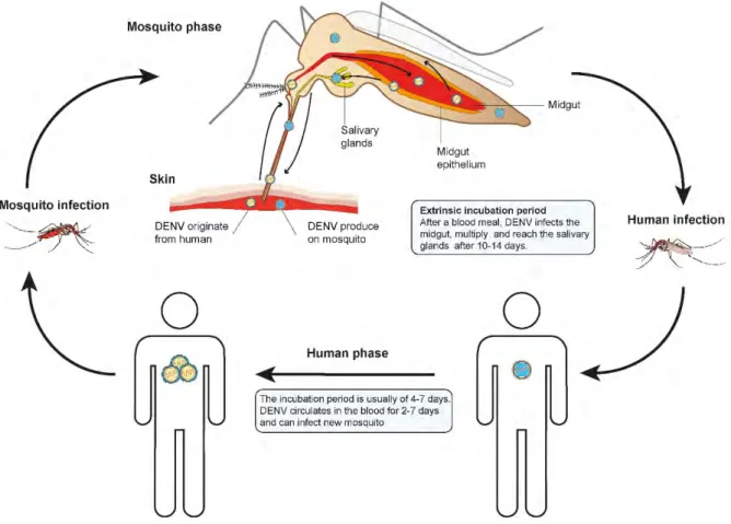
DENV is maintained at low level in endemic highly dense urban area. Epidemic episodes contribute to the persistence of the virus. Multiple DENV serotypes can circulate in the same area. Aedes mosquito will be infected by blood-feeding on a human in the viremic phase of DENV infection (Fig 2). Extrinsic incubation period, the time taken by the virus to multiply and be present in mosquito saliva, is usually 10-14 days. Infected mosquitoes can infect several humans during subsequent bites. About 5-10 days after being bitten by DENV-infected mosquito, a person develops high viremia that persists approximately 7 days. During this period, an infected person can transmit the virus to a new mosquito.
24
DENV sylvatic cycle exists between Non-Human Primates (NHPs) and Aedes forest mosquitoes in rainforests of Southeast Asia (Malaysia) an West Africa (Senegal) [22]. Contrary to the YFV, no evidence exist for a sylvatic cycle in America [23]. DENV found in NHPs in south America are likely spill back from human viruses [24].
1.2.2. DENV cycle in mosquito
Following a blood meal on an infected human, DENV reach the mosquito midgut where it infects and multiplies in epithelium [25] (Fig 2). Blood meal is digested within 48 hours, while replication in the midgut continues and reach a peak at 7 days after ingestion. DENV then disseminates in the whole mosquito body, including salivary glands which are fully infected at 10-14 days post infectious blood meal. Mosquitoes can then transmit DENV to human via saliva during subsequent blood feeding. Throughout its infection cycle in the mosquito, DENV replicates in different tissues and cell types, involving specific physiological changes at each stage.
In mosquitoes, DENV is confronted to complex barriers from the midgut to salivary glands [26]. After the blood meal, the virus needs to infect the midgut epithelium and to overpass an extracellular matrix called basal lamina (BL) to disseminate from midgut to secondary tissues. After crossing the midgut barrier, new virion can infect fat body and nerve tissues, but hemocytes are likely an important target for the next step of arbovirus amplification [27]. DENV then infect lateral and median lobes of salivary glands [25] where viral replication will lead to the release of new viral particles in the excretory canal of the gland. At that point, the virus is associated with apoptosis and mosquito saliva containing proteins and enzymes having immune response modulating properties [28], which suggest that saliva is an important factor in virus transmission to human host.
25
The ability of a mosquito to acquire and propagate DENV through all these steps is defined as vector competence. The competence is related to host genetic factors such as innate immune response and tissue barriers [29]. For instance, Ae.
aegypti endogenous RNA interference pathway is involved in arbovirus infection
in the midgut and can be virus dose-dependent to overtake host defense [30]. Enhancing RNAi pathway in mosquito can improve the midgut infection barrier (MIB) to DENV after cell entry and decrease the vector competence [31]. Mosquito RNAi effector polymorphism is also a key factor to DENV resistance [32]. The MIB can also be associated to resistance of midgut epithelial cells to viral infection, due to filtration by the extracellular matrix or a lack of host membrane receptor of targeted cells. Precisely, the 67kDa protein described as a DENV receptor in midgut cells, is identified as a marker of vector competence for DENV in Ae.
aegypti mosquito [33,34]. Infection of salivary glands is likely receptor-mediated
and dependent on mosquito strain, similarly to midgut infection. Compatibility between mosquito strain and virus strain to bind and infect tissue especially at the midgut and salivary gland levels are part of endogenous factor that are independent of virus-dose interaction and define vector competence [35].
Among the host endogenous factor, it can be hypothesized that the metabolite composition of certain tissues, especially in lipid constituting cell membranes, such as the midgut or the salivary glands play a major role in such barriers. Furthermore, exogenous nutritional content in lipid and sugar could affect vector competence for arbovirus, as observed for West Nile virus [36]
26
Figure 2. DENV cycle in humans and mosquitoes.
1.3. Dengue virus (DENV)
1.3.1. DENV biology
DENV belongs to the Flavivirus genus of the Flaviviridae family. Other pathogenic Flaviviruses include Yellow Fever virus (YFV), West Nile virus (WNV), Japanese encephalitis virus (JEV), tick-borne encephalitis virus (TBEV) and Zika virus (ZIKV) (Fig 3). DENV is composed of four antigenically and genetically different subtypes, called serotype 1 to 4. DENV has the ability to infect and propagate in two different hosts, human and mosquito.
DENV presumably evolved from sylvatic strains in Africa or Asia from non-human primates [37,38]. The four independent DENV serotypes likely involved different host switch events.
27 Figure 3. Phylogenetic tree of mosquito borne flavivirus. The dendrogram is based
on amino acid sequence of the virus polyprotein. Colors differentiate the vectors [39].
Aedes mosquitoes vector DENV, ZKV, YFV and the Spondweni virus. Culex genus
mosquitoes vector WNV, JEV and St. Louis encephalitis virus. Ticks (Ixodes genus) are vectors of the tick-borne encephalitis virus.
DENV are enveloped viruses with a spherical shape of 50 nm diameter. The virus is composed by a lipid envelope associated with 2 structural proteins, the envelope (E) and membrane (M) proteins. Within the viral envelope, the RNA viral genome is associated with the capsid (C) protein. Importantly, the lipid bilayer is the only component that does not come from the viral genome, but from the endomembrane compartment of the host cell.
The genome is a single-stranded positive sense RNA ((+)ssRNA) of about 11kb that encodes ten proteins. The + genome is used as template for the – genome synthesis, protein synthesis and for packing into new viral particles. Three structural proteins, E, pr-membrane (prM) and C; and 7 non-structural (NS) proteins, NS1, NS2A,
28
NS2B, NS3, NS4A, NS4B and NS5 are encoded in a polyprotein. During assembly, a RNA-capsid complex recruits a lipid bilayer membrane, in which the E and prM are embedded. Virions exist in mature and immature form depending on the presence of pr protein, which is shed during maturation.
E protein (53 kDa) is involved in viral entry into targeted cells by binding to cellular receptors and triggering viral and cellular membranes fusion [40]. In mature form, 90 homodimers of E proteins are arranged on the surface, by sets of 3 E homodimers in 30 rafts. E ectodomain is composed of 3 connected domains (DI-III) and a fusion loop at the tip of DII [41]. DI is the central structure, DII contains a fusion loop important for the fusion of viral and cell membranes, and DIII is exposed at the particle surface with cellular-binding motifs. In immature form, E and prM protein are associated in heterodimers forming spike. The prM protein (21 kDa) participate in the formation and the maturation of new virion. During maturation of newly produced particles, pr peptide is cleaved from the M peptide through the secretory pathway involving pH-dependent reaction and host protease. In mature virion, M protein is anchored into the viral membrane by two transmembrane helices under the E protein. It is also important to note that temperature induces structural changes in viral particles. A virus incubated at 28°C, daytime mosquito body temperature, will have a smooth surface, while at human body temperature of 37°C, the virus has a rough and heterogeneous structure due to specific arrangement of the E proteins [42]. This underlines that the virus surface is not a static but a dynamic structure. This conformational amplitude is called viral breathing [43]. Different conformations can exist and this does not have any consequences on the interaction of the virus with target cells [44]. The C proteins (12kDa) are associated in homodimer and act as RNA
29
chaperone with RNA-binding activity. This complex forms the viral ribonucleoprotein (RNP).
The NS proteins are involved in viral replication and packing, operating in endoplasmic reticulum (ER) and secretory pathway of the cell. NS1 is associated with intracellular membrane and can be secreted (sNS1). NS1 participates in early viral genome replication [45]. The secretory form activates the innate immune system and is associated with vascular leakage in severe dengue [46]. NS2A is a membrane protein involved in RNA replication and viral assembly [47]. NS2B act as a cofactor of NS3 which has several functions, especially during RNA synthesis with helicase and capping activities. NS4A is a membrane protein associated with the formation of the replication complex (RC). NS4B has inhibitory capacity against interferon (IFN) response [48]. NS5 has the biggest size and is highly conserved. Several functions are associated with NS5, such as suppression of IFN system, RNA synthesis and capping. DENV genome contains also two untranslated regions UTR: a short 5’ UTR of ~100 nucleotides and a longer 3’ UTR of ~450 nucleotides, both highly structured. Those UTR are involved in genome replication [49].
1.3.2. DENV cellular life cycle
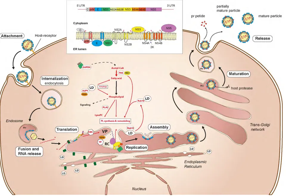
1.3.2.1 Virus entry
Susceptible cells contain attachment factors on the surface that allow contact with viral particles through E binding. This promotes virus entry. Multiple cell types can be infected in vitro, including epithelial, endothelial, muscular, dendritic, mast cells and hepatocytes, monocytes and mosquito cells [8]. Different DENV receptors have been candidates in mammalian and mosquito cells, consistent with the ability of the virus to
30
infect a diversity of cells, as well as 2 different hosts [50,51]. Glycosaminoglycans (GAG) such as heparan sulfate, dendritic cells lectin receptor (DC-SIGN), macrophage mannose receptor (MR), lipopolysaccharide (CD14), heat-shock protein 70 and 90 (HSP70/90), binding-immunoglobulin protein GRP78 and TIM/TAM phospholipid receptors were identified on mammalian cells. On mosquito cells, the chaperone prohibitin protein was identified as receptor by interaction with E [52]. A set of proteins and glycoproteins, some of which related to heat shock protein family, were also candidates as mosquito cell receptor for DENV [51].
After virus adsorption to cell surface, entry occurs mainly by clathrin-dependent endocytosis. DENV has been preferentially found on cell surface of clathrin-coated pit [53]. Alternative entry pathways exist but are minor and found only on mammalian Vero cell line [54]. On mosquitoes cells, only the clathrin-mediated endocytosis pathway was observed four the four DENV serotypes [55,56]. An invagination in the plasma membrane is created and closed by dynamin to form a clathrin-coated vesicle. The endosomal vesicle is transported inside the cell by a mechanism involving actin filaments [57].
At this step the enveloped virus is contained in a vesicle delimited by a lipid bilayer. The endosomal low-pH induces molecular changes on the E proteins leading to viral and endosomal membrane fusion and release of the viral RNP into the cytoplasm. The homodimer of E rearranges in trimers under the acidification, which makes the fusion loop accessible and facilitates interaction with the outer lipid layer of the endosome membrane [40]. The E protein then folds back and induces hemifusion of monolayers followed by pore formation. Cellular vacuolar ATPase are important for endosome acidification and its inhibition blocks DENV infection [58,59].
31
1.3.2.2 Polyprotein translation
After release from the endosomes, the RNP is in cytoplasm and undergoes capsid uncoating. The single open reading frame (ORF) is translated into a long polyprotein associated with ER membranes, processed by viral and host proteases into the 3 structural proteins (C, prM, E) and the 7 NS proteins (NS1, NS2A, NS2B, NS3, NS4A, NS4B, and NS5) [58]. The genome is likely recruited to the ER to initiate translation with ribosomes. It is also suggested that translation starts into the cytosol and continues in ER when the transmembrane domain of the C protein emerges from ribosomes. Actually, the full-length polyprotein has yet to be observed, suggesting rapid cleavage of viral proteins. The polyprotein intertwines with the ER membrane: NS2A, NS2B, NS4A and NS4B have transmembrane domains and are anchored in the ER bilayer. C, NS3 and NS5 are on the cytoplasmic side, when prM, E and NS1 are on the lumen side. Polyprotein processing is realized by viral and cellular proteases. NS3/NS2B complex cleaves proteins on the cytoplasmic side, while a host peptidase cleaves those in the lumen. A small part of immature C remains in the ER after the cleavage. The cleavage of NS4A and NS4B leave a 2K peptide inserted in the ER membrane. The translation process highly involves ER membrane, and several proteins remain anchored in ER.
1.3.2.3 Replication
Viral replication requires viral proteins, host factors and viral RNA. The positive strand viral genome ((+)ssRNA) is copied into antigenome ((-)ssRNA), which is then used as a template for genome replication. Viral synthesis involves membrane rearrangements via lipid membrane invagination to ensure efficient RNA synthesis in replication complex (RC) [60,61]. RC may also protect viral replication from host defense mechanisms.
32
DENV replication complex structure has been characterized in human and mosquito cells [62,63]. In mammalian cells, membrane alterations induced by DENV have different shapes, from convoluted membranes (CM), double-membrane vesicles (Ve), tubular structures (T) and vesicle packets (VP), derivating from ER. Open pores were observed in Ve, allowing probably transport of building block from the cytoplasm for RNA synthesis and/or release of newly synthetized RNA for encapsidation. VP are composed of small groups of Ve formed by ER membranes rearrangement containing viral replication sites. Vesicles induced by DENV contain NS proteins and dsRNA intermediates in RC, suggesting active RNA synthesis [63]. Those distinct structures were also observed in mosquito cells, except for CM. Formation of different structures suggest specific modifications of host membranes induced by DENV replication. Vesicle formation may be induced by NS4A protein due to its transmembrane domain acting on the luminal leaflet on the ER [64,65]. Membrane invagination close a cytoplasmic window containing NS1, NS3 and NS5 [64]. Inside a vesicle, replication complex organization is represented as a complex containing dsRNA associated with NS3/NS2B protease/helicase and NS5 methyl-transferase-polymerase [66]. NS3 has helicase activity to unfold dsRNA during RNA synthesis, while NS5 is the RNA-dependent-RNA polymerase (RdRp) and methyltransferase involved in newly synthetized RNA, that also cap RNA by its triphosphatase activity. NS4B ER-anchored protein binds the NS3/NS2B enzymes as factor to support replication. It was observed that the cleavage of the 2K peptide associated with NS4A is important to induce membrane arrangement in RC [64]. Furthermore, NS4A induces rearrangement and phosphorylation of vimentin intermediate filaments to support DENV RC at the perinuclear site [67], suggesting the involvement of the cytoskeleton. NS2A transmembrane protein is essential for RNA synthesis and involved in the RC
33
organization [47]. NS1 is found in the lumen side of the ER, and connect to the RC via transmembrane interactions, probably by interaction with NS4B as shown during WNV infection [68]. Role of NS1 in viral replication is currently not reported.
1.3.2.4 Assembly
DENV assembly occurs at the ER with C, prM and E proteins assembling with viral genomes to bud towards ER lumen. NS2A as well may be required for viral assembly [47]. NS3 is required for flavivirus RNA packing into viral particles, without involving helicase and protease activity [69,70]. The C protein is enough to fold RNA and participates in spontaneous encapsidation [71]. Encapsidated viral RNA is probably released from RC through the pores, but viral RNA transport to assembly sites remain unclear. Once assembled, flaviviruses form immature particles characterized by spikes of trimeric prM and E form [72]. Maturation of flaviviruses occurs through Golgi and trans-Golgi networks and requires an acidic environment [73]. pH induces molecular rearrangement of prM and E protein, enabling access to prM for furin host protease. Cleavage of prM releases pr and maintain M on the envelope. Immature or partially immature virions are also produced and have alter infectious capacity.
1.4. Prevention and treatment
1.4.1. Antiviral developments
No antiviral against DENV is currently available and only symptomatic care is provided to patients [74]. It is recommended to stay hydrated and avoid anticoagulant such as aspirin-containing drugs. For severe dengue patients in shock syndrome,
34
intravenous fluid supplementation is essential and prophylactic platelet transfusion is performed, although the latter does not prevent bleeding [75].
Multiple drugs with in vitro antiviral activity against DENV have been tested in clinical trials (chloroquine, balapiravir, celgosivir, lovastatin), without success in preventing disease or lowering viremia [76]. Ivermectin is currently in clinical trial in phase II/III in children and adult patients (ClinicalTrials.gov number NCT02045069). DENV-2-infected Aedes albopictus mosquito treated with ivermectin have shown a 50% decrease in infection rate and almost complete clearance of DENV RNA [77]. Ivermectin also inhibits in vitro replication of flaviviruses, mainly YFV and DENV with a lower effect for the later, by targeting NS3 helicase activity [78]. Ivermectin is usually used to treat parasite infections in humans such as malaria [79,80]. Other drugs that target NS proteins such as NS3 and NS5 have been studied, but none reached clinical trials [81]. Recently a DENV inhibitor targeting NS4B was under development [82]. Neutralizing monoclonal antibodies are also candidates for dengue treatment [83]. Several bioactive compounds from natural products display anti-dengue activity, but have not been studied further [84–86].
1.4.2. Vaccines
Dengue vaccine against the four DENV serotypes is needed for the prevention and control strategy. Dengue vaccine development have been on the road since the end of the first half of the 20th century [87].
1.4.2.1. Dengvaxia, a partially efficacious vaccine
Dengvaxia is a DENV vaccine produced by Sanofi-Pasteur. It is a live attenuated tetravalent vaccine composed of the nonstructural genes of yellow fever vaccine strain 17D and envelope (E) and pre-membrane (prM) genes of the four DENV
35
serotypes. Four chimeric yellow fever dengue (CYD) vaccine are combined into a single formulation. The vaccination scheme remains long with 0, 6 and 12 months boosters. The vaccine is registered in 20 dengue-endemic countries and in the United States of America (USA) in May 2019 [88] and in the European Union [89]. However, its use remains low as it has poor efficacy in protecting against all dengue serotypes and seronegative people [90–92].
During the immunization programs in Brazil and Philippines, an increase risk of severe dengue in vaccinated seronegative group was observed, resulting in a low vaccine uptake. In September 2018, WHO recommended that only people with past DENV infection be vaccinated, after highly specific screening test, in the age range 9-45 years [93]. The U.S. Food and Drug Administration (FDA) approved the vaccine in people ages of 9-16 years who have laboratory-confirmed previous dengue infection and who live in endemic areas of the US [94]. In case when screening is not possible, vaccination could be administrated in areas with 80% seroprevalence or more by 9-year-old. Furthermore, DENV seropositive traveler in a high endemic country could consider the vaccination. This vaccine limitations highlights the urgent needs for rapid diagnostic of dengue serostatus in endemic countries.
1.4.2.2. Other vaccine in clinical trial
Two other vaccines are currently in clinical trial phase III [89]. The vaccine developed by Takeda (TDV) is in clinical trials in several countries in Asia and Latin America (ClinicalTrials.gov, NCT02747927). This vaccine is a live attenuated tetravalent dengue vaccine based on the backbone of DENV-2 and contains the E and prM genes of the other three serotypes. The vaccine developed by National Institute of Allergy and Infectious Diseases (NIAID) and Butantan Institute (TV003/TV005) and
36
licensed by Merck in the USA, is currently in clinical trials in Brazil (ClinicalTrials.gov, NCT02406729). This vaccine is a live attenuated tetravalent vaccine with mutations in 3’ UTR, while DENV-2 is a chimeric virus containing the capsid and NS protein genes from DENV-4 and prM and E genes from DENV-2. The vaccine induces seroconversion to four serotypes up to 90% of naïve adults [95]. Estimated study completion are respectively by 2021 and 2025 for Takeda and NIAID vaccines. These new candidate vaccines will be challenged on seronegative patients with particular scrutiny on safety issues.
1.4.3. Vector control
Control of mosquito vectors and diminution of human-vector contact is a strategic approach to reduce DENV infection [9]. Vector management targets the two main vectors, A. aegypti and A. albopictus. Vector control includes environmental, biological and chemical methods.
1.4.3.1. Environmental methods
Environmental methods intent to reduce mosquito breeding sites by elimination of larval habitats [9]. Modification of water supply and water storage in household is essential to control vector population. It implies the use of water pipes instead of free access water such as wells and traditional storage systems. Otherwise, water-storage containers need improvement to avoid mosquito access for oviposition by using covers or polystyrene beads. Solid waste such as used tires and plastic containers need efficient environmental management to decrease larval habitats in urban areas.
37
Some urbanization programs, such as in Singapore [96], take into account the urban disease vector problem during housing construction, on construction sites and through specialized infrastructure. For example, roof gutters are prohibited and home owners must ensure maintenance of water storages.
Vector control can also be managed by using mosquito traps. Mosquito traps use carbon dioxide or ultraviolet-A as attractants and aspirate mosquitoes with a vacuum fan [74]. Other traps use water or organic lure.
1.4.3.2. Biological method Wolbachia-infected mosquitoes
Wolbachia is a gram-negative bacteria naturally present in 70% of insect
species [97], including certain mosquito disease vectors [98]. The bacteria modify host reproduction to ensure its dispersion. Males infected with Wolbachia produce a sterile progeny, meaning no hatching, after mating with non-infected females. The phenomenon is called Cytoplasmic Incompatibility (CI). Wolbachia-induced sterility is a population suppression strategy used to eradicate A. aegypti populations [99]. Release of Wolbachia-infected A. aegypti males, exploiting the CI phenotype, is used in programs in Singapore [100], US and China. Furthermore, Wolbachia infection in female mosquitoes reduces virus multiplication [101] and is transmitted maternally. Because of these two traits, replacement of the wild population with Wolbachia-infected mosquitoes has been used to decrease transmission. Wolbachia-Wolbachia-infected mosquitoes have been released in large scales in Australia, Brazil, Colombia [102], Vietnam and Indonesia [103].
38
Genetically modified mosquitoes
Insect genetic engineering is another strategy used to control mosquito populations [104]. The most common genetically modified mosquitoes make use of a lethal gene insertion to sterilize progeny with wild populations, hence reduce population sizes. Male A. aegypti are introgressed with dominant lethal gene (i.e., RIDL) [105]. When engineered males mate with wild-type females, offspring are not viable and die at larval stages. This strategy has been tested in the field and showed encouraging result to suppress local mosquito population [106]. These strategies require very large quantities of modified mosquitoes, as well as increased knowledge of the vector ecology to foresee impacts of the modifications.
Larvicide organisms
Mosquito larvae control is conducted using bacteria and animals. Bacillus thuringiensis
israelensis (Bti) produced endotoxins are able to kill mosquito larvae, by permeabilizing
cell membranes and inducing cell death [107]. However, mosquitoes that survived Bti treatment will have fitness benefit compared to congeners not under larvicide pressure [108]. Larvivorous fish and small crustaceans (Copedod) have been used to reduce larvae in container [74]. Some fish species (Gambusa affinis) are insecticide tolerant, allowing combination with chemical control. Nevertheless, those species do not target specifically Aedes larvae and can affect other insect species, and their introduction into a new environment can affect biodiversity.
39
1.4.3.3. Chemical methods Insecticides
Insecticide use is the most common method to control mosquito population. Chemical control targets either the larval or adult stage, mainly by neurotoxic insecticides. However, insecticides have adverse environmental and health effects and are less effective due to evolution of insecticide resistance.
Larvicides are used to control larval stage and are amenable for water containers [9]. Those compounds are complementary to environmental interventions and restricted to area where water containers are difficult to reach. The challenge is to target only mosquito vector and not to alter water quality. Three classes of larvicides are generally used. Insect growth regulators affect hormones and disrupt the development of young insect. Organophosphates act as nerve agents by disrupting acetylcholine action, resulting in mosquito death. Biopesticides from bacteria toxin such as Bti toxin or the neurotoxin Spinosad can be used. Some larvicides are used for treatment of drinking-water, such as pyriproxyfen, temephos and methoprene at specific dosages.
Adulticides target adult vector to impact mosquito densities in large scale by spraying or fogging [9]. Space spraying is used to prevent epidemic or in emergency situation. Spraying is usually focused where dengue cases have been reported or in high-density population area (school, hospitals, housing). In emergencies, treatments can be applied every 2-3 days for 2 weeks. Significant suppression of adult mosquito populations in high risk area may require application once or twice a week. Organophosphates (Fenitrothion, Malathion) and pyrethroids (cyphenothrin, metofluthrin) are generally used for spraying application against vector. Pyrethroids alter signaling in nervous system by alteration of sodium channels. Insecticide derived
40
from bioactive compounds (terpenoids, alkaloids, pyrethrins, anthraquinones, saponins, monoterpenes) have also shown larvicidal, ovicidal and insecticidal activities [74]. Carbamates, organochlorines (i.e., DDT) are other classes of insecticides but are now rarely used.
Individual protection against vector uses mosquito repellent or Insecticide-treated materials (ITMs). The most widely used repellents are DEET (N, N-diethyl- 3-methylbenzamide), IR3533 (3-[N-acetyl-N-butyl]-aminopropionic acid ethyl ester) or Picardin (1-piperidinecarboxylic acid, 2-(2-hydroxyethyl)-1-methylpropylester). Oil extract from lemongrass (Cymbopogon nardus, Cymbopogon citratus) have repellant activity against A. aegypti and are used in sprays, lotion and bracelet for temporary individual protection [109]. Insecticide-treated bed nets (ITN) and long-lasting insecticides-nets (LLIN) incorporate insecticide in the fabric and reduces vector borne disease transmission.
Insecticide resistance
Chemical control with insecticides has been efficient in controlling Aedes mosquito population before resistance to all four classes of insecticides occurred [110– 114]. Rising of mosquito resistance to insecticides have been observed in more than 60 countries, with resistance to 2 or more classes [115]. It is proposed that low density of mosquito carry resistance genes that allow them to survive after insecticide exposure. The offspring of resistant mosquitoes will have a fitness advantage and be the dominant group of mosquito population [116].
Multiple resistance mechanisms have been identified in A. aegypti mosquito [117], including metabolic detoxification and modification of the insecticide-target site. More precisely, mutations in the sodium channel, on acetylcholinesterase or on GABA
41
receptors will induce insecticide resistance against pyrethroids, organophasphate and cyclodiene, respectively. Metabolic detoxification is due to overexpression/modification of cytochrome P450 genes, esterases and glutathione transferases (GST), which are able to metabolize different types of insecticides [110].
The variety of insecticides for public health is limited and their use led to evolution of resistant populations. Insecticide resistance management is essential to slow down the evolution of resistance and maintain efficiency of vector control. Reduction of insecticide pressure in vector control, agriculture and domestic use is essential. Alternation of insecticides with different modes of action, spread over different areas, use of mixture of insecticides, and nonchemical alternatives are strategies to manage resistances [118]. Development of novel chemicals focusing not on annihilating the vector but on blocking viral transmission is another unexploited strategy to fight the dengue global burden.
Vector competence and tolerance to infection
It is commonly reported that viral infection barely impacts physiology and fitness of mosquitoes [119,120]. However, rather than resisting infection, it seems that the mosquito tolerates infection by limiting damages [121]. This tolerance allows mosquito survival and high viral load, essential for virus transmission to humans. Different tolerance mechanisms have been suggested in midgut, such as immune pathways, tissue repair via stem cell division, metabolic adaptation, stress responses and microbiota-induced tolerance [122].
42
2. The metabolome
2.1. Overview
Metabolites are small molecules that determine the physiological state of cells and include carbohydrates, amino acids, lipids, nucleotides, hormones and vitamins. The metabolome is the set of all metabolites. Metabolites are usually presented as the end product of processes related to genes, transcripts and proteins. Consequently, any upstream bio cellular alterations result in metabolic changes and metabolome profiling brings information about the phenotype.
The metabolism is separated in several pathways, which are interconnected and converge on the tricarboxylic (TCA) cycle (Fig 4). TCA produces metabolic intermediates and contributes to energy production in the form of adenosine triphosphate (ATP). TCA cycle starts with acetyl-CoA, which is produced by catabolism of carbohydrates, mainly glucose, via glycolysis. In the TCA cycle, the acetyl group is oxidized to produce energy. Acetyl-CoA is also used for fatty acid and lipid biosynthesis, including phospholipids, sphingolipids and glycerolipids. Acetyl-CoA can be reversely produced by fatty acid catabolism via the β-oxidation. TCA cycle is connected with amino acid metabolism through shared intermediates and, thus, indirectly influences protein translation. Eventually, nucleic acids originate from the pentose phosphate pathway, which derives from glycolysis.
43 Figure 4. Overview of the metabolism. The general metabolism is composed of
metabolic pathways that converge onto the tricarboxylic cycle (TCA) via acetyl-CoA intermediate production. Breakdown of carbohydrates by the glycolysis, oxidation of fatty acids and amino acids pathways lead to acetyl-CoA production. Acetyl-CoA is used as precursor for fatty acids generation and structural lipid pathways. Glycolysis results in the production of pentose phosphates involved in nucleotide production. The TCA cycle produces precursors of amino acids and the reducing agent NADH, which feeds into the electron transport chain to produce chemical energy in the form of ATP. TCA, tricarboxylic cycle; PL, phospholipid; SL, sphingolipid; GL, glycerolipid; NADH, nicotinamide adenine dinucleotide; ADP, adenosine diphosphate: ATP, adenosine triphosphate.
![Figure 1. The global distribution of dengue . Adapted from [1,8], CDC Dengue heatmap ( /www.healthmap.org/dengue ), and data from the](https://thumb-eu.123doks.com/thumbv2/123doknet/2233588.16245/20.1262.120.1131.125.652/figure-global-distribution-dengue-adapted-dengue-heatmap-healthmap.webp)