DOI: 10.1126/scitranslmed.3006258
, 197ra104 (2013);
5
Sci Transl Med
et al.
Carla G. Silva
Development in Mice
Editor's Summary
pregnancy.
longitudinal prospective human studies will be needed to evaluate the consequences of caffeine consumption during effects of adenosine receptor antagonists including caffeine on brain development in humans. Retrospective and neuronal types as well as impaired memory on certain types of memory tests. This study raises questions about the that adult offspring of pregnant mice treated with adenosine receptor antagonists had reduced numbers of certain antagonists were more susceptible to seizures when exposed to a seizure-inducing agent. They further demonstrated into target regions. They then showed that 1-week-old offspring of pregnant mice treated with adenosine receptor delayed the migration of specific populations of neurons during brain maturation, resulting in their delayed insertion They found that caffeine or an adenosine receptor antagonist that specifically blocks type 2A adenosine receptors
added caffeine to the drinking water of female mice throughout pregnancy and lactation. et al.
this question, Silva
available about the effects of adenosine receptor antagonists such as caffeine on neural development. To address Neural development is strongly influenced by environmental factors including certain drugs. Little information is
Adenosine Receptor Antagonists and Fetal Brain Development
http://stm.sciencemag.org/content/5/197/197ra104.full.html
can be found at:
and other services, including high-resolution figures,
A complete electronic version of this article
http://stm.sciencemag.org/content/suppl/2013/08/05/5.197.197ra104.DC1.html
can be found in the online version of this article at:
Supplementary Material
http://www.sciencemag.org/about/permissions.dtl
in whole or in part can be found at:
article
permission to reproduce this
of this article or about obtaining
reprints
Information about obtaining
last week in December, by the American Association for the Advancement of Science, 1200 New York Avenue (print ISSN 1946-6234; online ISSN 1946-6242) is published weekly, except the Science Translational Medicine
on August 7, 2013
stm.sciencemag.org
B R A I N D E V E L O P M E N T
Adenosine Receptor Antagonists Including
Caffeine Alter Fetal Brain Development in Mice
Carla G. Silva,1,2,3* Christine Métin,4,5Walid Fazeli,6 Nuno J. Machado,3Sanja Darmopil,1,2,7 Pierre-Serge Launay,4,5Antoine Ghestem,1,2 Marie-Pascale Nesa,1,2 Emilie Bassot,1,2Eszter Szabó,3Younis Baqi,8 Christa E. Müller,8Angelo R. Tomé,3,9 Anton Ivanov,1,2Dirk Isbrandt,6Yuri Zilberter,1,2Rodrigo A. Cunha,3,10
Monique Esclapez,1,2†Christophe Bernard1,2*†
Consumption of certain substances during pregnancy can interfere with brain development, leading to deleterious long-term neurological and cognitive impairments in offspring. To test whether modulators of adenosine receptors affect neural development, we exposed mouse dams to a subtype-selective adenosine type 2A receptor (A2AR)
an-tagonist or to caffeine, a naturally occurring adenosine receptor anan-tagonist, during pregnancy and lactation. We ob-served delayed migration and insertion of g-aminobutyric acid (GABA) neurons into the hippocampal circuitry during the first postnatal week in offspring of dams treated with the A2AR antagonist or caffeine. This was associated with
increased neuronal network excitability and increased susceptibility to seizures in response to a seizure-inducing agent. Adult offspring of mouse dams exposed to A2AR antagonists during pregnancy and lactation displayed loss
of hippocampal GABA neurons and some cognitive deficits. These results demonstrate that exposure to A2AR
antago-nists including caffeine during pregnancy and lactation in rodents may have adverse effects on the neural develop-ment of their offspring.
INTRODUCTION
Neural development is strongly influenced by external (for example, environmental) factors including certain drugs (1). These drugs are known to modify cell migration (2, 3) and synapse formation (4, 5), re-sulting in improper wiring of neuronal circuits, ultimately leading to behavioral modifications in offspring later in life (6). There have been few studies about the effects on fetal neural development of other sub-stances such as the naturally occurring adenosine receptor antagonist caffeine, which is ubiquitously consumed in coffee and tea. Mild to moderate caffeine consumption (<200 mg/day) by mothers during pregnancy and lactation is not considered harmful to the fetus and ne-onate from the perspective of miscarriage or risk of prematurity. This is reflected in the current guidelines for caffeine consumption during pregnancy by the American College of Obstetricians and Gynecologists (7–9). Although caffeine can cross the placenta and blood-brain barrier (7), its impact on fetal and early postnatal development remains in-conclusive (10). Caffeine has several molecular targets. At high concen-trations (millimolar range), caffeine can inhibit phosphodiesterases resulting in mobilization of calcium stores or it may have a direct effect on g-aminobutyric acid type A (GABAA) receptors (11). Such high
con-centrations, considered toxic, are rarely found in the blood of
consum-ers of mild to moderate amounts of caffeine (7). In the micromolar range, a concentration regularly found in humans (7), caffeine acts as an antagonist of adenosine receptors, which modify numerous intra-cellular signaling pathways including cyclic adenosine monophosphate (cAMP)–dependent signaling (12). We set out to investigate the effects of caffeine consumption by pregnant rodent dams on brain develop-ment in their offspring. Because caffeine absorption takes place in the gastrointestinal tract (7), we used a protocol of oral caffeine administra-tion in mouse dams throughout pregnancy and lactaadministra-tion. We investi-gated the short-term (during the first postnatal week) and long-term (during adulthood) consequences of exposure to caffeine or a subtype-selective adenosine type 2A receptor (A2AR) antagonist during fetal
development in the dam’s offspring.
RESULTS
General effects of caffeine treatment on female mouse dams and their pups
To mimic a routine daily consumption in human subjects, we exposed female mice to caffeine (0.3 g/liter) in their drinking water during preg-nancy and lactation. This dose (0.3 g/liter) leads to a plasma concentra-tion of caffeine in rat dams similar to that found in the blood of humans drinking three to four cups of coffee per day (13) and to a plasma con-centration of caffeine in postnatal day 7 (P7) rat pups similar to that found in the umbilical cord of human neonates of moderate coffee-drinking (up to three cups per day) mothers (14). We found a similar concentration of caffeine in female mice that were exposed to caffeine (0.3 g/liter) in their drinking water [caffeine (1.0 ± 0.7 mg/liter) in serum]. Treated females (n = 7) adapted quickly to the addition of caffeine to the drinking water and thus showed no difference in water intake from 2 days after the start of caffeine treatment onward (fig. S1A). Food intake of caffeine-treated and untreated female mice was similar 1Aix Marseille Université, INS, 13005 Marseille, France.2Inserm, UMR_S 1106, 13005 Marseille,
France. 3Center for Neuroscience and Cell Biology, University of Coimbra, 3004-517 Coimbra, Portugal.4INSERM UMR-S 839, Institut du Fer à Moulin, 75005 Paris, France.
5Université Pierre et Marie Curie, 75005 Paris, France.6Experimental Neuropediatrics,
University Medical Center Hamburg-Eppendorf, 20246 Hamburg, Germany.7Department
of Anatomy, Croatian Institute for Brain Research, School of Medicine, University of Zagreb, 10000 Zagreb, Croatia.8PharmaCenter Bonn, Pharmaceutical Institute, Pharmaceutical
Chemistry I, Pharmaceutical Sciences Bonn, University of Bonn, 53121 Bonn, Germany.
9
Department of Life Sciences, Faculty of Sciences and Technology, University of Coimbra, 3030-790 Coimbra, Portugal.10Faculty of Medicine, University of Coimbra, 3004-504 Coimbra, Portugal.
*Corresponding author. E-mail: [email protected] (C.G.S.); christophe.bernard@ univ-amu.fr (C.B.)
†These authors contributed equally to this work.
on August 7, 2013
stm.sciencemag.org
(untreated, 3.48 ± 0.12 g/day; treated, 3.48 ± 0.13 g/day;P = 1.0). There was no difference in weight increase throughout pregnancy between control and caffeine-treated dams (fig. S1B).
Addition of caffeine to the drinking water led to enhanced activity during the active (dark) period, but not during the sleep (light) period of the day. The day-night cycle was not affected by the treatment (fig. S1C). During the first 2 weeks of life, the general condition of pups was qual-itatively assessed during the dark period every second day. In both caffeine-treated and control groups, all dams built a nest and pups were in all cases found with the mother within the nest. The gestational pe-riod of the control and caffeine-treated females was similar (19 to 20 days), and the litter sizes were not different between the two groups (fig. S1D). Caffeine was present in the brain of pups, where it reached concentra-tions of 1.06 ± 0.19 ng/mg together with its metabolic products, namely, theobromine (0.62 ± 0.02 ng/mg) and 1,3-dimethyluric acid (0.21 ± 0.04 ng/mg) (n = 5, P1 to P8 pups). Pups born from caffeine-treated dams displayed a transient body weight reduction between P1 and P11 that was gender-independent and that was normalized at P24 (fig. S1E). The weight of 13-week-old adult offspring was not changed by caffeine exposure (caffeine-treated group, 22.2 ± 3.5 g; control group, 24.2 ± 3.2 g;P = 0.22). Motor development and development of neuro-logical reflexes during the first 11 days of life were not affected by caf-feine exposure because the righting reflex (fig. S1F) and cliff avoidance behavior (fig. S1G) were not significantly different between caffeine-exposed and control offspring.
Together, these data indicate that caffeine treatment during preg-nancy and lactation has a major influence neither on female mice nor on pups’ sensorimotor development. We next assessed whether the construction of neuronal networks was affected by caffeine treatment, focusing on GABAergic circuits, which are often altered by various types of insults.
Caffeine treatment of dams alters GABA neuron migration in their offspring
Using GIN mice [green fluorescent protein (GFP)–expressing inhibito-ry neurons] that express GFP in a subpopulation of somatostatin-containing GABA neurons (15, 16), we found a consistent decrease in GFP-labeled neurons in the hippocampus and superficial cortical layers of all caffeine-exposed offspring (n = 11 pups) compared to controls (n = 10 pups) at P6 (Fig. 1, A and B). There was a 41% (P < 0.05) decrease in GABA neurons in the hippocampus of caffeine-exposed offspring at P6 (Fig. 1H and Table 1). In contrast, the distribution pattern (Fig. 1, D and E) and estimated number (Fig. 1H and Table 1) of hippocampal GFP-expressing neurons were similar in 3-month-old adult control and caffeine-exposed offspring. We thus hypothesized that caffeine may delay the migration of GABA neurons.
Cortical GABA neurons are generated within the ventral telencephalon by the medial and caudal ganglionic eminences (MGE and CGE) in mice (17–19); somatostatin-containing neurons are generated in the MGE (20, 21). Cortical GABA neurons first migrate tangentially before switching to a radial migration as they invade the cortical plate. Many GABA neurons keep migrating postnatally to reach their target layers (18, 22, 23). In keeping with this rule of migration, in control GIN mice, some GFP-labeled neurons were observed in the deep cortical layers at P1 (fig. S2, A and E), as they started their radial mi-gration to the superficial layers. At P3, few GFP-labeled neurons were positioned in the superficial supragranular layer (fig. S2, B and F) and in the stratum oriens of the hippocampus (fig. S2I). At P6, many
mor-phologically differentiated GFP-labeled neurons were evident in the superficial cortical layers (fig. S2, D and H) and in the hippocampus (Fig. 1A and fig. S2K). The distribution patterns of GFP-labeled neu-rons and axon terminals in caffeine-exposed offspring at P6 (fig. S2, C, G, and J) were similar to that observed at P3 in control mice (fig. S2, B, F, and I), supporting the hypothesis that caffeine delayed the mi-gration and insertion of interneurons in cortical networks.
Caffeine treatment affected the migration of the entire population of somatostatin-containing GABA neurons (fig. S3, A to F). Somatostatin-containing neurons displayed an immature distribution pattern in caffeine-exposed offspring at P6, illustrated by the presence of many somatostatin-containing neurons, including migrating neurons in the stratum radiatum (control, 25 neurons/mm2,n = 3 pups from three dams; caffeine-exposed, 58 neurons/mm2,n = 3 pups from three dams) similar to that observed at an earlier developmental stage in control mice (fig. S3, A to F). To assess the general state of the GABAergic circuitry, we investi-gated the distribution pattern of the vesicular GABA transporter (VGAT), which labels GABAergic terminals. VGAT labeling displayed immature features in caffeine-exposed offspring at P6, namely, low con-centrations of labeled terminals in the CA3-CA1 pyramidal cell (PC) layer and high concentrations in dendritic layers, as found in control P3 animals (fig. S3, G and H) (24). In contrast, control P6 mice displayed a high concentration of terminal labeling in the PC layer typ-ical of more mature networks (fig. S3I).
The delayed insertion of GABA neurons in cortical circuits suggests that caffeine may interfere with processes that control GABA neuron migration. This hypothesis was directly tested in vitro using time-lapse video microscopy (Fig. 1I). In cocultures prepared at embryonic stage 13.5 (E13.5), GABA neurons from the MGE migrate radially away from their explants of origin on a substrate of dissociated cortical cells (25, 26). Thirty to 45 min after caffeine application, the mean migration speed of MGE neurons was decreased by 53% (P < 0.001, n = 46 of 52 cells), an effect that persisted until the end of the recording session, up to 10 hours in some experiments (Fig. 1, J and K). A small percentage of MGE cells (11.5%,n = 6 of 52 cells) did not respond to caffeine application or re-sponded by increasing their migration speed. We then investigated the adenosine receptor subtype(s) involved in the control of GABA neuron migration.
A2ARs may control the migration of GABA neurons
Chronic consumption of caffeine exerts its central action mostly via its antagonistic action on A2ARs (27, 28). We thus substituted caffeine with
the selective A2AR antagonist KW6002 (istradefylline, 2 mg/kg per day).
Exposure to KW6002 (Fig. 1, C, F, and H), but not to its vehicle solution (Fig. 1G), reproduced the effects of caffeine, suggesting that caffeine delayed GABA neuron migration via its antagonistic action on A2AR
(Table 1). Application of the selective A2AR antagonist SCH58261
(200 nM) to MGE neurons reproduced the effect of caffeine, reducing the migration speed of MGE cells by 56% (P < 0.001, n = 57 of 62 cells), with a small percentage of cells (8%,n = 5 of 62) not responding to the antagonist or increasing their migration speed (Fig. 1, I, L, and M). In contrast, the A1receptor antagonist DPCPX (200 nM) did not change
the migration speed of MGE cells (Fig. 1, N and O). In the absence of drug, the migration speed of MGE neurons was not modified through-out the recording period (Fig. 1P).
These results demonstrate that the endogenous activity of A2AR is
important for GABA neuron migration. We thus determined whether A2ARs are indeed expressed by migrating GABA neurons. Using
on August 7, 2013
stm.sciencemag.org
nohistochemistry, we found A2AR in somatostatin-containing GABA
neurons, leaving the ganglionic eminence at E13 in rats, in particular within the leading process, a key element of cell migration (29) (Fig. 2, A to F). A similar scenario may be at play in primates as we also found A2AR in tangentially and pseudo-radially migrating GABA neurons (30)
in macaque fetal brain at E75 (Fig. 2, G and H). The intracellular path-ways linking A2AR activation and cell motility, as well as the source of
adenosine remain to be determined.
To further confirm that caffeine interferes with A2AR-dependent
mi-gration of somatostatin-containing GABA neurons, we performed caf-feine and KW6002 treatments in mouse dams constitutively lacking A2AR (A2AR-KO). All A2AR-KO pups showed a decreased number of
somatostatin-containing GABA neurons in both the hippocampus and superficial cortical layers (fig. S4, A to F). Somatostatin-containing neurons of A2AR-KO pups displayed poorly developed dendritic
pro-files compared to wild-type pups of the same strain (fig. S4, G and H),
Fig. 1. Caffeine exposure during the embryonic and early postnatal period impairs the migration of hippocampal GABA neuron subpop-ulations in mouse brain. (A to F) Representative coronal sections (6 to 10 examined per animal) from con-trol (A and D), caffeine-exposed (B and E), and KW6002-exposed (C and F) offspring of GIN mice at postnatal day 6 (P6) (A to C) and at 3 months (adult) (D to F). Photomicrographs show the distribution pattern of GFP-containing neurons (white arrowheads) in the hippocampus (HP) and cortex (CX). (A) Many GFP-containing neurons were already present in the superficial layers of the cortex and hippocampus in control pups. (B and C) Caffeine or KW6002 exposure during gestation resulted in a marked decrease in GFP-labeled cell bodies (white arrow-heads), axons, and dendrites in the hippocampus and cortex at P6. In con-trast, no major differences in GFP la-beling of the hippocampus and cortex were observed between control (D) and caffeine-exposed (E) or KW6002-exposed (F) adult offspring. (G) Expo-sure to vehicle during gestation did not modify the pattern of distribution of GFP-containing neurons in offspring. (H) Estimated total number of GFP-expressing neurons in the hippocam-pus of caffeine-exposed (n = 3) or KW6002-exposed (n = 2) P6 offspring was smaller (P < 0.05) than in control (n = 4). In contrast, the estimated num-ber of GFP-containing neurons was similar in the three adult groups (n = 3, 3, and 4, respectively; P = 0.508). (I) Representative time-lapse video se-quence illustrating a GFP-expressing MGE mouse neuron (white arrow) in vitro migrating on a substrate of
disso-ciated cortical cells in response to the application of SCH58261 [5-amino-7-(2-phenylethyl)-2-(2-furyl)pyrazolo(4,3-e)-1,2,4-triazolo(1,5-c)pyrimidine] (vertical black arrow) (time is in hours:minutes). (J to O) Caffeine or SCH58261, but not DPCPX (8-cyclopentyl-1,3-dipropylxanthine), decreased the migration speed of MGE cells in coculture. (J, L, and N) Bar graphs compare the mean migration speed of more than 50 MGE cells measured during a 2-hour period before and after application of caffeine (J, n = 52), SCH58261 (L, n = 62), or DPCPX (N, n = 83). P < 0.001 in (J) and (L), not significant in (N), Student’s t test. (K, M, and O) Time course of instantaneous migration speed for
repre-sentative MGE cells exposed to caffeine (K), SCH58261 (M), or DPCPX (O). Ver-tical black arrow indicates when the drug was applied. (P) In the absence of drug, the mean migration speed of MGE cells did not change during the recording period as demonstrated by the curves on the left, comparing the time course of nuclear movements in two MGE cells recorded in two contiguous cocultures: one without treatment (top) and one treated with SCH58261 (bottom). Right: The decrease in the mean nuclear migration speed was significant 1 hour after SCH58261 application (gray bars). *P < 0.05, **P < 0.01, ***P < 0.001. Scale bars, 250 mm (A to G) and 20 mm (I).
on August 7, 2013
stm.sciencemag.org
suggesting that loss of A2ARs impairs the migration and insertion of
GABA neurons in cortical and hippocampal layers. Neither caffeine nor KW6002 treatment further decreased the number of GABA neurons in the hippocampus of A2AR-KO pups (fig. S4I), further supporting the
notion that the phenotypic traits found in caffeine or KW6002-treated wild-type animals are due to a selective action of these drugs on A2ARs.
Abnormal GABA neuron migration may lead to altered network function (31). We thus assessed whether exposing dams to caffeine modified electrophysiological activity in the hippocampus of their offspring.
The hippocampus is hyperexcitable in caffeine-treated rodent offspring
During the first postnatal week, in vitro hippocampal activity is charac-terized by the occurrence of giant depolarizing potentials (GDPs), which constitute a good readout of network activity and excitability (32). The frequency of GDPs in the hippocampal CA3 region was 217% higher in caffeine-treated pups compared to controls and 206% higher in KW6002-treated compared to vehicle-KW6002-treated pups (Fig. 3, A and B, and table S1). The properties of GDPs depend on the activation of glutamate and GABAAreceptors (32). Measuring both GABAergic and glutamatergic
synaptic inputs (drives) received by CA3 PCs during GDPs, we found a 74% increased contribution of the glutamatergic synaptic input to the overall synaptic input (glutamate + GABA) (Fig. 3C and table S2) in caffeine-exposed animals (n = 7 cells from five pups, four dams) com-pared to controls (n = 6 cells from three pups, three dams) and a 130% increase in KW6002-treated pups (n = 3 cells from three pups, three dams) compared to vehicle-exposed pups (n = 5 cells from three pups, two dams). In contrast, in stratum radiatum interneurons, the glutamatergic synaptic input was similar in control (n = 10 cells from three pups, two dams), vehicle-exposed (n = 6 cells from three pups, two dams), caffeine-exposed (n = 15 cells from four pups, four dams), and KW6002-exposed (n = 14 cells from four pups, four dams) ani-mals (Fig. 3C and table S2).
In CA3 PCs, the frequency of spontaneous inhibitory postsynaptic currents (sIPSCs) and spontaneous excitatory postsynaptic currents (sEPSCs), which are measured between GDPs, was increased by 238 and 466%, respectively, in caffeine-exposed, and by 241 and 350%, re-spectively, in KW6002-exposed pups (Fig. 3, D to G, and table S3). In stratum radiatum interneurons, the frequency of sIPSCs and sEPSCs was increased by 428 and 325%, respectively, in caffeine-exposed, and by 407 and 245%, respectively, in KW6002-exposed animals (Fig. 3, D to G, and table S3). The corresponding decrease in the mean interevent interval (IEI), which is the time between two consecutive synaptic in-puts, is displayed in the histograms below the frequency curves. The contribution of the glutamatergic synaptic input carried by sEPSCs to
the overall synaptic input in CA3 PCs was increased by 134% in caffeine-exposed and 125% in KW6002-caffeine-exposed pups (Fig. 3H and table S4). However, it remained unchanged in stratum radiatum interneurons (Fig. 3H and table S4) because both glutamatergic and GABAergic drives were similarly increased in caffeine- or KW6002-exposed ani-mals. There is thus a general increased synaptic (glutamatergic and GABAergic) and network (GDP) activity in caffeine-exposed offspring compared to controls. Although appearing counterintuitive given the delayed insertion of GABA neurons, an increase in GABAergic activity is consistently found in several pathological contexts. For example, mice lacking lissencephaly-1 protein (33, 34) are characterized by defects in GABA neuron migration (35), which are overcompensated for by hy-peractivity of the remaining GABA neurons (33, 34).
Network hyperactivity can induce neuronal injury, in particular in GABA neurons (36), and is typified by dendritic swellings. In keeping with the increased network activity (GDPs, sEPSCs, and sIPSCs) re-ceived by GABA neurons, we consistently found large varicosities (sug-gesting swelling) along the dendrites of all GFP-expressing neurons in GIN mice (Fig. 3J) and recorded biocytin-filled interneurons (Fig. 3L) in caffeine-exposed offspring at P6. These varicosities were not observed in interneurons in controls (Fig. 3, I and K) or in PCs from caffeine-exposed offspring (Fig. 3M). Such selective effects on GABA neurons may result in cell dysfunction, perhaps contributing to the shift in the balance between excitatory and inhibitory inputs in principal cells.
We conclude that the anatomical alterations induced by caffeine (or KW6002) exposure are paralleled by physiological modifications in the developing brain of offspring. The hippocampal circuitry is characterized by a general hyperactivity, as evidenced by an increase in the frequency of GDPs, sEPSCs, and sIPSCs, as well as a shift in the balance between the GABAergic and glutamatergic synaptic inputs in favor of the latter in CA3 PCs.
Immature neuronal networks are seizure-prone (37), and defects in GABA neuron migration or in GABAergic signaling during develop-ment facilitate the emergence of an epileptic phenotype (31, 38). We thus tested seizure sensitivity in caffeine-exposed offspring during the first postnatal week.
Caffeine exposure increases susceptibility of mouse pups to seizures
We used flurothyl, a seizure-inducing agent (39), to assess seizure sus-ceptibility in caffeine-exposed and control offspring at P6. Pairs of caffeine-exposed/control and KW6002-exposed/control pups at P6 were exposed to flurothyl in two independent sessions (four treated and four control animals per session). None of the pups analyzed (eight controls from two dams, four caffeine-exposed pups from one dam, and four KW6002-exposed pups from one dam) displayed seizures when
Table 1. Estimated number of GFP-containing neurons in control, caffeine-exposed, and KW6002-exposed pup and adult offspring. The number of GFP-containing neurons was decreased in the hippo-campus of caffeine- and KW6002-exposed P6 offspring but not adult
offspring. Given that the morphological pattern of distribution, as well as the immunoreactivity, in hippocampus from vehicle-exposed animals was similar to controls, a stereological quantification was not performed in the vehicle group.
P6 control P6 caffeine P6 KW6002 Adult control Adult caffeine Adult vehicle
Experimental number 4 pups, 3 dams 3 pups, 3 dams 2 pups, 3 dams 4 3 3
Estimated number of GFP-containing neurons 4239 ± 398 2838 ± 317* 1971 ± 315*† 9336 ± 1173 8213 ± 1825 7152 ± 500
*P < 0.05 versus control, one-way analysis of variance (ANOVA). †P < 0.05 versus caffeine, one-way ANOVA.
on August 7, 2013
stm.sciencemag.org
0.1 ml of flurothyl was applied in the cage (Table 2). However, two pups from each of the treated group (caffeine and KW6002) displayed aber-rant movements, whereas none of the control animals did. When 0.2 ml of flurothyl was applied in the cage, only 6 abnormal movements were observed in the control group, whereas all treated pups made a total of 43 abnormal movements. At this flurothyl concentration, 100% of treated pups displayed seizures versus 38% in the control group. The time to trigger seizures, as well as the duration of seizures, was similar in treated and nontreated offspring (Table 2). We conclude that animals exposed to caffeine or KW6002 during the developmental period are more susceptible to seizures than controls, in agreement with the hyper-excitability of hippocampal networks found in vitro.
Interfering with GABAergic circuits during development alters neu-ronal networks in the adult at both structural and functional levels, and leads to cognitive deficits (31, 40, 41). We thus analyzed whether caf-feine or KW6002 exposure led to long-term sequelae in adult hippo-campal networks and assessed possible functional consequences. Hippocampal neuronal networks are altered in adult rodent offspring
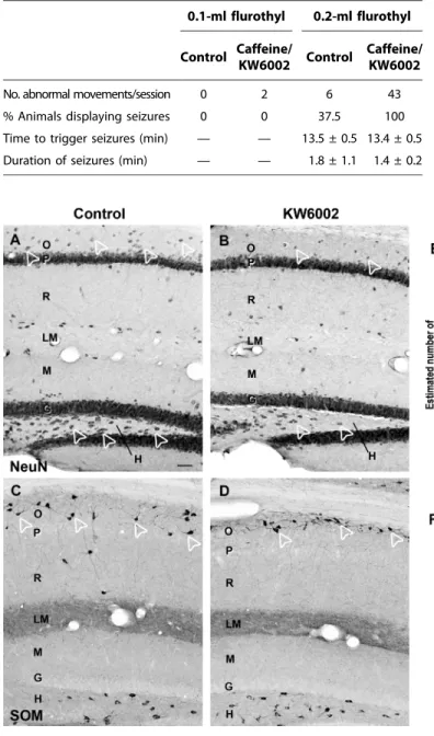
Using immunohistochemistry for the neuron-specific nuclear protein (NeuN), we found a marked neuronal loss in the stratum oriens and stratum radiatum/lacunosum moleculare of the CA1-CA3 region as well as in the hilus of the dentate gyrus of caffeine-exposed (n = 7)
Fig. 2. A2ARs are expressed by
migrat-ing GABA neurons in the rat and monkey fetal telencephalon. (A to C) Represent-ative adjacent coronal sections (of five to six examined per animal) of the rat tel-encephalon at E13. (A) Most newly generated somatostatin-containing neurons (SOM; black arrows) were located in the preplate (PP) of the ventral telencephalon, at the level of the ganglionic eminence (GE) with a few in the preplate of the adjacent dorsal tel-encephalon (black arrowheads). (B) Immu-nolabeling of A2ARs was detected in these
same regions (arrows). (C) Cresyl violet– stained section showing the ganglionic emi-nence and the proliferative zone of the dorsal telencephalon including the subventricular (SVZ) and ventricular (VZ) zones. (D) Higher magnification of a somatostatin-containing neuron from (A) with a characteristic mor-phology of a migrating neuron including a leading process (black arrows). (E) Accu-mulation of A2AR immunolabeling in the
cell body and leading process (black arrows) of a migrating somatostatin-containing neuron. (F) Confocal image of a migrating somatostatin-containing neuron in the preplate (white asterisk, cell body; white arrows, leading process). Cell is double-labeled for somatostatin (green) and A2AR
(red). (G and H) A2AR immunolabeling of
a representative coronal section of the monkey fetal telencephalon at E75. (G) A2AR immunoreactivity was detected in
the lower intermediate zone (IZ)/upper subventricular zone (arrowheads). (H) High-er magnification of the region in (G) showing many tangentially oriented migrating neuron-like cells immunolabeled for A2AR
(black arrows). Scale bars, 50 mm (A to C), 5 mm (D to F), 250 mm (G), and 25 mm (H).
on August 7, 2013
stm.sciencemag.org
and KW6002-exposed (n = 4) adult offspring (3 months old) compared to control (n = 12) mice (Fig. 4, A and B, and Table 3). The estimated total number of neurons in caffeine- or KW6002-exposed animals was
decreased, respectively, by 38 and 19% in the stratum oriens, by 34 and 24% in stratum radiatum/lacunosum moleculare, and by 39 and 22% in the hilus of the dentate gyrus (Fig. 4E and Table 3). Given that, in
con-Fig. 3. Caffeine exposure increases spontaneous and network glutamatergic and GABAergic activities in P6 mouse offspring. (A) Representative traces of spon-taneous activity recorded at a holding potential of +10 mV in a hippocampal CA3 PC from a brain slice of a control or a caffeine-exposed pup at P6. Black asterisks rep-resent GDPs. Note the in-crease in the frequency of GDPs and of spontaneous synaptic currents recorded between GDPs in caffeine-exposed offspring. Inset: De-tail of a GDP event indicated by #. (B) Bar graphs showing a decreased mean IEI be-tween GDPs recorded in hip-pocampal brain slices from caffeine-exposed pups com-pared to control pups (**P < 0.01) and from KW6002-exposed pups compared to vehicle (*P < 0.05). (C) The glutamatergic contribution to the overall synaptic input during GDPs was increased in PCs (**P < 0.01) but not in the stratum radiatum (R) interneurons (IN) (P = 0.82) from caffeine- or KW6002-exposed pups. The top panel shows representative glu-tamatergic and GABAergic currents recorded at−60 and +10 mV, respectively, dur-ing GDPs in a CA3 PC and a stratum radiatum inter-neuron. The area under the curve was smaller in GDPs recorded at +10 mV in PCs
of caffeine-exposed pups, which corresponds to a lower inhibitory charge transfer to the cells. (D and E) Representative traces of sIPSCs and sEPSCs recorded in vitro in a stratum radiatum interneuron and a PC from control and caffeine-exposed offspring. (F and G) Cumulative
probability distributions of IEIs for sIPSCs and sEPSCs recorded in vitro in a stratum radiatum interneuron and a PC from control and caffeine-exposed mouse offspring. Bar graphs showing the mean IEI are displayed below the cumulative probability distributions. In hippocampal slices from caffeine- or KW6002-exposed offspring, the IEI of sIPSCs (left panels) and sEPSCs (right panels) was decreased in stratum radiatum interneurons and in PCs com-pared to control or vehicle-exposed animals. (H) The contribution of the glutamatergic synaptic input to the overall synaptic input was increased selectively in PCs from caffeine- and KW6002-exposed offspring, but this was not observed for stratum radiatum interneurons (PCs, *P < 0.01; inter-neurons, P = 0.23). (I and J) GFP-labeled neurons in a control (I) and in a caffeine-exposed (J) P6 pup (same as in Fig. 1). (K to M) Biocytin-labeled interneurons (I and J) and PCs (K) recorded in vitro in a control (K) and a caffeine-exposed (L and M) P6 pup. In neurons from caffeine-exposed offspring, varicosities were consistently observed along dendrites of interneurons labeled with GFP (J, black arrows) or with biocytin (L, black arrows) but not along dendrites of PCs (M). These varicosities on dendrites are reminiscent of swelling profiles observed after excitotoxic damage. Scale bars, 50 mm (I and J) and 25 mm (K to M).
on August 7, 2013
stm.sciencemag.org
trol GIN mice, 87% of the stratum oriens neurons contain somatostatin, a population of neurons known to be particularly vulnerable in patho-logical conditions (42–45), we specifically analyzed their fate in adult caffeine-exposed offspring. Most of the cell loss observed in the stratum oriens corresponded to a 30% decrease in somatostatin-containing GABA neurons (Fig. 4, C, D, and F, and Table 3). The loss of somatostatin-containing neurons was region-specific because no difference in the number of somatostatin-containing neurons was found in the hilus of the dentate gyrus between treated (Fig. 4, D and F, and Table 3) and control mice (Fig. 4, C and F, and Table 3). The cell loss was also cell type–specific, because the number of GFP-containing interneurons, which represent a subpopulation of somatostatin-containing neurons in GIN mice, was not modified in adult animals (Fig. 1).
We then investigated whether the loss of somatostatin-containing interneurons in the stratum oriens was associated with electrophysio-logical modifications in CA1 PCs, the main target of these neurons and the output region of the hippocampal network. The frequency of sIPSCs was increased by 155 and 252% in caffeine- and KW6002-exposed adult offspring, respectively (Fig. 5, A and C, and table S5), whereas the
Table 2. Seizure susceptibility of control and caffeine- or KW6002-exposed pups to the convulsing agent flurothyl. Caffeine- or KW6002-exposed P6 pups were more vulnerable to flurothyl than control pups of the same age (eight controls from two dams, four caffeine-exposed pups from one dam, and four KW6002-exposed pups from one dam). Treated pups displayed a higher number of abnormal (spasm-like) movements after exposure to flurothyl, and the proportion of treated pups that manifested clonic seizures at a lower concentration of flurothyl (0.2 ml) was greater. No differences were found be-tween the time to trigger seizures and the duration of seizures bebe-tween groups.
0.1-ml flurothyl 0.2-ml flurothyl
Control Caffeine/
KW6002 Control
Caffeine/ KW6002
No. abnormal movements/session 0 2 6 43
% Animals displaying seizures 0 0 37.5 100
Time to trigger seizures (min) — — 13.5 ± 0.5 13.4 ± 0.5
Duration of seizures (min) — — 1.8 ± 1.1 1.4 ± 0.2
Fig. 4. Caffeine treatment leads to GABAergic cell loss in adult offspring. (A to D) Representative hippocampal coronal sections (of 10 to 12 examined per animal) from control (A and C) and KW6002-exposed (B and D) adult mouse off-spring immunolabeled for NeuN (A and B) and somatostatin (C and D). (A and B) NeuN-labeled cell bodies (arrowheads) were decreased in the stratum oriens (O) and the hilus (H) of the dentate gyrus in KW6002-exposed offspring (B) compared to controls (A). (D) KW6002-exposed offspring showed a decrease in somatostatin-containing neurons (arrowheads) compared to control (C). (E) Bar graphs of the estimated number of NeuN-containing neurons in control and caffeine- or
KW6002-exposed offspring show a significant decrease in the number of neurons in caffeine- and KW6002-exposed adult offspring compared to control in the stratum oriens (caffeine, **P < 0.01; KW6002, *P < 0.05), radiatum/lacunosum moleculare (R/LM) (caffeine, **P < 0.01; KW6002, *P < 0.05), and hilus (caffeine, **P < 0.01; KW6002, *P < 0.05). (F) Bar graphs of the estimated number of somatostatin-containing neurons show a significant decrease in the number of neurons in caffeine-and KW6002-exposed offspring compared to control in the stratum oriens (*P < 0.05) but not in the hilus (P = 0.132). P, stratum pyramidale; M, molecular layer of the den-tate gyrus; G, granular cell layer. *P < 0.05, **P < 0.01. Scale bars, 50 mm (A to D).
on August 7, 2013
stm.sciencemag.org
frequency of sEPSCs was decreased by 79 and 82% in adult caffeine- and KW6002-exposed adult offspring, respectively (Fig. 5, B and D, and table S5). The corresponding mean decrease in the IEI between consec-utive synaptic events is displayed by the histograms below the frequency curves. As a result, the contribution of the glutamatergic synaptic input to the overall synaptic input was decreased by 48% in caffeine- and KW6002-exposed compared to control and vehicle-treated animals (Fig. 5E). This may reflect a reorganization of the hippocampal circuitry in adult caffeine-exposed offspring, which may involve a hypoactivity of glutamatergic networks as well as a hyperactivity of GABAergic net-works (33, 34, 42).
These modifications were not associated with changes in long-term potentiation (LTP). LTP was induced in 6 of 9 hippocampal slices in controls, in 8 of 12 slices in caffeine-exposed, and in 8 of 8 slices in KW6002-exposed adult offspring (Fig. 5F). The increase in the slope of the field excitatory postsynaptic potential (that is, LTP) was not significant-ly different (P = 0.74) between controls (47%), caffeine-exposed (37%), and KW6002-exposed (51%) adult offspring (Fig. 5F). The stimulus inten-sity used to evoke field potentials was similar in the three groups, suggest-ing that the decrease in sEPSC frequency (Fig. 5, B and D) reflects a hypoactivity of presynaptic glutamatergic networks rather than their loss. Caffeine-exposed rodent offspring display cognitive deficits as adults
Given that the reorganization of hippocampal circuits may be associated with deficits in spatial memory (46), we assessed hippocampus-dependent memory in treated and control adult offspring using an object dis-placement and substitution task. Animals exposed to caffeine during development showed a lower preference for the displaced object com-pared to control animals, indicating a deficit in spatial, hippocampus-dependent memory (P < 0.05, nonparametric t test) (Fig. 6A). Their performance was also decreased on the nonspatial memory task, as they spent similar times exploring the familiar and new object (P < 0.05, non-parametrict test) (Fig. 6B).
Locomotor activity was not altered in caffeine-exposed adult off-spring (Fig. 6C). However, caffeine-exposed adult offoff-spring displayed impairment in the novel object recognition test (Fig. 6D) and two-trial reference memory test in the Y maze (Fig. 6E), in keeping with the re-sults obtained in rats exposed to caffeine during the embryonic period only (47). The recognition index for the novel object was decreased by 40% in caffeine-exposed and by 64% in KW6002-exposed adult off-spring (Fig. 6D;P < 0.05 control versus caffeine, and P < 0.01 vehicle
Table 3. Loss of neurons in treated adult offspring. Stereological quanti-fication of the total number of neurons (identified by NeuN immunoreactivity) and somatostatin-expressing neurons in the hippocampus is shown for con-trol, caffeine-exposed, and KW6002-exposed adult mouse offspring. NeuN-labeled neurons were quantified in the hippocampal regions (stratum oriens,
hilus of the dentate gyrus, and stratum radiatum). Somatostatexpressing in-terneurons were quantified in the hilus of the dentate gyrus and stratum oriens, where they were more abundantly located. Given that vehicle treatment did not modify any morphological parameter measured at P6, a stereological quantification was not performed in this group of adult animals.
Stratum oriens Dentate gyrus (hilus) Stratum radiatum
NeuN Control Caffeine KW6002 Control Caffeine KW6002 Control Caffeine KW6002
19,404 ± 658 (n = 4) 11,938 ± 1,506 (n = 4)† 15,744 ± 1,113 (n = 3)* 17,670 ± 629 (n = 4) 10,866 ± 411 (n = 4)† 13,776 ± 1,815 (n = 3)* 29,154 ± 1,988 (n = 4) 19,152 ± 1,152 (n = 4)† 22,064 ± 3,778 (n = 4)*
Somatostatin Control Caffeine/KW6002 Control Caffeine/KW6002
16,817 ± 1,072 (n = 3)
12,296 ± 592 (n = 3) * 5,884 ±
140 (n = 3)
5,141 ± 368 (n = 3)
*P < 0.05, one-way ANOVA. †P < 0.01, one-way ANOVA.
Fig. 5. GABAergic and glutamatergic activity in hippocampal CA1 PCs in adult offspring. (A) The frequency of sIPSCs was increased in caffeine-exposed adult offspring representing an increase in GABAergic activity. (B) The frequency of sEPSCs was decreased in caffeine-exposed adult offspring representing a decrease in glutamatergic activity. (C and D) Cumulative probability distributions of IEIs for sIPSCs and sEPSCs in caffeine- and KW6002-exposed adult offspring. Histograms showing the mean IEIs are depicted below. (E) The glutamatergic synaptic input was decreased in PCs of caffeine- and KW6002-exposed animals (*P < 0.05). (F) LTP is similar in control and caffeine- and KW6002-exposed adult mouse offspring. Left: Time evolution of field postsynaptic potential (fPSP) slopes after tetanic stimulation applied 10 min after obtaining stable baseline. Right: Averaged fPSP slopes before (gray trace) and 30 min after (black trace) tetanic stimulation. *P < 0.05, **P < 0.01.
on August 7, 2013
stm.sciencemag.org
versus KW6002). In the modified Y-maze test, all treated animals spent less time exploring the novel arm during the test trial. The exploration time decreased from 41 to 36% in caffeine-exposed and from 43 to 37% in KW6002-exposed adult offspring (Fig. 6E;P < 0.05). Altered perform-ances in these tasks were not related to increased anxiety because there was no difference between control, vehicle-treated, caffeine-treated, and KW6002-treated mice in the time spent in the open arm in the elevated plus maze (Fig. 6F). These data demonstrate that early life exposure to caffeine (or KW6002) may lead to altered memory function.
DISCUSSION
The results presented here suggest that maternal caffeine consumption by mouse dams during gestation and lactation has effects on neuronal development and adult behavior of their offspring. This developmental effect of caffeine may involve the blockade of A2ARs because
caffeine-induced modifications could be reproduced with a selective A2AR
an-tagonist, although we cannot rule out contributions of the other targets
of caffeine, for example, A1receptors, as shown during parturition (48).
Caffeine treatment did not have a major influence on food and water intake or on the day/night rhythm of female mice. However, we cannot fully exclude that treatment-associated physiological or behavioral changes in dams might have influenced fetal and/or neonatal develop-ment, for example, through changes in placental perfusion (49). During the dark, that is, active phase, female caffeine-treated mice showed increased activity. However, dams and pups were always found in the nest, suggesting that increased maternal activity did not impair mater-nal care for pups. The fact that we were able to reproduce our data in A2AR-KO pups suggests that the impact of perinatal caffeine exposure
on brain development may be linked to antagonism of A2AR by caffeine.
It has been reported that A2AR-KO adult mice display enhanced
working memory (50) but not reference memory (51). This finding does not contradict our results as we stopped the transient interference of A2ARs after weaning by withdrawing caffeine from the drinking water,
whereas A2ARs are absent throughout life in the A2AR-KO animals.
Thus, our results suggest a new role for A2ARs in the control of
hippo-campal development. However, we cannot fully rule out that the
Fig. 6. Caffeine exposure during early life leads to memory deficits in adult offspring. (A) Caffeine-exposed adult offspring (6 months of age, n = 5) displayed a deficit in hippocampus-dependent spatial memory on the object displacement task compared to control (n = 7) (A, control, nondisplaced: −5.46 ± 2.96, displaced: 2.85 ± 3.22, *P < 0.05; caffeine, nondisplaced: 0.36 ± 0.72, displaced: 2.61 ± 2.36, P = 0.286). (B) Caffeine-exposed adult offspring (6 months of age) also displayed a deficit on the hippocampal-dependent nonspatial object substitution task compared to control (con-trol, n = 7, familiar:−6.02 ± 4.01, novel: 11.51 ± 4.20, *P < 0.05; caffeine, n = 5, familiar: 3.55 ± 3.87, novel: 8.32 ± 3.31, P = 0.090). (C to E) Behavior anal-ysis of control, caffeine-exposed, vehicle-exposed, and KW6002-exposed adult GIN mouse offspring (at 3 months of age). (C) Locomotion (number of cross-ings) of animals exposed to caffeine or KW6002, analyzed during the first session in the open-field arena, was similar to respective controls (control:
166 ± 12, n = 9; caffeine: 156 ± 10, n = 8, P = 0.588; vehicle: 131 ± 9, n = 10; KW6002: 147 ± 8, n = 7, P = 0.227). (D and E) In contrast to their respective controls, both caffeine- and KW6002-exposed adult offspring displayed impaired performance on memory tasks in the object recognition test (D, control: 0.48 ± 0.04, n = 9, caffeine: 0.29 ± 0.06, n = 8, *P < 0.05, vehicle: 0.39 ± 0.003, n = 10, KW6002: 0.14 ± 0.08, n = 7, **P < 0.01) and by spending less time exploring the novel arm in the Y maze (E, control: 41 ± 1%, n = 8, caf-feine: 36 ± 2%, n = 10, *P < 0.05; vehicle: 43 ± 2%, n = 10, KW6002: 37 ± 2%, n = 7, *P < 0.05). (F) All treated and untreated adult offspring spent a similar amount of time in the open arm of the elevated plus maze, indicating no change in anxiety (control: 37 ± 7%, n = 9, caffeine: 29 ± 5%, n = 10, P = 0.423; vehicle: 28 ± 5%, n = 10, KW6002: 26 ± 3%, n = 7, P = 0.817). *P < 0.05, **P < 0.01, one-way ANOVA followed by a Tukey test, comparing treated with control mice.
on August 7, 2013
stm.sciencemag.org
caffeine-induced maternal hyperactivity and weight loss of the pups might be a potentially confounding factor. In keeping with our observations, maternal caffeine intake during pregnancy in humans is associated with lower birth weight but no change in gestational length (52). In humans, lower birth weight correlates with enhanced seizure susceptibility (53) and cognitive deficits (54).
The delayed migration of GABA neurons induced by caffeine expo-sure was associated with a general increase in neuronal network activity, although causality between both observations remains to be established. These alterations share some similarities with those found in animals exposed to drugs of abuse during developmental periods (6). After weaning, animals were not exposed to caffeine any longer, but the early effects of caffeine on neuron migration and integration ultimately led to long-term sequelae in hippocampal circuits. The disruption of develop-mental programs usually leads to long-term alterations in GABAergic circuits, typified by neuronal loss (including subpopulations of somatostatin-containing neurons), and cognitive deficits in adult animals (40, 55–57). The phenotypic traits observed after caffeine exposure show some similarities to those found in models of neuronal migration dis-orders (31) and models of psychiatric disorders linked to GABA neuron dysfunction (41). This suggests that interfering with GABA neurons during development may have adverse consequences.
The mechanisms underlying the changes in glutamatergic and GABAergic activities in adult caffeine-exposed offspring may involve an imbalance between excitation and inhibition; a correct balance is im-portant for the proper functioning of cortical networks (58, 59). Cogni-tive deficits in certain psychiatric disorders are also associated with hyperactivity of inhibitory circuits or hypoactivity of glutamatergic cir-cuits (41). The altered balance between excitation and inhibition found in adult caffeine-exposed offspring may contribute to the impaired memory observed on several memory tests.
The challenge with rodent models for studying the effects of psycho-active drugs is whether the results can be extrapolated to humans given the differences in development and maturation across species. The ex-pression of A2ARs in migrating neurons in macaques indicates that
A2AR-related pathways can be potentially affected by caffeine in
pri-mates. Note that caffeine citrate is widely used for the treatment of apnea in premature human neonates up to 33 to 34 weeks of gestation. No deleterious consequences of this treatment have been identified in treated premature neonates up to the age of 5 years (60, 61). Given our findings, it will be important to develop longitudinal clinical studies to assess short- and long-term consequences in human neonates born to women who consumed caffeine on a regular basis during pregnancy and lactation, as well as in neonates treated with caffeine citrate.
MATERIALS AND METHODS
Study design
The overall objective of the study was to determine the effects of caffeine consumption (caffeine was added to the drinking water) by mouse dams on the brain development of their offspring. Hippocampal circuits of male offspring at P6 from treated and untreated dams were assessed with electrophysiological, morphological, and immunohistochemical techniques. Seizure susceptibility was also assessed. When the caffeine-exposed and control offspring reached adulthood, we assessed hippo-campal circuits and cognitive performance. A specific antagonist of A2ARs was also used to assess whether the effects of caffeine could be
due to its antagonizing action on A2ARs. Time-lapse imaging was used
to measure the effects of caffeine and A2AR antagonists on the migration
of GABA neurons in medial ganglionic eminence cell cocultures. Final-ly, several cognitive functions, including spatial memory, were assessed in caffeine-exposed adult offspring.
Animals
Caffeine and KW6002 treatments were performed in GIN mice and in A2AR constitutive knockout dams. Caffeine was given to animals dissolved
in tap water at a concentration of 0.3 g/liter. KW6002 (istradefylline) was prepared and given to animals at a concentration of 2 mg/kg per day in a vehicle solution containing 0.4% methylcellulose and 0.9% NaCl. Both treatments were started 15 days before mating and prolonged during all gestational period up to P15. Caffeine treatment started at gestational day 0.5 for experiments assessing the activity of females and behavioral analysis of pups of treated dams.
Electrophysiology
Transverse hippocampal slices from pups and from adults. IPSCs were recorded at a holding potential of +10 mV, the reversal potential for glutamatergic events, and EPSCs were recorded at−60 mV, the reversal potential for GABAergic events (42). For LTP induction protocol, fPSPs were recorded in the stratum radiatum of CA1 region and evoked upon tetanic stimulation of the Schaffer collaterals.
Immunohistochemistry
Immunohistochemistry was performed as previously described (30, 62) with tissue from rat embryos, GIN mouse pups, A2AR-KO pups, and
adult GIN mice. Immunohistochemical localization of A2ARs was performed
in E13 Wistar rats and E75 cynomolgus monkey (Macaca fascicularis). Stereological quantification
The numbers of neurons labeled for NeuN, GFP, and somatostatin were estimated with the optical fractionator method, as previously described (43).
Cell culture of MGE and analysis of migration speed GFP-expressing MGE explants (E13.5) were cultured on E13.5-dissociated cortical cells (26) and monitored by time-lapse video mi-croscopy before and after drug application.
Measurement of caffeine and its metabolites in brain tissue The tissue of all brain was homogenized and mixed with 30% of per-chloric acid, and the supernatant obtained after centrifugation was separated by high-performance liquid chromatography (HPLC). Behavioral analysis
Locomotor activity was evaluated in an open-field arena by counting the total number of crossings for 5 min. Spatial memory performance was evaluated with the modified Y-maze test (63, 64). The object recog-nition test was performed, and the recogrecog-nition index was calculated as previously described (65, 66). Anxiety was evaluated in an elevated plus maze (67, 68). The object displacement and substitution task tests were performed in three stages (69).
Seizure induction with flurothyl
The evaluation of spasm-like movements and seizures was based on (70). Animals were video-monitored.
on August 7, 2013
stm.sciencemag.org
Statistical analysis
One-way ANOVA was used to compare the number of neurons and the physiological activity in the hippocampus of control, vehicle-exposed, caffeine-exposed, and KW6002-exposed pups and adults. Eventually, a Tukey test was used as a post hoc test. A Student’s t test was used in the experiments of cell migration and in the object displacement and substitution task test. The behavioral parameters of female mice and pups were analyzed with a three-way or two-way mixed ANOVA, respectively, followed by Newman-Keuls post hoc tests when appropri-ate. Statistical significance was considered whenP < 0.05. Levels of sig-nificance are indicated as follows: *P < 0.05, **P < 0.01, and ***P < 0.001.
SUPPLEMENTARY MATERIALS
www.sciencetranslationalmedicine.org/cgi/content/full/5/197/197ra104/DC1 Methods
Fig. S1. Physiological and behavioral consequences of caffeine treatment.
Fig. S2. Caffeine exposure during pregnancy and the early postnatal period delays the migra-tion and insermigra-tion of GFP-containing neurons in cortical layers and in the hippocampus. Fig. S3. Caffeine exposure during pregnancy and the early postnatal period delays the migra-tion and insermigra-tion of somatostatin-containing neurons and the maturamigra-tion of the hippocampal GABAergic network.
Fig. S4. Decreased numbers of somatostatin-containing neurons in the hippocampus and in cortical layers of mice lacking A2ARs.
Table S1. Properties of the spontaneous activity (GDPs) in hippocampal slices from P5 to P7 control, caffeine-exposed, vehicle-exposed, and KW6002-exposed mouse pup offspring. Table S2. Imbalance between excitatory and inhibitory synaptic inputs in PCs from caffeine-and KW6002-exposed pup offspring during GDPs.
Table S3. Properties of the spontaneous activity recorded in vitro in hippocampal neurons of P5 to P7 mouse pup offspring.
Table S4. Imbalance between excitatory and inhibitory synaptic inputs in PCs from caffeine-and KW6002-exposed pup offspring during spontaneous activity outside GDPs.
Table S5. Properties of the spontaneous activity recorded in vitro in hippocampal neurons from control, caffeine-exposed, vehicle-exposed, and KW6002-exposed adult mouse offspring.
REFERENCES AND NOTES
1. A. L. Salisbury, K. L. Ponder, J. F. Padbury, B. M. Lester, Fetal effects of psychoactive drugs. Clin. Perinatol. 36, 595–619 (2009).
2. M. W. Miller, Effects of alcohol on the generation and migration of cerebral cortical neu-rons. Science 233, 1308–1311 (1986).
3. J. E. Crandall, H. E. Hackett, S. A. Tobet, B. E. Kosofsky, P. G. Bhide, Cocaine exposure de-creases GABA neuron migration from the ganglionic eminence to the cerebral cortex in embryonic mice. Cereb. Cortex 14, 665–675 (2004).
4. S. Anavi-Goffer, J. Mulder, The polarised life of the endocannabinoid system in CNS de-velopment. Chembiochem 10, 1591–1598 (2009).
5. P. Berghuis, A. M. Rajnicek, Y. M. Morozov, R. A. Ross, J. Mulder, G. M. Urbán, K. Monory, G. Marsicano, M. Matteoli, A. Canty, A. J. Irving, I. Katona, Y. Yanagawa, P. Rakic, B. Lutz, K. Mackie, T. Harkany, Hardwiring the brain: Endocannabinoids shape neuronal connectivity. Science 316, 1212–1216 (2007).
6. B. L. Thompson, P. Levitt, G. D. Stanwood, Prenatal exposure to drugs: Effects on brain devel-opment and implications for policy and education. Nat. Rev. Neurosci. 10, 303–312 (2009). 7. B. B. Fredholm, K. Bättig, J. Holmén, A. Nehlig, E. E. Zvartau, Actions of caffeine in the brain
with special reference to factors that contribute to its widespread use. Pharmacol. Rev. 51, 83–133 (1999).
8. P. Nawrot, S. Jordan, J. Eastwood, J. Rotstein, A. Hugenholtz, M. Feeley, Effects of caffeine on human health. Food Addit. Contam. 20, 1–30 (2003).
9. http://www.acog.org/~/media/Committee%20Opinions/Committee%20on%20Obstetric% 20Practice/co462.pdf?dmc=1&ts=20130313T0649414568
10. U. Adén, Methylxanthines during pregnancy and early postnatal life. Handb. Exp. Pharmacol. 200, 373–389 (2011).
11. J. A. Ribeiro, A. M. Sebastião, Caffeine and adenosine. J. Alzheimers Dis. 20 (Suppl.), S3–S15 (2010).
12. G. Schulte, B. B. Fredholm, Signalling from adenosine receptors to mitogen-activated pro-tein kinases. Cell. Signal. 15, 813–827 (2003).
13. U. Adén, E. Herlenius, L. Q. Tang, B. B. Fredholm, Maternal caffeine intake has minor effects on adenosine receptor ontogeny in the rat brain. Pediatr. Res. 48, 177–183 (2000). 14. O. Bjorklund, J. Kahlström, P. Salmi, B. B. Fredholm, Perinatal caffeine, acting on maternal
adenosine A1receptors, causes long-lasting behavioral changes in mouse offspring. PLoS One 3, e3977 (2008).
15. A. A. Oliva Jr., M. Jiang, T. Lam, K. L. Smith, J. W. Swann, Novel hippocampal interneuronal subtypes identified using transgenic mice that express green fluorescent protein in GABAergic interneurons. J. Neurosci. 20, 3354–3368 (2000).
16. P. P. Quilichini, M. Le Van Quyen, A. Ivanov, D. A. Turner, A. Carabalona, H. Gozlan, M. Esclapez, C. Bernard, Hub GABA neurons mediate g-frequency oscillations at ictal-like event onset in the immature hippocampus. Neuron 74, 57–64 (2012).
17. J. G. Corbin, S. Nery, G. Fishell, Telencephalic cells take a tangent: Non-radial migration in the mammalian forebrain. Nat. Neurosci. 4 (Suppl.), 1177–1182 (2001).
18. G. Miyoshi, G. Fishell, GABAergic interneuron lineages selectively sort into specific cortical layers during early postnatal development. Cereb. Cortex 21, 845–852 (2011). 19. S. Nery, G. Fishell, J. G. Corbin, The caudal ganglionic eminence is a source of distinct
cortical and subcortical cell populations. Nat. Neurosci. 5, 1279–1287 (2002).
20. G. Miyoshi, S. J. Butt, H. Takebayashi, G. Fishell, Physiologically distinct temporal cohorts of cortical interneurons arise from telencephalic Olig2-expressing precursors. J. Neurosci. 27, 7786–7798 (2007).
21. S. J. Pleasure, S. Anderson, R. Hevner, A. Bagri, O. Marin, D. H. Lowenstein, J. L. Rubenstein, Cell migration from the ganglionic eminences is required for the development of hippocam-pal GABAergic interneurons. Neuron 28, 727–740 (2000).
22. S. T. Dupuy, C. R. Houser, Developmental changes in GABA neurons of the rat dentate gyrus: An in situ hybridization and birthdating study. J. Comp. Neurol. 389, 402–418 (1997). 23. Y. M. Morozov, A. E. Ayoub, P. Rakic, Translocation of synaptically connected interneurons across the dentate gyrus of the early postnatal rat hippocampus. J. Neurosci. 26, 5017–5027 (2006). 24. R. Tyzio, A. Represa, I. Jorquera, Y. Ben-Ari, H. Gozlan, L. Aniksztejn, The establishment of
GABAergic and glutamatergic synapses on CA1 pyramidal neurons is sequential and correlates with the development of the apical dendrite. J. Neurosci. 19, 10372–10382 (1999).
25. J. P. Baudoin, C. Alvarez, P. Gaspar, C. Métin, Nocodazole-induced changes in microtubule dynamics impair the morphology and directionality of migrating medial ganglionic eminence cells. Dev. Neurosci. 30, 132–143 (2008).
26. A. Bellion, J. P. Baudoin, C. Alvarez, M. Bornens, C. Métin, Nucleokinesis in tangentially migrating neurons comprises two alternating phases: Forward migration of the Golgi/centrosome associated with centrosome splitting and myosin contraction at the rear. J. Neurosci. 25, 5691–5699 (2005).
27. R. A. Cunha, P. M. Agostinho, Chronic caffeine consumption prevents memory disturbance in different animal models of memory decline. J. Alzheimers Dis. 20 (Suppl. 1), S95–S116 (2010). 28. S. Ferré, An update on the mechanisms of the psychostimulant effects of caffeine. J. Neurochem.
105, 1067–1079 (2008).
29. C. Métin, R. B. Vallee, P. Rakic, P. G. Bhide, Modes and mishaps of neuronal migration in the mammalian brain. J. Neurosci. 28, 11746–11752 (2008).
30. Z. Petanjek, B. Berger, M. Esclapez, Origins of cortical GABAergic neurons in the cynomolgus monkey. Cereb. Cortex 19, 249–262 (2009).
31. R. Guerrini, E. Parrini, Neuronal migration disorders. Neurobiol. Dis. 38, 154–166 (2010). 32. Y. Ben-Ari, Developing networks play a similar melody. Trends Neurosci. 24, 353–360 (2001). 33. D. L. Jones, S. C. Baraban, Inhibitory inputs to hippocampal interneurons are reorganized
in Lis1 mutant mice. J. Neurophysiol. 102, 648–658 (2009).
34. D. L. Jones, S. C. Baraban, Characterization of inhibitory circuits in the malformed hippo-campus of Lis1 mutant mice. J. Neurophysiol. 98, 2737–2746 (2007).
35. M. F. McManus, I. M. Nasrallah, M. M. Pancoast, A. Wynshaw-Boris, J. A. Golden, Lis1 is necessary for normal non-radial migration of inhibitory interneurons. Am. J. Pathol. 165, 775–784 (2004).
36. A. A. Oliva Jr., T. T. Lam, J. W. Swann, Distally directed dendrotoxicity induced by kainic acid in hippocampal interneurons of green fluorescent protein-expressing transgenic mice. J. Neurosci. 22, 8052–8062 (2002).
37. G. L. Holmes, The long-term effects of neonatal seizures. Clin. Perinatol. 36, 901–914, vii–viii (2009).
38. S. W. Briggs, A. S. Galanopoulou, Altered GABA signaling in early life epilepsies. Neural Plast. 2011, 527605 (2011).
39. M. D. Krasowski, Differential modulatory actions of the volatile convulsant flurothyl and its anesthetic isomer at inhibitory ligand-gated ion channels. Neuropharmacology 39, 1168–1183 (2000).
40. K. Ramamoorthi, Y. Lin, The contribution of GABAergic dysfunction to neurodevelopmental disorders. Trends Mol. Med. 17, 452–462 (2011).
41. O. Marín, Interneuron dysfunction in psychiatric disorders. Nat. Rev. Neurosci. 13, 107–120 (2012). 42. R. Cossart, C. Dinocourt, J. C. Hirsch, A. Merchan-Perez, J. De Felipe, Y. Ben-Ari, M. Esclapez, C. Bernard, Dendritic but not somatic GABAergic inhibition is decreased in experimental epilepsy. Nat. Neurosci. 4, 52–62 (2001).
on August 7, 2013
stm.sciencemag.org
43. C. Dinocourt, Z. Petanjek, T. F. Freund, Y. Ben-Ari, M. Esclapez, Loss of interneurons innervating pyramidal cell dendrites and axon initial segments in the CA1 region of the hippocampus following pilocarpine-induced seizures. J. Comp. Neurol. 459, 407–425 (2003).
44. D. Arabadzisz, T. F. Freund, Changes in excitatory and inhibitory circuits of the rat hippo-campus 12–14 months after complete forebrain ischemia. Neuroscience 92, 27–45 (1999). 45. C. Perez-Cruz, M. W. Nolte, M. M. van Gaalen, N. R. Rustay, A. Termont, A. Tanghe, F. Kirchhoff, U. Ebert, Reduced spine density in specific regions of CA1 pyramidal neurons in two trans-genic mouse models of Alzheimer’s disease. J. Neurosci. 31, 3926–3934 (2011).
46. L. Chauvière, N. Rafrafi, C. Thinus-Blanc, F. Bartolomei, M. Esclapez, C. Bernard, Early deficits in spatial memory and theta rhythm in experimental temporal lobe epilepsy. J. Neurosci. 29, 5402–5410 (2009).
47. D. E. Soellner, T. Grandys, J. L. Nuñez, Chronic prenatal caffeine exposure impairs novel object recognition and radial arm maze behaviors in adult rats. Behav. Brain Res. 205, 191–199 (2009).
48. V. Dzhala, L. Desfreres, Z. Melyan, Y. Ben-Ari, R. Khazipov, Epileptogenic action of caffeine during anoxia in the neonatal rat hippocampus. Ann. Neurol. 46, 95–102 (1999). 49. P. Kirkinen, P. Jouppila, A. Koivula, J. Vuori, M. Puukka, The effect of caffeine on placental
and fetal blood flow in human pregnancy. Am. J. Obstet. Gynecol. 147, 939–942 (1983). 50. S. J. Zhou, M. E. Zhu, D. Shu, X. P. Du, X. H. Song, X. T. Wang, R. Y. Zheng, X. H. Cai, J. F. Chen,
J. C. He, Preferential enhancement of working memory in mice lacking adenosine A2A recep-tors. Brain Res. 1303, 74–83 (2009).
51. P. M. Canas, L. O. Porciúncula, G. M. Cunha, C. G. Silva, N. J. Machado, J. M. Oliveira, C. R. Oliveira, R. A. Cunha, Adenosine A2Areceptor blockade prevents synaptotoxicity and memory dysfunction caused by b-amyloid peptides via p38 mitogen-activated protein kinase pathway. J. Neurosci. 29, 14741–14751 (2009).
52. V. Sengpiel, E. Elind, J. Bacelis, S. Nilsson, J. Grove, R. Myhre, M. Haugen, H. M. Meltzer, J. Alexander, B. Jacobsson, A. L. Brantsaeter, Maternal caffeine intake during pregnancy is as-sociated with birth weight but not with gestational length: Results from a large prospective observational cohort study. BMC Med. 11, 42 (2013).
53. Y. Sun, M. Vestergaard, C. B. Pedersen, J. Christensen, O. Basso, J. Olsen, Gestational age, birth weight, intrauterine growth, and the risk of epilepsy. Am. J. Epidemiol. 167, 262–270 (2008). 54. B. J. Jefferis, C. Power, C. Hertzman, Birth weight, childhood socioeconomic environment,
and cognitive development in the 1958 British birth cohort study. BMJ 325, 305 (2002). 55. G. Kerjan, H. Koizumi, E. B. Han, C. M. Dubé, S. N. Djakovic, G. N. Patrick, T. Z. Baram, S. F. Heinemann,
J. G. Gleeson, Mice lacking doublecortin and doublecortin-like kinase 2 display altered hippo-campal neuronal maturation and spontaneous seizures. Proc. Natl. Acad. Sci. U.S.A. 106, 6766–6771 (2009).
56. I. Cobos, M. E. Calcagnotto, A. J. Vilaythong, M. T. Thwin, J. L. Noebels, S. C. Baraban, J. L. Rubenstein, Mice lacking Dlx1 show subtype-specific loss of interneurons, reduced inhibition and epilepsy. Nat. Neurosci. 8, 1059–1068 (2005).
57. R. Grantyn, C. Henneberger, R. Jüttner, J. C. Meier, S. Kirischuk, Functional hallmarks of GABAergic synapse maturation and the diverse roles of neurotrophins. Front. Cell. Neurosci. 5, 13 (2011).
58. Y. Shu, A. Hasenstaub, D. A. McCormick, Turning on and off recurrent balanced cortical activity. Nature 423, 288–293 (2003).
59. T. P. Vogels, L. F. Abbott, Gating multiple signals through detailed balance of excitation and inhibition in spiking networks. Nat. Neurosci. 12, 483–491 (2009).
60. B. Schmidt, P. J. Anderson, L. W. Doyle, D. Dewey, R. E. Grunau, E. V. Asztalos, P. G. Davis, W. Tin, D. Moddemann, A. Solimano, A. Ohlsson, K. J. Barrington, R. S. Roberts; Caffeine for Apnea of Prematurity (CAP) Trial Investigators, Survival without disability to age 5 years after neonatal caffeine therapy for apnea of prematurity. JAMA 307, 275–282 (2012). 61. B. Schmidt, R. S. Roberts, P. Davis, L. W. Doyle, K. J. Barrington, A. Ohlsson, A. Solimano, W. Tin;
Caffeine for Apnea of Prematurity Trial Group, Long-term effects of caffeine therapy for apnea of prematurity. N. Engl. J. Med. 357, 1893–1902 (2007).
62. R. Soussi, N. Zhang, S. Tahtakran, C. R. Houser, M. Esclapez, Heterogeneity of the supramammillary– hippocampal pathways: Evidence for a unique GABAergic neurotransmitter phenotype and regional differences. Eur. J. Neurosci. 32, 771–785 (2010).
63. Y. Akwa, N. Ladurelle, D. F. Covey, E. E. Baulieu, The synthetic enantiomer of pregnenolone sulfate is very active on memory in rats and mice, even more so than its physiological neurosteroid counterpart: Distinct mechanisms? Proc. Natl. Acad. Sci. U.S.A. 98, 14033–14037 (2001).
64. F. Dellu, W. Mayo, J. Cherkaoui, M. M. Le, H. Simon, A two-trial memory task with auto-mated recording: Study in young and aged rats. Brain Res. 588, 132–139 (1992). 65. M. S. Costa, P. H. Botton, S. Mioranzza, D. O. Souza, L. O. Porciúncula, Caffeine prevents
age-associated recognition memory decline and changes brain-derived neurotrophic
factor and tirosine kinase receptor (TrkB) content in mice. Neuroscience 153, 1071–1078 (2008).
66. R. A. Bevins, J. Besheer, Object recognition in rats and mice: A one-trial non-matching-to-sample learning task to study‘recognition memory’. Nat. Protoc. 1, 1306–1311 (2006). 67. S. L. Handley, S. Mithani, Effects of alpha-adrenoceptor agonists and antagonists in a
maze-exploration model of‘fear’-motivated behaviour. Naunyn Schmiedebergs Arch. Pharmacol. 327, 1–5 (1984).
68. A. A. Walf, C. A. Frye, The use of the elevated plus maze as an assay of anxiety-related behavior in rodents. Nat. Protoc. 2, 322–328 (2007).
69. M. Pratte, M. Jamon, Impairment of novelty detection in mice targeted for the Chl1 gene. Physiol. Behav. 97, 394–400 (2009).
70. E. Isaeva, D. Isaev, R. Khazipov, G. L. Holmes, Long-term suppression of GABAergic activity by neonatal seizures in rat somatosensory cortex. Epilepsy Res. 87, 286–289 (2009). 71. J. F. Chen, Z. Huang, J. Ma, J. Zhu, R. Moratalla, D. Standaert, M. A. Moskowitz, J. S. Fink,
M. A. Schwarzschild, A2Aadenosine receptor deficiency attenuates brain injury induced by transient focal ischemia in mice. J. Neurosci. 19, 9192–9200 (1999).
72. E. Bona, U. Adén, B. B. Fredholm, H. Hagberg, The effect of long term caffeine treatment on hypoxic-ischemic brain damage in the neonate. Pediatr. Res. 38, 312–318 (1995). 73. G. P. Cognato, P. M. Agostinho, J. Hockemeyer, C. E. Müller, D. O. Souza, R. A. Cunha, Caffeine
and an adenosine A2Areceptor antagonist prevent memory impairment and synaptotoxicity in adult rats triggered by a convulsive episode in early life. J. Neurochem. 112, 453–462 (2010). 74. M. J. West, L. Slomianka, H. J. Gundersen, Unbiased stereological estimation of the total number of neurons in the subdivisions of the rat hippocampus using the optical fractionator. Anat. Rec. 231, 482–497 (1991).
75. M. Esclapez, J. C. Hirsch, Y. Ben-Ari, C. Bernard, Newly formed excitatory pathways provide a substrate for hyperexcitability in experimental temporal lobe epilepsy. J. Comp. Neurol. 408, 449–460 (1999).
76. E. Isaeva, D. Isaev, R. Khazipov, G. L. Holmes, Selective impairment of GABAergic synaptic transmission in the flurothyl model of neonatal seizures. Eur. J. Neurosci. 23, 1559–1566 (2006).
77. M. G. Price, J. W. Yoo, D. L. Burgess, F. Deng, R. A. Hrachovy, J. D. Frost Jr., J. L. Noebels, A triplet repeat expansion genetic mouse model of infantile spasms syndrome, Arx(GCG)10+7, with interneuronopathy, spasms in infancy, persistent seizures, and adult cognitive and behavioral impairment. J. Neurosci. 29, 8752–8763 (2009).
Acknowledgments: We thank M. Picciotto, D. Kullmann, D. Johnston, and F. Morellini for their helpful comments on the manuscript. Funding: Supported by Fundação para a Ciência e para a Tecnologia (grant PTDC/SAU-NEU/74318/2006), INSERM, DARPA (09-68-ESR-FP-010), Fédération de la Recherche sur le Cerveau, Fondation Jérôme Lejeune, the Deutsche Forschungsgemeinschaft (IS63/3-1,2,IS63/4-1), and neurodapt (Landesexzellenzinitiative Hamburg). Author contributions: C.G.S. performed most of the experiments, interpreted the results, and wrote and prepared the manuscript. C.M. performed video-microscopy experiments, interpreted the results, and prepared the manuscript. W.F. measured and evaluated physiological/ behavioral parameters of females and pups receiving caffeine treatment and prepared the man-uscript. N.J.M. performed behavioral experiments, interpreted the results, and prepared the manuscript. S.D. performed immunohistochemical experiments in primates and interpreted the results. E.B. performed immunohistochemical experiments in A2AR-KO mice. P.-S.L., A.G., and M.-P.N. provided technical help. Y.B. and C.E.M. synthesized and provided KW6002. A.I. and Y.Z. performed LTP experiments, interpreted the results, and prepared the manuscript. E.S. and A.R.T. carried out the HPLC determinations of caffeine levels. D.I. supervised the behavioral analyses of females and pups and prepared the manuscript. R.A.C. supervised the project and revised the manuscript. M.E. and C.B. supervised the project and wrote the manuscript. Competing interests: R.A.C. participates in medical education about coffee consumption or-ganized by the nonprofit organization Associação Industrial e Comercial do Café. The other authors declare that they have no competing interests.
Submitted 1 April 2013 Accepted 19 July 2013 Published 7 August 2013 10.1126/scitranslmed.3006258
Citation: C. G. Silva, C. Métin, W. Fazeli, N. J. Machado, S. Darmopil, P.-S. Launay, A. Ghestem, M.-P. Nesa, E. Bassot, E. Szabó, Y. Baqi, C. E. Müller, A. R. Tomé, A. Ivanov, D. Isbrandt, Y. Zilberter, R. A. Cunha, M. Esclapez, C. Bernard, Adenosine receptor antagonists including caffeine alter fetal brain development in mice. Sci. Transl. Med. 5, 197ra104 (2013).
on August 7, 2013
stm.sciencemag.org