Improved serodiagnosis of alveolar echinococcosis of
humans using an in vitro-produced Echinococcus
multilocularis antigen
N. MU¨ LLER*, E. FREI, S. NUN˜ EZ and B. GOTTSTEIN*
Institute of Parasitology, University of Berne, Berne, Switzerland
(Received 30 October 2006; revised 3 November 2006; accepted 3 November 2006; first published online 19 February 2007 )
S U M M A R Y
Serology is an important tool for the diagnosis of alveolar echinococcosis (AE) in humans. In order to improve serodiagnostic performance, we have developed an in vitro-produced Echinococcus mulilocularis metacestode vesicle fluid (EmVF) antigen for application in an immunoblot assay. Immunoblot analysis of EmVF revealed an abundant immunoreactive band triplet of 20–22 kDa, achieving a sensitivity of 100 % based on the testing of sera from 62 pre-operative and pre-treatment cases of active and inactive AE. Thus, the EmVF-immunoblotting allowed the specific detection of cases seronegative by the Em2- and/or EmII/3–10-ELISA, usually attributable to abortive, inactive cases of AE. The specificity of the EmVF-immunoblotting did not allow discrimination between AE and cystic echinococcosis (CE) but was 100 % with respect to non-Echinococcus parasitic infections or cancer malignancies. Based on the findings of this study, it is recommended that the current ELISA test combination (Em2- and II/3–10-ELISA) be complemented with EmVF-immunoblotting, allowing an improved diagnosis of both clinical and subclinical forms of AE, including those associated with E. multilocularis-specific antibody reactivities not detectable by ELISA.
Key words : Echinococcus multilocularis, in vitro cultivation, immunoblotting, enzyme-linked immunosorbent assay (ELISA).
I N T R O D U C T I O N
Human alveolar echinococcosis (AE) is caused by the infection with the metacestode (larval stage) of E. multilocularis (see Gottstein and Reichen, 2002 ; Gottstein, 2004 ; Ito and Craig, 2003). Serological tests based on the enzyme-linked immunosorbent assay (ELISA) are useful for the early diagnosis of AE (i.e. before the disease becomes clinically mani-fest), at a time point when radical surgical resection of the parasitized tissue is more likely (Gottstein et al. 2001). Several years ago, the early immunodiagnosis of AE was improved via the isolation and application in ELISA of an affinity-purified, native metacestode Em2 antigen (Gottstein et al. 1983) and the re-combinant antigen II/3–10 (Mu¨ ller et al. 1989 ; Felleisen and Gottstein, 1993) representing a highly antigenic subfragment of E. multilocularis antigen II/3 (Vogel et al. 1988). The combined application of these antigens allowed discrimination between AE and cystic echinococcosis (CE, caused by the metacestode of E. granulosus) as well as cysticercosis
(caused by the metacestode of Taenia solium) and other diseases caused by various parasitic nematodes and trematodes. Other research groups have also produced recombinant E. multilocularis antigens (EM10, Frosch et al. 1991 ; Em4, Hemmings and McManus, 1991 ; rEm18, Xiao et al. 2003) which exhibited similar diagnostic operating characteristics and all proved to represent the same ERM-gene family (Brehm et al. 1999). Despite the excellent diagnostic qualities of, for example, the ELISA utilizing the antigens Em2 and II/3–10, either a lack of reactivity or some cross-reactivity was observed in a small number of confirmed AE cases, thus ham-pering interpretation in view of these peculiar cases (Gottstein et al. 1993). Weak antibody reactivity is a relatively common feature in AE cases, particularly when related to certain clinical scenarios, such as a recently acquired infection or an abortive course of infection (i.e. degradation or death of the parasite in tissues). Another issue is that cross-reactive serum antibodies, frequently occurring in cancer patients (e.g. Pfister et al. 1999 ; Poretti et al. 1999) may complicate the interpretation of results.
Immunoblot analysis is an important approach that suitably complements the ELISA-based im-munodiagnosis of CE (Poretti et al. 1999 ; Liance et al. 2000 ; Doiz et al. 2001 ; Furuya et al. 2004 ; Akisu et al. 2005). An immunoblot for CE, previously
* Corresponding authors : Institute of Parasitology, La¨nggass-Strasse 122, CH-3012 Berne, Switzerland. Tel : +41 31 6312474 or +41 31 6312418. Fax: +41 31
6312622. E-mail : [email protected] or bruno.
developed in our laboratory based on the use of E. granulosus hydatid cyst fluid (EgHF) antigen, relied on the detection of a genus-specific 8 kDa subunit of antigen B and 2 other immunodiagnostic antigens of 29 and 34 kDa (Poretti et al. 1999). In this study, antigen B and, if detectable, the 29 kDa and 34 kDa antigens were shown to be genus-specific and allowed the diagnosis of AE and CE, with a sensi-tivity of 91 % and a specificity of 97 % (antigen B) or 94 % (29 and 34 kDa antigens), respectively. Some years ago, Ito et al. (1999) established 2 immunoblots for the diagnosis of AE, which were based on the use of fractions containing either semi-purified 8 kDa E. granulosus antigen B or an 18 kDa antigen (Em18), enriched by isoelectric focusing from an E. multi-locularis protoscolex extract. A large-scale serological evaluation of this ‘ dual immunoblot system ’ re-vealed the genus-specific characteristics of antigen B in that 92 % of the CE sera and 79 % of the AE sera reacted with this antigen. Conversely, the Em18 antigen was shown to detect AE with a diagnostic sensitivity of 97 % and a moderate specificity of 74 %, and had potential to differentiate AE from CE. Liance et al. (2000) evaluated a commercial immunoblot (Echinococcus Western Blot IgGTM,
LDBIO Diagnostics, Lyon, France) that employs a whole larval antigen from E. multilocularis. In this test system, the serological reactivity with a 7 kDa band and/or a diffuse 26 to 28 kDa band was indicative of the presence of Echinococcus-specific antibodies. The diagnostic sensitivities of the immunoblot were determined to be 96.7 % for AE and 98 % for CE, respectively. Cross-reactivity was shown using sera from patients with neurocysti-cercosis (7 of 20) or Schistosoma mansoni infection (3 of 18 sera). In a more recent study, Korkmaz et al. (2004) tested 2 E. multilocularis metacestode anti-gens, Em70 and Em90, and achieved a sensitivity of 100 % and a specificity of 99.1 % for the diagnosis of AE. Most of these studies did or could not include (due to unavailability) ‘ diagnostically critical ’ sera, such as those derived from patients with abortive AE or with very low levels of anti-E. multilocularis serum antibodies due to degenerative or dead metacestodes. Such sera frequently remain negative when tested against native or recombinant E. multilocularis anti-gens. Therefore, any improvement of the sensitivity and specificity of an antigen or a test for the sero-diagnosis of AE is considered a significant advance. In the present study, we developed an improved immunoblot assay for the specific diagnosis of AE, irrespective of the form of disease. This assay utilizes an in vitro-produced E. multilocularis vesicle fluid (EmVF) antigen, as an alternative to native anti-gens usually obtained from alveolar cysts from experimentally infected rodents. Together with the present Em2- and II/3–10-ELISA (Gottstein et al. 1993), this immunoblot method allows the accurate diagnosis of both clinical and subclinical cases of AE.
M A T E R I A L S A N D M E T H O D S
Preparation of E. multilocularis vesicle fluid (EmFV ) antigen
The EmVF antigen was obtained by aseptic aspir-ation of fluid from fertile vesicular cysts, axenically cultivated in vitro as described previously (Hemphill and Gottstein, 1995). EmVF was complemented with 1 mmol of the proteinase inhibitor phenyl-methyl-sulfonyl fluoride (PMSF), centrifuged at 14 000 g for 10 min and the supernatant sub-sequently stored frozen atx80 xC until use. In order to validate individual batches of fluid, the protein/ carbohydrate ratio was required to range between 4 : 1 and 5 : 1, the protein concentration between 0.7 and 0.9 mg/ml (EmVF with higher protein con-centrations were discarded). Also, the immunoblot characteristics of the EmVF had to be consistent with those described herein.
Human sera
Well-defined sera from 62 Central European AE patients (54 of them with active hepatic lesions, and 8 of them had fully calcified lesions and thus rep-resented inactive or ‘ abortive ’ cases) were used for assessing the primary diagnostic sensitivity of the tests for AE. Inactivity of metacestodes was proven either by post-diagnostic surgical removal of parasite material, with a subsequent viability assessment as described by Rausch et al. (1987) or by extensive clinical and subsequent computer tomography or X-ray imaging of the patients (see Gottstein et al. 2001). Therefore, all sera were sampled prior to any therapeutic or surgical intervention. Each diagnosis of AE had been confirmed by clinical, histological (if feasible) and serological means.
Sera used for assessing test specificities were obtained from patients with clinically, parasit-ologically and/or histparasit-ologically proven infections, involving the following parasites (disease and num-ber of sera obtained from patients are in parenth-eses) : E. granulosus (cystic echinococcosis, 22 sera), Fasciola hepatica (fascioliasis, 7 sera), Schistosoma spp. (schistosomiasis, 7 sera), Taenia solium (cysti-cercosis, 10 sera), Ascaris lumbricoides (ascariasis, 7 sera), Strongyloides stercoralis (strongyloidiasis, 6 sera), Toxocara canis (toxocariasis ; visceral larva migrans, 7 sera), Trichinella spiralis (trichinellosis, 7 sera) ; Onchocerca volvulus (filariasis, 6 sera), and Entamoeba histolytica (amoebiasis, 7 sera). All of these sera had been pre-selected based on their ‘ positivity ’ using homologous parasite antigens in tests carried out routinely in our diagnostic labora-tory in the University of Berne. Sera from 18 cancer patients were also selected from a previous study (Pfister et al. 1999) based on their serological re-activity in Echinococcus tests, as described by Poretti et al. (1999).
Enzyme-linked immunosorbent assays (ELISAs) These assays were carried out essentially as described by Gottstein et al. (1993), by testing sera at a 1 : 100 dilution and using the following antigens (at optimized coating concentrations) : EmVF-antigen (2.8mg protein per ml) ; EgHF antigen (5 mg protein per ml) ; Em2 antigen (2.8mg carbohydrates per ml) ; II/3–10 antigen (1mg protein per ml). All Echinococcus-antigen-ELISAs employed in the present study were first calibrated to determine the threshold for the discrimination between positive and negative results. The actual cut-off value was thus determined by testing blood donor sera, and the threshold was set at a mean of the respective OD405nm values plus 3 standard deviations (cf.
Gottstein et al. 2001). When plotted, the respective OD405nm values essentially represented a normal
distribution. The blood donor sera were from healthy Swiss persons and were selected based on an even male-female ratio and an age range of between 20 and 65 years. By using the present selection criteria, the negative control groups thus matched the corresponding parameters of the 62 sera from the Central European AE-patients. Inter-test and intra-test variations were assessed by determining the coefficients of variation for reference negative and positive sera, all having been tested in triplicate on each test plate ; variation of f15% was recorded, which is considered acceptable for immunological assays (Pfister et al. 1999). Since this latter finding was essentially the same as described previously (Gottstein et al. 1993), no modification was required for the present study.
Immunoblotting
Immunoblotting was performed as described pre-viously for E. granulosus hydatid fluid (EgHF) antigen (Poretti et al. 1999), with the exception that EmVF (7mg per cm slot) was used instead of EgHF. Sera were used at a dilution of 1 : 100.
Affinity purification of specific antibodies against the 20–22 and 8 kDa antigens
The affinity purification of specific antibodies against the 2 immunodominant bands in the EmVF antigen was performed essentially as described by Mu¨ ller et al. (1992). Briefly, the region corresponding to the 20–22 and 8 kDa antigens was excised from the nitrocellulose, following sodium dodecyl-sulfate polyacrylamide gel electrophresis (SDS-PAGE) and blotting of the EmVF antigen. After washing and blocking with phosphate-buffered saline (PBS, pH 7.2) containing 0.3 % Tween 20 (Sigma) for 1 h at room temperature (22–24 xC), a strongly positive serum from an AE patient, applied at a dilution of 1 : 20 in PBS containing 0.3 % Tween 20, was
incubated overnight at 4 xC. After washing the strips in PBS containing 0.3 % Tween 20 (3 times for 10 min each), the bound antibodies were eluted in 900ml of low-pH buffer (50 mMTris, 50 mMglycine,
0.05 % Tween 20, pH 2.6) for 5 min on ice, with occasional vortexing. Subsequently, the strips were removed, and 100ml of 1MTris base was
immedi-ately added to neutralize the pH. The eluted antibody fractions were centrifuged (10 000 g for 20 min) to remove any small nitrocellulose particles, and bovine serum albumin (BSA) was added to a final concentration of 0.1 %. The affinity-purified antibodies were stored at x20 xC and used for immunoblotting at a dilution of 1 : 10.
Determination of protein and carbohydrate concentrations
Soluble protein concentrations were determined using a commercial assay (Bradford Kit, Bio-Rad, Switzerland), employing bovine plasma gamma-globulin as a standard. Carbohydrate concentrations were estimated using the orcinol-sulfuric acid assay, employing Dextran T-2000 as a standard (Miguez et al. 1996).
R E S U L T S
Comparative serological analyses of various Echinococcus antigen-ELISAs
The sensitivities for the ELISAs using different antigens for the serological diagnosis of AE or CE are given in Table 1. The testing of ‘ crude ’ Echinococcus Table 1. Diagnostic sensitivities of crude, purified, and recombinant Echinococcus antigens for
immunodiagnosis of alveolar (AE) and cystic (CE) echinococcosis by ELISA and immunoblot in patients with confirmed AE or CE
Assay Sensitivity ( %)* AE active (n=62) AE calcified (n=8) CE (n=22) ELISA : EmVF 89 (55) 1/8 95 (21) EgHF 81 (50) 0/8 91 (20) Em2 90 (56) 6/8 n.d.# II/3–10 79 (49) 0/8 n.d.# EmVF+EgHF 89 (55) 1/8 95 (21) Em2+II/3–10 97 (60) 6/8 n.d.# All antigens 97 (60) 6/8 n.d.# Immunoblot : EmVF 100 (62) 8/8 50 (11)
* The number of positive samples is given in parenthesis ; individual test results, see Fig. 1.
-81 64-50 37 26- 21- 15- 8-M kDa 28 28 27 2 21 29 79 66 0 34 30 75 77 0 83 31 107 84 26 49 32 78 83 87 39 33 129 92 115 59 34 74 42 11 79 35 121 96 3 96 36 88 77 0 2 37 82 50 23 101 38 78 78 18 67 39 89 41 71 0 40 12 0 1 0 41 5 2 29 11 42 13 28 2 80 43 45 39 89 62 44 69 86 22 76 45 21 0 2 0 46 76 60 92 32 47 83 81 107 1 48 50 46 28 13 49 94 90 12 51 50 98 97 39 108 51 111 96 34 35 52 110 80 10 114 53 70 21 1 15 P O S N E G C O N P O S 54 93 87 26 53 81- 64- 50- 37- 26- 21- 15-M kDa 8-1 121 100 60 104 2 125 93 23 100 3 149 124 21 92 4 62 39 15 57 5 92 73 42 116 6 81 57 19 23 7 100 78 4 73 8 65 38 103 19 9 110 91 14 98 10 120 82 27 106 11 125 102 57 97 12 129 91 61 59 13 15 0 0 68 14 100 103 102 69 15 31 18 34 18 16 110 118 37 61 17 127 108 23 75 18 26 0 59 0 19 96 67 13 110 20 48 19 12 6 21 84 79 21 0 22 96 68 1 72 23 87 70 20 33 24 121 89 36 103 25 71 52 10 41 26 113 87 62 71 27 130 108 56 40 P O S N E G C O N P O S 81- 64- 50- 37- 26- 21- 15- 8-M kDa 7 0 0 3 0 6 0 0 0 0 5 0 0 0 0 4 0 0 0 0 3 0 0 0 0 2 0 0 0 0 1 0 0 0 0 P O S 22 128 106 0 0 21 32 63 0 0 20 23 41 0 0 19 40 58 0 0 18 51 40 0 0 17 46 62 3 0 16 122 86 0 0 15 55 99 0 1 14 53 76 0 0 13 54 54 0 0 12 3 0 0 0 11 129 100 0 0 10 0 0 0 0 9 94 88 0 0 8 35 19 0 0 7 12 6 0 0 6 103 55 0 3 5 11 24 0 0 4 18 31 0 0 3 89 55 0 0 2 154 102 0 0 1 69 50 0 0 P O S N E G C O N P O S AE CE amoe-biasis 65 kDa 20-22 kDa 8 kDa 64- 50- 37- 26- 21- 15- 8-ELISA EmVF EgHF Em2 EmII/3-10 65 kDa 20-22 kDa 8 kDa AE 55 0 0 0 0 P O S N E G C O N 81-M kDa ELISA EmVF EgHF Em2 EmII/3-10 57 0 0 31 0 56 0 0 32 0 50- 37- 26- 21- 15- 8-65 kDa 20-22 kDa 8 kDa AE deg. AE deg. 58 11 0 5 0 P O S N E G ELISA EmVF EgHF Em2 EmII/3-10 60 0 0 3 0 59 0 0 3 0 64- 81-M kDa C O N 61 0 0 9 0 62 0 0 0 0 A 1: A 2:
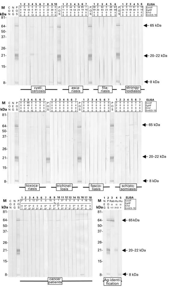
Fig. 1. EmVF-immunoblot with sera from 62 patients with active alveolar echinococcosis (AE) (54 sera) ; degenerate alveolar echinococcosis (non-viable metacestode) (AE, deg ; 8 sera in total : 3 tested in analysis 1 [A1, 2nd blot strip panel] and 5 in analysis 2 [A2, 4th blot strip panel]) ; cystic echinococcosis (CE, 22 sera) ; amoebiasis (7 sera) ;
cysticercosis (10 sera) ; ascariasis (7 sera) ; filariasis (6 sera) ; strongyloidiasis (7 sera) ; toxocariasis (7 sera) ; trichinellosis (7 sera), fascioliasis (7 sera) ; and schistosomiasis (7 sera), and sera from cancer patients (18 sera). Molecular weight markers (M) (left) are given in kilodaltons (kDa) and positions of the major immunoreactive bands (right) representing 65 kDa, 20–22 kDa, and 8 kDa antigens are indicated by arrows. As controls, blot strips incubated exclusively with anti-human immunoglobulin-peroxidase conjugate (CON), or with negative (NEG) or postive (POS) serum were included.
1 0 0 0 0 2 0 18 0 0 3 0 43 0 0 4 0 6 0 12 5 0 61 0 0 6 0 0 0 0 7 0 0 0 0 8 2 22 0 0 9 0 39 0 17 10 0 0 0 0 P O S 1 8 11 0 0 2 0 0 0 0 3 44 18 0 0 4 9 54 0 0 5 16 24 0 0 6 65 19 0 0 7 7 10 0 0 P O S 1 30 59 0 0 2 2 20 0 0 3 82 93 0 0 4 0 23 0 0 5 48 88 0 0 6 87 98 0 0 P O S 1 9 25 0 0 2 0 12 0 0 3 11 49 0 0 4 57 48 0 0 P O S N E G C O N 81- 64- 50- 37- 26- 21- 15-5 0 0 0 0 6 10 10 0 0
cysti-cercosis riasisasca- riasisfila- strongy-loidiasis
8-65 kDa 20–22 kDa 8 kDa ELISA EmVF EgHF Em2 EmII/3–10 M kDa 81- 64- 50- 37- 26- 21- 15- 8-6 0 7 0 2 5 0 0 0 0 4 0 0 0 0 3 0 0 0 0 2 0 0 0 0 1 0 0 2 0 7 0 0 3 0 P O S 6 0 0 0 0 5 0 0 0 0 4 0 0 0 0 3 0 0 0 0 2 0 0 0 0 1 0 0 0 0 7 7 4 0 2 6 0 0 0 0 5 0 0 0 0 4 0 0 0 0 3 0 0 0 0 2 0 0 0 0 1 0 0 0 0 7 0 0 0 0 6 0 0 0 0 5 0 0 0 0 4 0 2 0 15 3 60 49 0 0 2 0 0 0 0 1 0 0 0 0 7 0 0 0 0 N E G C O N P O S P O S P O S
toxoca-riasis trichinel-losis fascio-liasis somiasis
schisto-65 kDa 20–22 kDa 8 kDa EmVF EgHF Em2 EmII/3–10 ELISA M kDa 64- 37- 26- 21- 15- 8-M kDa 81-cancer patients 1 0* 0 7 2 0 0* 0* 3 0 0* 0* 4 12 0 0* 5 0* 0 0* 6 0 0 0* 7 0 0* 0* 8 15 0 0* 9 5 0* 0* 10 1 0* 9 11 0* 0* 0* 12 0 0 0* 13 0 0 0* 14 0 0* 0* 15 9 0 0* P O S N E G C O N 50- 64- 50- 37- 26- 21- 15- 8- Ag-identi-fication 16 4 0* 0* 17 16 0 0 18 10 0 0 81-2 P O S 5 Hu α 8 1 N E G 4 Hu α 20-22 3 Rab α II/3 8 kDa EmVF EgHF Em2 EmII/3–10 ELISA M kDa an.d. 65 kDa 20–22 kDa
Fig. 1 (Cont.). EmVF-, EgHF, Em2-, and II/3–10-ELISA values of the individual sera are listed on top of each strip. For sera from cancer patients, borderline-negative ELISA results are indicated with asteriscs. Identification of the major antigens (Ag-identification) and assessment of serological cross-reactivities among the corresponding bands on the blot was performed by demonstrating the immunoreactivity with a polyclonal rabbit anti-II/3–10 antibody (Raba II/3) and antibodies affinity-purified on bands representing the 22–20 kDa (Hu a 20–22), and 8 kDa (Hu a 8) antigens.aNot done (n.d.), as pre-selected based on EgHF-positivity according to Pfister et al. (1999).
metacestode antigens using well-defined sera from untreated AE patients revealed that the EmVF-ELISA had a better diagnostic sensitivity than the EgHF-ELISA (89 % vs 81 %) ; most of the sera yielded positive results in the 2 ELISAs and also exhibited similar values. Furthermore, all sera positive in the EgHF-ELISA were also positive in the EmVF-ELISA. However, the accumulation of results achieved from both assays did not improve the diagnostic sensitivity. The individual diagnostic sensitivities of the Em2-ELISA and the II/3–10-ELISA were 90 % and 79 %, respectively. Interest-ingly, the Em2-ELISA scored positive for 6 of 8 sera from inactive AE cases with necrotic lesions (see Fig. 1). The cumulative diagnostic sensitivity from the Em2- and II/3–10-ELISAs (as achieved in the Em2plus-ELISA reported earlier, see Gottstein et al. 1993) was high, and allowed detection of 97 % of AE cases included in the present study (Table 1). Hence, no further improvement in diagnostic sensitivity was achieved when the results from all 4 ELISAs assessed were accumulated.
Upon diagnosis, patients with CE demonstrated EmVF-ELISA reactivity, in that 21 of 22 sera scored positive values. Conversely, 20 sera from this panel were positive in the EgHF-ELISA (see Table 1). Accordingly, the EmVF-ELISA exhibited a better diagnostic sensitivity than the EgHF-ELISA (95 % vs. 91 %). Furthermore, most of sera positive in the 2 ELISAs showed similar values, and the accumulation of the results from both tests did not improve the diagnostic sensitivity (Table 1). As expected, the antigens Em2 and II/3–10 previously proven to be suitable for the specific serodiagnosis of AE did not react with the majority of sera from CE patients (see Fig. 1).
Diagnostic specificities linked to the different ELISAs and to potentially cross-reactive sera from
patients with other parasitoses are listed in Table 2. Sera from patients with Taenia solium cysticercosis (n=10) showed low to moderate cross-reactivity in the EmVF-, Em2- and II/3–10-ELISAs (f2 cross-reactive sera) but high cross-reactivity in the EgHF-ELISA (6 cross-reactive sera). Also, some cross-reactivity was observed for sera from patients with nematode (n=33; 17, 20, 0 and 2 cross-reactive sera in the EmVF-, EgHF-, Em2-, and II/3–10-ELISAs, respectively) and trematode infections (n=14; 0, 1, 2 and 1 cross-reactive serum/sera in the EmVF-, EgHF-, Em2- and II/3–10-ELISAs, respectively). Sera from patients with hepatic amoebiasis were also assessed for possible cross-reactivity, because diagnostic images (by computer tomography or X-ray) of liver lesions due to this disease can be confused with those caused by AE. In this case, cross-reactivity was restricted to 1 serum sample, which was weakly positive in the Em2-ELISA. In summary, the overall specificities of the different ELISAs related to non-Echinococcus parasitic infections were determined to be 72 % (EmVF-ELISA), 58 % (EgHF-ELISA), 95 % (Em2-ELISA) and 92 % (II/3–10-(Em2-ELISA).
Since the EgHF-ELISA used previously for the screening of CE was associated with relatively frequent false-positive reactions in cancer patients (Pfister et al. 1999 ; Poretti et al. 1999), 18 such cross-reactive sera were specifically selected and tested in the different E. multilocularis-ELISAs. In the EmVF-ELISA, 8 of these sera scored positive and another 3 exhibited marginal reactivity (ELISA-value :fx2 antibody units, AU). Conversely, in the Em2-ELSA, all samples from the panel of cross-reactive sera were test negative, but 9 of them showed marginal reactivity (fx2 AU). Within this panel, 2 sera were test positive in the II/3–10-ELISA, whereas 14 other sera gave low values (fx2 AU). Table 2. Diagnostic specificities of crude, purified, and recombinant Echinococcus antigens in view of potential cross-reactions with serum antibodies from patients with various parasite infections or cancer malignancies
Assay
Number of positive samples*
Relative overall specificity ( %)· related to non-Echinococcus parasitic infections (n=64) Amoebiasis (n=7) Cysticercosis (n=10) Nematodes** (n=33) Trematodes# (n=14) Cancer (n=18) ELISA : EmVF 0 1 17 0 8 72 (18) EgHF 0 6 20 1 n.d.b 58 (27) Em2 1 0 0 2 0 95 (3) II/3–10 0 2 2 1 2 92 (5) Immunoblot : EmVF 0 0 0 0 0 100 (0)
* Individual test results, see Fig. 1.
** Diseases (n) : ascariasis (7), filariasis (6), strongyloidiasis (6), toxocariasis (7), trichinellosis (7). # Diseases (n) : fascioliasis (7), schistosomiasis (7).
· The number of positive samples is given in parenthesis.
Serological evaluation of the EmVF-immunoblot EmVF-immunoblot analysis using sera from AE patients revealed major immunoreactive bands of y65, 20–22 (triplet), and 8 (doublet) kDa (Fig. 1). While the molecules representing the 20–22 triplet and the 8 kDa doublet require biochemical charac-terization (see Discussion section), the 65 kDa band represents a full-length antigen II/3 (Felleisen and Gottstein, 1993), supported by demonstrating the reactivity of this band employing a hyperimmune serum raised against antigen recII/3–10, a recom-binant subfragment of E. multilocularis antigen II/3 (see Vogel et al. 1988 ; Mu¨ ller et al. 1989). A pre-liminary evaluation of the blotting method in our diagnostic laboratory (data not shown) had indicated that neither the 65 kDa antigen II/3 nor the 8 kDa antigen achieved optimum diagnostic sensitivity or specificity. Therefore, all immunoblot strips obtained in the present study were scored with the criterion of positivity being a detectable signal for the 20–22 kDa triplet.
Immunoblot data and the corresponding ELISA-results of individual sera are shown in Fig. 1. The performance of immunoblotting with regard to sen-sitivity and specificity is summarized in Tables 1 and 2, respectively. When 62 sera from AE patients were tested in terms of their reactivity with the 20–22 kDa band triplet, the immunoblot revealed a diagnostic sensitivity of 100 %. In particular, the immunoblot also detected all 8 inactive AE cases with degenerate liver lesions (Fig. 1). Here, respective testing con-sisted of 2 separate analyses (see Fig. 1 ; analysis 1 [panel of strips indicated as A1] and 2 [panel of strips indicated as A2]) that were performed with 2 differ-ent batches of EmVF antigen. As displayed in Fig. 1, in the second immunoblot analysis (A2), a few diagnostically irrelevant bands (e.g. a 10 kDa and 35 kDa bands) were more pronounced as compared with the first analysis (A1). Furthermore, the 20–22 kDa band triplet was not adequately resolved in the blot strips from analysis A2. However, despite this inconsistency, sera from all 8 inactive cases exhibited an appropriate profile for the 20–22 kDa band triplet.
The relatively high rate of cross-reactivity (50 %) determined for sera from patients with CE (Table 1) demonstrated that the immunoblot was not suitable for a serological differentiation between AE and CE. However, the specificity of the immunoblot, con-sidering sera from patients with parasitic infections other than Echinococcus, was 100 % (Table 2). In this case, the scoring of strips incubated with sera from patients with T. solium cysticercosis was somewhat affected by the occasional detection of 2 faint bands above the diagnostically relevant 20–22 kDa triplet. However, strictly considering the diagnostic criteria (i.e. the position and nature of the immuno-reactivity of bands), such sera were scored as negative.
None of the 18 sera from cancer patients tested had non-specific reactivity with the diagnostic 20–22 kDa triplet. However, some of them reacted weakly with the 65 kDa band (9 positive sera) and the 8 kDa doublet (1 positive serum).
A possible antigenic relationship between the 65 kDa antigen II/3, the 20–22 kDa and the 8 kDa antigens was tested by incubating EmVF-blot strips with polyclonal antibodies specific to E. multi-locularis antigen II/3- and affinity-purified poly-clonal antibodies directed against the 20–22 or 8 kDa antigen. In this immunoblot (see Fig. 1, last panel), each of the 3 monospecific antibody fractions reacted individually with the corresponding antigen but did not exhibit detectable cross-reactivity with any of the other antigens on the blot. This result indicated that the 65, 20–22 and 8 kDa antigens are distinct and do not represent different breakdown products and/or conformational variants or isoforms of the same molecule.
D I S C U S S I O N
Substantial efforts have been undertaken to maximize, or at least optimize, the perfomance of serological tests for the diagnosis of echinococcosis, and many studies have yielded novel test systems or antigens to enhance both diagnostic sensitivity and specificity (Gottstein et al. 1993 ; Poretti et al. 1999 ; Liance et al. 2000 ; Sako et al. 2002 ; Ito and Craig, 2003 ; Xiao et al. 2003 ; Kormaz et al. 2004 ; Mamuti et al. 2004). New epidemiological and clinical findings have shown unusual presentations of infec-tions that also exhibit unusual serological profiles upon diagnosis. For example, hepatic lesions con-taining degrading or dead E. multilocularis cysts may stimulate weak anti-Em2-antibody responses, whereas anti-II/3–10 (and thus also anti-EM10, Em4 and/or Em18) responses are not detectable in such patients (Gottstein et al. 1996 ; Zingg et al. 2004 ; Matsumoto et al. 2006). Also, the technique of immunoblotting has gained increased attention for the diagnosis of echinococcosis (Ito et al. 1999 ; Poretti et al. 1999 ; Kormaz et al. 2004). Crude or semi-purified antigens produced from the meta-cestode stage obtained from experimentally infected rodents have been used for these immunoblots. Two problems can be associated with this approach. One relates to the presence of host proteins in such extracts, which may relate to non-specific antibody reactivity to host epitopes or which may sterically cover and thus hide parasite epitopes otherwise accessible to antibodies.
In the present study, a significant improvement was achieved in that a highly sensitive (in relation to AE) and specific (regarding diseases other than AE and CE) immunoblot using axenically in vitro-produced vesicle fluid from E. multilocularis meta-cestodes as an antigen was developed. Although the
corresponding E. granulosus hydatid fluid antigen can be readily obtained from fertile hydatid cysts, it has been shown to contain substantial amounts of host proteins that can lead to false-positive (anti-host-protein) reactions (Gottstein et al. 1987). The in vitro culture of E. multilocularis metacestodes has been established as a valuable and commonly used alternative to animal experimentation, as reflected by numerous publications in the field (e.g. reviewed by Hemphill et al. 2002 ; Brehm et al. 2006). Preliminary experience with the EmVF antigen produced using the same procedure has already been gained pre-viously by ELISA (Romig et al. 1999) and within the context of cell-immunological investigations of cytokines in both AE patients (Sturm et al. 1995) and mice experimentally infected with E. multilocularis metacestode vesicles (Dai et al. 2001).
The present culture system serves as a continuous source for an almost unlimited supply of EmVF antigen from a well-defined line of E. multilocularis, which is devoid of potentially interfering host components. This offers an excellent basis for the standardization of serological tests, as we have shown that antigenic variation can be expressed among different isolates of E. multilocularis from naturally infected rodents (Gottstein, 1991). Compared with the other Echinococcus immunoblots established thus far (Ito et al. 1999 ; Poretti et al. 1999 ; Liance et al. 2000 ; Kormaz et al. 2004), the EmVF-blot exhibits a relatively simple immunoreactive banding profile, dominated by the diagnostic 20–22 kDa triplet. Although not yet proven experimentally, the simi-larity in antigenic abundance among the bands within the triplet suggests that the 20–22 kDa antigen represents a protein with differential con-formations, post-translational modification (such as glycosylation) and/or degradation forms rather than a set of different proteins of similar sizes. Consistent with differential glycosylation is the finding that individual bands within this pattern are smeary.
In the present study, we tested the hypothesis that the protein(s) represented in the 20–22 kDa triple band correspond to the E. multilocularis protoscolex antigen Em18, identified previously (Sako et al. 2002) as an 18 kDa major proteolytic degradation product of the 65 kDa antigen II/3. However, specific antibodies purified to antigen II/3 bound to intact antigen II/3 (appearing on the EmVF-blot as a 65 kDa band) did not react with the 20–22 kDa triplet, refuting this hypothesis. Hence, the 20–22 kDa triplet does not appear to contain any antigen II/3 components. Interestingly, the 18 kDa band did not represent components of the EmVF-antigen. This may either be due to 1 or more biological characteristics of E. multilocularis, in that this band is detected in crude extracts of protoscoleces and/or when PMSF is added as a protease inhibitor to pre-vent proteolytic degradation (following the isolation
of the fluid). Also, by demonstrating a lack of anti-body cross-reactivity between the 20–22 kDa and the 8 kDa antigen (a subunit of antigen B) in the same immunoblot analysis, any immunological relation-ship between these 2 abundant antigens was not supported.
The present and previous investigations (Mu¨ ller et al. 1989 ; Gottstein et al. 1993) have shown that recombinant antigen II/3–10 is an excellent diag-nostic reagent, allowing a sensitive and specific diagnosis of AE by ELISA, confirmed by other researchers (Frosch et al. 1991 ; Hemmings and McManus, 1991 ; Xiao et al. 2003). Using immuno-reactivity to the native 65 kDa antigen II/3 as a diagnostic criterion, we showed herein that the majority of AE and some CE sera tested positive, but also sera from patients with some other disorders did not completely lack reactivity. Such apparent cross-reactivity was also observed with the 8 kDa band known as the 8 kDa subunit of E. multilocularis antigen B (cf. Mamuti et al. 2004). Since cross-reactivity complicates the scoring of the EmVF-immunoblot and the interpretation of the results, only the abundant 20–22 kDa bands were considered as diagnostic markers. This approach allowed the diagnosis of 100 % of AE cases and also the serological detection of all cases with an inactive E. multilocularis infection. The immunoblot analysis did not con-sistently discriminate between AE and CE but ex-hibited a maximal specificity compared with other parasitoses. This finding was supported by results obtained by testing sera from patients suffering from various cancers. In a previous evaluation focused on the diagnostic specificity of the EgHF-ELISA (Poretti et al. 1999), a substantial number (6.3 %) of these sera tested false-positive, and this result was considered to complicate the serological diagnosis of echinococcosis. In the present study, only the EmVF- and II/3–10-ELISAs occasionally produced similar false-positive results, but this was not the case for the EmVF-immunoblot.
Taken together, the present study revealed that the EmVF-immunoblot ideally complements the Em2- and II/3–10-ELISAs for the serological diag-nosis of AE. In combination with these 2 ELISAs, a reliable serodiagnosis of both active and inactive E. multilocularis infections in humans could be achieved. Prospectively, we plan to identify the nature of these 20–22 kDa proteins, to address their role in the host-parasite interplay and, if appropriate, to clone respective gene(s). We also plan to determine the value of the EmVF-immunoblot to monitor AE patients serologically after surgery and during long-term benzimidazole chemotherapy.
This work was supported by grants from the Swiss National Science Foundation (grant no. 31-11780/1), the Hans-Sigrist-Foundation of the University of Berne and the EU EchinoRisk-project QLK2-CT-2001-01995 (BBW no. 00.0586-1).
R E F E R E N C E S
Akisu, C., Bayram Delibas, S., Yuncu, G., Aksoy, U., Ozkoc, S., Bicmen, C., Sevinc, S. and Yaldiz, S. (2005). Evaluation of IHA, ELISA and Western Blot tests in diagnosis of pulmonary cystic hidatidosis. Tuberculosis and Torax53, 156–160.
Brehm, K., Jensen, K., Frosch, P. and Frosch, M. (1999). Characterization of the genomic locus expressing the ERM-like protein of Echinococcus multilocularis. Molecular and Biochemical Parasitology100, 147–152. Brehm, K., Spiliotis, M., Zavala-Gongora, R.,
Konrad, C. and Frosch, M. (2006). The molecular mechanisms of larval cestode development : first steps into an unknown world. Parasitology International55 (Suppl. 1), S15–S21.
Dai, W. J., Hemphill, A., Waldvogel, A., Ingold, K., Deplazes, P., Mossmann, H. and Gottstein, B. (2001). Major carbohydrate antigen of Echinococcus multilocularis induces an immunoglobulin G response independent of alphabeta+ CD4+ T cells. Infection and Immunity69, 6074–6083.
Doiz, O., Benito, R., Sbihi, Y., Osuna, A., Clavel, A. and Gomez-Lus, R. (2001). Western blot applied to the diagnosis and post-treatment monitoring of human hydatidosis. Diagnostic Microbiology and Infectious Diseases41, 139–142.
Felleisen, R. and Gottstein, B. (1993). Echinococcus multilocularis : molecular and immunochemical characterization of diagnostic antigen II/3-10. Parasitology107, 335–342.
Frosch, M., Frosch, P., Pfister, T., Schaad, V. and Bitter-Suermann, D. (1991). Cloning and characterisation of an immunodominant major surface antigen of Echinococcus multilocularis. Molecular and Biochemical Parasitology48, 121–130.
Furuya, K., Kawanaka, M., Yamano, K., Sato, N. and Honma, H. (2004). Laboratory evaluation of commercial immunoblot assay kit for serodiagnosis of Echinococcus infections using sera from patients with alveolar hydatidosis in Hokkaido. Kansenshogaku Zasshi78, 320–326.
Gottstein, B. (1991). Echinococcus multilocularis : antigenic variance between different parasite isolates. Parasitology Research77, 359–361.
Gottstein, B. (2004). Hydatid Disease. In Infectious Diseases (ed. Armstrong, D. and Cohen, J.),
pp. 1601–1606. 2nd Edn, Vol. II, Mosby, London, UK. Gottstein, B., Eckert, J. and Fey, H. (1983). Serological
differentiation between Echinococcus granulosus and E. multilocularis infections in man. Zeitschrift fu¨r Parasitenkunde69, 347–356.
Gottstein, B., Eckert, J., Michael, S. A. and Thompson, R. C. A. (1987). Echinococcus granulosus antigens : Immunoelectrophoretic and Western blot analysis of hydatid cyst fluids. Parasitology Research 73, 186–189.
Gottstein, B., Jacquier, P., Bresson-Hadni, S. and Eckert, J. (1993). Improved primary immunodiagnosis of alveolar echinococcosis in humans by an enzyme-linked immunosorbent assay using the Em2plus-antigen.
Journal of Clinical Microbiology31, 373–376. Gottstein, B. and Reichen, J. (2002). Echinococcosis/
Hydatidosis. In Manson’s Tropical Diseases (ed. Cook,
G. C. and Zumla, A.), pp. 1561–1582. 21st Edn. Elsevier Science Ltd. Philadelphia, PA, USA. Gottstein, B., Saucy, F., Deplazes, P., Reichen, J.,
Demierre, G., Zu¨ rcher, C., Busato, A. and Pugin, P. (2001). Is a high prevalence of Echinococcus multilocularis in wild and domestic animals associated with increased disease incidence in humans ? Emerging Infectious Diseases7, 408–412.
Gottstein, B., Bettens, F., Parkinson, A. J. and Wilson, F. (1996). Immunological parameters associated with susceptibility or resistance to alveolar hydatid disease in Yupiks/Inupiats. Arctic Medical Research55, 14–19. Hemmings, L. and McManus, D. P. (1991). The
diagnostic value and molecular characterisation of an Echinococcus multilocularis antigen gene clone. Molecular and Biochemical Parasitology44, 53–61.
Hemphill, A. and Gottstein, B. (1995). Immunological and morphological studies on the proliferation of in vitro cultivated Echinococcus multilocularis metacestode. Parasitology Research81, 605–614. Hemphill, A., Stettler, M., Walker, M., Siles-Lucas,
M., Fink, R. and Gottstein, B. (2002). Culture of Echinococcus multilocularis metacestodes : an alternative to animal use. Trends in Parasitology18, 445–451. Ito, A. and Craig, P. S. (2003). Immunodiagnostic and
molecular approaches for the detection of taeniid cestode infections. Trends in Parasitology19, 377–381.
Ito, A., Ma, L., Schantz, P. M., Gottstein, B., Liu, Y. H., Chai, J. J., Abdel-Hafez, S. K., Altintas, N., Joshi, D. D., Lightowlers, M. W. and Pawlowski, Z. S. (1999). Differential serodiagnosis for cystic and alveolar echinococcosis using fractions of Echinococcus granulosus cyst fluid (antigen B) and E. multilocularis protoscolex (EM18). American Journal of Tropical Medicine and Hygiene60, 188–192.
Korkmaz, M., Inceboz, T., Celebi, F., Babaoglu, A. and Uner, A. (2004). Use of two sensitive and specific immunoblot markers, em70 and em90, for diagnosis of alveolar echinococcosis. Journal of Clinical Microbiology 42, 3350–3352.
Liance, M., Janin, V., Bresson-Hadni, S., Vuitton, D. A., Houin, R. and Piarroux, R. (2000). Immunodiagnosis of Echinococcus infections : confirmatory testing and species differentiation by a new commercial Western Blot. Journal of Clinical Microbiology38, 3718–3721.
Mamuti, W., Yamasaki, H., Sako, Y., Nakao, M., Xiao, N., Nakaya, K., Sato, N., Vuitton, D. A., Piarroux, R., Lightowlers, M. W., Craig, P. S. and Ito, A. (2004). Molecular cloning, expression, and serological evaluation of an 8-kilodalton subunit of antigen B from Echinococcus multilocularis. Journal of Clinical Microbiology42, 1082–1088.
Matsumoto, J., Mu¨ ller, N., Hemphill, A., Oku, Y., Kamiya, M. and Gottstein, B. (2006). 14-3-3- and II/3-10-gene expression as molecular markers to address viability and growth activity of Echinococcus multilocularis metacestode. Parasitology132, 83–94. Miguez, M., Baz, A. and Nieto, A. (1996).
Carbohydrates on the surface of Echinococcus granulosus protoscoleces are immunodominant in mice. Parasite Immunology18, 559–569.
Mu¨ ller, N., Gottstein, B., Vogel, M., Flury, K. and Seebeck, T. (1989). Application of a recombinant
Echinococcus multilocularis antigen in an enzyme-linked immunosorbent assay for immunodiagnosis of human alveolar echinococcosis. Molecular and Biochemical Parasitology36, 151–159.
Mu¨ ller, N., Hemphill, A., Imboden, M., Duvallet, G., Dwinger, R. H. and Seebeck, T. (1992). Identification and characterization of two repetitive non-variable antigens from African trypanosomes which are recognized early during infection. Parasitology104, 111–120.
Pfister, M., Gottstein, B., Cerny, T. and Cerny, A. (1999). Immunodiagnosis of echinococcosis in cancer patients. Clinical Microbiology and Infection5, 693–697. Poretti, D., Felleisen, E., Grimm, F., Pfister, M.,
Teuscher, F., Zu¨ rcher, C., Reichen, R. and Gottstein, B. (1999). Differential immunodiagnosis between cystic hydatid disease and other cross-reactive pathologies. American Journal of Tropical Medicine and Hygiene60, 193–198.
Rausch, R. L., Wilson, J. F., Schantz, P. M. and McMahon, B. J. (1987). Spontaneous death of Echinococcus multilocularis : cases diagnosed serologically (by Em2 ELISA) and clinical significance. American Journal of Tropical Medicine and Hygiene36, 576–585.
Romig, T., Kratzer, W., Kimmig, P., Frosch, M., Gaus, W., Flegel, W. A., Gottstein, B., Lucius, R., Beckh, K. and Kern, P. (1999). An epidemiological survey of human alveolar echinococcosis in
southwestern Germany. American Journal of Tropical Medicine and Hygiene6, 566–573.
Sako, Y., Nakao, M., Nakaya, K., Yamasaki, H., Gottstein, B., Lightowers, M. W., Schantz, P. M. and Ito, A. (2002). Alveolar echinococcosis : characterization of diagnostic antigen Em18 and serological evaluation of recombinant Em18. Journal of Clinical Microbiology40, 2760–2765.
Sturm, D., Menzel, J., Gottstein, B. and Kern, P. (1995). Interleukin-5 is the predominant cytokine produced by peripheral blood mononuclear cells in alveolar echinococcosis. Infection and Immunity63, 1688–1697.
Vogel, M., Gottstein, B., Mu¨ ller, N. and Seebeck, T. (1988). Production of a recombinant antigen of
Echinococcus multilocularis with high immunodiagnostic sensitivity and specificity. Molecular and Biochemical Parasitology31, 117–125.
Xiao, N., Mamuti, W., Yamasaki, H., Sako, Y., Nakao, M., Nakaya, K., Gottstein, B., Schantz, P. M., Lightowlers, M. W., Craig, P. S. and Ito, A. (2003). Evaluation of use of recombinant Em18 and affinity-purified Em18 for serological differentiation of alveolar echinococcosis from cystic echinococcosis and other parasitic infections. Journal of Clinical Microbiology41, 3351–3353.
Zingg, W., Renner-Schneiter, E. C., Pauli-Magnus, C., Renner, E. L., van Overbeck, J., Schla¨pfer, E., Weber, M., Weber, R., Opravil, M., Gottstein, B., Speck, R. F. – the Swiss HIV Cohort Study. (2004). Alveolar echinococcosis of the liver in an adult with human immunodeficiency virus type-1 infection. Infection32, 299–302.
![Fig. 1. EmVF-immunoblot with sera from 62 patients with active alveolar echinococcosis (AE) (54 sera) ; degenerate alveolar echinococcosis (non-viable metacestode) (AE, deg ; 8 sera in total : 3 tested in analysis 1 [A1, 2nd blot strip panel] and 5 in anal](https://thumb-eu.123doks.com/thumbv2/123doknet/14889117.648343/4.892.106.765.77.981/immunoblot-patients-alveolar-echinococcosis-degenerate-alveolar-echinococcosis-metacestode.webp)