UNIVERSITÉ DE GENÈVE
Département de zoologie et de biologie animale FACULTÉ DES SCIENCES
Professeur J.-L. Bény
Département de morphologie FACULTÉ DE MÉDECINE
Professeur M.S. Pepper Professeur R. Montesano _____________________________________________________________________
Le rôle des récepteurs à VEGF
dans la régulation de l'angiogenèse
THÈSE
présentée à la Faculté des sciences de l’Université de Genève pour obtenir le grade de Docteur ès sciences, mention biologique
par Jean-Christophe TILLE de Ormont-Dessous (Vaud) Thèse N° 3513 Genève
Atelier de reproduction de la Section de physique 2004
REMERCIEMENTS
J'aimerai remercier
le Professeur Lelio Orci pour m'avoir accueilli au sein du département de morphologie.
mon superviseur de thèse la Professeur Michael Pepper pour son enthousiasme, sa disponibilité, sa stimulation intellectuelle et pour la mise à ma disposition de tous les moyens techniques nécessaires.
Mylène Amherdt et Alain Perrelet pour la relecture attentionnée de ma thèse.
Stefano Mandriota pour son initiation à la manipulation des souris et son encouragement constant.
Corinne Di Sanza, Mireille Quayzin, Jacqueline Rial-Robert, Nadine Dupont, Dany Baetens, Danielle Ben Nasr et Esther Sutter pour leurs excellents conseils et aides techniques.
Riccardo Emilio Nisato, Benoît Jenny et Hélène Mottaz pour avoir partagé l'espace vital du laboratoire et du bureau.
TABLE DES MATIÈRES
Pages
Introduction 1
Chapitre 1: Vascular endothelial growth factor (VEGF) receptor-2 15 antagonists inhibit VEGF- and basic fibroblast growth
factor-induced angiogenesis in vivo and in vitro
Chapitre 2: Vascular endothelial growth factor (VEGF) receptor-2 30 signaling mediates VEGF-C∆N∆C- and VEGF-A-induced
angiogenesis in vitro
Chapitre 3: Mesenchymal cells potentiate vascular endothelial growth 45 factor-induced angiogenesis in vitro
Chapitre 4: Familial predisposition to tufted angioma: identification of 60 blood and lymphatic vascular components
INTRODUCTION
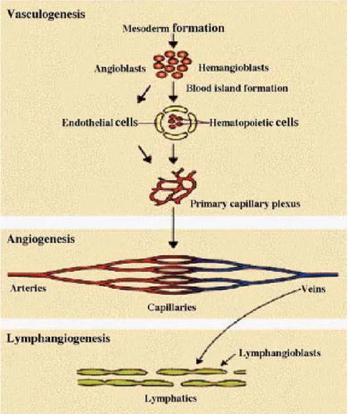
Le système vasculaire est le premier système à se former chez l'embryon de mammifère. Son développement commence par la différenciation in situ d'hémangioblastes, précurseurs des cellules endothéliales et hématopoïétiques, à partir du mésenchyme embryonnaire. Il s'ensuit une prolifération et une migration des cellules endothéliales pour former l'arbre vasculaire primitif au cours d'un processus appelé vasculogenèse (Risau and Flamme, 1995). Cet arbre vasculaire primitif est remodelé en un réseau comprenant des vaisseaux sanguins de différents calibres. Avec la croissance de l'embryon, de nouveau vaisseaux sanguins se forment à partir de la microcirculation pré-existante, lors d'un processus appelé angiogenèse (Flamme et al., 1997; Risau, 1997).
Les capillaires sont les plus petits vaisseaux, ils permettent la diffusion de l'oxygène et de nutriments vers les tissus environnants. Le sang arrive aux capillaires par des artères, des vaisseaux de plus grand calibre, et repart par les veines. Ces trois types de vaisseaux sanguins diffèrent par la constitution de leur paroi vasculaire. En effet, suite au battement du cœur, un flux sanguin traverse les vaisseaux induisant les cellules endothéliales à émettre un signal vers les cellules mésenchymateuses environnantes. Ces dernières vont être induites à proliférer, migrer vers les cellules endothéliales puis se différencier en cellules péri-vasculaires appelées péricytes pour les capillaires et cellules musculaires lisses pour les artères et les veines.
Il s'ensuit une phase de maturation pendant laquelle une interaction s'établit entre les cellules endothéliales et les cellules péri-vasculaires. Le contact entre ces deux types de cellules induit une stabilisation des cellules endothéliales en les rendant réfractaires au stimulus pro- ou anti-angiogénique (Benjamin et al., 1999; Benjamin et al., 1998). Les cellules musculaires lisses vont s'apposer concentriquement autour des tubes de cellules endothéliales afin de former une paroi vasculaire fonctionnelle.
Plus tardivement par rapport à la formation des vaisseaux sanguins, les vaisseaux lymphatiques se développent en bourgeonnant, puis en se détachant des veines pré-existantes pour former un réseau parallèle, selon un processus que l'on appelle
lymphangiogenèse. Les vaisseaux lymphatiques se forment aussi à partir de précurseurs des cellules endothéliales lymphatiques, les lymphangioblastes, qui se sont différenciés in situ dans le mésenchyme (Rodriguez-Niedenfuhr et al., 2001).
Figure 1. Le développement du réseau sanguin et lymphatique (Wilting et al., 2003).
Chez l'adulte, l'angiogenèse s'observe physiologiquement par exemple lors du cycle menstruel féminin, pendant la gestation au niveau de la glande mammaire et du placenta, au cours de l'inflammation, des processus de réparation tissulaire et lors de l'ischémie tissulaire. L'on peut également observer la formation de nouveau vaisseaux sanguins dans des conditions pathologiques tel que la rétinopathie diabétique, l'artériosclérose, l'arthrite rhumatoïde, la croissance tumorale et les malformations vasculaires (Carmeliet, 2003). La connaissance des mécanismes moléculaires qui règle l'angiogenèse est importante pour le développement de
nouveaux moyens thérapeutiques, avec comme but d'empêcher ou de stimuler la formation de nouveaux vaisseaux sanguins selon les circonstances.
RÉGULATION DE L'ANGIOGENÈSE
La recherche de molécules impliquées dans la régulation de l'angiogenèse a conduit à l'identification de plusieurs familles de protéines, en particulier des facteurs de croissance, des molécules d'adhésion et des enzymes protéolytiques (Carmeliet, 2000). Parmi les cytokines impliquées dans la formation de nouveaux vaisseaux sanguins et dans l'assemblage correct de la paroi vasculaire, on trouve la famille des vascular endothelial growth factor (VEGF), des fibroblast growth factor (FGF), platelet-derived growth factor (PDGF), des angiopoïétines (Ang) et les transforming growth factor-β (TGF-β). Les malformations ou les dysfonctions vasculaires compromettent de nombreuses fonctions organiques résultant à des maladies congénitales et acquises. Des mutations dans les gènes codant pour le récepteur des Ang, Tie-2 ou TEK, ainsi que pour deux récepteurs auxiliaires des TGF-β, endoglin (ENG) et activin receptor-like kinase 1 (ACVRL1), ont été identifiés chez des familles atteintes de malformations vasculaires (Brouillard and Vikkula, 2003). Les mutations de Tie-2 sont impliquées dans la survenue de malformations veineuses et les mutations d'ENG et ACVLR-1 dans l'apparition de télangiectasies hémorragiques héréditaires. Ces deux maladies ont en commun un défaut dans l'assemblage de la paroi vasculaire.
Le processus d'angiogenèse à partir d'un vaisseau pré-existant peut être divisé en plusieurs phases. Dans la partie de la paroi vasculaire destinée à émettre un nouveau capillaire, il y a tout d'abord une perte de contact entre les cellules endothéliales et les péricytes avec une augmentation de la perméabilité vasculaire par Ang-2 et VEGF-A. Suite à cette activation, les cellules endothéliales dégradent protéolytiquement leur lame basale, prolifèrent et migrent à travers la matrice extra-cellulaire vers la source angiogénique en formant un capillaire dépourvu de cellules vasculaires. Ultérieurement, il y a recrutement et différenciation des cellules péri-vasculaires par TGF-β1, PDGF-BB et Ang-2 afin de former un vaisseau sanguin avec une paroi non perméable et fonctionnelle. Parmi les divers facteurs angiogéniques la
famille la plus étudiée est celle des VEGF qui a pour cible principale les cellules endothéliales (Ferrara et al., 2003).
LA FAMILLE DES VEGF
La famille des VEGF consiste en des dimères de glycoprotéines qui ont en commun un domaine d'homologie des VEGF. Celui-ci est composé de 8 cystéines impliquée dans les liaisons disulfures des dimères (McDonald and Hendrickson, 1993). La famille des VEGF comprend plusieurs membres de protéines sécrétées, jouant un rôle important dans la physiologie et la pathologie du système vasculaire, incluant la vasculogenèse, l'hématopïèse, l'angiogenèse et la lymphangiogenèse (Jussila and Alitalo, 2002). Cette famille se compose de VEGF-A, VEGF-B, VEGF-C, VEGF-D et PlGF. L'effet des VEGF se fait par l'intermédiaire de trois récepteurs tyrosine kinase nommé VEGFR-1/Flt-1, VEGFR-2/KDR/Flk-1 et VEGFR-3/Flt-4. Le profil de liaisons des VEGF à ces récepteurs n'est pas spécifique à un membre en particulier. En effet, VEGFR-1 lie VEGF-A, -B et PlGF-1,-2 , VEGFR-2 lie VEGF-A, -C et -D tandis que VEGFR-3 lie VEGF-C et -D.
Figures 2. La famille des VEGF et ses récepteurs (Ferrara et al., 2003).
VEGF-A fut le premier facteur découvert ayant une activité mitotique spécifique sur les cellules endothéliales (Ferrara and Henzel, 1989). Il existe plusieurs isoformes de VEGF-A produites par épissage alternatif, la plus abondante et la plus étudiée étant celle contenant 165 acides aminés (Ferrara et al., 2003). L'importance de VEGF-A dans l'angiogenèse a été montré dans divers modèles expérimentaux in vivo. Chez la souris, l'inactivation du gène de VEGF-A conduit à une mort embryonnaire et ceci même à l'état hétérozygote (Carmeliet et al., 1996; Ferrara et al., 1996). Si l'on inactive VEGF-A spécifiquement à partir de la période néonatale, il en résulte, entre autres, un retard de la croissance létal (Gerber et al., 1999). Ceci démontre bien la nécessité de VEGF-A en ce qui concerne le développement physiologique de l'organisme. De même, son importance dans l'induction angiogénique lors de la croissance tumorale a été mise en évidence dans un modèle animal de transformation oncogénique des cellules bêta des îlots pancréatiques (Bergers et al., 2000; Inoue et al., 2002).
VEGF-B est un ligand spécifique pour VEGFR-1 (Olofsson et al., 1998). Son transcrit subit un épissage alternatif produisant deux isoformes de 167 et 186 acides aminés. Suite à leur sécrétion, VEGF-B167 reste associé avec la surface cellulaire par un
domaine liant l'héparine, tandis que VEGF-B186 est librement diffusible (Olofsson et
al., 1996). Basé sur la distribution tissulaire ainsi que sur le phénotype, suite à l'inactivation du gène codant pour VEGF-B, celui-ci participe au recrutement de cellules inflammatoires dans un modèle d'arthrite, au développement de la conduction cardiaque, mais n'est pas nécessaire à l'angiogenèse embryonnaire (Aase et al., 2001; Bellomo et al., 2000; Mould et al., 2003; Olofsson et al., 1996).
PlGF est transcrit en 2 isoformes par épissage alternatif, PlGF-1 et PlGF-2 chez l'homme, seul PlGF-2 est exprimé chez la souris (DiPalma et al., 1996). PlGF-2 contient 21 acides aminés supplémentaire dans sans sa partie C-terminale lui permettant de lié l'héparine (Hauser and Weich, 1993; Migdal et al., 1998). Chez la souris, l'inactivation du gène codant pour PlGF-2 affecte l'angiogenèse pathologique (Luttun et al., 2002a). PlGF-2 agit sur l'activation des cellules endothéliales, le recrutement des cellules musculaire lisse et le recrutement des cellules inflammatoires à partir de la moelle osseuse (Hattori et al., 2002; Heissig et al.,
2002). PlGF est nécessaire à l'augmentation de la perméabilité induite par VEGF-A (Carmeliet et al., 2001).
VEGF-C et VEGF-D sont deux membres de la famille des VEGF impliqués dans la lymphangiogenèse en plus d'un possible rôle dans l'angiogenèse (Jussila and Alitalo, 2002). Ces deux cytokines sont sécrétés sous forme d'homodimère, puis sous l'action d'enzymes protéolytiques comme la plasmine et les furins PC5 et PC7 (McColl et al., 2003; Siegfried et al., 2003), ils subissent extracellulairement un clivage de leur partie N- et C-terminales. Ces clivages successifs modulent la capacité de VEGF-C et VEGF-D à lier VEGFR-2 et VEGFR-3 et par la même modifient l'effet biologique de ces cytokines. La forme partiellement transformée de 31 kDa, ayant subit le clivage de la partie C-terminale, lie et active VEGFR-3 tandis que la protéine mature de 21 kDa, qui a subit le clivage des parties N- et C-terminales, lie et active VEGFR-2 en plus de VEGFR-3 (Joukov et al., 1997). L'inactivation de gène codant pour VEGF-C résulte en une altération du développement des vaisseaux lymphatiques sans affecté le développement vasculaire (Karkkainen et al., 2003).
Les récepteurs à VEGF sont de type tyrosine kinase. Après le dimérisation suite à la stimulation par le ligand, il y a autophosphorylation des récepteurs. Les VEGFR sont exprimés à un faible niveau dans beaucoup de tissus adultes mais leur niveau d'expression dans les cellules endothéliales est augmenté pendant le développement ainsi que dans des pathologies où l'angiogenèse joue un rôle important. L'inactivation des gènes codant pour VEGFR-1, VEGFR-2 ou VEGFR-3 résultent chacune en une létalité embryonnaire chez la souris.
L'inactivation du gène codant pour VEGFR-1 induit un surnombre de cellules endothéliales ayant comme conséquence une altération de la vasculogenèse (Fong et al., 1995; Fong et al., 1999). Par contre, l'inactivation de la partie tyrosine kinase de VEGFR-1 est compatible avec le développement normal chez la souris, alors qu'elle affecte l'angiogenèse tumorale (Hiratsuka et al., 2001; Hiratsuka et al., 1998). Pendant l'embryogenèse, VEGFR-1 servirait de récepteur "réservoir" pour VEGF-A réglant sa disponibilité pour VEGFR-2 ainsi que son activité transmise par cette liaison à VEGFR-2. Dans l'angiogenèse pathologique VEGFR-1 participe au
développement de lésions artéromateuses et dans l'arthrite (Luttun et al., 2002b). L'inhibition de VEGFR-1 affecte l'infiltration monocytaires diminuant ainsi la progression des plaques. Dans un modèle murin d'arthrite, l'inhibition de VEGFR-1 diminue l'œdème, l'érythème et l'ankylose (Luttun et al., 2002b). Dans ces deux modèles, VEGFR-1 est nécessaire à la mobilisation de précurseurs de cellules endothéliales à partir de la moelle osseuse (Carmeliet et al., 2001; Lyden et al., 2001). Il semble que VEGF-B et PlGF soit nécessaire pour la réponse biologique induite par la phosphorylation de VEGFR-1 (Autiero et al., 2003).
L'inactivation du gène codant pour VEGFR-2 chez la souris résulte en un manque de développement d'hémangioblastes, ce qui affecte la vasculogenèse (Shalaby et al., 1995). Des données in vitro et in vivo ont démontré l'importance de l'activation par VEGF-A, de VEGFR-2 dans les cellules endothéliales en ce qui concerne l'induction spécifique de gènes, la migration, la prolifération, la perméabilité vasculaire, ainsi que dans l'angiogenèse (Ferrara et al., 2003). La forme mature de C et VEGF-D lie et active également VEGFR-2 en plus de VEGFR-3 (Jussila and Alitalo, 2002). Actuellement, il n'est pas certain que l'activation de VEGFR-2 par ces différents ligands induise les mêmes voies de signalisation et donc une réponse physiologique identique. Une mutation somatique dans le gène codant pour VEGFR-2 a été démontrée au niveau des cellules endothéliales d'hémangiome, une pathologie vasculaire ayant comme caractéristique une prolifération de capillaires sanguins (Walter et al., 2002).
VEGFR-3 est traduit en un précurseur qui subit un clivage protéolytique dans sa partie extra-cellulaire, celle-ci restant liée à sa partie trans-membranaire par des ponts disulfures (Jussila and Alitalo, 2002). Au stade embryonnaire, VEGFR-3 est exprimé au niveau de tout l'arbre vasculaire puis se restreint aux vaisseaux lymphatiques et à certains capillaires fenestrés (Kaipainen et al., 1995; Partanen et al., 2000). L'inactivation du gène codant pour VEGFR-3 résulte en un défaut dans le remodelage de l'arbre vasculaire avant l'apparition des vaisseaux lymphatiques (Dumont et al., 1998). La différenciation des cellules endothéliales, ainsi que la vasculogenèse, ne sont pas affectées par l'absence de VEGFR-3, par contre le développement de l'hématopoïèse au niveau de la membrane vitelline est diminuée, suggérant un rôle pour VEGFR-3 dans l'hématopoïèse (Hamada et al., 2000). Dans
l'angiogenèse tumorale, VEGFR-3 est ré-exprimé au niveau des cellules endothéliales des vaisseaux sanguins (Clarijs et al., 2002; Valtola et al., 1999). Le blocage de VEGFR-3, lors du processus tumoral, résulte en une altération de l'angiogenèse et entraîne une diminution de la croissance tumorale (Kubo et al., 2000). VEGFR-3 est impliqué dans le lymphoedème héréditaire de type I (Irrthum et al., 2000; Karkkainen et al., 2000). Cette maladie se transmet de manière autosomale dominante et les mutations décrites se trouvent dans la partie kinase de VEGFR-3. Ces mutations affectent la capacité d'autophosphorylation du récepteur empêchant ainsi l'activation de la voie de signalisation (Irrthum et al., 2000; Karkkainen et al., 2000). De plus, dans les hémangiomes, une mutation somatique dans la partie kinase de VEGFR-3 a été démontrée dans les cellules endothéliales de la lésion, mais pas dans celles du tissu sain environnant (Walter et al., 2002).
OBJECTIF DE LA THÈSE
L'importance de la famille des facteurs de croissance de l'endothélium vasculaire (vascular endothelial growth factor, VEGF) dans la physiologie et la pathologie du système vasculaire nous a amené à étudier les différents récepteurs de cette famille (VEGFR) exprimés par les cellules endothéliales.
Dans une première partie, j'ai étudié le rôle de VEGFR-1, -2 et -3 dans l'angiogenèse
in vivo et in vitro, ainsi que dans la régulation des gènes induits par la famille des
VEGF et FGF-2.
Dans une deuxième partie, j'ai étudié l'interaction entre cellules endothéliales et cellules mésenchymateuses, précurseurs des cellules péri-vasculaires, lors du processus d'angiogenèse in vitro induite par VEGF-A et FGF-2.
Dans la dernière partie, j'ai étudié une famille atteinte d'une malformation vasculaire héréditaire en caractérisant le type de vaisseaux présents dans la lésion. De plus, j'ai étudié l'association entre une prédisposition pour cette pathologie et la transmission de gènes impliqués dans le développement vasculaire.
RÉFÉRENCES
Aase, K., von Euler, G., Li, X., Ponten, A., Thoren, P., Cao, R., Cao, Y., Olofsson, B., Gebre-Medhin, S., Pekny, M., et al. (2001). Vascular endothelial growth factor-B-deficient mice display an atrial conduction defect. Circulation 104, 358-364.
Autiero, M., Luttun, A., Tjwa, M., and Carmeliet, P. (2003). Placental growth factor and its receptor, vascular endothelial growth factor receptor-1: novel targets for stimulation of ischemic tissue revascularization and inhibition of angiogenic and inflammatory disorders. J Thromb Haemost 1, 1356-1370.
Bellomo, D., Headrick, J. P., Silins, G. U., Paterson, C. A., Thomas, P. S., Gartside, M., Mould, A., Cahill, M. M., Tonks, I. D., Grimmond, S. M., et al. (2000). Mice lacking the vascular endothelial growth factor-B gene (Vegfb) have smaller hearts,
dysfunctional coronary vasculature, and impaired recovery from cardiac ischemia. Circ Res 86, E29-35.
Benjamin, L. E., Golijanin, D., Itin, A., Pode, D., and Keshet, E. (1999). Selective ablation of immature blood vessels in established human tumors follows vascular endothelial growth factor withdrawal. J Clin Invest 103, 159-165.
Benjamin, L. E., Hemo, I., and Keshet, E. (1998). A plasticity window for blood vessel remodelling is defined by pericyte coverage of the preformed endothelial network and is regulated by PDGF-B and VEGF. Development 125, 1591-1598.
Bergers, G., Brekken, R., McMahon, G., Vu, T. H., Itoh, T., Tamaki, K., Tanzawa, K., Thorpe, P., Itohara, S., Werb, Z., and Hanahan, D. (2000). Matrix metalloproteinase-9 triggers the angiogenic switch during carcinogenesis. Nat Cell Biol 2, 737-744. Brouillard, P., and Vikkula, M. (2003). Vascular malformations: localized defects in vascular morphogenesis. Clin Genet 63, 340-351.
Carmeliet, P. (2000). Mechanisms of angiogenesis and arteriogenesis. Nat Med 6, 389-395.
Carmeliet, P. (2003). Angiogenesis in health and disease. Nat Med 9, 653-660. Carmeliet, P., Ferreira, V., Breier, G., Pollefeyt, S., Kieckens, L., Gertsenstein, M., Fahrig, M., Vandenhoeck, A., Harpal, K., Eberhardt, C., et al. (1996). Abnormal blood vessel development and lethality in embryos lacking a single VEGF allele. Nature
380, 435-439.
Carmeliet, P., Moons, L., Luttun, A., Vincenti, V., Compernolle, V., De Mol, M., Wu, Y., Bono, F., Devy, L., Beck, H., et al. (2001). Synergism between vascular
endothelial growth factor and placental growth factor contributes to angiogenesis and plasma extravasation in pathological conditions. Nat Med 7, 575-583.
Clarijs, R., Schalkwijk, L., Hofmann, U. B., Ruiter, D. J., and de Waal, R. M. (2002). Induction of vascular endothelial growth factor receptor-3 expression on tumor microvasculature as a new progression marker in human cutaneous melanoma. Cancer Res 62, 7059-7065.
DiPalma, T., Tucci, M., Russo, G., Maglione, D., Lago, C. T., Romano, A., Saccone, S., Della Valle, G., De Gregorio, L., Dragani, T. A., et al. (1996). The placenta growth factor gene of the mouse. Mamm Genome 7, 6-12.
Dumont, D. J., Jussila, L., Taipale, J., Lymboussaki, A., Mustonen, T., Pajusola, K., Breitman, M., and Alitalo, K. (1998). Cardiovascular failure in mouse embryos deficient in VEGF receptor-3. Science 282, 946-949.
Ferrara, N., Carver-Moore, K., Chen, H., Dowd, M., Lu, L., O'Shea, K. S., Powell-Braxton, L., Hillan, K. J., and Moore, M. W. (1996). Heterozygous embryonic lethality induced by targeted inactivation of the VEGF gene. Nature 380, 439-442.
Ferrara, N., Gerber, H. P., and LeCouter, J. (2003). The biology of VEGF and its receptors. Nat Med 9, 669-676.
Ferrara, N., and Henzel, W. J. (1989). Pituitary follicular cells secrete a novel
heparin-binding growth factor specific for vascular endothelial cells. Biochem Biophys Res Commun 161, 851-858.
Flamme, I., Frolich, T., and Risau, W. (1997). Molecular mechanisms of vasculogenesis and embryonic angiogenesis. J Cell Physiol 173, 206-210.
Fong, G. H., Rossant, J., Gertsenstein, M., and Breitman, M. L. (1995). Role of the Flt-1 receptor tyrosine kinase in regulating the assembly of vascular endothelium. Nature 376, 66-70.
Fong, G. H., Zhang, L., Bryce, D. M., and Peng, J. (1999). Increased hemangioblast commitment, not vascular disorganization, is the primary defect in flt-1 knock-out mice. Development 126, 3015-3025.
Gerber, H. P., Hillan, K. J., Ryan, A. M., Kowalski, J., Keller, G. A., Rangell, L., Wright, B. D., Radtke, F., Aguet, M., and Ferrara, N. (1999). VEGF is required for growth and survival in neonatal mice. Development 126, 1149-1159.
Hamada, K., Oike, Y., Takakura, N., Ito, Y., Jussila, L., Dumont, D. J., Alitalo, K., and Suda, T. (2000). VEGF-C signaling pathways through VEGFR-2 and VEGFR-3 in vasculoangiogenesis and hematopoiesis. Blood 96, 3793-3800.
Hattori, K., Heissig, B., Wu, Y., Dias, S., Tejada, R., Ferris, B., Hicklin, D. J., Zhu, Z., Bohlen, P., Witte, L., et al. (2002). Placental growth factor reconstitutes
hematopoiesis by recruiting VEGFR1(+) stem cells from bone-marrow microenvironment. Nat Med 8, 841-849.
Hauser, S., and Weich, H. A. (1993). A heparin-binding form of placenta growth factor (PlGF-2) is expressed in human umbilical vein endothelial cells and in placenta. Growth Factors 9, 259-268.
Heissig, B., Hattori, K., Dias, S., Friedrich, M., Ferris, B., Hackett, N. R., Crystal, R. G., Besmer, P., Lyden, D., Moore, M. A., et al. (2002). Recruitment of stem and progenitor cells from the bone marrow niche requires MMP-9 mediated release of kit-ligand. Cell 109, 625-637.
Hiratsuka, S., Maru, Y., Okada, A., Seiki, M., Noda, T., and Shibuya, M. (2001). Involvement of Flt-1 tyrosine kinase (vascular endothelial growth factor receptor-1) in pathological angiogenesis. Cancer Res 61, 1207-1213.
Hiratsuka, S., Minowa, O., Kuno, J., Noda, T., and Shibuya, M. (1998). Flt-1 lacking the tyrosine kinase domain is sufficient for normal development and angiogenesis in mice. Proc Natl Acad Sci U S A 95, 9349-9354.
Inoue, M., Hager, J. H., Ferrara, N., Gerber, H. P., and Hanahan, D. (2002). VEGF-A has a critical, nonredundant role in angiogenic switching and pancreatic beta cell carcinogenesis. Cancer Cell 1, 193-202.
Irrthum, A., Karkkainen, M. J., Devriendt, K., Alitalo, K., and Vikkula, M. (2000). Congenital hereditary lymphedema caused by a mutation that inactivates VEGFR3 tyrosine kinase. Am J Hum Genet 67, 295-301.
Joukov, V., Sorsa, T., Kumar, V., Jeltsch, M., Claesson-Welsh, L., Cao, Y., Saksela, O., Kalkkinen, N., and Alitalo, K. (1997). Proteolytic processing regulates receptor specificity and activity of VEGF-C. Embo J 16, 3898-3911.
Jussila, L., and Alitalo, K. (2002). Vascular growth factors and lymphangiogenesis. Physiol Rev 82, 673-700.
Kaipainen, A., Korhonen, J., Mustonen, T., van Hinsbergh, V. W., Fang, G. H., Dumont, D., Breitman, M., and Alitalo, K. (1995). Expression of the fms-like tyrosine kinase 4 gene becomes restricted to lymphatic endothelium during development. Proc Natl Acad Sci U S A 92, 3566-3570.
Karkkainen, M. J., Ferrell, R. E., Lawrence, E. C., Kimak, M. A., Levinson, K. L., McTigue, M. A., Alitalo, K., and Finegold, D. N. (2000). Missense mutations interfere with VEGFR-3 signalling in primary lymphoedema. Nat Genet 25, 153-159.
Karkkainen, M. J., Haiko, P., Sainio, K., Partanen, J., Taipale, J., Petrova, T. V., Jeltsch, M., Jackson, D. G., Talikka, M., Rauvala, H., et al. (2003). Vascular
endothelial growth factor C is required for sprouting of the first lymphatic vessels from embryonic veins. Nat Immunol.
Kubo, H., Fujiwara, T., Jussila, L., Hashi, H., Ogawa, M., Shimizu, K., Awane, M., Sakai, Y., Takabayashi, A., Alitalo, K., et al. (2000). Involvement of vascular endothelial growth factor receptor-3 in maintenance of integrity of endothelial cell lining during tumor angiogenesis. Blood 96, 546-553.
Luttun, A., Brusselmans, K., Fukao, H., Tjwa, M., Ueshima, S., Herbert, J. M., Matsuo, O., Collen, D., Carmeliet, P., and Moons, L. (2002a). Loss of placental growth factor protects mice against vascular permeability in pathological conditions. Biochem Biophys Res Commun 295, 428-434.
Luttun, A., Tjwa, M., Moons, L., Wu, Y., Angelillo-Scherrer, A., Liao, F., Nagy, J. A., Hooper, A., Priller, J., De Klerck, B., et al. (2002b). Revascularization of ischemic tissues by PlGF treatment, and inhibition of tumor angiogenesis, arthritis and atherosclerosis by anti-Flt1. Nat Med 8, 831-840.
Lyden, D., Hattori, K., Dias, S., Costa, C., Blaikie, P., Butros, L., Chadburn, A., Heissig, B., Marks, W., Witte, L., et al. (2001). Impaired recruitment of bone-marrow-derived endothelial and hematopoietic precursor cells blocks tumor angiogenesis and growth. Nat Med 7, 1194-1201.
McColl, B. K., Baldwin, M. E., Roufail, S., Freeman, C., Moritz, R. L., Simpson, R. J., Alitalo, K., Stacker, S. A., and Achen, M. G. (2003). Plasmin activates the
lymphangiogenic growth factors VEGF-C and VEGF-D. J Exp Med 198, 863-868. McDonald, N. Q., and Hendrickson, W. A. (1993). A structural superfamily of growth factors containing a cystine knot motif. Cell 73, 421-424.
Migdal, M., Huppertz, B., Tessler, S., Comforti, A., Shibuya, M., Reich, R., Baumann, H., and Neufeld, G. (1998). Neuropilin-1 is a placenta growth factor-2 receptor. J Biol Chem 273, 22272-22278.
Mould, A. W., Tonks, I. D., Cahill, M. M., Pettit, A. R., Thomas, R., Hayward, N. K., and Kay, G. F. (2003). Vegfb gene knockout mice display reduced pathology and synovial angiogenesis in both antigen-induced and collagen-induced models of arthritis. Arthritis Rheum 48, 2660-2669.
Olofsson, B., Korpelainen, E., Pepper, M. S., Mandriota, S. J., Aase, K., Kumar, V., Gunji, Y., Jeltsch, M. M., Shibuya, M., Alitalo, K., and Eriksson, U. (1998). Vascular endothelial growth factor B (VEGF-B) binds to VEGF receptor-1 and regulates plasminogen activator activity in endothelial cells. Proc Natl Acad Sci U S A 95, 11709-11714.
Olofsson, B., Pajusola, K., von Euler, G., Chilov, D., Alitalo, K., and Eriksson, U. (1996). Genomic organization of the mouse and human genes for vascular endothelial growth factor B (VEGF-B) and characterization of a second splice isoform. J Biol Chem 271, 19310-19317.
Partanen, T. A., Arola, J., Saaristo, A., Jussila, L., Ora, A., Miettinen, M., Stacker, S. A., Achen, M. G., and Alitalo, K. (2000). VEGF-C and VEGF-D expression in
neuroendocrine cells and their receptor, VEGFR-3, in fenestrated blood vessels in human tissues. Faseb J 14, 2087-2096.
Risau, W. (1997). Mechanisms of angiogenesis. Nature 386, 671-674.
Risau, W., and Flamme, I. (1995). Vasculogenesis. Annu Rev Cell Dev Biol 11, 73-91.
Rodriguez-Niedenfuhr, M., Papoutsi, M., Christ, B., Nicolaides, K. H., von Kaisenberg, C. S., Tomarev, S. I., and Wilting, J. (2001). Prox1 is a marker of ectodermal placodes, endodermal compartments, lymphatic endothelium and lymphangioblasts. Anat Embryol (Berl) 204, 399-406.
Shalaby, F., Rossant, J., Yamaguchi, T. P., Gertsenstein, M., Wu, X. F., Breitman, M. L., and Schuh, A. C. (1995). Failure of blood-island formation and vasculogenesis in Flk-1-deficient mice. Nature 376, 62-66.
14
Siegfried, G., Basak, A., Cromlish, J. A., Benjannet, S., Marcinkiewicz, J., Chretien, M., Seidah, N. G., and Khatib, A. M. (2003). The secretory proprotein convertases furin, PC5, and PC7 activate VEGF-C to induce tumorigenesis. J Clin Invest 111, 1723-1732.
Valtola, R., Salven, P., Heikkila, P., Taipale, J., Joensuu, H., Rehn, M., Pihlajaniemi, T., Weich, H., deWaal, R., and Alitalo, K. (1999). VEGFR-3 and its ligand VEGF-C are associated with angiogenesis in breast cancer. Am J Pathol 154, 1381-1390. Walter, J. W., North, P. E., Waner, M., Mizeracki, A., Blei, F., Walker, J. W., Reinisch, J. F., and Marchuk, D. A. (2002). Somatic mutation of vascular endothelial growth factor receptors in juvenile hemangioma. Genes Chromosomes Cancer 33, 295-303. Wilting, J., Christ, B., Yuan, L., and Eichmann, A. (2003). Cellular and molecular mechanisms of embryonic haemangiogenesis and lymphangiogenesis.
CHAPITRE 1
VASCULAR ENDOTHELIAL GROWTH FACTOR (VEGF) RECEPTOR-2
ANTAGONISTS INHIBIT VEGF- AND BASIC FIBROBLAST GROWTH
FACTOR-INDUCED ANGIOGENESIS IN VIVO AND IN VITRO
En collaboration avec:
J. Wood1, S.J. Mandriota, C. Schnell1, S. Ferrari1, J. Mestan1, Z. Zhu2, L. Witte2 and M.S. Pepper.
Department of Morphology, University Medical Center, Geneva, Switzerland;
1Oncology Research, Novartis Pharma, Basel, Switzerland; 2ImClone Systems Inc., New York, USA.
Publié dans The Journal of Pharmacology and Experimental Therapeutics 299:1073-1085, 2001.
16
Je remercie Novartis pour la synthèse des inhibiteurs, pour la caractérisation in vitro des kinases qu'elles affectent ainsi que pour les tests d'inhibitions de la prolifération des HUVEC et les tests d'inhibition de l'angiogenèse in vivo.
Je remercie ImClone pour m'avoir donné accès au anticorps neutralisant VEGFR-2.
J'ai personnellement effectué les tests d'inhibition de l'angiogenèse in vitro, de prolifération des cellules endothéliales bovines, la migration cellulaire, les
zymographies, les RNAse protection, les immunoprécipitation, les western blot ainsi que la rédaction du manuscrit.
Vascular Endothelial Growth Factor (VEGF) Receptor-2
Antagonists Inhibit VEGF- and Basic Fibroblast Growth
Factor-Induced Angiogenesis in Vivo and in Vitro
J.-C. TILLE, J. WOOD, S.J. MANDRIOTA, C. SCHNELL, S. FERRARI, J. MESTAN, Z. ZHU, L. WITTE, and M. S. PEPPER
Department of Morphology, Geneva University Medical Center, Basel, Switzerland (J.-C.T., S.J.M., M.S.P.); Oncology Research, Novartis Pharma, Basel, Switzerland (J.W., C.S., S.F., J.M.); and ImClone Systems Inc., New York, New York (Z.Z, L.W.)
Received May 24, 2001; accepted September 4, 2001 This paper is available online at http://jpet.aspetjournals.org
ABSTRACT
Exponential tumor growth is angiogenesis-dependent. Vascular endothelial growth factor (VEGF) and basic fibroblast growth factor (bFGF) are potent angiogenic inducers that act synergis-tically in vivo and in vitro. We assessed the effect of specific inhibitors of VEGF receptor (VEGFR)-2 tyrosine kinase activity in in vivo and in vitro models of VEGF- and bFGF-induced angiogenesis. In an implant mouse model of angiogenesis, VEGFR-2 inhibitors completely blocked angiogenesis induced by VEGF, and, surprisingly, also inhibited the effect of bFGF to various extents. In vitro, VEGF- and bFGF-induced bovine mi-crovascular and aortic endothelial (BME and BAE) cell collagen gel invasion could be blocked by the VEGFR-2 inhibitors by 100
and ⬃90%, respectively. Similar results were obtained with VEGFR-1-IgG and VEGFR-3-IgG fusion proteins and with VEGFR-2 blocking antibodies. Both BME and BAE cells pro-duce VEGF and VEGF-C, which is not modulated by bFGF. Thus, the unexpected inhibition of bFGF-induced angiogenesis by VEGFR-2 antagonists reveals a requirement for endogenous VEGF and VEGF-C in this process. These findings broaden the spectrum of mediators of angiogenesis that can be inhibited by VEGFR-2 antagonists and highlight the importance of these compounds as agents for inhibiting tumor growth sustained by both VEGF and bFGF.
Angiogenesis, the process by which new capillary blood vessels originate from pre-existing vessels, is an absolute requirement for the establishment of a vascular supply in both normal and neoplastic tissues. During angiogenesis, previously quiescent endothelial cells are stimulated to de-grade their basement membrane and to invade the surround-ing stroma, initially as solid endothelial cell cords. Later, these cords develop a lumen and deposit a new basement membrane, thus resulting in functional new capillaries. Sev-eral molecules affect one or more endothelial cell functions involved in these processes. Many are polypeptide growth factors, among which acidic and basic fibroblast growth fac-tors (bFGF) and vascular endothelial growth factor/vascular permeability factor (VEGF/VPF; referred as VEGF in this paper) are the best characterized. FGFs and VEGF are
mi-togenic for endothelial cells and stimulate endothelial cell migration and pericellular proteolysis (for review, see Ger-wins et al., 2000).
VEGF is an endothelial cell-specific mitogen that acts through two tyrosine kinase receptors, VEGFR-1/Flt and VEGFR-2/Flk-1/KDR, whose expression is restricted to endo-thelial cells, monocytes, and hematopoietic precursors. Dis-ruption of either the VEGFR-1 or the VEGFR-2 gene results in lethal embryonic vascular and/or hematopoietic abnormal-ities. Similar defects have been described in mice lacking a single copy of the VEGF gene, which indicates a crucial dose-dependent requirement for VEGF during early develop-ment (for review, see Ferrara, 2001). VEGF-C, a protein with structural homology to VEGF, was the first VEGFR-3/Flt-4 ligand to be described (for review, see Veikkola et al., 2000). VEGFR-3 is expressed widely in embryonic endothelium, but in postnatal life becomes restricted to lymphatic endothelial cells and some venules (Kaipainen et al., 1995). Based on its This work was supported by Grant 3100-064037.00 from the Swiss National
Science Foundation and by grants-in-aid from the Fondation Suisse de Cardi-ologie and the Recherche Suisse Contre le Cancer.
ABBREVIATIONS: bFGF, basic fibroblast growth factor; BME, bovine microvascular endothelial; BAE, bovine aortic endothelial; FGF, fibroblast growth factor; FGFR, fibroblast growth factor receptor; VEGF, vascular endothelial growth factor; VEGFR, vascular endothelial growth factor receptor; PA, plasminogen activator; BSA, bovine serum albumin; DCS, donor calf serum; DMEM, Dulbecco’s modified Eagle’s medium; CHO, Chinese hamster ovary; DTT, dithiothreitol; GST, gold sodium thiomalate; BrdU, bromodeoxyuracil; FCS, fetal calf serum; PVDF, polyvinylidene
difluoride;␣MEM, minimal essential medium, ␣-modification; HUVE, human umbilical vein endothelial; RT-PCR, reverse transcription-polymerase
chain reaction; bp, base pairs. 0022-3565/01/2993-1073–1085$3.00
THEJOURNAL OFPHARMACOLOGY ANDEXPERIMENTALTHERAPEUTICS Vol. 299, No. 3 Copyright © 2001 by The American Society for Pharmacology and Experimental Therapeutics 4172/947459
JPET 299:1073–1085, 2001 Printed in U.S.A.
expression profile and binding to VEGFR-3, VEGF-C has been implicated in the development of the lymphatic system, and tissue-specific overexpression of VEGF-C in the mouse results in a specific lymphangiogenic effect (Jeltsch et al., 1997; Mandriota et al., 2001). However, the role of VEGF-C/ VEGFR-3 is not restricted to lymphangiogenesis. First, tar-geted inactivation of the VEGFR-3 gene resulted in embry-onic lethal vascular defects, before the emergence of lymphatic vessels (Dumont et al., 1998). Second, VEGFR-3 is re-expressed in blood vessels of some tumors (Valtola et al., 1999; Kubo et al., 2000). Third, VEGF-C can induce angio-genesis under certain circumstances (Cao et al., 1998; Wit-zenbichler et al., 1998). Interestingly, both VEGF and VEGF-C have been reported to synergize with bFGF in the induction of angiogenesis in vitro (Pepper et al., 1992a, 1998). The observation that VEGFR expression correlates well with tumor angiogenesis points to VEGFR tyrosine kinase activities as potential targets for anti-angiogenic tumor ther-apies. Because VEGFR-2 is the main signal transducing re-ceptor for VEGF, much effort has gone into the development of VEGFR-2 tyrosine kinase inhibitors. Low molecular weight compounds of different chemical classes (Fong et al., 1999; Wood et al., 2000) have been developed that are potent inhibitors of VEGFR tyrosine kinases. A series of 1-anilino-phthalazines have been synthesized that not only inhibit VEGFR-2 but also VEGFR-1 and VEGFR-3 and that do not affect FGFR tyrosine kinase activity in the same dose range (Bold et al., 2000a). These compounds completely block VEGF activity in in vivo and in vitro models of angiogenesis (Bold et al., 2000a; Wood et al., 2000). They also inhibit tumor angiogenesis, tumor growth, and the formation of me-tastasis in rodent tumor models (Wood et al., 2000).
In the present studies, we assessed the effects of several 1-anilino-phthalazines on VEGF- and bFGF-induced angio-genesis in vivo and in vitro. Surprisingly, we observed inhi-bition of angiogenesis induced by either cytokine, despite the fact that 1-anilino-phthalazines specifically block the activity of VEGFR-2 without effecting FGFR-1, -3, or -4. This prompted us to assess whether bFGF-induced in vitro angio-genesis may be dependent on endogenous VEGF and/or VEGF-C. Using a variety of intra- and extracellular antago-nists, we were able to demonstrate that bFGF-induced inva-sion is indeed partially dependent on endogenous VEGF and VEGF-C.
Materials and Methods
Reagents. The VEGFR-2 inhibitors PTK787/ZK222584, AAC789/ ZK202650, and AAD777/ZK202664 come from a series of anilino-phthalazines discovered and synthesized by Novartis Pharma AG (Basel, Switzerland) as part of a joint research collaboration with Schering AG (Berlin, Germany). The discovery, structure, and profile of these compounds is described in detail elsewhere (Bold et al., 2000a, 2000b; Wood et al., 2000; Manley et al., 2001). Recombinant human bFGF (155 amino acid form) was provided by Dr P. Sarmien-tos (Farmitalia Carlo Erba, Milan, Italy). Recombinant human VEGF (165-amino acid homodimeric isoform) was purchased from Peprotech Inc. (Rocky Hill, NJ). Recombinant human VEGF-C
(⌬N⌬C) was provided by Dr. K. Alitalo (Molecular/Cancer Biology
Laboratory, Helsinki, Finland). VEGFR-1-IgG (Flt-IgG) and CD4-IgG were provided by Dr. N. Ferrara (Genentech Inc., South San Francisco, CA). VEGFR-3-IgG (Flt-4-IgG) was provided by Dr. W. I. Wood (Genentech). Anti-human VEGFR-2 antibodies (p1C11 and
6.64) and an isotype control for p1C11 (C225) have been described previously (Witte et al., 1998; Zhu et al., 1998).
Cell Culture. Human VEGFR-2/KDR-transfected CHO cells were obtained from the Institute of Molecular Medicine, Tumor Biology Center (Freiburg, Germany). Human umbilical vein endothelial (HUVE) cells (Promo Cell Nr. C-12250, BioConcept AG, Switzerland) were cultivated in endothelial cell growth medium (Promo Cell Nr. C-22110). Bovine adrenal cortex-derived microvascular endothelial (BME) cells (Furie et al., 1984) were grown in minimal essential
medium,␣-modification (␣MEM; Invitrogen, Carlsbad, CA),
supple-mented with 5% heat-inactivated donor calf serum (DCS) (Flow Laboratories, Baar, Switzerland), penicillin (110 U/ml), and
strepto-mycin (110 g/ml). Bovine aortic endothelial (BAE) cells, isolated
from scrapings of adult bovine thoracic aortas and cloned by limiting dilution as previously described (Pepper et al., 1992b), were cultured in low glucose Dulbecco’s modified minimal essential medium (DMEM) (Invitrogen) supplemented with 10% DCS and antibiotics. VEGF-Receptor Tyrosine Kinase Assays. In vitro kinase as-says were performed in 96-well plates as a filter binding assay using recombinant GST-fused kinase domains expressed in baculovirus
and purified over glutathione-Sepharose. [␣-33P]ATP was used as the
phosphate donor and poly-(Glu:Tyr 4:1) peptide (Sigma, Buchs, Swit-zerland) was used as the acceptor. Recombinant GST-fusion proteins were diluted in 20 mM Tris-HCl, pH 7.5, containing 1 to 3 mM
MnCl2, 3 to 10 mM Mg Cl2, 0.25 mg/ml PEG 20000, and 1 mM DTT,
according to their specific activity. Each GST-fused kinase was in-cubated under optimized buffer conditions (20 mM Tris-HCl buffer,
pH 7.5, 1–3 mM MnCl2, 3–10 mM MgCl2, 3– 8g/ml poly-(Glu:Tyr
4:1), 0.25 mg/ml PEG 20000, 8M ATP, 10 M sodium vanadate, 1
mM DTT, and 0.2Ci [␣-33P]ATP) in a total volume of 30l in the
presence or absence of the test substance for 10 min at room
tem-perature. The reaction was stopped by adding 10 l of 250 mM
EDTA. Using a 96-well filter system, half of the volume (20l) was
transferred onto an Immobilon-PVDF membrane (Millipore,
Bed-ford, MA). The membrane was washed extensively in 0.5% H3PO4
and then soaked in ethanol. After drying, Microscint cocktail (Pack-ard, Meriden, CT) was added, and scintillation counting was
per-formed. IC50 values in these and all assays described below were
calculated by linear regression analysis of the percentage inhibition. Kinase Selectivity Assays. To determine the enzyme selectivity profile of the compounds, their effects on the kinase activity of human VEGFR-1, human and mouse VEGFR-2, human VEGFR-3,
human PDGFR-␣, human PDGFR-, human FGFR-1, human
FGFR-3, human FGFR-4, and human Tie-2 were measured using the same substrate and procedure as described above for VEGF receptor kinases.
In Vitro Cell Proliferation Assays. HUVE cell proliferation was determined by measuring the incorporation of the pyrimidine analog bromodeoxyuracil (BrdU) into DNA using the Biotrak Cell Proliferation Elisa System V.2 (Amersham Pharmacia Biotech AB, Uppsala, Sweden). Cells were seeded into 1.5% gelatin-coated
96-well plates (5⫻ 103cells per well) and incubated in endothelial cell
growth medium containing 5% FCS for 24 h. The medium was replaced with essential basic medium (1.5% FCS), and the cells were incubated for another 24 h. Essential basic medium was then re-placed with fresh medium containing either 50 ng/ml VEGF or 0.5 ng/ml bFGF. Inhibitors were added just before addition of growth factors. The cells were incubated for a further 24 h before adding the BrdU labeling solution. Twenty four hours later, the labeling solu-tion was removed, the cells were fixed, and the incorporated BrdU was visualized with a peroxidase-labeled anti-BrdU antibody and TMB substrate, as described by the manufacturer.
BME cells were seeded into 23-mm wells (Costar, Cambridge, MA)
at 10,000 cells per well in ␣MEM ⫹ 5% DCS. Two hours later,
inhibitors and cytokines were added at the indicated concentrations. Medium and cytokines were renewed after 2 days, and cells were trypsinized and counted after a further 2 days using a FACScan
analyzer (Becton-Dickinson, San Jose, CA). Results are shown as the mean of duplicate wells from two independent experiments.
VEGFR-2 Phosphorylation Assays. Human VEGFR-2-trans-fected CHO cells were seeded into 6-well plates and grown to about 80% confluency. Inhibitor was added in serial dilutions and the cells incubated for 2 h at 37°C in medium without FCS. VEGF (20 ng/ml) was then added. After a 5-min incubation at 37°C, the cells were washed twice with ice-cold phosphate-buffered saline and lysed (50 mM Tris-HCl, pH 7.4, 150 mM sodium chloride, 5 mM EDTA, 1 mM EGTA, 1% NP-40, 2 mM sodium ortho-vanadate, 1 mM phenylmethylsulfonyl fluoride, 50 mg/ml aprotinin, and 80 mg/ml leupeptin). Nuclei were removed by centrifugation for 10 min at 4°C. Protein concentrations of the lysates were determined using BSA as a standard. Microtiter plates were coated with a monoclonal antibody to human VEGFR-2/KDR (Mab 1495.12.14,
Novartis), which served as a capture antibody. Cell lysates (20g
of protein per well) were added in triplicate together with PY-20(AP), an alkaline phosphatase-labeled anti-phosphotyrosine an-tibody (Transduction Laboratories, Lexington, KY). After an over-night incubation at 4°C, the bound PY-20(AP) was detected by chemiluminescence with a luminescent alkaline phosphatase sub-strate (TROPIX, Bedford, MA).
In Vivo Growth Factor-Induced Angiogenesis Model. Porous Teflon chambers (volume, 0.5 ml) filled with 0.8% w/v
agar-contain-ing heparin (20 U/ml) with or without VEGF (2g/ml) or bFGF (0.3
g/ml) were implanted subcutaneously on the dorsal flank of female mice (MAG; Novartis). The mice were treated with compounds (p.o. once daily) or vehicle (5% dimethyl sulfoxide, 1% Tween 80 in water) starting 1 day before implantation of the chamber and continuing for 5 days thereafter. At the end of the treatment period, the mice were killed, and the chambers were removed. The vascularized tissue growing around the chamber was removed carefully and weighed, and the blood content was assessed by measuring hemoglobin levels (Drabkins method; Sigma). The percentage inhibition of the angio-genic response (increase in tissue weight or total blood) was calcu-lated as follows for individual animals receiving the drug treatment
and chambers containing growth factor: (A⫺ B)/(C ⫺ D) ⫻ 100,
where A is the weight (or blood volume) of the tissue from a drug-treated mouse with a chamber containing growth factor; B is the mean weight (or blood volume) of the tissue from the group of drug-treated mice with chambers not containing growth factor; C is the mean weight (or blood volume) of the tissue from the group of vehicle-treated mice with chambers containing growth factor; and D is the mean background weight (or blood volume) of the tissue from the group of vehicle-treated mice with chambers not containing
growth factor. EC50values were estimated from the dose response
curves (% inhibition versus dose) plotted using a sigmoidal curve fitting program (Origin 6.0; Microcal Software, Inc., Northampton, NA). Each experiment was performed with six animals per dose group and each dose was tested in at least two independent experi-ments.
In Vitro Angiogenesis Assay. The in vitro angiogenesis assay was performed as described (Montesano and Orci, 1985). BME cells
were seeded onto 500-l three-dimensional rat type I collagen gels in
16-mm tissue culture wells (Nunc), at 0.5 to 1.0⫻ 105cells/well in
500l of ␣MEM ⫹ 5% DCS. Upon reaching confluence (3 days), DCS
was reduced to 2%, and the cells were treated with bFGF; VEGF; VEGF-C; neutralizing polyclonal anti-VEGF antibodies (R&D Sys-tems, Minneapolis, MN); anti-trinitrophenol antibody (Pharmingen,
San Diego, CA); preimmune rabbit ␥-globulins; soluble
VEGFR-1-IgG, VEGFR-3-VEGFR-1-IgG, or CD4-IgG; anti-VEGFR-2 antibodies; or VEGF-R2 kinase inhibitors. Medium, cytokines, and antagonists were renewed after 2 days, and after a further 2 days cultures were photographed under phase contrast microscopy using a Nikon Dia-phot TMD inverted Dia-photomicroscope (Nikon, Tokyo, Japan). Quan-titation was performed as described (Pepper et al., 1992a). Results
are shown as the mean additive sprout length⫾ S.E.M. (in
microme-ters) for at least three experiments per condition. Mean values were
compared using the Student’s unpaired t test, and a significant p
value was taken as⬍0.05. ED50values were estimated from the dose
response curves plotted using a sigmoidal curve fitting program (Origin 6.0).
RNA Purification and RNase Protection Assay. Total cellular RNA was purified using Trizol reagent (Invitrogen). RNase
protec-tion assays were as described (Pepper et al., 1993a). [␣-32P]dUTP
cRNA probes were generated from partial bovine VEGF164 (this
paper), bovine VEGF-B, bovine VEGF-C, and bovine VEGF-D (M. S. Pepper and S. J. Mandriota, manuscript in preparation) and bovine acidic ribosomal phosphoprotein P0 cDNAs, the latter of which was used as an internal control (Pepper and Mandriota, 1998). Autora-diograms were scanned with a Laser ScanJet IIex Instrument (Hewlett Packard, Palo Alto, CA) and bands were quantitated using ImageQuant 3.3 (Molecular Dynamics, Sunnyvale, CA).
Reverse Transcription-Polymerase Chain Reaction (RT-PCR). Two micrograms of total cellular RNA were combined with random exanucleotides (Boehringer Mannheim, Mannheim, Ger-many) and Moloney Murine Leukemia Virus reverse transcriptase (Promega, Madison, WI) according to the manufacturer’s instruc-tions. VEGF cDNAs were amplified from 1/20 of the volume of the RT product using PrimeZyme DNA polymerase (Biometra, Go¨ttingen, Germany) and a Biometra TRIO-Thermoblock instrument. The
primer sequences used were: for VEGF, forward: 5
⬘-CCGGAATTC-CAGGAGTACCCAGATGAG; reverse: 5
⬘-CGCGGATCCGGCTCAC-CGCCTCGGCTTGTC, located in the NH2-terminal region or in the
translational-end region, respectively, of the bovine VEGF cDNA, the former upstream and the latter downstream of the alternative splicing site (Leung et al., 1989). In a parallel PCR reaction, 1/20 of the volume of the same RT products was amplified by a pair of
primers (forward: 5⬘-CCGGAATTCATGCGTTACGTTGCCTCCTAC;
reverse: 5⬘-CGCGGATCCGGCCAGCTTGCCGATACCCTG) located
in the coding region of the bovine acidic ribosomal protein P2 (Gen-Bank accession number U17836). Cycles employed for PCR were as follows: for VEGF: 95°C, 3 min (one time); 95°C, 1 min; 62°C, 1 min; 72°C, 1 min (27 times); 72°C, 10 min (one time); for P2: 95°C, 3 min (one time); 95°C, 45 s; 64°C, 45 s; 72°C, 45 s (27 times); 72°C, 10 min (one time). PCR products were electrophoresed in 6% (VEGF) or 8% (P2) polyacrylamide gels and stained with ethidium bromide. For all the semiquantitative RT-PCR reactions, preliminary experiments were performed to determine the number of PCR cycles at which saturation occurred, and the experiments mentioned were performed
with a number of cycles that precedes saturation. A⬃400-bp
RT-PCR product from bovine brain, amplified with the VEGF primer
pair described above, and corresponding in size to the expected Mrof
the 164-amino acid VEGF isoform (411 bp), was purified, digested
with EcoRI and BamHI (which are restriction sites included in the 5⬘
ends of the primers), and subcloned into the corresponding sites of
pBluescriptKS(⫺) (Stratagene, La Jolla, CA). The insert was
se-quenced partially on both strands by the chain termination method (Sanger et al., 1977). Analysis of a total of 323 bases revealed 99%
identity with the bovine VEGF164cDNA (Leung et al., 1989).
Immunoprecipitation and Western Blotting. Immunoprecipi-tation and detection of VEGFR-2 phosphorylation were performed as previously described (Pepper and Mandriota, 1998). BME and BAE cell VEGFR-2 was immunoprecipitated from cell lysates with a poly-clonal antibody (sc-504; Santa Cruz Biochemicals, Santa Cruz, CA) recognizing amino acids 1158 to 1345 in the mouse VEGFR-2 car-boxy terminus. Immunoprecipitates were boiled for 3 min in the presence of 100 mM DTT, run in a 5% SDS polyacrylamide gel, and transferred to a PVDF membrane (Bio-Rad, Hercules, CA). Western blotting analysis was performed with an phosphotyrosine anti-body (catalog number 05-321; Upstate Biotechnology, Lake Placid, NY) and a polyclonal anti-VEGFR-2 antibody (sc-315; Santa Cruz Biochemicals) recognizing the mouse carboxyl-terminal amino acids 1348 to 1367. For detection of VEGF and VEGF-C, confluent BME and BAE cell monolayers in 100-mm tissue culture dishes were incubated with bFGF (10 ng/ml) or cytokine-free medium for 8 and VEGFR-2 Antagonists Inhibit Angiogenesis 1075
15 h. The cells were lysed in 50 mM Tris-HCl, pH 8.0, 0.5% SDS, 5 mM EDTA, 0.5% NP40, 1 mM phenylmethylsulfonyl fluoride, 200
Kallikrein Inhibitory Units aprotinin, and 1g/ml pepstatin A. Cell
extracts (100g) were electrophoresed in 12% SDS polyacrylamide
gels under nonreducing conditions and then transferred to a PVDF membrane for 1 h at 100 V. The membrane was incubated overnight at 4°C in 1% skim milk and 1% BSA in 10 mM Tris-HCl, pH 7.4, 0.15 M NaCl, and 0.1% Tween 20. VEGF or VEGF-C were detected by incubating the membrane with a rabbit polyclonal anti-human
VEGF (1 g/ml; sc-507; Santa Cruz) or anti-human VEGF-C (1
g/ml; sc-9047; Santa Cruz) antibody for 1 h at room temperature. After incubation with horseradish peroxidase-conjugated goat anti-mouse IgG (diluted 1:10,000; sc-2004; Santa Cruz) for 1 h at room temperature, immune complexes were detected with the enhanced chemiluminescence ECL detection system (Amersham Pharmacia Biotech). Membranes were exposed to audiographic films (Kodak, Rochester, NY). Recombinant human VEGF or normal rabbit serum were used as a control.
Zymography and Reverse Zymography. BME cells were
grown to confluence in 35-mm dishes in ␣MEM ⫹ 5% DCS. At
confluence, medium was removed and the cells were washed twice with phosphate-buffered saline and then incubated with serum-free medium containing 200 Kallikrein Inhibitory Units/ml Trasylol (Bayer-Pharma AG, Zurich, Switzerland), cytokines and inhibitors for 15 h. Supernatants and cells lysates were harvested and analyzed by zymography and reverse zymography as previously described (Pepper et al., 1990).
Migration Assay. Migration assays was performed in a 48-well micro-chemotaxis chamber (AP48; NeuroProbe, Gaithersburg, MD). Eight-micrometer polyvinylpyrrolidine-free polycarbonate filters
(NeuroProbe) were coated with collagen type I (100g/ml) in 0.2%
acetic acid for 30 min at room temperature and air dried. The filter was placed over the bottom chamber containing VEGF (100 ng/ml) or
bFGF (10 ng/ml) in DMEM⫹ 0.1% BSA-serum-free medium. BAE
cells were suspended in the same medium at 50,000 cells in 50l and
were added to the upper chamber. For assessment of the inhibitory activity of VEGFR-2 kinase inhibitors, BAE cells were preincubated in DMEM before addition to the upper chamber. The chamber was incubated for 24 h at 37°C. Nonmigrated cells on the upper surface of the filter were removed by scraping, and migrated cells on the lower surface were fixed for 1 min in ethanol and stained for 1 min in 4% Toludine blue. Filters were scanned, and spots were quantitated using Scion Image software. Results are shown as the number of
migrating cells⫾ S.E.M. (as a percentage of controls) for at least
three wells per condition.
Results
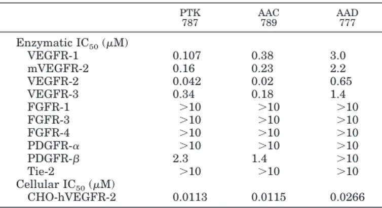
Characterization of the VEGF Receptor Tyrosine Ki-nase Inhibitors. All compounds are potent inhibitors of the human VEGFR-2 tyrosine kinase with similar or weaker activity against its mouse counterpart (Table 1). These com-pounds are also moderately active against VEGFR-1 and VEGFR-3, but have little or no activity against PDGFR- tyrosine kinases, and are all inactive against FGFR-1, FGFR-3, FGFR-4, PDGFR-␣, and Tie-2 tyrosine kinases at concentrations of up to 10M (Table 1).
Because a kinase inhibitor must enter cells to inhibit the kinase domain of the receptor, the effects of the inhibitors was assessed on HUVE cell proliferation by measuring the incorporation of the pyrimidine analog BrdU. All compounds were found to be potent inhibitors of VEGF-induced HUVE cell proliferation with an IC50of 1.6 nM for AAC789, 5.8 nM for PTK787, and 19.6 nM for AAD777. Furthermore, all compounds were inactive against bFGF-induced proliferation in the same concentration range; inhibition of bFGF-induced HUVE cell proliferation was only observed at concentrations 100- to 1000-fold higher than those required to inhibit VEGF-induced proliferation (data not shown).
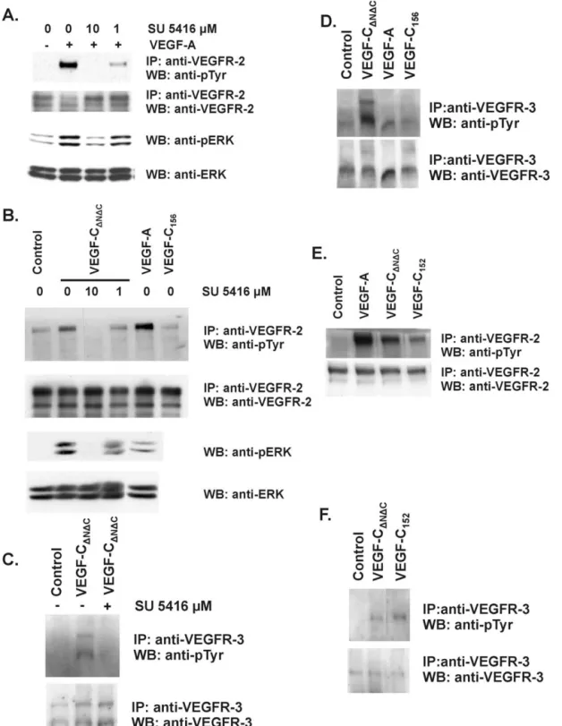
All compounds inhibited VEGF-induced VEGFR-2 auto-phosphorylation in a double antibody chemiluminescence assay using CHO cells transfected with human VEGFR-2; this was observed at the same concentrations at which VEGF-induced HUVE cell proliferation was inhibited (Ta-ble 1). To demonstrate the effect of PTK787 on VEGF-induced receptor phosphorylation in endothelial cells
ex-pressing VEGFR-2 (Pepper and Mandriota, 1998),
confluent BAE cell monolayers were incubated with 10M PTK787 in the absence or presence of VEGF or bFGF. The phosphorylation of VEGFR-2 was then examined by West-ern blotting of VEGFR-2 immunoprecipitates. VEGF in-duced phosphorylation of VEGFR-2 in BAE cells, and this could be inhibited completely by 10 M PTK787 (Fig. 1). No phosphorylation of VEGFR-2 by bFGF was observed at longer incubation times (up to 8 h) and identical results were obtained with BME cells (data not shown). To confirm the specificity of the 200-kDa band, the Western blot was reprobed with a second anti-VEGFR-2 antibody raised
TABLE 1
Inhibitory activity of receptor tyrosine kinase inhibitors
The enzymatic kinase assays were performed using recombinant GST-fused human kinase domains of the receptor, excepted for VEGFR-2 were both mouse (mVEGFR-2) and human (VEGFR-2) kinase domains were examined. VEGF-induced cellular receptor phosphorylation was performed on CHO cells transfected with human VEGFR-2. Each value represents the mean calculated from at least three independent experiments. PTK 787 AAC 789 AAD 777 Enzymatic IC50(M) VEGFR-1 0.107 0.38 3.0 mVEGFR-2 0.16 0.23 2.2 VEGFR-2 0.042 0.02 0.65 VEGFR-3 0.34 0.18 1.4 FGFR-1 ⬎10 ⬎10 ⬎10 FGFR-3 ⬎10 ⬎10 ⬎10 FGFR-4 ⬎10 ⬎10 ⬎10 PDGFR-␣ ⬎10 ⬎10 ⬎10 PDGFR- 2.3 1.4 ⬎10 Tie-2 ⬎10 ⬎10 ⬎10 Cellular IC50(M) CHO-hVEGFR-2 0.0113 0.0115 0.0266
Fig. 1. Inhibition of VEGFR-2 tyrosine phosphorylation by VEGFR-2
tyrosine kinase inhibitors. BAE cells were incubated with 10M PTK787 in the presence or absence of 50 ng/ml VEGF or 10 ng/ml bFGF for 1 h at 4°C and then for 8 min at 37°C. VEGFR-2 was immunoprecipitated from cell lysates. Western blot analysis was performed on immunoprecipitates with an anti-phosphotyrosine antibody (upper panel) and a polyclonal anti-VEGFR-2 antibody (lower panel). Three experiments were per-formed and similar results were obtained.
against a different peptide than the antibody used for immunoprecipitation. A band of 200 kDa and a second band of approximately 190 kDa were detected (Fig. 1), which is consistent with our previous findings (Mandriota et al., 1996; Mandriota and Pepper, 1997; Pepper and Mandriota, 1998).
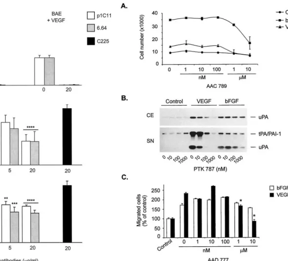
VEGFR Tyrosine Kinase Inhibitors Inhibit Angio-genesis in Vivo. The VEGFR-2 tyrosine kinase inhibitors (Table 1) were tested in an in vivo model of angiogenesis. In this model, VEGF and bFGF induce growth of vascularized tissue around a subcutaneous implant. The angiogenic re-sponse is quantitated by measuring the weight and blood content of this tissue. All compounds given in daily oral doses for 6 days blocked VEGF-induced angiogenesis in a dose-dependent manner (Table 2; Fig. 2). All of the compounds also inhibited the response to bFGF to some extent, but the dose-response curve was not linear for all compounds (Fig. 2). The unexpected inhibition of bFGF-induced angiogenesis prompted us to study this effect in more detail, using pure cultures of endothelial cells and an in vitro model of angio-genesis (Montesano and Orci, 1985).
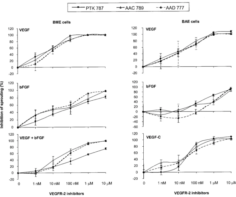
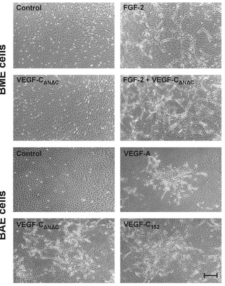
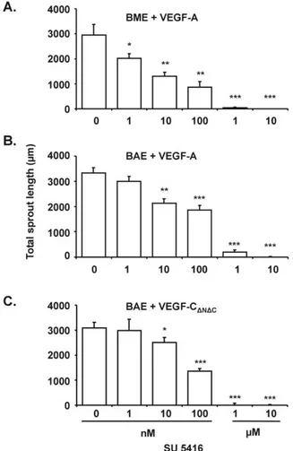
VEGFR Tyrosine Kinase Inhibitors Inhibit Angio-genesis in Vitro. We have shown previously that VEGF family members and bFGF induce BME and BAE cells to invade three-dimensional collagen gels as capillary-like, tubular structures (in vitro angiogenesis). More precisely, VEGF and bFGF induced invasion of BME cells (which express VEGFR-1 and -2 and FGFR-1), whereas VEGF, VEGF-C, and bFGF induced invasion of BAE cells (which express VEGFR-1, -2, and -3 and FGFR-1) (Pepper et al., 1992a, 1993b, 1998; Mandriota et al., 1996; Pepper and Mandriota, 1998). In addition, bFGF and VEGF induced a synergistic response in BME and BAE cells, VEGF-C po-tentiated the effect of bFGF in BME cells, and VEGF and VEGF-C had a synergistic effect on BAE cells (Pepper et al., 1992a, 1995, 1998). Using a variety of VEGF mutants that specifically bind VEGFR-1, -2, or -3 and neutralizing anti-VEGFR-2 antibodies, we have observed that VEGF-and VEGF-C (⌬N⌬C) in vitro angiogenic activities are mediated by VEGFR-2 (J.-C. Tille and M. S. Pepper, manu-script in preparation). We therefore investigated the effect of the VEGFR-2 tyrosine kinase inhibitors listed in Table 1 on VEGF-, VEGF-C-, and bFGF- induced BME or BAE cell collagen gel invasion.
All compounds completely inhibited VEGF-induced BME and BAE cell invasion and VEGF-C-induced BAE cell inva-sion. The inhibition was dose-dependent in both cells types with a maximal effect from 1 M. bFGF-induced invasion was reduced by 92% at 10M in BME cells and by 88% at the
same concentration in BAE cells (Table 2; Figs. 3 and 4). These findings are consistent with our in vivo results (Table 2; Fig. 2). Moreover, the synergistic angiogenic effect of VEGF and bFGF was inhibited by 91% at 10M (Fig. 4). No overt cytotoxicity was observed with any of the VEGFR-2 kinase inhibitors used at concentrations of up to 10M (Fig. 3).
Because the VEGFR-2 tyrosine kinase inhibitors have no effect on FGFR-1, which is expressed by both BME and BAE cells (Mandriota et al., 1996; Pepper and Mandriota, 1998; Pepper et al., 1998), we hypothesized that bFGF-induced angiogenesis may be mediated by the autocrine activity of endogenous VEGF and/or VEGF-C in these cells.
TABLE 2
Inhibitory activity of VEGF receptor tyrosine kinase inhibitors on in vitro and in vivo angiogenesis
Value represent the IC50for in vitro assays and ED50for in vivo assays calculated from the means of at least three independent experiments.
ED50In vivo (mg/kg)
IC50In vitro (M)
BAE BME
bFGF VEGF bFGF VEGF-C VEGF bFGF VEGF
PTK 787 30 21 0.027 0.032 0.057 0.08 0.009 AAC 789 9 26 1.102 0.035 0.016 0.035 0.0055 AAD 777 23 6 1.435 0.041 0.0117 0.012 0.009
Fig. 2. Inhibition of VEGF- and bFGF-induced angiogenesis in vivo by
VEGFR-2 tyrosine kinase inhibitors. Porous Teflon chambers (volume, 0.5 ml) filled with 0.8% w/v agar containing 20 U/ml heparin with or without 2g/ml VEGF or 0.3 g/ml bFGF were implanted subcutane-ously on the dorsal flank of female mice. The mice were treated with VEGFR-2 tyrosine kinase inhibitors at the indicated dose (p.o. once daily) or vehicle. Five days after implantation, the mice were killed, and the chambers were removed. The vascularized tissue growing around the chamber was removed carefully and weighed, and the blood content assessed by measuring hemoglobin. Results are shown as percent inhibition of increase in blood content around the chamber. Each experiment was performed with six animals per dose group, and each dose was tested in at least two independent experiments.
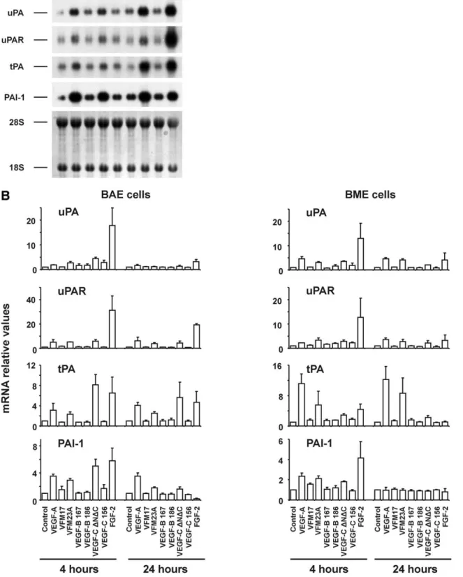
Effect of bFGF on Endogenous VEGF and VEGF-C mRNA and Protein Expression in Endothelial Cells. Based on the observation that bFGF increases VEGFR-2 expression at the levels of mRNA and total protein in BME cells (Pepper and Mandriota, 1998), we hypothesized 1) that BME and BAE cells may express VEGF and/or VEGF-C; 2) that bFGF increases VEGF and VEGF-C in these cells; and 3) that the concomitant expression of VEGF, VEGF-C, and VEGFR-2 promotes an autocrine loop of VEGFR-2 activation in endothelial cells.
To assess the profile of expression of VEGF family mem-bers by BME and BAE cells, total cellular RNAs from con-fluent monolayers were analyzed by RNase protection. Both cell lines were found to express the VEGF164and VEGF120 isoforms, VEGF-B, and VEGF-C but not VEGF-D (Fig. 5). RT-PCR analysis using a pair of oligonucleotide primers, which are external to the alternative splicing region of bovine VEGF, revealed four bands in the kidney (Fig. 6A), two of which, corresponding in size to the VEGF164and VEGF120 isoforms (411 or 279 bp, respectively) were also found in BME and BAE cells. The size of the two larger bands (⬃470 and ⬃520 bp, respectively) in kidney may represent the bovine counterparts of the VEGF189 and VEGF206isoforms previ-ously described in humans (for review, see Ferrara, 2001). No bands were detected if RT was omitted (Fig. 5A), indicating that the bands were not derived from contaminating genomic DNA.
We next assessed whether bFGF affects expression of VEGF-A, -B, and -C in our cells. RNase protection and semi-quantitative RT-PCR analysis of BME and BAE cell RNAs
revealed that both the VEGF120and the VEGF164isoforms were more abundant in bFGF-treated cells after 4 h and to a lesser extent after 15 h incubation with bFGF (Figs. 5 and 6A and data not shown). By RNase protection assay, VEGF164 mRNA was increased by 1.6- and 3.1-fold, respectively, in BME and BAE cells treated with bFGF for 4 h, when nor-malized to the internal control acidic ribosomal phosphopro-tein P0 (Fig. 5). bFGF did not affect the relative proportion of the two VEGF isoforms (Fig. 6A). After 4 h of exposure to bFGF, VEGF-C mRNA was increased by 1.7- and 2.0-fold in BME and BAE cells (Fig. 5 and 6C). bFGF did not increase VEGF-B in BME or BAE cells, and VEGF-D was not induced by bFGF in either cell line (Fig. 5 and 6B).
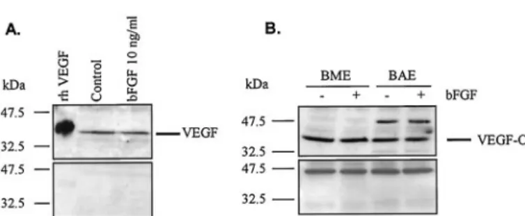
To assess the effect of bFGF on endogenous VEGF and VEGF-C at the protein level, confluent monolayers of BME and BAE cells were incubated with serum-free medium in the presence or absence of bFGF for 8 h, and cell extracts were analyzed by Western blot. Fig. 7A (upper panel) shows the presence of a prominent band of⬃42 kDa, which corre-sponds to the predicted molecular mass of bovine VEGF164 under nonreducing conditions. When recombinant human VEGF165was added to the incubation buffer at a 5-fold molar excess over the primary antibody, the ⬃42 kDa band was undetectable, thus confirming its specificity (Fig. 7A, lower panel). Similar results were obtained with BAE cells (data not shown). No significant differences in the level of VEGF protein were observed between bFGF-treated and untreated BME or BAE cells, after 8 or 15 h of incubation (Fig. 7A and data not shown). With respect to VEGF-C, a prominent band at the expected size of ⬃43 kDa could be detected in both BME and BAE cells (Fig. 7B, upper panel). An additional, higher molecular mass band was detected in BAE cells, which may represent an unprocessed or partially processed form of VEGF-C. After treatment with bFGF for 8 h, no modulation was seen in the level of the ⬃43 kDa VEGF-C band in either cell line (Fig. 7B, upper panel). The specificity of the band was assessed by replacing the anti-VEGF-C an-tibody with normal rabbit serum (Fig. 7B, lower panel).
Taken together, these data demonstrate that bFGF in-creases VEGF and VEGF-C mRNA expression in a transitory manner in BME and BAE cells, without significantly altering the expression of either gene at the protein level.
VEGF and VEGF-C Antagonists Inhibit bFGF-In-duced Angiogenesis in Vitro. To assess the possibility that the in vitro angiogenic effect of bFGF may be dependent in part on an autocrine VEGF and/or VEGF-C loop, a chi-meric molecule consisting of the extracellular domain of hu-man VEGFR-1 fused to a fragment of the heavy chain of human IgG␥1 (VEGFR-1-IgG), which is a potent inhibitor of VEGF activity (Aiello et al., 1995; Shima et al., 1995), was tested for its effect on bFGF-induced BME cell collagen gel invasion. VEGFR-1-IgG, but not a CD4-human IgG␥1 fusion protein, inhibited bFGF-induced in vitro angiogenesis in a dose-dependent manner, with a maximal inhibition of 51% at 10g/ml (Figs. 8 and 9A). Similarly, up to 48% inhibition was observed with a neutralizing anti-human VEGF polyclonal antibody, whereas preimmune rabbit␥-globulins had no ef-fect (Figs. 8 and 9B). No toxicity was observed in VEGFR-1-IgG- or anti-VEGF antibody-treated cells (Fig. 8). VEGF-induced BME cell invasion was inhibited almost completely by both VEGFR-1-IgG and the anti-human VEGF antibody (Fig. 9, A and B). To assess the role of endogenous VEGF-C in Fig. 3. Inhibition of VEGF-induced in vitro angiogenesis by PTK787.
Confluent monolayers of BME cells on three-dimensional collagen gels were treated for 4 days with 30 ng/ml VEGF (c) or 10 ng/ml bFGF (e) alone or in combination with 10 M PTK787 (d and f). The resulting capillary-like tubular structures were viewed by phase-contrast micros-copy. In b, in which the plane of focus is at the level of the surface of the collagen gel, cells treated with 10M PTK787 show no signs of cytotox-icity (similar results were obtain with the other inhibitors). No invasion occurred in untreated cultures (a). Bar, 75m.
bFGF-induced in vitro angiogenesis, a similar chimeric mol-ecule (VEGFR-3-IgG) (Lee et al., 1996) was tested on induced BAE cell invasion. VEGFR-3-IgG inhibited bFGF-induced in vitro angiogenesis in a dose-dependent manner, with a maximal inhibition of 50% at 10g/ml, whereas con-trol IgGs had no effect (Fig. 9C). No toxicity was observed in VEGFR-3-IgG-treated cells (data not shown). VEGFR-1-IgG did not alter significantly VEGF-C-induced invasion, and or VEGFR-3-IgG had no effect on invasion induced by VEGF, thereby demonstrating that VEGF-C- induced angiogenesis is not dependent on endogenous VEGF, and vice versa (data not shown).
To assess directly the role of VEGFR-2 in bFGF-induced in vitro angiogenesis, VEFGR-2 blocking antibodies (Witte et al., 1998; Zhu et al., 1998) were added to the system. VEGF-induced BME cell invasion was inhibited in a dose-dependent manner by the two anti-human VEGFR-2 antibodies, p1C11 and 6.64, with a maximal inhibition of 99.0 and 99.9% at 20 g/ml (Fig. 10A and data not shown). Similarly, up to 99.5
and 98.5% inhibition was observed with p1C11 and 6.64 on VEGF-induced BAE cell invasion (Fig. 10A and data not shown). bFGF-induced BME cell collagen gel invasion was inhibited partially by both antibodies with a maximal inhi-bition of 48 and 49% at 20g/ml of p1C11 and 6.64, respec-tively (Fig. 10B). bFGF-induced BAE cell in vitro angiogen-esis was inhibited by 23% with p1C11 and by 37.5% by 6.64 at 20g/ml (Fig. 10C). The control isotope antibody, C225, had no effect (Fig. 10B and 10C) and no toxicity was observed in any of the antibody-treated cultures (data not shown).
These findings point to a role for VEGFR-2 in the in vitro angiogenic effect mediated by bFGF in bovine endothelial cells. We are aware that there are differences between endothelial cells from different species and vascular beds and that the VEGF/VEGFR-2 autocrine loop may not be relevant to all cell types. Nonetheless, to extend this ob-servation, we next assessed the effect of VEGFR-2 tyrosine kinase inhibitors on endothelial cell functions that are required for angiogenesis.
Fig. 4. Inhibition of in vitro angiogenesis by VEGFR-2 inhibitors. Confluent monolayers of BME cells on three-dimensional collagen gels were treated
with 10 ng/ml bFGF, 30 ng/ml VEGF, or both cytokines in combination, whereas BAE cells were treated with 3 ng/ml bFGF, 30 ng/ml VEGF, or 30 ng/ml VEGF-C. VEGFR-2 inhibitors were added at the concentrations indicated. Invasion was measured after 4 days and is expressed as percent inhibition of sprouting induced by cytokines alone. Results are shown as the means⫾ S.E.M. from at least three experiments per condition.