HAL Id: dumas-02153482
https://dumas.ccsd.cnrs.fr/dumas-02153482
Submitted on 12 Jun 2019HAL is a multi-disciplinary open access archive for the deposit and dissemination of sci-entific research documents, whether they are pub-lished or not. The documents may come from teaching and research institutions in France or abroad, or from public or private research centers.
L’archive ouverte pluridisciplinaire HAL, est destinée au dépôt et à la diffusion de documents scientifiques de niveau recherche, publiés ou non, émanant des établissements d’enseignement et de recherche français ou étrangers, des laboratoires publics ou privés.
Identification de cibles cellulaires impliquées dans
l’hypertension artérielle pulmonaire induite par les
inhibiteurs de protéines kinases par une analyse de
pharmacovigilance / pharmacodynamie
Lucie Cornet
To cite this version:
Lucie Cornet. Identification de cibles cellulaires impliquées dans l’hypertension artérielle pulmonaire induite par les inhibiteurs de protéines kinases par une analyse de pharmacovigilance / pharmacody-namie. Sciences pharmaceutiques. 2018. �dumas-02153482�
AVERTISSEMENT
Ce document est le fruit d'un long travail approuvé par le
jury de soutenance et mis à disposition de l'ensemble de la
communauté universitaire élargie.
Il n’a pas été réévalué depuis la date de soutenance.
Il est soumis à la propriété intellectuelle de l'auteur. Ceci
implique une obligation de citation et de référencement
lors de l’utilisation de ce document.
D’autre part, toute contrefaçon, plagiat, reproduction illicite
encourt une poursuite pénale.
Contact au SID de Grenoble :
bump-theses@univ-grenoble-alpes.fr
LIENS
LIENS
Code de la Propriété Intellectuelle. articles L 122. 4
Code de la Propriété Intellectuelle. articles L 335.2- L 335.10
http://www.cfcopies.com/juridique/droit-auteur
UNIVERSITÉ GRENOBLE ALPES
UFR DE PHARMACIE DE GRENOBLE
Année : 2018
IDENTIFICATION DE CIBLES CELLULAIRES IMPLIQUÉES DANS
L'HYPERTENSION ARTÉRIELLE PULMONAIRE INDUITE PAR LES
INHIBITEURS DE PROTEINES KINASES PAR UNE ANALYSE DE
PHARMACOVIGILANCE / PHARMACODYNAMIE.
MÉMOIRE DU DIPLÔME D’ÉTUDES SPÉCIALISÉES DE PHARMACIE HOSPITALIERE ET DES COLLECTIVITES
Conformément aux dispositions du décret N° 90-810 du 10 septembre 1990, tient lieu de
THÈSE
PRÉSENTÉE POUR L’OBTENTION DU TITRE DE DOCTEUR EN PHARMACIE DIPLÔME D’ÉTAT
Lucie CORNET
MÉMOIRE SOUTENU PUBLIQUEMENT À LA FACULTÉ DE PHARMACIE DE GRENOBLE
Le : 26/04/2018
DEVANT LE JURY COMPOSÉ DE
Président du jury : Monsieur le Professeur Christophe RIBUOT Membres : Monsieur le Professeur Michel TOD
Monsieur le Professeur Jean-Luc CRACOWSKI Monsieur le Docteur Sébastien QUETANT Directeur de thèse : Monsieur le Docteur Charles KHOURI
L’UFR de Pharmacie de Grenoble n’entend donner aucune approbation ni improbation aux opinions émises dans les mémoires ; ces opinions sont considérées comme propres à leurs auteurs.
REMERCIEMENTS
A Messieurs les Professeurs Jean-Luc CRACOWSKI et Michel TOD, ainsi qu’à Monsieur le Docteur Sébastien QUETANT,
Merci de m’avoir fait l’honneur d’évaluer mon travail.
A Monsieur le Professeur Christophe RIBUOT,
Merci d’avoir accepté de présider mon jury de thèse.
A Monsieur le Docteur Charles Khouri
Un grand merci de m’avoir proposé ce travail et de m’avoir permis de le réaliser.
Merci également pour ta disponibilité et tes conseils judicieux, c’est un réel plaisir de travailler
Merci à tous ceux qui m’ont accueillie pendant 6 mois dans leur PUI, dans leur espace de pratique.
A Dreux, merci à Patricia qui m’a donné cette image du pharmacien hospitalier, merci à Christelle, à Claire, à Sandrine et à Marie-Claire. Merci à Joseph et Anne-So à qui je souhaite plein de bonheur, merci à Baptiste sans qui cela n’aurait pas été pareil.
A Vichy, Merci à Magali de m’avoir si bien encadrée. Merci à Françoise, à Mélanie, à Helena, et un immense merci à Jérôme. Merci aux agents de sté d’avoir fait de mon départ un moment inoubliable et à toute l’équipe de la pharmacie. Merci à Toto, Micka, Alex, Olivier, Vincent et tous les autres d’avoir fait de ce semestre un des meilleurs.
A Lyon Sud, merci de m’avoir montré l’excellence et l’exigence et de m’avoir permis de me rendre compte de ce que je voulais vraiment.
A Grenoble, un immense merci à Chacha, Loulou, Océ, Mélo-mélo, Morgane et Samia d’avoir tenu bon contre l’immensité du monde. Merci à Thomas pour ces discussions toujours plus intéressantes.
A Sainté, merci à Xavier, Fabien, Sandrine, Agnès, et un grand merci à Marion, vous êtes géniaux ! Merci aussi à Maellou, qui a rendu ce semestre d’une douceur inouïe. Merci à Caro1, d’avoir tant fait pour moi, à Jean, Aurélie, Camille, Lucille, les sangsues, l’Euro 2016, et tout ce qui a fait que c’était quand même très cool.
A Grenoble again, merci à Jean-Luc de m’avoir permis de faire ce stage et ce master, j’en suis sortie plus grande. Merci à Bertrand, Hélène, Atanur, Marie-Claire, Yan, aux Apicuriens, à Dalil, à Toto, à Orianou et à Ben évidemment, merci pour ta présence, ton soutien et tes bons conseils.
Merci à Caro2 pour tous ces moments partagés avec toi, ces tartes aux brocolis et ces tisanes quotidiennes.
Aux pharmacovigilants de Grenoble, merci à Brubru, Marion, Nath, Michel, vous m’avez fait aimer la biblio, et par-dessus tout merci à Charles, je n’aurais souhaité personne d’autre pour encadrer ma thèse.
A Grenoble again bis, merci à Isabelle d’avoir partagé ton savoir, merci à Dorothée, à Stéphanie, à Virginie, à Marion et à Anne-Laure et à Paupau pour avoir fait de ce semestre une fin en beauté.
Merci à mes colocs d’amour, Loulou, Soph’, Thib (dit Titi) de m’avoir supportée et d’avoir rendu la vie si douce.
Enfin merci aux copains Oriane (re), Doudou, Etienne, Paupau, Laulau, Sylvette, Mimie, Titi et tous les autres. Merci à Alex pour la poule au pot dans le jardin et la course de roulades.
Merci à tous les internes avec qui j’ai un jour partagé un verre : Gwendal, Thomas, Clémence, Mathieu, Arnaud, Béné, Alicia, Julianne, Ariane, Elisa…
Merci à Marine pour ces soirées de debrief intense et ces saucissons partagés.
Merci à mon Doudou (Eddy pour les intimes), à Sarou, Rémos, Clem, Suze, Paul, Wiwi, et tant d’autres pour ces années pharmas et ces retrouvailles semestrielles.
Merci au Chèvre Show pour cette bouffée d’été. Merci à Laurent qui m’a éduquée et à Francis, qui m’a dés-éduquée.
Merci à LéaAu d’exister dans ma vie. Merci à Yo et Triphon de m’avoir accueillie.
Merci à mes parents Pascale et Daniel, de m’avoir soutenue jusqu’au bout, dans les doutes, les larmes, la joie, les rires, et tout le temps.
Merci à ma sœur Laure pour tout ce que tu es et ce que tu représentes pour moi, tu es merveilleuse.
Merci à mon frèro Quentin, dit Roupet, toi aussi tu es fantastique.
Merci à toutes les personnes que j’ai croisées durant mon parcours professionnel, qui m’ont donné envie de devenir comme eux, ou qui m’ont montré ce que je ne voulais pas devenir.
1
TABLE DES MATIERES
Introduction ... 5 HTAP ... 5 Définition ... 5 Classification ... 7 Physiopathologie ... 8 Epidémiologie ... 10 Traitement de l’HTAP ... 10
HTAP iatrogène/induite par les toxiques ... 12
Inhibiteurs de protéines kinases ... 13
Objectif ... 16
Identification of cellular targets involved in PAH induced by PKI by a pharmacovigilance / pharmacodynamics analysis ... 17 Abstract ... 18 Introduction ... 19 Methods ... 19 1. Data sources ... 19 Pharmacovigilance database ... 19 Selection of cases ... 19
Selection of protein kinase inhibitors ... 20
Identification of protein kinases involved in PAH ... 20
Affinity data ... 20 Co-medication of interest ... 20 2. Analysis ... 20 Disproportionality analysis ... 20 Correlation analysis ... 20 Results ... 21
2
Selection of cases ... 21
Description of PAH cases ... 21
Identification of protein kinases involved in PAH ... 21
Disproportionality analysis ... 21 Correlation analysis ... 22 Co-medication analysis ... 23 Clusters ... 23 Temporal analysis ... 23 Discussion ... 25 Conclusion ... 27 References ... 27 Supplementary Material ... 30
Appendix 1. Details of co-reported Meddra term excluded from the analysis ... 31
Appendix 2. Most relevant references about the protein kinases of interest ... 33
Appendix 3. Results of sensitivity analysis ... 35
Appendix 4. Correlation analysis ... 36
Appendix 5. Results of the co-medication analysis of dasatinib PAH cases. ... 38
Discussion ... 40
Conclusion...45
3
INDEX DES FIGURES ET TABLEAUX
Figure a. Cathétérisme cardiaque droit ... 6
Tableau a. Définition hémodynamique de l'hypertension pulmonaire ... 6
Tableau b. Classification de l'HTP ... 7
Figure b. Physiopathologie de l'HTAP. ... 9
Figure c. Cancer-like théorie de l'HTAP. ... 10
Figure d. Les trois voies de traitement de l'HTAP. ... 11
Tableau c. Traitement des HTAP ... 12
Tableau d. Les inhibiteurs de protéines kinases, cibles et indications ... 13
Figure 1. Forest plot of the disproportionality analysis of PAH induced by PKI. ... 22
Figure 2. Manhattan plot synthetizing the correlation analysis... 23
Figure 3. Results of the co-medication analysis between PAH and non-PAH cases related to dasatinib in Vigibase®. ... 24
Figure 4. Clusters dendrogram of protein kinases inhibitors based on their affinity profile. .. 24
Figure 5. Proportion of reported PAH cases for 1000 reported adverse events per year. ... 25
Figure S1. Flow chart of PAH cases selection for analysis ... 31
Figure S2. Identification of protein kinases involved in pulmonary function. ... 32
Table S1. Most relevant references about target of interest ... 33
Figure S3. Disproportionality signal of PAH induce by PKI from the first five years after FDA approval versus all medication in pharmacovigilance database. ... 35
Table S2. Correlation and p-value of all sensitivity analysis. ... 36
Table S3. Result of co-medication analysis ... 38
Figure e. Targeting the kinome: bosutinib versus dasatinib ... 43
4
LISTE DES ABREVIATIONS
DCI : Dénomination commune internationale
HCP : hémangiomatose capillaire pulmonaire HTAP: Hypertension artérielle pulmonaire HTAPP : hypertension artérielle pulmonaire persistante du nouveau-né
HTP : Hypertension pulmonaire
HTPC : HTP thromboembolique chronique IPK: Inhibiteurs de protéines kinases LMC : leucémie myéloïde chronique
MVO : maladies veino-occlusives
PAPm : pression artérielle pulmonaire moyenne PAPO : pression artérielle pulmonaire d’occlusion
RCP : Résumé des caractéristiques du produit UW : unité Wood
FDA : Food and Drug administration
PAH : Pulmonary arterial hypertension
PHT : Pulmonary Hypertension
PKI : Protein Kinase Inhibitor CML: chronic myeloid leukemia MeSH: Medical Subject Heading
PAPm : mean Pulmonary arterial pressure PAWP: pulmonary arterial wedge pressure ROR: Reported Odds Ratio
5
Identification de cibles cellulaires impliquées dans
l'Hypertension artérielle pulmonaire, induite par les
Inhibiteurs de Protéines Kinases par une analyse de
pharmacovigilance / pharmacodynamie.
Introduction
HTAP
Définition
Les dernières recommandations relatives aux définitions, à la classification, au diagnostic et au traitement des patients atteints d’hypertension pulmonaire (HTP) ont été conjointement
publiées par la Société européenne de cardiologie (European Society of Cardiology, ESC) et la
Société européenne respiratoire (European Respiratory Society, ERS) en 2015 (Galiè et al.,
2016). Ces mises à jour succèdent aux deux premières recommandations publiées en 2004 et
2009.
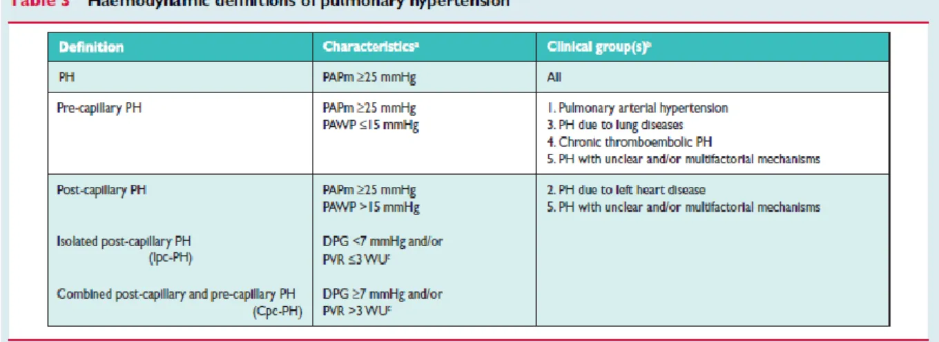
La définition hémodynamique de l’hypertension pulmonaire est basée sur l’élévation de la
pression artérielle pulmonaire moyenne (PAPm) au-dessus de 25 mmHg (valeurs normales au
repos = 14 ± 3 mmHg, et valeurs limite haute = 20 mmHg) mesurée par cathétérisme cardiaque droit. Le cathétérisme cardiaque droit est un examen invasif, permettant d’explorer les cavités
cardiaques droites afin de mesurer les pressions de l'oreillette droite, du ventricule droit, de
l'artère pulmonaire et des capillaires pulmonaires. Il consiste en l’introduction d’un cathéter
6
Figure a. Cathétérisme cardiaque droit
La distinction entre l’HTP pré-capillaire et post-capillaire est faite par la mesure de la pression
artérielle pulmonaire d’occlusion (PAPO ou PAWP pulmonary arterial wedge pressure) ≤ 15
mmHg pour une HTP précapillaire ou > 15 mmHg pour une HTP postcapillaire (Tableau a).
7
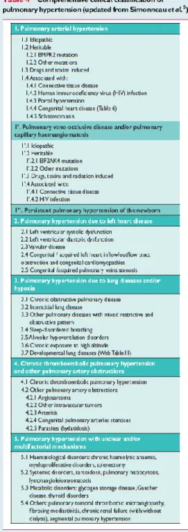
Classification
La classification de l’HTP vise à définir cinq groupes homogènes en regroupant plusieurs
conditions physiopathologiques selon leurs présentations cliniques, leurs caractéristiques
hémodynamiques et leurs traitements (Tableau b).
Le groupe 1 caractérise les HTAP pour lesquelles les
changements pathologiques affectent
préférentiellement les artères pulmonaires distales
(<500 µm). L’hypertension artérielle pulmonaire
regroupe les HTAP idiopathiques, héritable, induites
par des médicaments / toxiques, et les HTAP associées aux connectivites, au VIH, à l’hypertension portale,
aux maladies cardiaques congénitales et aux schistosomiases. Le groupe 1’ inclut principalement
les maladies veino-occlusives (MVO) pulmonaires et
hémangiomatoses capillaires pulmonaires (HCP) et le groupe 1’’ regroupe les hypertensions artérielles
pulmonaires persistantes du nouveau-né (HTAPP).
Le groupe 2 décrit les HTP associées à une pathologie
du cœur gauche, le groupe 3 caractérise les HTP liées
aux maladies pulmonaires chroniques et/ou à l’hypoxie et le groupe 4 se rapporte aux HTP
thromboembolique chronique (HTPC) impliquant une
obstruction chronique des artères pulmonaires. Le
groupe 5 caractérise les HTP de mécanisme incertain
ou multifactoriel (Galiè et al., 2016). Tableau b. Classification de l'HTP
8
La définition de l’hypertension artérielle pulmonaire (HTAP, groupe 1 de la classification
impliquant les HTAP iatrogènes) inclut une résistance vasculaire pulmonaire (RVP) de > 3
unités Wood (UW). Ce qui permet de la distinguer des situations entraînant une augmentation
de la PAPm par élévation du débit cardiaque sans remodelage vasculaire pulmonaire spécifique.
Le terme HTAP désigne ainsi un sous-groupe de patients présentant
- pression artérielle pulmonaire moyenne (PAPm) ≥ 25 mmHg
- une HTP précapillaire (PAPO ou PAWP) ≤ 15 mmHg
- et une élévation des résistances vasculaires pulmonaires (RVP > 3 UW),
- en l’absence d’autres causes d’HTP précapillaire (maladies respiratoires chroniques et l’HTP thromboembolique chronique).
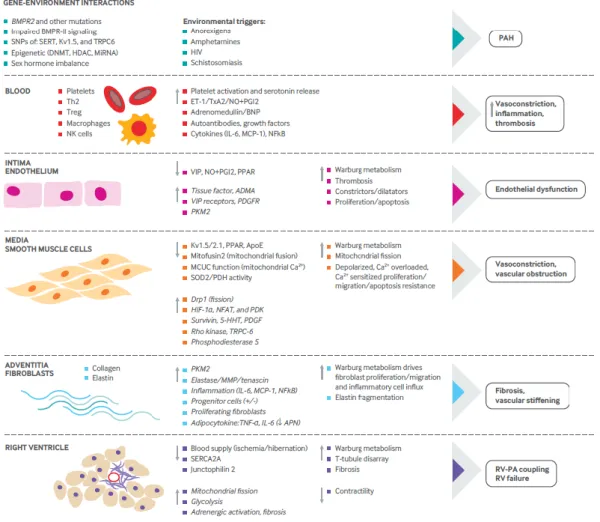
Physiopathologie
La physiopathologie de l’HTAP reste encore incertaine, plusieurs concepts émergent quant aux
origines de cette pathologie. L’origine semble multifactorielle avec l’intervention de multiples
voies dérégulées. Un aspect génétique est retrouvé avec la découverte de nombreuses mutations
permissives mais non suffisantes de la maladie. Une dysfonction endothéliale associée à une augmentation de l’inflammation et une vasoconstriction semble également intervenir (Figure
9
Figure b. Physiopathologie de l'HTAP. Extrait de Thenappan et al., 2018
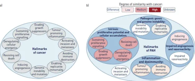
Un parallèle avec le phénotype de cancer a également été réalisé (cancer-like theory ou théorie
du cancer) associant remodelage vasculaire et prolifération cellulaire associée à une
10
Figure c. Cancer-like théorie de l'HTAP. Extrait de Guignabert et al., 2013
Epidémiologie
La prévalence de l’HTP atteint 97 cas par million de personnes avec un ratio femme/homme de
1,8. La prévalence de l’HTAP représente quant à elle 15 à 60 cas par million de personne (Galiè
et al., 2016). L’HTAP peut être idiopathique (30 à 50%) incluant 15 à 20% de forme héritable,
liées aux connectivites (15 à 30%), au VIH (1 à 6,2%), aux maladies cardiaques congénitales
(10 à 43%) ou iatrogènes (3 à 9,5%) avec une distinction selon la zone géographique (Prins and
Thenappan, 2016 ; Lau et al., 2017).
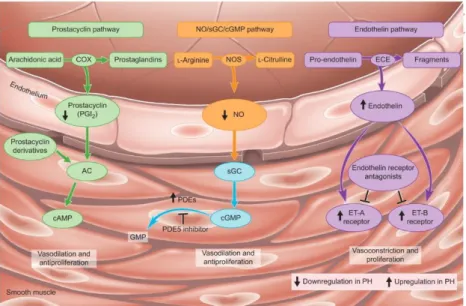
Traitement de l’HTAP
Actuellement, le traitement de l’HTAP repose sur trois approches thérapeutiques :
- Les mesures générales incluant l’activité physique, la réhabilitation, l’immunisation
contre la grippe et le pneumocoque et les soins de soutiens (anticoagulants oraux,
diurétiques, oxygénothérapie)
- Les traitements pharmacologiques de l’HTAP incluent plusieurs classes
11
Les antagonistes des récepteurs à l’endothéline (ambrisentan, bosentan, macitentan) reversent la vasoconstriction et les effets mitogènes de l’endothéline
sur les tissus pulmonaires des patients.
Les inhibiteurs de la phosphodiéstérase-5 (sildenafil, tadalafil, vardenafil) et un stimulateur direct de la guanylate cyclase soluble (riociguat) induisent une
vasodilatation via la voie monoxyde d’azote (NO) / GMPc.
Les analogues de la prostacycline (betaprost, epoprostenol, iloprost, treprostinil) et les agonistes des récepteurs IP aux prostacyclines (selexipag) entrainent une
vasodilatation du lit vasculaire (Hoeper et al., 2016; Galiè et al., 2016).
Figure d. Les trois voies de traitement de l'HTAP. Extrait de Humbert, 2010
De fortes doses d’inhibiteurs calciques sont également utilisées chez les patients répondeurs,
dans le cadre d’HTAP idiopathique. Étonnamment, certains IPK ont été testés comme
traitement des HTAP (Mucke, 2013; Kimura et al., 2017). De nouvelles molécules telles que l’olaparib (PARP-1 inhibiteurs), l’anakinra (inhibiteur de l’interleukine-1alpha et -12beta), le
fulvestrant (antagoniste compétitif des récepteurs aux estrogènes) ou le fasudil (Rho-kinase
inhibiteur utilisé pour favoriser l’apoptose cellulaire) sont en cours d’essais tandis que d’autres
12
telles que la combinaison de thérapeutiques ou l’utilisation de nouvelles molécules agissant sur
les voies de signalisation de l’HTAP sont également en cours d’investigation (Vaidya et al.,
2017).
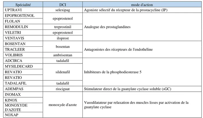
Tableau c. Traitement des HTAP
Spécialité DCI mode d'action
UPTRAVI selexipag Agoniste sélectif du récepteur de la prostacycline (IP) EPOPROSTENOL
epoprostenol
Analogue des prostaglandines FLOLAN REMODULIN treprostinil VELETRI epoprostenol VENTAVIS iloprost BOSENTAN bosentan
Antagonistes des récepteurs de l'endothéline TRACLEER VOLIBRIS ambrisentan ADCIRCA tadalafil Inhibiteurs de la phosphodiesterase 5 MYSILDECARD sildenafil REVATIO REVATIO TADALAFIL tadalafil
ADEMPAS riociguat Stimulateur direct de la guanylate cyclase soluble (sGC) INOMAX
monoxyde d'azote Vasodilatateur par relaxation des muscles lisses par activation de la guanylate cyclase
KINOX MONOXYDE D'AZOTE NOXAP
HTAP iatrogène/induite par les toxiques
Plusieurs molécules sont connues pour induire des HTAP. Les amphétamines et leurs dérivés,
tels que l’aminorex, la fenfluramine, la dexfenfluramine, le benfluorex, la phentermine, et le
mazindol, ainsi que les interférons alpha et beta sont connus pour induire des HTAP. Plus
récemment, un lien entre le dasatinib et la survenue de modifications vasculaires réversibles a
été mis en évidence, suite à plusieurs cas d’HTAP attribués à cet inhibiteur multikinase indiqué
dans la leucémie myéloïde chronique (Quintás-Cardama et al., 2007) ; (Mattei et al., 2009). Par
la suite, des cas d’HTAP ont été décrits avec d’autres IPK, comme le bosutinib, le ponatinib ou
13
Inhibiteurs de protéines kinases
Les inhibiteurs de protéines kinases sont des petites molécules conçues pour interférer avec une
cible moléculaire spécifique qui jouerait un rôle crucial dans la croissance ou la progression
tumorale (Sawyers, 2004). De nombreuses molécules sont maintenant approuvées dans
plusieurs maladies. Chaque IPK se lie à un profil unique de cibles moléculaires plus ou moins
sélectives sur PK d'intérêt.
Tableau d. Les inhibiteurs de protéines kinases, cibles et indications
DCI Specialité Laboratoire
Date d'autorisatio
n par la FDA
Cible d'action selon les RCP (Résumé de caractéristique du produit) Indications Afatinib GIOTRIF Boehringer Ingelheim Pharmaceuticals 12-Jul-2013 ErbB - EGFR (ErbB1=HER1) - HER2 (ErbB2) - ErbB3 - ErbB4 cancer bronchique non à petites cellules
Axitinib INLYTA Pfizer 27-Jan-2012
VEGFR - VEGFR-1 - VEGFR-2 - VEGFR-3 adénocarcinome rénal
Bosutinib BOSULIF Pfizer 4-Sep-2012
Bcr-Abl Src, Lyn, Hck PDGF c-kit c-Fms EphA et B Trk, Axl et Tec ErbB Csk Ste20 PK dépendantes de la calmoduline. Leucémie myéloïde chronique
Cabozantinib CABOMETYX Ipsen 29-Nov-2012
MET VEGF
GAS6 (AXL), RET, ROS1, TYR03, MER KIT, TRKB Fms-3 (FLT3) TIE-2. adénocarcinome rénal carcinome thyroïdien
Ceritinib ZYKADIA Novartis 29-Apr-2014 ALK
cancer bronchique non à petites
cellules
14
Crizotinib XALKORI Pfizer 26-Aug-2011
ALK RTK HGFR, c-Met ROS1 (c-ros) (RON) RTK cancer bronchique non à petites cellules
Dabrafenib TAFINLAR GlaxoSmithKline,
Pharmaceuticals 29-May-2013 RAF RAS/RAF/MEK/ERK cancer bronchique non à petites cellules mélanome malin
Dasatinib SPRYCEL Bristol-Myers
Squibb Co 28-Jun-2006 BCR-ABL SRC family (LYN, HCK) c-KIT EPH PDGFbeta Leucémie myéloïde chronique
Erlotinib TARCEVA Roche 18-Nov-2004 EGFR (ErbB1=HER1)
cancer bronchique non à petites
cellules cancer du pancréas
Gefitinib IRESSA AstraZeneca 1-Jan-2003 EGFR (ErbB1=HER1)
cancer bronchique non à petites
cellules
Ibrutinib IMBRUVICA Janssen Cilag 13-Nov-2013 BTK Leucémie myéloïde
chronique
Imatinib GLIVEC Novartis 1-Jan-2001
Bcr-Abl Kit DDR1 -DDR2 CSFR1 PDGFalpha, beta Leucémie myéloïde chronique
Lapatinib TYVERB Novartis 13-Mar-2007
ErbB
- EGFR (ErbB1=HER1) - HER2 (ErbB2)
cancer du sein
Lenvatinib LENVIMA Eisai 13-Feb-2015
VEGF - VEGFR1 (FLT1) - VEGFR2 (KDR) - VEGFR3 (FLT4) FGF - FGFR1, 2, 3 et 4 PDGF - PDGFR alpha KIT RET carcinome thyroïdien Lestaurtinib Teva Pharmaceutical Industries (Cephalon)
1-Oct-2003 FLT3 Leucémie myéloïde
15
Lorlatinib Pfizer 1-Oct-2016 ALK/ROS1
cancer bronchique non à petites cellules Nilotinib TASIGNA Novartis Pharmaceuticals Corporation 29-Oct-2007 Bcr-Abl PDGFR Kit Ephrine Leucémie myéloïde chronique
Osimertinib TAGRISSO AstraZeneca
Pharmaceuticals 13-Nov-2015 EGFR muté : -EGFRm et T790M cancer bronchique non à petites cellules
palbociclib IBRANCE Pfizer 3-Feb-2015 CDK4/6 cancer du sein
Pazopanib VOTRIENT GlaxoSmithKline,
Pharmaceuticals 19-Oct-2009 VEGFR-1, VEGFR-2 VEGFR-3 PDGFR-α et PDGFR-ß c-KIT carcinome rénal
Ponatinib ICLUSIG Ariad
pharmaceuticals 11-Dec-2012 Bcr-Abl RET, FLT3 et KIT FGFR, PDGFR, VEGFR. Leucémie myéloïde chronique
Regorafenib STIVARGA Bayer HealthCare
Pharmaceuticals 27-Sep-2012
VEGFR1, 2, 3 TIE2
KIT, RET, RAF-1, BRAF, BRAFV600E PDGFR FGFR CSF1R cancer colorectal GIST (tumeurs stromales gastro-intestinales) carcinome hépatocellulaire
Ruxolitinib JAKAVI Novartis 16-Nov-2011 JAK1 et JAK2
myélofibrose (MF) et maladie de
Vaquez
Sorafenib NEXAVAR Bayer HealthCare
Pharmaceuticals 20-Dec-2005
CRAF, BRAF, V600E BRAF, c-KIT, et FLT-3) 2, VEGFR-3, et PDGFR-bêta c-KIT, FLT-3
carcinome rénal
Sunitinib SUTENT Pfizer 26-Jan-2006
PDGFR alpha et bêta VEGFR1/2/3 KIT FLT3 CSF-1R RET GIST, carcinome rénal, cancer du pancréas
16
Trametinib MEKINIST GlaxoSmithKline,
Pharmaceuticals 29-May-2013 MEK1 et MEK2
cancer bronchique non à petites
cellules mélanome malin
Vandetanib CAPRELSA AstraZeneca,
Pharmaceuticals 6-Apr-2011 VEGFR-2 VEGFR-23 EGFR RET carcinome thyroïdien
Vemurafenib ZELBORAF Genentech 17/08/2011 BRAF mélanome malin
Objectif
Nous avons cherché à identifier les cibles cellulaires impliquées dans la physiopathologie des HTAP induites par les IPK, via une analyse de corrélation entre l’affinité des IPK sur certaines
cibles impliquées dans la physiologie de l’HTAP, et les signaux de disproportionnalité, calculés
17
Identification of cellular targets involved in Pulmonary
Arterial Hypertension induced by Protein Kinase
Inhibitors by a pharmacovigilance / pharmacodynamics
analysis.
18
Abstract
Pulmonary arterial hypertension (PAH) remains a rare and incurable, life-threatening disease, induced by several etiologies. Few drugs are known to induce PAH. Among them, the multi tyrosine kinase inhibitor dasatinib was recently linked to PAH followed by some other protein kinases inhibitors (PKI) (e.g. bosutinib, ponatinib, nilotinib). Nevertheless the pathophysiology of PAH induced by PKI remains unclear. To gain knowledge into this rare and severe pathology we performed a study combining a pharmacovigilance approach (disproportionality analysis on the WHO pharmacovigilance database) and the pharmacodynamics properties of PKI. A positive disproportionality signal was found for dasatinib, bosutinib, ponatinib, ruxolitinib, nilotinib and imatinib. Four non-receptors proteins kinase disclosed a significant correlation with the development of PAH: c-src (r= 0.85 and p= 0.00003), c-yes (r= 0.82 and p= 0.00012), Lck (r= 0.8 and p= 0.00038) and Lyn (r= 0.80 and p= 0.00023), all belonging to the SRC family kinases. Kinases of the BMP signaling seems also to play a role in the pathophysiology of PAH induced by PKI. Moreover, a co-medication analysis revealed that diuretics, insulins, statins and antihypertensive drugs are more frequently associated in dasatinib PAH cases compared to other adverse event cases. Interestingly, the dasatinib profile seems different from other PKI in the results of the cluster analysis. Consistently and based on in vitro and in vivo findings, PKI increase the risk of developing PAH but require a comparable genetic, epigenetic or environmental “second hit”. This is to our knowledge, the first pharmacovigilance analysis which investigate the risk of PAH associated with PKI. The study highlights the role of SRC protein kinases family in PAH induced by PKI. This approach combining pharmacovigilance and pharmacodynamics data allowed us to generate some hypothesis about the pathophysiology of the disease, however the results have to be confirmed by further studies. Overall, this study contributes to a better understanding of PAH induced by PKI.
19
Introduction
Pulmonary hypertension (PH) is defined as an increase in mean arterial pressure (PAPm) ≥ 25 mmHg assessed by right heart catheterization (RHC) (1). Pathophysiology is characterized by remodeling processes through an increased migration and proliferation of pulmonary arterial smooth muscle cells leading to vascular remodeling (2). The classification proposed by the Fifth World Symposium on Pulmonary Hypertension defines five groups of different pathological features which characterize the diverse clinical PH groups (3). Group 1 characterize pulmonary arterial hypertension (PAH). PAH a has multifactorial pathobiology leading to pulmonary remodeling of pulmonary arteries (4). PAH may be idiopathic (30-50%) including 15-20% of heritable PAH, associated with conditions like connective tissue disease (15-30%), HIV infection (1–6.2%), congenital heart disease (in 10-43%) or drug and toxin induced (3-9.5%) with worldwide distinctions (5,6). Among drug induced PAH, the multi tyrosine kinase inhibitor dasatinib was recently linked to PAH (5,6). Several other cases reported induction or aggravation of PAH with several protein kinases inhibitors (PKI) such as ponatinib, bosutinib, and lapatinib but the pathophysiology remains unclear (7). To gain knowledge into this rare and severe adverse event we performed a study mixing a pharmacovigilance approach and pharmacodynamics properties of PKI. We used a disproportionality analysis from pharmacovigilance data of the WHO pharmacovigilance database Vigibase as a proxy of the risk of ADR (8). In a second step we identified cellular target of interest
through a systematic literature review. Finally, we correlated the pharmacovigilance signals with the affinity for the different PKI.
Methods
1. Data sources
Pharmacovigilance database
VigiBase® is the WHO (World Health Organization) global database of individual case safety reports (ICSRs). This database contained at the time of extraction approximatively 16 million reports of suspected adverse effects of medicines, from more than 150 countries since 1968. VigiBase® provide ICSRs with patient information such as gender, age, medical history, country, drug and concomitant drug taken with chronological information, indication of the drug, adverse effects and its severity and outcomes.
Selection of cases
We use the standardized High Level Term (HLT) “Pulmonary Hypertensions” of MedDRA (Medical Dictionary for Regulatory Activities) terminology to identify PH cases from Vigibase®. To select drug induced type 1 PAH we excluded all ICSRs of PH associated with cardiac, pulmonary or thrombotic disorders, connective tissue disease, HIV infection, congenital heart disease or schistosomiasis. Details are available on supplementary appendix 1.
Then, ICSRs containing drugs or toxins known to induce PAH (aminorex, fenfluramine, dexfenfluramine, benfluorex, amphetamines (dexamfetamine), phenter-mine, mazindol, and IFN alpha and beta) were also excluded.
20
Selection of protein kinase inhibitors
To get enough data on each PKI, we included in the analysis PKI with more than 100 reports in the WHO pharmacovigilance database between January 1st 2002 and December 31 2017: Afatinib, Alectinib, Axitinib, Bosutinib, Cabozantinib, Ceritinib, Cobimetinib, Crizotinib, Dabrafenib, Dasatinib, Erlotinib, Gefitinib, Ibrutinib, Imatinib, Lapatinib, Lenvatinib, Lestaurinib, Osimertinib, Nilotinib, Palbociclib, Pazopanib, Ponatinib, Regorafenib, Ruxolitinib, Sorafenib, Sunitinib, Trametinib, Vandetanib and Vemurafenib.
Identification of protein kinases involved in PAH
Cellular targets of interest involved in PAH pathophysiology were identified through a systematic literature review in Medline with the Medical Subject Headings ("Familial Primary Pulmonary Hypertension"[Mesh]) AND "Protein Kinases"[Mesh].
Affinity data
Affinity data for the targets of interest were extracted from the IUPHAR/BPS Guide to PHARMACOLOGY 2018 developed by the « International Union of Basic and Clinical Pharmacology » and the « British Pharmacological Society » (9).
Co-medication of interest
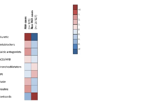
To better characterize patient profiles at risk of developing PAH, we extracted all drugs other than PKI associated with dasatinib ICSRs. Then we compared the proportion of cases associated with some drug classes of interest (e.g. diuretics, beta-blockers, calcium antagonists, statin, proton pump
inhibitors, insulins, corticoids, bronchodilators, ACEi/ARB) between PAH cases and all other adverse events.
2. Analysis
Disproportionality analysis
We first performed a disproportionality analysis with the Reporting Odds Ratio (ROR) method for each PKI of interest considered as suspect or concomitant (10). We compared the proportion of PAH reported for each PKI with the proportion of PAH associated with all other drugs used as non-cases. The cut-off for signal detection was defined as a ROR lower boundary 95% confidence interval greater or equal to 1 and number of cases (n) greater or equal to 3. Results are expressed as ROR (95% CI). We also performed a temporal analysis to asses to influence of media safety alerts on reporting rate of PAH among reported adverse events.
Correlation analysis
To assess the link between the identified cellular targets of interest and pharmacovigilance signals, we calculated the Pearson correlation coefficients (r) using pKd, (negative logarithm of the dissociation constant Kd) and ROR.
We assumed that higher the affinity for the cellular target was, higher was the ‘risk’ of drug-induced PAH. From multiple comparisons, the threshold P-value of the test was adapted using a Bonferroni correction (11).
Secondary analyses were performed to assess the robustness of the results: i) A sensibility analysis excluding PKI which had less than 3 cases of PAH; ii) standardizing
21 the time on the market of the different PKI at six years after FDA approval date, corresponding to the time between dasatinib approval and the first published safety alert; iii) performing the correlation analysis among PKI used in hematological pathologies.
Finaly, we performed a hierarchical cluster analysis to assess the similarity among receptor binding affinity profile of the included PK.
All analyses were performed using R statistical software (version 3.2.3).
Results
Selection of cases
To December 31 2017, among more than 16 million ADR were reported in Vigibase®, a total of 286 834 ICSRs were related to the 29 selected PKI (suspected or concomitant). Among them, 733 cases of pulmonary hypertension were extracted. The exclusion of cases associated with other PAH etiologies and concomitant drugs leaded to 482 final ISCRs included in the analysis (Figure S1).
Description of PAH cases
482 cases of PAH were included in analysis, 222 were women (44.2%), 230 men (45.8%) and 50 had an unknown status (10%). The mean age was 58.2 ± 16.1 years (median = 60) with 58.6 ± 15.1 years for women and 58.02 ± 16.2 years for men. Among 78 cases, a pleural effusion was associated with PAH
(15.5%). PAH emerged approximately 28 months after beginning of PKI (data available for 197 ISCRs, 6 months for bosutinib, 24 months for dasatinib, 14 months for nilotinib, 10 months for ponatinib and 21 months for ruxolitinib).
Identification of protein kinases involved in PAH
36 PKs involved in PAH pathophysiology were identified through the literature review (Figure S2): ALK1/5, AMPKa1/2, BMPR-1/2, B-Raf, c-yes, DDR1, EIF2K4, ERB-b1, FAK, FGFR1/2, HER2, IGF-1R, JAK1/2, JNK1/2, KIT, Lck, Lyn, HGF, p38MAPK, PDGFRα/β, PKG, RAF1, ROCK-2, Src, TEC, TIE2, and VEGFR-1/2/3. Most relevant references about target of interest are reported in Appendix 2.
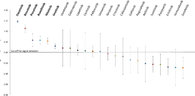
Disproportionality analysis
A positive disproportionality signal was found for dasatinib, bosutinib, ponatinib, ruxolitinib, nilotinib and imatinib, with a ROR of 28.55 (25.53, 31.93), 13.43 (8.65, 20.87), 3.73 (1.86, 7.46), 3.71 (2.44, 5.65), 3.39 (2.43, 4.73), and 1.97 (1.50, 2.57) respectively. ROR are represented in Figure 1. Results of the sensibility analysis (standardizing on time on the market) are presented in Supplementay Material (Appendix 3. Figure S3). The results were consistent with the main analysis except for imatinib, indeed the signal for imatinib disappeared in the sensibility analysis standardizing on time on the market.
22
Figure 1. Forest plot of the disproportionality analysis of PAH induced by PKI showing the ROR (95% CI) for included PKI. Results were log transformed for drawing purpose. PKI associated with positive disproportionnality signals are triangle, others are circles and PKI without PAH cases are lines.
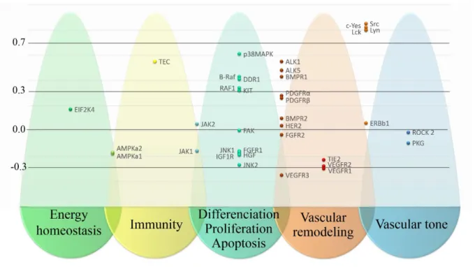
Correlation analysis
Correlation results are presented in Figure 2. Four non-receptors proteins kinase disclosed a significant correlation with the development of PAH: c-src (r= 0.85 and p= 0.00003), c-yes (r= 0.82 and p= 0.00012), Lck (r= 0.8 and p= 0.00038) and Lyn (r= 0.80 and p= 0.00023). Proportion of variance (r²) explained by the model were respectively 0.72, 0.67, 0.64 and 0.64 for c-src, c-yes, Lck and Lyn. In sensitivity analysis (see Appendix 4) standardizing the time on the market of the different PKI at six years (r c-src= 0.88, rc-yes= 0.83, rLyn= 0.83 and rLck=
0.82) and including only PKI which had more than 3 cases of PAH (rc-src= 0.89, rc-yes= 0.87, rLyn= 0.85) showed comparative result of correlation with a high but non-significant correlation for Lck (rLck= 0.82; p=0.0037). In sensitivity analysis including only PKI used in hematology the SRC family protein kinases remained highly correlated (rc-src= 0.92, rc-yes= 0.94, rLyn= 0.87) but non-significantly. Similarly, ALK5, TEC and HER2 were also highly correlated (r=0.99, 0.97 and 0.96) with non-significant p-values.
23
Figure 2. Manhattan plot synthetizing the correlation analysis. Pearson coefficients of each target classified according to their cellular function.
Co-medication analysis
In the co-medication analysis we found that diuretics, insulins, statins and antihypertensive drugs are more frequently associated in dasatinib PAH cases compared to other adverse event cases. Incidence of co-medication are represented in Figure 3 (details are available on supplementary appendix 5).
Clusters
Results of hierarchical clustering are presented in Figure 4.
Temporal analysis
Results of the temporal analysis associated with firsts publications reporting an
association between a PKI and PAH is presented in Figure 5. Notably, a huge increase in the rate of notification for dasatinib can be seen after first media alert.
24
Figure 3. Results of the co-medication analysis between PAH and non-PAH cases related to dasatinib in Vigibase®.
25
Discussion
To our knowledge this is the first pharmacovigilance analysis assessing the reporting risk of PAH associated with PKI use. Among more than 16 million ADR reported in the WHO pharmacovigilance database Vigibase® at the date of the extraction, 286 834 ICSRs were associated the 29 selected PKI including 482 PAH cases. Disproportionality analysis showed that dasatinib, bosutinib, ponatinib, ruxolitinib, nilotinib and imatinib had a significant pharmacovigilance signal. Those signals remained positive in sensitivity analysis except for imatinib (supplementary material). Those results are mainly in accordance with the literature data about PKI inducing PAH. Dasatinib is commonly implicated in induction or aggravation of PAH (6,12)(13)(14). More recently, bosutinib, imatinib, ponatinib and ruxolitinib were linked to PAH (15,16). A recent published case series suggested that lapatinib, a PKI used in breast cancer with human epidermal growth factor receptor mutations, might also cause PAH, but only one of the six patients presented in this article
had RHC confirming precapillary PAH (17). In our study, lapatinib showed a weak and non-significant dispropor-tionality signal with a ROR of 1.13 (0.61, 2.10). To date, there is no publication linking imatinib to PAH, imatinib has even been tested as a potential therapy in patients with PAH with an unfavorable benefit-to-risk balance (18). The pharmacovigilance signal found in our analysis for imatinib may be provoked by confusion bias owning to the higher prevalence of PAH in patients with a myeloproliferative disease (19). Accordingly, the signal for imatinib disappeared in a sensitivity analysis. In pharmacodynamic correlation analysis, c-src, c-yes, lck and lyn were highly correlated to the PAH reporting risk. The Src tyrosine kinases family contain nine members: three of them (Src, Fyn and Yes) are ubiquitously distributed and six (Blk, Yrk, Fgr, Hck, Lck and Lyn) are variously expressed depending on tissues. It has been shown that Src tyrosine kinases are crucial for TWIK-related acid sensitive potassium 1 (TASK-1) potassium channel functioning, acting as a cofactor (7). Mimicking hypoxia condition, inhibition of SRC kinases decrease TASK-1 activity result in intracellular calcium level Figure 5. Proportion of reported PAH cases for 1000 reported adverse events per year.
26 increase thus enhancing vasoconstriction and vascular remodeling (7). However, beyond inhibition of such protein kinases, it has recently been shown that dasatinib might induce apoptosis and endothelial cell dysfunction through an increase of mitochondrial reactive oxygen species that is independent from Src family kinases inhibition (14).
Members of the BMP signaling pathway
showed heterogeneous results in our study
.
Whereas ALK1, ALK5 and BMPR-1 showed positive correlation in the main or in sensitivity analysis, BMPR-2, the first cause of heritable PAH, does not shown any correlation in our study. The BMP signaling pathway is involved in cell proliferation, mitochondrial dysfunction and inflammation (20). Mutation of BMPR2 the gene coding for the receptor BMPR2 account for 70-80% of heritable PAH, furthermore BMPR2 concentration has also been shown to be reduced in lung tissue from patients with PAH (21). However, estimates indicate that only approximately 20% of individuals with a known genetic mutation in BMPR2 will develop PAH during their life, thus, BMPR2 mutation is required but is not sufficient alone for phenotypic expression and increase an individual’s chance of developing PAH (22)(20). Interestingly, it has recently been shown that BMPR2 reduction, through micro-RNA 124, lead to mitochondrial Warbung phenotype, thus should explain the mitochondrial increased of reactive oxygen species found by Guinabert et al (14,23). Overall, we cannot exclude that BMP signaling pathway may participate to PAH induced by PKI but it seems not primarily involved in the pathophysiology. Other target such as TEC and p38MAPK showed
moderate correlation (0.3 < p < 0.7) with non-significant results within adjusted analysis considering only PKI with more than three cases; their implication remains to be further elucidated.
The absence of association between PDGF and VEGF protein kinases reinforce the fact that vascular remodeling is not a major component of PAH induced by PKI; which is consistent with the observations of PAH reversal upon PKI discontinuation.
Genetic mutations are considered to be permissive of disease, and require additional epigenetic, inflammatory or environmental factors for the development of PAH in people with those mutations (24). Consistently and based on in vitro and in vivo findings, PKI increase the risk of developing PAH but require a comparable genetic, epigenetic or environmental “second hit” (14). Our exploration of associated co-medication found that antihypertensive drugs (diuretics, beta-blockers, ACEi/ARB and calcium antagonists), statins and insulins are more represented in PAH patients. This result suggest the possibility that diabetes, hypercholesterolemia and arterial hypertension would be a risk factor for dasatinib induced PAH. We could hypothesize that endothelial dysfunction and cellular hypoxia associated with diabetes, dyslipidemia and arterial hypertension could trigger PAH in those patients (25). Accordingly to published cases series, a higher proportion of men may develop PKI induced PAH whereas the incidence of PAH is fourfold higher in the general population (20). It is known that men have worse prognostic mainly because of a maladaptive response of the right ventricle to PAH, we
27 thus cannot exclude a participation of hormones and sex in triggering PAH (26). Another interesting point is that a PKI such as vandetanib which have a similar affinity profile to bosutinib, nilotinib and imatinib (Figure 4) but indicated in prostatic cancer do not provoke PAH. Such observation strongly highlights the role of the hematological underlying pathology in the genesis of PAH.
The results of the cluster analysis are mainly in accordance with the literature data, such as the analogy between imatinib and nilotinib (27). Interestingly the dasatinib profile seems different from other PKI. Given that pharmacovigilance notifications are based on a spontaneous reporting system, amount and proportion of cases reported for a medicinal product may variate depending of many factors such as media safety alerts, time since marketing or selective notification. Illustrating this variability, the time trend analysis showed a dramatic increase in rate of reporting after the firsts case-series and case-reports publications (Figure 5). However, despite those bias a correlation between relative risks and measure of disproportionality was found (8). Moreover, despite the case selection performed for this study (Figure S1) we cannot exclude that spurious PAH were included, indeed only two cases the results of right heart catheterization were reported. For this analysis, we assumed that PAH was caused by a single PK and that the pathophysiological mechanism is the same for all PKI. Our study was not able to detect co-inhibition of multiple pKs or inhibition/activation of non-PK cellular targets (e.g. proteasomes, G protein-coupled receptors, voltage-gated ion channels or ligand-gated ion channels). Therefore the
implication of other target in the pathogenesis of PKI-induced PAH cannot be ruled out.
Conclusion
This is to our knowledge, the first pharmacovigilance analysis to investigate the risk of PAH associated with PKI. The disproportionality analysis showed that dasatinib, but also bosutinib, ponatinib, ruxolitinib, and nilotinib had a significant disproportionality signal. This study highlights the role of SRC protein kinases family in PAH induced by PKI. Given the biased nature of the pharmacovigilance data, the results have to be confirmed by further analysis. Overall, this study contributes to a better understanding of PAH induced PKI.
References
1. Galiè N, Humbert M, Vachiery J-L, Gibbs S, Lang I, Torbicki A, et al. 2015 ESC/ERS Guidelines for the diagnosis and treatment of pulmonary hypertension: The Joint Task Force for the Diagnosis and Treatment of Pulmonary Hypertension of the European Society of Cardiology (ESC) and the European Respiratory Society (ERS)Endorsed by: Association for European Paediatric and Congenital Cardiology (AEPC), International Society for Heart and Lung Transplantation (ISHLT). Eur Heart J. 2016 Jan 1;37(1):67– 119.
2. Shimoda LA, Laurie SS. Vascular remodeling in pulmonary hypertension. J Mol Med. 2013 Mar;91(3):297–309.
28 3. Simonneau G, Gatzoulis MA, Adatia I, Celermajer D, Denton C, Ghofrani A, et al. Updated clinical classification of pulmonary hypertension. J Am Coll Cardiol. 2013 Dec 24;62(25 Suppl):D34-41.
4. Humbert M, Morrell NW, Archer SL, Stenmark KR, MacLean MR, Lang IM, et al. Cellular and molecular pathobiology of pulmonary arterial hypertension. J Am Coll Cardiol. 2004 Jun;43(12):S13–24.
5. Mattei D, Feola M, Orzan F, Mordini N, Rapezzi D, Gallamini A. Reversible dasatinib-induced pulmonary arterial hypertension and right ventricle failure in a previously allografted CML patient. Bone Marrow Transplant. 2009 Jun;43(12):967–8. 6. Rasheed W, Flaim B, Seymour JF. Reversible severe pulmonary hypertension secondary to dasatinib in a patient with chronic myeloid leukemia. Leuk Res. 2009 Jun;33(6):861–4.
7. Nagaraj C, Tang B, Balint Z, Wygrecka M, Hrzenjak A, Kwapiszewska G, et al. Src tyrosine kinase is crucial for potassium channel function in human pulmonary arteries. Eur Respir J. 2013 Jan 1;41(1):85–95.
8. Maciá-Martínez M-A, de Abajo FJ, Roberts G, Slattery J, Thakrar B, Wisniewski AFZ. An Empirical Approach to Explore the Relationship Between Measures of Disproportionate Reporting and Relative Risks from Analytical Studies. Drug Saf. 2016 Jan;39(1):29–43.
9. Harding SD, Sharman JL, Faccenda E, Southan C, Pawson AJ, Ireland S, et al. The IUPHAR/BPS Guide to PHARMACOLOGY in 2018: updates and expansion to encompass the new guide to IMMUNOPHARMACOLOGY. Nucleic Acids Res. 2018 Jan 4;46(D1):D1091–106.
10. van Puijenbroek EP, Bate A, Leufkens HGM, Lindquist M, Orre R, Egberts ACG. A comparison of measures of disproportionality for signal detection in spontaneous reporting systems for adverse drug reactions. Pharmacoepidemiol Drug Saf. 2002 Jan;11(1):3–10.
11. Bland JM, Altman DG. Multiple significance tests: the Bonferroni method. 1995. BMJ : British Medical Journal. 12. Mattei D, Feola M, Orzan F, Mordini N, Rapezzi D, Gallamini A. Reversible dasatinib-induced pulmonary arterial hypertension and right ventricle failure in a previously allografted CML patient. Bone Marrow Transplant. 2009 Jun;43(12):967–8. 13. Hennigs JK, Keller G, Baumann HJ, Honecker F, Kluge S, Bokemeyer C, et al. Multi tyrosine kinase inhibitor dasatinib as novel cause of severe pre-capillary pulmonary hypertension? BMC Pulm Med [Internet]. 2011 Dec [cited 2017 Nov 21];11(1). Available from: http://bmcpulmmed.biomedcentral.com/arti cles/10.1186/1471-2466-11-30
14. Guignabert C, Phan C, Seferian A, Huertas A, Tu L, Thuillet R, et al. Dasatinib induces lung vascular toxicity and predisposes to pulmonary hypertension. J Clin Invest. 2016 Aug 2;126(9):3207–18. 15. Low AT, Howard L, Harrison C, Tulloh RMR. Pulmonary arterial hypertension exacerbated by ruxolitinib. Haematologica. 2015 Jun 1;100(6):e244–5. 16. Weatherald J, Chaumais M-C, Montani D. Pulmonary arterial hypertension induced by tyrosine kinase inhibitors: Curr Opin Pulm Med. 2017 Jul;1.
17. Alkhatib Y, Albashaireh D, Al-Aqtash T, Awdish R. The role of tyrosine kinase inhibitor “Lapatinib” in pulmonary
29 hypertension. Pulm Pharmacol Ther. 2016 Apr;37:81–4.
18. Frost AE, Barst RJ, Hoeper MM, Chang H-J, Frantz RP, Fukumoto Y, et al. Long-term safety and efficacy of imatinib in pulmonary arterial hypertension. J Heart Lung Transplant. 2015 Nov;34(11):1366– 75.
19. Cortelezzi A, Gritti G, Del Papa N, Pasquini MC, Calori R, Gianelli U, et al. Pulmonary arterial hypertension in primary myelofibrosis is common and associated with an altered angiogenic status. Leukemia. 2008 Mar;22(3):646–9.
20. Thenappan T, Ormiston ML, Ryan JJ, Archer SL. Pulmonary arterial hypertension: pathogenesis and clinical management. BMJ. 2018 Mar 14;j5492. 21. Atkinson C, Stewart S, Upton PD, Machado R, Thomson JR, Trembath RC, et al. Primary pulmonary hypertension is associated with reduced pulmonary vascular expression of type II bone morphogenetic protein receptor. Circulation. 2002 Apr 9;105(14):1672–8.
22. Austin ED, Loyd JE. Genetics and Mediators in Pulmonary Arterial Hypertension. Clin Chest Med. 2007 Mar;28(1):43–57.
23. Caruso P, Dunmore BJ, Schlosser K, Schoors S, Dos Santos CC, Perez-Iratxeta C, et al. Identification of miR-124 as a Major
Regulator of Enhanced Endothelial Cell Glycolysis in Pulmonary Arterial Hypertension via PTBP1 and PKM2. Circulation. 2017 Sep 26;CIRCULATIONAHA.117.028034. 24. Pousada G, Baloira A, Valverde D. Complex inheritance in Pulmonary Arterial Hypertension patients with several mutations. Sci Rep [Internet]. 2016 Dec [cited 2018 Mar 12];6(1). Available from: http://www.nature.com/articles/srep33570 25. Sada K, Nishikawa T, Kukidome D, Yoshinaga T, Kajihara N, Sonoda K, et al. Hyperglycemia Induces Cellular Hypoxia through Production of Mitochondrial ROS Followed by Suppression of Aquaporin-1. Essop MF, editor. PLOS ONE. 2016 Jul 6;11(7):e0158619.
26. Shapiro S, Traiger GL, Turner M, McGoon MD, Wason P, Barst RJ. Sex differences in the diagnosis, treatment, and outcome of patients with pulmonary arterial hypertension enrolled in the registry to evaluate early and long-term pulmonary arterial hypertension disease management. Chest. 2012 Feb;141(2):363–73.
27. Green MR, Newton MD, Fancher KM. Off-Target Effects of BCR-ABL and JAK2 Inhibitors: Am J Clin Oncol. 2016 Feb;39(1):76–84.
30
Identification of cellular targets involved in Pulmonary Arterial
Hypertension induced by Protein Kinase Inhibitors by a
pharmacovigilance / pharmacodynamics analysis.
31
Appendix 1. Details of co-reported Meddra term excluded from the
analysis
-cardiac disorders (from MeDDRA classification: Cardiac disorders SOC - Cardiac and
vascular disorder congenital HGLT - Cardiac and vascular investigation HGLT).
- pulmonary disorders (respiratory and mediastinal neoplasms malignant and unspecified
HGLT - bronchial disorders (excl neoplasms) HGLT - lower respiratory tract inflammatory and
immunologic conditions HLT– parenchymal lung disorders HLT - pulmonary thrombotic and
embolic conditions HLT - respiratory tract disorders NEC HLT – tumour embolism / tumour
thrombosis PT) and
-thrombotic disorders (embolism and thrombosis HGLT).
32
Figure S2. Identification of protein kinases involved in pulmonary function through a literature review.
33
Appendix 2. Most relevant references about the protein kinases of
interest
Table S1. Most relevant references about target of interest
Target involved in pulmonary
pathophysiology Sources
ALK1 Activin receptor-like kinase-1
(Star et al., 2010) ; (Girerd et al., 2017) ; (Gore et al., 2014)
ALK5 transforming growth factor-β1 (TGFβ1)
(Tojais et al., 2017) (Upton and Morrell, 2013)
AMPKa1 (AMP-activated protein kinase)
(Ibe et al., 2013) (Omura et al., 2016) AMPKa2 (Ibe et al., 2013) BMPR-1 = ALK6 (Chida et al., 2012)
BMPR-2 (Tojais et al., 2017) B-Raf (Rapidly Accelerated Fibrosarcoma) (Awad et al., 2016) C-Raf = Raf1 (Hopper et al., 2015)
DDR1 Discoidin domain receptor
(Sakamoto et al., 2001)
EIF2AK4 eukaryotic translation initiation factor 2 alpha kinase 4
(Tenorio et al., 2015) (Eichstaedt et al., 2016) (Best et al., 2017) ERB-b1 = EGFR = Her1 (Dahal et al., 2010)
ERB-b2 = HER2 (Dahal et al., 2010) focal adhesion kinase FAK (Paulin et al., 2014)
FGFR1
(Zheng et al., 2015) (Kim, 2014) (Izikki et al., 2009) FGFR2 (Schermuly et al., 2011)
IGF-1R (insulin like growth factor)
(Sun et al., 2016) (Baumgart et al., 2017) (Dewachter et al., 2014)
34
JAK 1 (Lachmann et al., 2017) JAK2 (Mattar et al., 2016) JNK1/2 (c-Jun N-terminal kinase) =
mitogen-activated protein kinase 9
(Wilson et al., 2015) (Das et al., 2016) c-kit = KIT
stem cell growth factor receptor (SCFR)
(Montani et al., 2014) (Farha et al., 2014) Lck Leukocyte C-terminal Src kinase (Andruska et al., 2017)
lyn (Pullamsetti et al., 2012a) c MET = HGF (Schermuly et al., 2011) p38 MAPK = mitogen-activated protein
kinase 14 (Wilson et al., 2015) (Church et al., 2015) PDGFRα (Berghausen et al., 2013) (Schermuly, 2005) PDGFRβ (Cai et al., 2017) (Weatherald et al., 2017) PKG cGMP-dependent protein kinase (Patel et al., 2014)
ROCK-2 (Shimizu et al., 2013) Tyrosine-protein kinase c-Src (Guignabert et al., 2016)
TEC (de Lavallade et al., 2008) TEK receptor tyrosine kinase TIE2 (Guignabert et al., 2016)
VEGFR-1 (Derrett-Smith et al., 2013) VEGFR-2 (Nicolls et al., 2012) VEGFR-3 (Hwangbo et al., 2017)
35
Appendix 3. Results of sensitivity analysis
Sensitivity analysis were performed to compare the proportion of PAH reported for each PKI
with the proportion of PAH reported for all other PKI.
We performed an analysis using only reported cases from the first six years after the FDA
approval. A positive disproportionality signal was found for dasatinib with a ROR of 13.32
(8.56; 20.72), bosutinib 10.30 (6.63; 16.00), ponatinib 2.83 (1.41; 5.66), ruxolitinib 1.94 (1.20;
3.12) and nilotinib 2.07 (0.78; 5.53). Logarythmic value are represented in Figure S3.
Figure S3. Disproportionality signal of PAH induced by PKI from the first six years after FDA approval versus all medication in pharmacovigilance database. PRR and 95% CI were log transformed.
36
Appendix 4. Correlation analysis
In sensitivity analysis standardizing the time on the market of the different PKI at six years (r c-src= 0.90. rc-yes= 0.89. rLyn= 0.83) and including only PKI which had more than 3 cases of PAH (rc-src= 0.89. rc-yes= 0.87. rLyn= 0.84) showed comparative result of correlation with a high but non-significant correlation for Lck (rLck= 0.85; p=0.0021 and rLck= 0.82; p=0.037 respectively).
Table S2. Correlation and p-value of all sensitivity analysis.
Target 6 year after approval More than 3 cases PKI used in haematology
r p-value r p-value r p-value
ACVRL1 0.46 0.08400 0.90 0.00018 0.91 0.03000 ALK5 0.40 0.13000 0.76 0.00620 0.99 0.00074 AMPKa1 -0.18 0.52000 -0.16 0.63000 0.16 0.80000 AMPKa2 -0.21 0.44000 -0.18 0.60000 NA NA B_Raf 0.36 0.19000 0.35 0.29000 0.58 0.31000 BMPR_1 0.29 0.30000 0.59 0.05700 0.83 0.08000 BMPR_2 0.19 0.50000 0.08 0.82000 -0.096 0.88000 c_yes 0.83 0.00013 0.87 0.00047 0.94 0.01800 c-src 0.88 0.00002 0.89 0.00021 0.92 0.02800 DDR1 0.40 0.14000 0.38 0.25000 0.26 0.68000 EIF2K4 0.19 0.49000 0.42 0.20000 0.62 0.27000 ERB_b1 0.15 0.59000 0.24 0.49000 0.79 0.11000 FAK 0.07 0.80000 -0.04 0.91000 0.16 0.80000 FGFR1 -0.22 0.43000 -0.07 0.83000 0.91 0.03000 FGFR2 -0.11 0.69000 0.07 0.83000 0.91 0.03000 HER2 0.13 0.63000 0.15 0.66000 0.96 0.00960 HGF -0.19 0.50000 -0.17 0.63000 0.16 0.80000 IGF_1R -0.23 0.41000 -0.23 0.49000 NA NA
37 JAK1 -0.22 0.43000 -0.20 0.56000 -0.32 0.60000 JAK2 0.00 0.99000 0.07 0.83000 -0.14 0.83000 JNK1 -0.22 0.43000 -0.17 0.61000 -0.55 0.34000 JNK2 -0.28 0.32000 -0.23 0.50000 -0.34 0.58000 KIT 0.25 0.36000 0.32 0.34000 0.56 0.33000 Lck 0.82 0.00030 0.82 0.00370 0.81 0.09700 Lyn 0.83 0.00013 0.85 0.00094 0.87 0.05500 p38_MAPK 0.56 0.02900 0.61 0.04400 0.73 0.16000 PDGFRalfa 0.17 0.54000 0.32 0.34000 0.59 0.29000 PDGFRbeta 0.20 0.47000 0.29 0.38000 0.62 0.26000 PKG -0.15 0.60000 -0.05 0.90000 -0.32 0.59000 RAF1 0.30 0.28000 0.26 0.44000 0.57 0.32000 ROCK_2 0.02 0.94000 -0.01 0.97000 -0.21 0.73000 TEC 0.47 0.06800 0.55 0.06300 0.97 0.00640 TIE2 -0.24 0.38000 -0.22 0.52000 -0.28 0.65000 VEGFR_1 -0.34 0.21000 -0.33 0.32000 0.91 0.03000 VEGFR_2 -0.34 0.21000 -0.29 0.38000 0.91 0.03000 VEGFR_3 -0.32 0.23000 -0.38 0.25000 NA NA
38
Appendix 5. Results of the co-medication analysis of dasatinib PAH
cases.
Table S3. Result of co-medication analysis
Co-medication of interest PAH cases (n= 325)
Non PAH cases
(n=16 527) Ratio Diuretic 45 13.8% 783 4.74% 2.92 Furosemide 29 8.9% 610 3.69% 2.42 Spironolactone 9 2.8% 102 0.62% 4.49 Hydrochlorothiazide 7 2.2% 71 0.43% 5.01 Betablockers 17 5.2% 634 3.84% 1.36 Metoprolol 4 1.2% 345 2.09% 0.59 Bisoprolol 8 2.5% 116 0.70% 3.51 Atenolol 2 0.6% 127 0.77% 0.80 Nebivolol 3 0.9% 29 0.18% 5.26 Propranolol 0 0.0% 17 0.10% 0.00 Calcic antagonists 13 4.0% 444 2.69% 1.49 Amlodipine 8 2.5% 330 2.00% 1.23 Diltiazem 1 0.3% 70 0.42% 0.73 Nifedipine 1 0.3% 30 0.18% 1.70 Nisoldipine 1 0.3% 0 0.00% NA Lercanidipine 2 0.6% 15 0.09% 6.78 Nicardpine 0 0.0% 0 0.00% NA IEC/ARA2 18 5.5% 805 4.87% 1.14 Lisinopril 1 0.3% 281 1.70% 0.18 Perindopril 3 0.9% 32 0.19% 4.77 Losartan 1 0.3% 130 0.79% 0.39 Ramipril 6 1.8% 95 0.57% 3.21 Valsartan 3 0.9% 90 0.54% 1.70 Telmisartan 1 0.3% 30 0.18% 1.70 Olmesartan 0 0.0% 47 0.28% 0.00 Candesartan 1 0.3% 62 0.38% 0.82 Irbesartan 2 0.6% 38 0.23% 2.68 Bronchodilatators 6 1.8% 315 1.91% 0.97 Budesonide 2 0.6% 43 0.26% 2.37 Fluticasone 1 0.3% 144 0.87% 0.35 Salbutamol 1 0.3% 26 0.16% 1.96
39 Salmeterol 0 0.0% 72 0.44% 0.00 Formoterol 2 0.6% 30 0.18% 3.39 PPI 12 3.7% 755 4.57% 0.81 Lansoprazole 5 1.5% 106 0.64% 2.40 Omeprazole 2 0.6% 276 1.67% 0.37 Rabeprazole 1 0.3% 34 0.21% 1.50 Pantoprazole 1 0.3% 235 1.42% 0.22 Esomeprazole 3 0.9% 104 0.63% 1.47 Statin 11 3.4% 428 2.59% 1.31 Simvastatin 3 0.9% 216 1.31% 0.71 Atorvastatin 2 0.6% 1 0.01% 101.70 Rosuvastatin 4 1.2% 106 0.64% 1.92 Pravastatin 2 0.6% 80 0.48% 1.27 Pitavastatin 0 0.0% 7 0.04% 0.00 Lovastatin 0 0.0% 18 0.11% 0.00 Insulins 5 1.5% 174 1.05% 1.46 Insulin aspart 1 0.3% 38 0.23% 1.34 Insulin glulisine 1 0.3% 0 0.00% NA Insulin 1 0.3% 46 0.28% 1.11 Insulin detemir 2 0.6% 0 0.00% NA Insulin lispro 0 0.0% 0 0.00% NA Insulin human 0 0.0% 21 0.13% 0.00 Insulin glargine 0 0.0% 72 0.44% 0.00 Corticoids 4 1.2% 455 2.75% 0.45 Prednisolone 2 0.6% 158 0.96% 0.64 Prednisone 2 0.6% 222 1.34% 0.46 Methylprednisolone 0 0.0% 75 0.45% 0.00 Vasodilatators 2 0.6% 12 0.07% 8.48 Sildenafil 2 0.6% 9 0.05% 11.30 Tadalafil 0 0.0% 3 0.02% 0.00 Other 0 Levothyroxine 10 3.1% 419 2.49% 1.24 Allopurinol 8 2.5% 438 2.60% 0.95 Acetylsalicylicacid 8 2.5% 585 3.47% 0.71 Metformine 4 1.2% 242 1.44% 0.86 Digoxin 2 0.6% 54 0.32% 1.92
40
DISCUSSION
Il s’agit, à notre connaissance, de la première analyse de pharmacovigilance évaluant le risque
d’HTAP associé à l’utilisation des IPK. Parmi plus de 16 millions d’effets indésirables
rapportés dans la base de données internationales de pharmacovigilance de l’OMS, 286 834
effets indésirables étaient rapportés sous IPK dont 482 cas d’HTAP qui ont été extraits. L’analyse de disproportionnalité a montré que le dasatinib, mais aussi le bosutinib, le ponatinib,
le ruxolitinib, le nilotinib et l’imatinib possédaient un signal de disproportionnalité significatif.
Les analyses de sensibilité confirment ces résultats (à l’exception de l’imatinib pour lequel le
signal de disproportionnalité disparait) et sont en accord avec la littérature (Moguillansky et
al., 2017 ; Minami et al., 2017 ; Low et al., 2015). Une série de cas publiés récemment suggère
que le lapatinib, un ITK utilisé dans le cancer du sein, pourrait également être à l’origine
d'HTAP iatrogènes, mais un seul des six patients inclus dans cette série de cas présentait une
HTP précapillaire confirmée (Alkhatib et al., 2016). Dans notre étude, le lapatinib a montré un
signal de disproportionnalité faible et non significatif avec un ROR de 1,13 (0,61; 2,10). À ce
jour, il n'existe aucune publication liant l'imatinib à l'HTAP, l'imatinib ayant même été testé
comme traitement potentiel chez les patients atteints d'HTAP avec un rapport bénéfice / risque
défavorable (Frost et al., 2015). Le signal de pharmacovigilance pour l'imatinib révélé par notre
étude, est probablement induit par un biais de confusion attribuable à la prévalence plus élevée
de l'HTAP chez les patients atteints de maladie myéloproliférative (Cortelezzi et al., 2008 ;
Guilpain et al., 2008).
L’analyse de corrélation pharmacodynamique, révèle une forte corrélation entre l’affinité pour
c-src, c-yes, lck et lyn et la disproportionnalité du signal de pharmacovigilance. La famille des
Src tyrosine kinases contient neuf membres : trois d'entre elles (Src, Fyn et Yes) sont
ubiquitaires et six (Blk, Yrk, Fgr, Hck, Lck et Lyn) sont exprimées différemment selon les
41
canal potassique lié à TWIK 1 (TASK-1), agissant comme un cofacteur. Imitant un état
d'hypoxie, l'inhibition des SRC tyrosines kinases diminue l'activité de TASK-1 entraînant une
augmentation du calcium intracellulaire, aggravant la vasoconstriction et le remodelage
vasculaire. Cependant, il a récemment été montré que le dasatinib pouvait induire une apoptose
et une dysfonction des cellules endothéliales par une augmentation mitochondriale des espèces
réactives de l'oxygène, qui est indépendante de l'inhibition des kinases de la famille Src. De plus, l’absence de corrélation entre les récepteurs PDGF et VEGF renforce l’hypothèse d’un
remodelage vasculaire au second plan dans la physiopathologie des HTAP aux IPK ; hypothèse
confortée par le fait que ces HTAP régressent lors de l’arrêt des IPK.
Les membres de la voie de signalisation BMP montrent des résultats hétérogènes dans notre
étude. Alors qu’ALK1, ALK5 et BMPR-1 ont montré une corrélation positive dans l'analyse
principale ou dans l'analyse de sensibilité, BMPR-2, première mutation génétique découverte à l’origine d'HTAP héritables, ne montre, dans notre étude, aucune corrélation. La voie de
signalisation BMP est impliquée dans la prolifération cellulaire, le dysfonctionnement
mitochondrial et l'inflammation. Une mutation de BMPR2, le gène codant pour le récepteur
BMPR2, représente 70-80% des HTAP héritables. Cependant, seulement 20% des personnes
ayant une mutation génétique connue de BMPR2 développeront une HTAP au cours de leur
vie. La mutation de BMPR2 constitue donc un facteur de risque mais n'est pas suffisante pour
induire une HTAP. Nous ne pouvons néanmoins pas exclure que la voie de signalisation BMP
participe à la physiopathologie des HTAP induite par les PK, mais elle ne semble pas être
principalement impliquée. D'autres cibles telles que TEC et p38MAPK ont montré une
corrélation modérée (0,3 < p <0,7) avec des résultats non significatifs dans l'analyse de
sensibilité considérant uniquement les IPK ayant plus de trois cas d’HTAP ; leur implication