O
riginal
A
rticle
Stem Cells
2005;23:392–402 www.StemCells.com
Plasticity of Cultured Mesenchymal Stem Cells: Switch from
Nestin-Positive to Excitable Neuron-Like Phenotype
Sabine Wislet-Gendebien,
aGrégory Hans,
a,bPierre Leprince,
aJean-Michel Rigo,
a,b,cGustave Moonen,
a,bBernard Rogister
a,ba
Center for Cellular and Molecular Neurobiology and
bDepartment of Neurology, University of Liège, Liège,
Belgium;
cDepartment of Physiology, Transnationale Universiteit Limburg / Limburgs
Universitair Centrum, Biomedisch Onderzoekinstituut, Diepenbeek, Belgium
Key Words. Action potential • Differentiation • Mesenchymal stem cells • Neurons • Neurotransmission
Correspondence: Sabine Wislet-Gendebien, Centre for Cellular and Molecular Neurobiology, University of Liège, 17 Place Del-cour, B-4020 Liège, Belgium. Telephone: 32-4-366-5917; Fax: 32-4-366-5912; E-mail: Sabine.Wislet@utoronto.ca Received July 2, 2004; accepted for publication November 3 2004. ©AlphaMed Press 1066-5099/2005/$12.00.0 doi: 10.1634/stem-cells.2004-0149
Abstract
Bone marrow mesenchymal stem cells (MSCs) can dif-ferentiate into several types of mesenchymal cells, includ-ing osteocytes, chondrocytes, and adipocytes, but, under appropriate experimental conditions, can also differenti-ate into nonmesenchymal cells—for instance, neural cells. These observations have raised interest in the possible use of MSCs in cell therapy strategies for various neurologi-cal disorders. In the study reported here, we addressed the question of in vitro differentiation of MSCs into func-tional neurons. First, we demonstrate that when they are co-cultured with cerebellar granule neurons, adult MSCs can express neuronal markers. Two factors are needed for the emergence of neuronal differentiation of the MSCs: the first one is nestin expression by MSCs (nestin is a marker for the responsive character of MSCs to extrinsic signals), and the second one is a direct cell–cell interaction between neural cells and MSCs that allows the integration
of these extrinsic signals. Three different approaches sug-gest that neural phenotypes arise from MSCs by a differ-entiation rather than a cell fusion process, although this last phenomenon can also coexist. The expression of sev-eral genes—including sox, pax, notch, delta, frizzled, and
erbB—was analyzed by quantitative reverse transcription
polymerase chain reaction (RT-PCR) in order to further characterize the nestin-positive phenotype compared to the nestin-negative one. An overexpression of sox2, sox10,
pax6, fzd, erbB2, and erbB4 is found in nestin-positive
MSCs. Finally, electrophysiological analyses demonstrate that MSC-derived neuron-like cells can fire single-action potentials and respond to several neurotransmitters such as GABA, glycine, and glutamate. We conclude that nes-tin-positive MSCs can differentiate in vitro into excitable neuron-like cells. Stem Cells 2005;23:392– 402
Introduction
A stem cell is an unspecialized cell with the ability to renew itself indefinitely; under appropriate conditions, stem cells can give rise to a wide range of mature cell types. Two types of stem cells can be distinguished according to their origin
and their potential of differentiation: embryonic stem cells (ESCs) and somatic stem cells (SSCs) [1]. ESCs are derived from the early blastocyst and the inner cell mass of the embryo and are able to differentiate into the three germ layer cell types (pluripotentiality) [2, 3, 4]. SSCs are isolated from
fetal (after gastrulation) or adult tissues, but classically they are programmed to produce only the cell types that belong to the tissue from which they originate. However, recent stud-ies suggest that SSCs might be able to exhibit more plasticity than previously thought, as they seem able to differentiate into many cell types. These observations were mainly reported when SSCs were grafted in a damaged tissue [5]. This phe-nomenon, also known as phenotypic plasticity of SSCs, has been described among others for mesenchymal stem cells (MSCs), which under appropriate in vivo or in vitro condi-tions can adopt a neural fate [6–8].
Over the past few years in parallel to stem cell research, there has been a growing interest in the molecular mecha-nisms involved in cell type determination and differentiation during development. The discovery of intrinsic and extrinsic factors leading to the differentiation into ectoderm, meso-derm, and endoderm during the early stages of development; and the identification of factors that are responsible for further differentiation into more restricted cell types later in develop-ment, would allow a better understanding of the mechanisms that control the phenotypic plasticity of stem cells.
Recently, we demonstrated that cultured MSCs from adult rat bone marrow are able to express nestin, an interme-diate filament protein that is predominantly expressed dur-ing neural development [9]. Two factors seem essential for this nestin expression to occur: the first one is the absence of serum-derived components in the culture medium, and the second one is an in vitro maturation of the MSCs. Indeed, a minimum of 25 population doublings are required for nestin to be expressed in 75% of cultured MSCs. In this study, we dem-onstrated that nestin-positive MSCs, when co-cultured with mouse cerebellar granule (mCG) neurons, are able to differ-entiate into excitable neuron-like cells. By using quantitative reverse transcription polymerase chain reaction (RT-PCR), we demonstrated that sox and pax transcription factors, as well as the frizzled (wnt receptor) and erbB (neuregulin receptors) receptors, are upregulated in nestin-positive MSCs compared with nestin-negative MSCs (nnMSCs), suggesting a possible role for these genes in the acquisition of a responsiveness to neural fate–inducing signals in a nestin-positive MSC popu-lation. Electrophysiological recordings of npMSC-derived neuron-like (MDN) cells show that in co-culture with CG neurons, MDN cells first express functional voltage-gated K+
channels followed after 8 days of co-culture by voltage-gated Na+ channels, allowing firing of action potentials. Finally,
several experiments strongly suggest that this neuronal fate in the nestin-positive MSCs is a consequence of a “transdif-ferentiation” process, although not completely ruling out that cell fusion processes could also contribute to the appearance of neuronal-like cells.
Materials and Methods
Preparation and Culture of Rat Mesenchymal Stem
Cells (rMSCs)
Adult (8–15-week-old) rat (Wistar Institute, University of Pennsylvania, State College, http://www.wistar.upenn.edu/) (housing and breeding are taken care of in intramural animal facilities) bone marrow was obtained from femoral and tibial bones by aspiration and was resuspended into 5 ml of Delbuc-co’s modified Eagle’s medium (DMEM; Invitrogen, Merel-beke, Belgium, http://www.invitrogen.com) [10]. Between 100 and 200 × 106 marrow cells were plated on 175-cm5 tissue
culture flasks in DMEM added with 10% (v/v) fetal bovine serum (Invitrogen). After 24 hours, the nonadherent cells were removed. When the MSCs became confluent, they were resuspended with 0.25% trypsin (w/v; Sigma-Aldrich Com-pany Ltd, Dorset, U.K., http://www.sigmaaldrich.com), in 1 mM EDTA (Fluka brand, Sigma; http://www.sigmaaldrich. com/Brands/Fluka) and then subcultured. Characterization of MSCs and induction of nestin expression are described in our previous paper [9].
Preparation and Culture of Mouse Cerebellar
Granule Neurons
Mouse cerebellar granule (mCG) neuron cultures were pre-pared from 3-day-old green C57BL/6 mice (Jackson Immu-noResearch Laboratories, West Grove, PA, http://www. jacksonimmuno.com), according to Lefebvre et al. [11]. Green mouse express green fluorescent protein (GFP) under control of the β-actin promoter [12]. Briefly, cerebella were removed and freed of meninges. They were then minced into small fragments and incubated at 37°C for 25 minutes in 0.25% trypsin and 0.01% DNAse (w/v, in a cation-free solu-tion). Fragments were then washed with minimum essen-tial medium (Invitrogen) supplemented with glucose (final concentration 6 g 1–1), insulin (Sigma; 5 μg ml–1) and
pyru-vate (Invitrogen; 1 mM). The potassium concentration was increased to 25 mM, while the sodium concentration was decreased in an equimolar amount (MEM-25HS). The dis-sociation was achieved mechanically by up-and-down aspira-tions in a 5-ml plastic pipette. The resulting cell suspension was then filtered on a 15-μM nylon sieve (pore size = 15 μm). Cells were then counted and diluted to a final concentration of 2.5 106 ml–1. The cell suspension was finally plated on a
sub-strate previously coated with polyornithine (0.1 mg ml–1). The
cells were cultured for 24 hours before any other experimental procedure was performed.
Culture of Nestin-Positive MSCs on
Paraformaldehyde-Fixed mCG Neurons
described above and cultivated for 5 days before fixation with 4% (v/v) sterile paraformaldehyde. Then the fixed cells were treated with gelatine (1% w/v, in Tris-buffered saline [TBS]; Bio-Rad Laboratories, Hercules, CA, http://www. bio-rad.com) for 1 hour at 37°C, under sterile conditions. Nestin-positive MSCs were transferred on these fixed cul-tures, and the co-cultures were grown in mCG neuron–con-ditioned medium for 5 days.
Immunocytochemistry
Nestin-positive MSCs were trypsinized and placed with mCG neurons on polyornithine-coated dishes, in guanine and cytosine (GC) medium, for 5–15 days and were processed for immunocytochemistry as already described [9]. Briefly, the cultures were fixed with 4% (v/v) paraformaldehyde for 15 minutes at room temperature and washed three times in TBS. They were then permeabilized in 1% triton-X100 (v/v) for 15 minutes and washed three times in TBS. Nonspecific binding was blocked by a 1 hour treatment in TBST (TBS plus 0.1% w/ v Tween) containing defatted milk powder (30 mg ml–1). The
cells were then incubated for 1 hour at room temperature with one of the following primary antibodies (diluted in blocking buffer):
anti-glial fibrillary acidic protein (GFAP[Dako, Glostrup, Denmark, http://www.ump.com/dako.html] mouse immunoglobulin [IgG], dilution 1:500)
anti-GLAST [13] (rabbit IgG, 1:4000)
anti-Tuj1 ([Eurogentec, Southampton, Hampshire, UK, http://uk.eurogentec.com/] mouse IgG, dilution 1:1,500) anti-NeuN ([Chemicon International, Temecula, CA, http://
www.chemicon.com] mouse IgG, dilution 1:50) anti-SMI31 ([Chemicon] mouse IgG, 1:400) anti-MAP2ab ([Sigma] mouse IgG, dilution 1:400) anti-synaptophysin ([Chemicon] mouse IgG, dilution 1:200) anti-M2 ([Developmental Studies Hybridoma Bank, DSHB,
Iowa City, IA, http://www.uiowa.edu/~dshbwww] rat IgG, dilution 1:500)
anti-M6 ([Developmental Studies Hybridoma Bank] rat IgG, dilution 1:300)
After three washes in TBS, cells were incubated in fluorescein isothiocyanate (FITC)–conjugated, rhodamine-conjugated, or Cy5-conjugated anti-mouse, anti-rat, or anti-rabbit IgGs (dilution 1:500; Jackson Immunoresearch) for 1 hour at room temperature and in the dark. The nuclei were stained with ethidium homodimer (0.2 μM; Sigma). Omission of primary antibodies served as negative control and resulted in no detect-able staining. The preparations were then mounted in Fluoprep (bioMérieux-Pierre Fabre, Boulonge, France, http://www. biomerieux.com/) and observed using a Bio-Rad MRC1024 laser scanning confocal microscope (Bio-Rad Laboratories). The fraction of positive cells was determined by counting 10
nonoverlapping microscopic fields (± 30 MSC-derived cells per field) for each coverslip in at least three separate experi-ments (n then corresponds to the number of coverslips).
FACS Analysis
Fluorescence-activated cell sorting (FACS) analysis of co-cul-tured rMSCs and CG neurons were performed with anti-GFAP and anti-Tuj1 antibodies. Briefly, after 5 days of co-culture, the cells were trypsinized and suspended in phosphate-buffered solution (PBS) containing 1% (v/v) fetal calf serum. After centrifugation, the pellet was resuspended (750,000 cells per ml) in FACS permeabilization solution (Becton, Dickin-son; BD Biosciences, Oxford, U.K., http://www.bd.com), for 30 minutes at room temperature. The cells were centrifuged and washed three times in PBS containing 1% (v/v) fetal calf serum before the incubation with anti-GFAP ([Dako] mouse IgG, dilution 1:500) or anti-Tuj1 ([Pharmingen; BD Biosci-ences; http://www.bdbiosciences.com/pharmingen], mouse IgG, dilution 1:400) antibodies, for 1 hour at room tempera-ture. The co-culture cells were then washed three times in PBS containing 1% (v/v) fetal calf serum and incubated with anti-mouse IgG (1:500) or anti-rabbit IgG (1:500) secondary antibodies coupled to rhodamine (Jackson ImmunoResearch) for 1 hour at room temperature and in the dark. The analysis was performed with a FACSort instrument (Becton, Dickin-son), and the results were analyzed using the Cellquest pro-gram (Becton, Dickinson).
DNA Ploidy
DNA content per cell was determined by FACS analysis after staining the cells with propidium iodide. After 5 days of co-culture, MSCs and mCG neurons were trypsinized and fixed into 70% ethanol, at 4°C for 16 hours. The cells were stained with propidium iodide (400 mg ml–1; Sigma)
just before FACS analysis.
RT-PCR and Quantitative RT-PCR Analysis
Total RNA from nestin-postive MSCs (15 passages in serum-free media) and nestin-negative MSCs (15 passages in serum condition) were prepared using the RNeasy total RNA purifi-cation kit (Qiagen, Westburg, U.K., http://www1.qiagen.com). For cDNA synthesis, random hexamer primers (Invitrogen) were used to prime RT reactions. The cDNA synthesis was carried out using Moloney-murine leukemia virus (M-MLV) Superscript II reverse transcriptase (Invitrogen) following the manufacturer’s instructions. The PCR mix contained 2.5 mM MgCl2, PCR nucleotide mix of dNTP at 0.2 mM each, 0.5 :
M primers (Table.1), 5 U/:l Taq DNA polymerase (Gibco),10 ng DNA template, and nuclease-free water to a final volume of 50 μl. Cycling parameters were as follows: denaturation at 94°C for 30 seconds, annealing (see Table 1) for 30 seconds,
and elongation at 72°C for 30 seconds, for 35 cycles on the PTC200 DNA Engine (MJ Research, Waltham, MA, http:// www.mjr.com/). To exclude the possibility of the presence of genomic DNA, we performed control reactions without the RT step. The fzd RT-PCR products were purified using a commercial kit (Qiagen) and subcloned using the TOPO TA cloning kit (Invitrogen). Positive clones were sequenced (Eurogentec), and genes were identified by BLAST search. Quantitative PCR was carried out using standard protocols with Quantitec SYBR Green PCR Kit (Qiagen). The PCR mix contained SYBR Green Mix, 0.5 μM primers (Table 1), 1 ng DNA template, and nuclease-free water to reach a final vol-ume of 25 μ1. Quantitative PCR was performed on the Rotor-Gene RG-3000 (Corbett Research, Sydney, Australia, http:// www.corbettresearch.com/) and analyzed with Rotorgene Software (Corbett Research). The percentage of gene expres-sion by nestin-positive MSCs was normalized in function of glyceraldehyde-3-phosphate dehydrogenase (GAPDH) gene expression and compared to the gene expression level of nes-tin-negative MSCs that was considered as 100%.
Solutions and Drugs for
Electrophysiological Analyses
Nestin-positive MSCs were stained with DiD Vybrant cell-labeling solution (red; Molecular Probes, Invitrogen,
Pais-ley, U.K., http://www.probes.com/) and co-cultivated with GFP-positive mCG neurons in order to easily identify the two types of cells. This procedure allowed us to be sure that the cells from which we recorded were not CG neurons, but prevented us from labeling a particular recorded cell for post hoc immunocytochemical characterization, which would have involved quadruple immunostainings. For whole-cell recordings, coverslips with co-cultured nestin-posi-tive MSCs and CG neurons were transferred to the stage of a Zeiss interferential contrast microscope equipped with fluorescence and perfused (3 ml per minute) with a solution containing NaCl (137 mM), KCl (5.4 mM), CaCl2 (2 mM),
H2O (1.8mM), D-glucose (22.2 mM), and HEPES; the pH
was adjusted to 7.2 with NaOH. The sodium and potassium channel blockers were, respectively, tetrodotoxin (TTX; Sigma, http://www.fisherscientific.com; 1 μM) and tetra-ethyl ammonium (TEA; 10 μM; Janssen Chemica, Geel, Belgium). The neurotransmitters used were GABA (1 mM), glycine (100 μM), and glutamate (100 μM), and the inhibi-tors of neurotransmitters were, respectively, gabazine (10 μM), strychnine (1 μM), and a mix of AP-5 (1 μM; Tocris Cookson, Avonmouth, Bristol, UK; http://www.tocris. com/) and CNQX (6-cyano-7-nitroquinoxaline-2,3-dione; 1 μM; Tocris). All drugs were applied by a microperfusion system (SPS-8, List Medical, Darmstadt, Germany).
Table 1. Primers used for reverse transcription polymerase chain reaction (RT-PCR) and quantitative RT-PCR experiments
Annealing
Gene Forward Reverse temperature (°) (°)
Sox1 AACCCCAAGATGCACAACTC TAGCCCAGCCGTTGACAT 54
Sox2 TAGCACTTGTTGCCAGAACG AAGCCGCTCTTCTCTTTTCC 54
Sox3 AGAACCCAAGATGCACAAC CTGCACGAGCGAGTAGGC 54
Sox4 AAGCACATGGCTGACTACCC GGAGTCCGCACCTTGTAGAC 54
Sox9 TGAAGAAGGAGAGCGAGGAA GGGGCTGGTACTTGTAATCG 54
Sox10 AGCCCAGGTGAAGACAGAGA CCCCTCTAAGGTCGGGATAG 54
Sox11 TCATGTTCGACCTGGCTTG CCCCACCATCCTCTTTATCC 54
Pax3 GCCAATCAACTGATGGCTTT CATTCGAAGGAATGGTGCTT 53
Pax6 AGTTCTTCGCAACCTGGCTA GGAGCTGATGGAGTTGGTGT 58
Notch1 TTACAGCCACCATCACAGCCACACC ATGCCCTCGGACCAATCAGA 51
Notch2 AACCCGTGTCTGAACCAAGG CCATTGTTCATACACGGCTTG 51
Notch3 ACACTGGGAGTTCTCTGT AGTAGTTCAGGTCTTGGTTGCAGAA 48
Delta1 TGTTCTAACGGTGCCAAGTG CTGCAGTTCCTGCCTGTGTA 48
ErbB2 GAGGTGGTGAGCTGACACT CCTCTGATTGGTTCACATACT 53
ErbB3 AGATCTGCACCATTGACGTC TAGGTCTAGGTCCAGTTCTG 52
ErbB4 GCTAGAGACCCTCAAAGATACC GCATGGGCATTCCTTGTTGGTGT 51
FZD NNNGAATTCTAYCCNGARMGNCCNAT NNNAAGCTTNGCNGCNARRAACCA 55
NF-L TACTCTCAGAGCTCGCAGGTCTT ACCATGCACAGGCTGGCAGCAA 55
GFAP ACATCGAGATCGCCACCTAC ACATCACATCCTTGTGCTCC 52
Electrophysiological Recordings
Borosilicate recording electrodes (5–10 MΩ) were made using a Flaming-Brown microelectrode puller (P97, Sutter Instrument Co., Novato, CA, http://www.sutter.com/). Micro-pipettes were filled with an intracellular-like solution contain-ing KCl (130 mM), CaC12 (2 mM), H2O (1 mM), D-glucose
(11.1 mM), EGTA (10 mM), Na2-ATP (2.5 mM), Mg-ATP (2.5
mM), HEPES (10 mM) at pH 7.4. Electrophysiological record-ings were performed with a patch-clamp amplifier (RK400; Bio-Logic, Claix, France, http://www.bio-logic.info/) using the whole-cell configuration of the patch-clamp recording technique. Series resistances (10–20 MΩ) were electronically compensated (up to 80%), and current traces were filtered at 3 kHz, acquired and digitized at 0.5 kHz for voltage-clamp recordings and at 20 kHz for current-clamp recordings, and stored on a personal computer system. Data recording and measures were achieved using the TIDA software (HEKA Elektronik Lambrecht/Pfalz, Germany, http://www.heka. com). Graphing and statistical analyses were performed with the aid of the GraphPad Prism software (version 4.0; Graph-Pad, San Diego, http://www.graphpadcom).
Results
Neuronal and Astroglial Marker Expression
by Nestin-Positive MSCs
MSCs were isolated from femoral and tibial bones of adult rats, propagated in culture, and immunologically and func-tionally characterized [9]. As we already demonstrated, a nes-tin-expressing stage is necessary to observe a GFAP+ cell
dif-ferentiation of MSCs co-cultured with neural stem cells [9]. To observe a possible neuronal differentiation of MSCs, we co-cultured nestin-positive MSCs for 5 days with GFP+ mCG
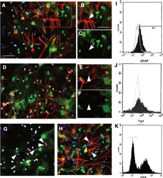
neurons obtained from 3-day-old green mice. After 5 days of co-culture, immunological labelings revealed that MSCs began to express differentiation markers: 40.17% ± 2.95% expressed GFAP (Fig. 1A–C), 19.00% ± 1.58% expressed Tuj1 (Fig. 1D–F), and 19.30% ± 1.38% expressed NeuN (Fig.1G) (n = 12 for each condition). These results were confirmed by FACS analyses (Fig. 1I,J) as we measured 44.57% ± 3.59% GFAP– and 18% ± 4.1% Tuj1+ cells, data which are not
statisti-cally different from that obtained by eye-counting immunola-beled cells. The FACS analyses were performed on GFP– cell
populations corresponding to the first spike of cells in Figure 1K. Moreover, the absence of GFAP and NeuN double-labeled cells suggests that nestin-positive MSCs are specifically ori-ented toward either an astroglial or a neuronal fate (Fig. 1H). Two conditions are needed before MSCs begin to express neuronal markers: first, for the appearance of GFAP+ cells in
co-cultures with neural stem cells, MSCs have to express nes-tin; second, a direct cell–cell contact between nestin-positive
MSCs and CG neurons is needed. Indeed, when nnMSCs are co-cultivated with CG neurons or when nestin-positive MSCs and CG neurons are co-cultivated with a physical separation (millicell device), no neuronal markers could be detected (data not shown).
Characterization of Nestin-Positive MSCs
The nestin expression by MSCs coincides with the acquisition by these cells of the ability to respond to extrinsic signals and cues driving their neural differentiation [9]. To get some clue into the molecular mechanism(s) involved in this differentia-tion, we decided to compare the expression in nestin-positive and nestin-negative MSCs of several factors known to have important roles during normal nervous system development. Using RT-PCR and quantitative RT-PCR, the nestin-positive MSC and nnMSC gene expression profiles of the following factors were compared, both qualitatively and quantitatively:
Figure 1. Neuronal and astroglial markers expression by nes-tin-positive mesenchymal stem cells (MSCs). Nesnes-tin-positive MSCs were co-cultured for 5 days with green fluorescent pro-tein (GFP)–positive cerebellar granule (CG) neurons (green). Immunocytochemical labeling and fluorescence-activated cell sorting (FACS) analysis showed that some nestin-positive MSCs expressed neural markers. GFAP (red, A–C) was expressed by about 40% of the nestin-positive MSCs, and Tuj1 (red, D–F) was expressed by about 20% of the nestin-positive MSCs, as was the NeuN marker (red, G). Those results were confirmed by FACS analysis (GFAP, I; Tuj1, J) on a GFP– population (K). A dou-ble labeling directed against GFAP (red) and NeuN (blue) (H) showed that nestin-positive MSCs specifically oriented toward an astroglial-like or a neuronal-like fate. Nuclei were counter-stained with EtD1 (blue or red, depending on the color used for the secondary antibody). Arrowheads show neural expression markers by nestin-positive MSCs. Scale bars = 40 μm (A, D, and G–K) and 30μm (B–C and E–F).
the sox and pax transcription factors, as well as frizzled (wnt receptors), erbB (neuregulin receptors) and the notch-delta signaling pathway. Figure 2 shows that MSCs express sox2,
sox9, sox10, sox11, pax6, notch1, notch2, delta1, erbB2, erbB4, and fzd and do not express sox1, sox3, pax3, delta2, and erbB3. In the analyses of fzd genes, we used degenerated
prim-ers that hybridize with all fzd genes [14]. The resulting prod-ucts were cloned using the TA cloning procedure, and ran-dom clones were analyzed by sequencing. A BLAST search allowed us to determine the identity of each clone: 80% of fzd receptors in nnMSCs were fzd2, and 20% were fzd1; like-wise, in nestin-positive MSCs, we obtained 75% of fzd2, 20% of fzd1, and 5% of fzd5 (data not shown). Finally, quantitative RT-PCR demonstrated that nestin-positive MSCs overexpress
sox2, sox10, pax6, fzd, erbB2, and erbB4 when compared with
the nnMSCs. We also observed that nestin-positive MSCs overexpress the GFAP gene compared with nnMSCs, but none of them express the neurofilament-light gene.
Electrophysiological Analysis of
Mesenchymal-Derived Neuron-Like (MDN) Cells
Using fluorescent microscopy, we selected for cells that pre-sented a neuron-like shape (rounded cell body with extended processes, hereafter termed MDN cells), that had incorpo-rated DiD Vybrant (red) and that were clearly GFP– (see
Meth-ods). Whole-cell patch-clamp recordings were obtained from 234 MDN cells that had been co-cultured with CG neurons for 4–15 days. Based on the appearance of different voltage-gated current profiles (see next paragraph), three culture time peri-ods were distinguished that, moreover, showed significantly (ANOVA; p < .001) different resting membrane potentials (Vrest): –37.6 ± 3.0 mV for MDNs co-cultured with CGs for 4–6
days in vitro (hereafter noted 5DIV; n = 61), –50.3 ± 2.0 mV for 7–9 days (8DIV; n = 76), and –55.7 ± 2.3 mV for 10–15 days (12.5DIV; n = 97).
Excitability
The activation of voltage-gated channels at the three develop-mental stages (5, 8, and 12.5DIV) was assessed by applying to whole-cell patch-clamped MDN cells hyperpolarizing and depolarizing voltage steps from a holding potential (VH) of
–80 mV (–80 to –125 mV and –80 to +30 mV in 15-mV incre-ments; Fig. 3A–D). At 5DIV, depolarizing steps positive to –40 mV induced outward currents in 95% of MDN cells that showed little desensitization and were blocked by 10 μM TEA, indicating the activation of potassium voltage-gated channels. Potassium current densities were stable throughout the culture time period (7.25 ± 1.16 pA.pF–1). At 5DIV, no inward
cur-rents were observed. Conversely, depolarizing voltage steps positive to –30 mV induced inward currents in 85% of 8DIV MDN cells and in 95% of 12.5DIV MDN cells that rapidly activate and inactivate and that were abolished by a perfusion of 1 μM TTX, showing that they are mediated by voltage-activated sodium channels. Between 8DIV and 12.5DIV, no significant differences were found regarding the sodium cur-rent densities (8.29 ± 0.87 pA.pF–1 (n = 51) at 8DIV compared
with 10.15 ± 1.04 pA.pF–1 (n = 59) at 12.5DIV). However—and
interestingly—the sodium current densities in MDN cells were two times lower (18.63 ± 2.45 pA.pF–1; n = 21) than the
one recorded in co-cultured CG neurons. Consistent with the occurrence of sodium and potassium voltage-gated currents, an injection of 600 nA current into whole-cell patch-clamped and current-clamped MDN cells elicited action potentials in 21.5% of 8DIV MDN cells (n = 34) (Fig. 4A) and in 25% of 12.5DIV MDN cells. These action potentials were always
Figure 2. Molecular characterization of nestin-positive mesen-chymal stem cells (MSCs). Reverse transcription polymerase chain reaction (RT-PCR) and quantitative RT-PCR were per-formed to better characterize the molecular profile of nestin-positive MSCs compared with nestin-negative MSCs (nnMSCs) regarding some genes known to have a role during neuro-onto-genesis. In this case, we observed that nestin-positive MSCs overexpressed sox2, sox10, pax6, erbB2, erbB4, fzd, and GFAP. The results of quantitative RT-PCR are expressed as percentage of gene expression in nestin-positive MSCs compared with the same gene expressed in nnMSCs after normalization with the GAPDH housekeeping gene.
present as single spikes and were reversibly inhibited by a 1-μM TTX application (Fig. 4).
Neurotransmitter Effects
Responses of whole-cell voltage-clamped MDNs (VH =
–80 mV) to microperfusion-applied neurotransmitters (1
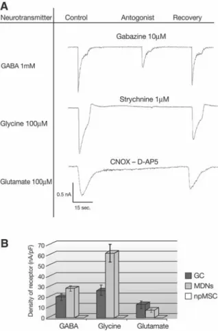
mM GABA, 100 μM glycine, and 100 μM glutamate) were subsequently measured. As soon as at 5DIV, neurotransmit-ters elicited inward currents in 98% of MDN cells (n = 78, Fig. 5A). Since no significant differences in current densities were observed between the different culture time periods, mea-sures of current densities were pooled for each neurotrans-mitter, yielding means of 28.2 ± 2.7 pA.pF–1 for GABA, 61.4
± 10.6 pA.pF–1 for glycine, and 7.5 ± 2.6 pA.pF–1 for glutamate.
The responses elicited by GABA, glycine, and glutamate were mediated by their respective ionotropic receptors—that is, GABAA receptors, glycine receptors, and AMPA/KA/NMDA
receptors—since transmitter-evoked currents were reversibly blocked by specific antagonists of these receptors: namely, by gabazine (10 μM), strychnine (1 μM), and a mix of CNQX and AP-5 (1 μM each), respectively. Before the co-culture of nes-tin-positive MSCs with CG neurons, inward currents elicited by those neurotransmitters were already present, but only in 35% of nestin-positive MSCs (n = 15, Fig. 5B) and with much lower mean current densities: 0.06 ± 0.02 pA.pF–1 for GABA,
0.07 ± 0.03 pA.pF–1 for glycine, and 0.13 ± 0.05 pA.pF–1 for
glutamate. Current densities of neurotransmitter-evoked responses were also measured in CG neurons with which nes-tin-positive MSCs were co-cultured. In CG neurons, GABA, glycine, and glutamate elicited currents in 100% of the cells (n = 15), with mean current densities of 19.71 ± 3.96 pA.pF–1
for GABA, 26.02 ± 5.55 pA.pF–1 for glycine, and 12.68 ± 4.54
pA.pF–1 for glutamate. Current density comparison between
MDN cells and CG neurons yielded a significant difference for glycine responses only (ANOVA; p < .01). Finally, we looked for a possible synaptic activation of the neurotrans-mitter receptors present in MDN cells. Although MDN cells expressed axonal (MAP2ab, Fig. 6A–C), dendritic (SMI31, Fig. 6D–F), and synaptic markers (synaptophysin, Fig. 6G–I), we never observed spontaneous synaptic events in cultured MDN cells.
Mechanisms of Neural Differentiation of Nestin-Positive MSCs
Recent studies have indicated that MSCs are able to fuse with mature neurons [8, 15, 16]. In this study, three types of experi-ments were performed which strongly suggest that MSCs dif-ferentiate into neuron-like cells rather than fuse with existing CG neurons. The first experiment involved the analysis of the DNA content of nestin-positive MSCs and CG neurons before and after the co-culture. In all cases, we did not observe a sig-nificant difference in ploidy, which could explain the 60% of neural differentiation (40% into astrocytes and 20% into neu-rons) in the MSC population (Fig. 7A). Although the sensitiv-ity of this technique does not allow us to completely rule out that some rare polyploid cells could be present, the majority of the observed cells were diploid. In a second set of experiments, we used the M2 and M6 markers, which specifically
recog-Figure 3. Voltage-gated channel activation in mesenchymal stem cell (MSC)–derived neuron-like (MDN) cells. (A): Typi-cal recordings of single whole-cell patch-clamped and volt-age-clamped MDN cells at three culture time periods (5, 8, and 12.5DIV). Hyperpolarizing and depolarizing steps were applied from a holding potential of –80 mV (–80 to –120 mV and –80 to +30 mV in 15-mV increments). The first and third lines show recordings in control conditions, and the second in the presence of 1 μM tetrodotoxin (TTX). The arrows indicate the time where the measures were performed (filled arrows for outward currents, and open arrows for inward currents). (B–D): Current-voltage relationships for outward (triangle; recordings obtained in the presence of 1 µM TTX) and inward (square; recording obtained in the presence of 10 μM tetraethyl ammonium [TEA]).
Figure 4. Excitability as shown in a typical recording of a single whole-cell patch-clamped and current-clamped mesen-chymal stem cell (MSC) neuron-like (MDN) cell at 8DIV. A depolarizing 600 nA current injection induced the firing of a single action potential that could be reversibly blocked by 1
nize respectively astrocytes and neurons from mouse but not from rat [17]. Rat npMSCs were co-cultivated for 5 days with mouse CG neurons. Thereafter, double labeling with GFAP and M2 antibodies, on one hand, and Tuj1 and M6, on the other hand, were performed. We could observe (1) that all GFAP+/
GFP– and Tuj1+/GFP– cells are also negative for M2 and M6,
respectively, allowing us to conclude that those cells are of rat origin and thus likely derive from the MSC population (Fig. 7B–G); and (2) that all the cells recognized either by the M2 or by the M6 antibodies are also GFP+, ruling out a
downregula-tion of GFP expression during the co-culture period. Finally, nestin-positive MSCs were cultured for 5 days on paraformal-dehyde-fixed GFP+ CG neurons in the presence of
CG-condi-tioned medium. In such conditions, we observed that 16.1% ± 2.6% (n = 8) nestin-positive MSC-derived cells were NeuN+
(Fig. 7H–J), and 23.1% ± 2.1% (n = 7) nestin-positive MSC-derived cells were GFAP+ (Fig. 7K–M). Note that the level
of GFP fluorescence in the fixed cells maintained for 5 days in culture remains stable. Although some rare fusion events cannot be excluded in these experiments, we can conclude that most of the MDN cells result from a true differentiation pro-cess of nestin-positive MSCs.
Discussion
Cellular therapies using stem cells are promising approaches for the treatment of several chronic or acute neurological dis-eases such as Parkinson’s [18] and Huntington’s disdis-eases [19] and spinal cord injuries [20]. One main problem relates to the origin and the nature of the cells to be used for such proce-dures. Although somatic stem cells derived from adult tissues seem to be good candidates [21], a better knowledge of the mechanisms underlying the phenotypic plasticity of somatic stem cells is a prerequisite before considering their use in the treatment of neurological diseases. In the study reported here, two major issues were addressed: (1) characterizing some molecular factors that could play a part in directing nestin-positive MSCs into a neural phenotype; and (2) determining the environmental conditions that allow nestin-positive MSCs to differentiate, at least in vitro, into functional neuron-like cells. Furthermore, we have tried to distinguish between true differentiation and cell fusion processes in the in vitro com-mitment of nestin-positive MSCs to a neural fate.
First, we analyzed the gene expression profile for differ-ent factors like sox, pax, notch, delta, frizzled, and erbB and observed that the nestin-positive MSCs express a higher level of sox2, sox10, pax6, fzd, erbB2, and erbB4. The expression of
sox2, which encodes a high-mobility group (HMG)-box–type
transcription factor, has been proposed to act by maintaining a neural progenitor identity [22]. It becomes downregulated in the neural plate when neural crest cells segregate from the dorsal neural tube, and it remains low during neural crest cell
Figure 6. (A–I): Expression of axonal (MAP2ab, blue, A–C), dendritic (SMI31, blue, D–F), and synaptic (synaptophysin, blue, G–I) markers by 8DIV cultured MDN cells.
Figure 5. Neurotransmitter sensitivities. (A): Typical recording of single whole-cell patch-clamped and voltage-clamped mes-enchymal stem cell (MSC) neuron-like (MDN) cell at 8DIV. Either 1 mM GABA, 100 μM glycine, or 100 μM glutamate was applied for 10 seconds every 30 seconds, and the resulting cur-rent was recorded in the absence or presence of specific antago-nists—namely, 10 μM gabazine, 1 μM strychnine, and a mix of 1 μM CNQX and 1 μM D-AP5, respectively. Antagonists were first applied alone for 10 seconds before being applied for 10 sec-onds with neurotransmitters. (B): Responses to GABA, glycine, and glutamate in mouse cerebellar granule (mCG) neurons, MSC-derived neuron-like (MDN) cells, and nestin-positive MSCs (before co-culturing with mCG neurons) are expressed as current densities—that is, mean peak amplitudes corrected for cell capacitance (pA.pF–1). **p < .01 (one-way ANOVA
migration. Sox2 expression is subsequently upregulated in some crest-derived cells in the developing peripheral nervous system and is later restricted to glial sublineages [23]. Sox10 is expressed in immature glial cells, in both the peripheral and the central nervous systems, where it is linked to Schwann cell [24] and oligodendrocyte [25] differentiation, respectively. Although it has been reported that, in vivo, MSCs are able to differentiate into oligodendrocytes [26], we never observed oligodendroglial differentiation of MSCs in our conditions and have not looked for a possible differentiation into Schwann cells. However, it would be interesting to search for a possible differentiation of Sox10+ MSCs specifically into GFAP+ and
GLAST+ cells. Pax6 is a paired-box–containing transcription
factor and is required for the development of the embryonic central nervous system, eye, and pancreas [27]. Recently, Talamillo et al. [28] suggested that the primary role of Pax6 is the regulation of the cell surface properties that are respon-sible for both cellular identity and radial migration. As only nestin-positive MSCs can grow in suspension culture, it would be interesting to test a possible role of Pax6 overexpression on these modified cell surface properties. ErbB4 is one of the neu-regulin receptors and is expressed with ErbB2 by neural stem cells when they proliferate [29]. Neuregulins have been sug-gested to stimulate neural stem cell [29] and oligodendroglial progenitor [30] proliferation and survival. Moreover, several reports implicate neuregulins in neuron and oligodendrocyte differentiation [29, 31]. Neuregulins form a large family of proteins: some being secreted, some being membrane-span-ning, and some being cleaved or shredded [32]. In the study reported here, we demonstrated that neural differentiation of nestin-positive MSCs requires both a direct cell-to-cell
Figure 7. Differentiation of nestin-positive mesenchymal stem cells (MSCs) into neural cells. (A): Nestin-positive MSCs (red) and mouse cerebellar granule (mCG) neurons (yellow) before and after co-culture (blue) were stained with propidium iodide and subjected to fluorescence-activated cell sorting (FACS) analy-sis to determine their ploidy. (B–D): A double-labeling with M2 (red) and GFAP (blue) antibodies allowed the confirmation of the mesenchymal origin of some GFAP+ cells. Nestin-positive
MSC-derived GFAP+ cells (C) are not recognized by the M2 antibody
(B), but the GFP+ astrocytes (green astrocytes) are recognized by
the M2 antibody (D). (E–G): Likewise, a double-labeling with M6 (blue) and Tuj1 (red) antibodies allows the confirmation of the mesenchymal origin of some Tuj1+ cells. Nestin-positive
MSC-derived Tuj1+ cells (E) are not recognized by the M6 antibody
(F), but the GFP+ neurons (green neurons) are recognized by the
M6 antibody (G). (H–M): Finally, we cultivated nestin-positive MSCs onto paraformaldehyde-fixed green fluorescent protein (GFP)–positive mCG neurons in centrifuged and filtrated mCG-conditioned medium for 5 days. GFAP+ cells (red, H–J) and
NeuN+ cells (red, K–M) derived from nestin-positive MSCs were
observed. Arrowheads indicate the nestin-positive MSC-derived cells that express neural markers. Scale bars (B–C, E–F, H–I, and K–L) 20 μm and (D, G, J, and M) 40 μm.
contact and the presence of conditioned medium. This observation suggests that both membrane proteins and soluble factors play a part in the induction of neural phenotypic plas-ticity of MSCs. This molecular comparison should be pursued using a more global approach such as a proteomic analysis of both nestin-negative and nestin-positive protein extracts.
One of the most exciting finding of this work is the abil-ity of some nestin-positive MSCs to differentiate into excitable MSC neuron-like (MDN) cells. Those cells were functionally characterized, and three maturation stages were observed. At 4–6 days of co-culture, MDN cells showed some neurotrans-mitter sensitivities (GABA, glycine, serotonin, and glutamate) and K+ currents inhibited by TEA. At that culture time period,
MDN cells do not express functional sodium voltage-gated channels and have a low membrane potential (Vrest) (–37.6 ± 3,
n = 61). After 7–9 days to 10–15 days of co-culture, MDN cells
displayed Na+ currents reversely inhibited by TTX and were
able to fire single-spike action potentials. In those older co-cul-tures, the Vrest reaches a more negative value, which is closer to the value usually measured in neurons (7–9 days, –50.3 ± 2, n = 76; 10–15 days, –56.7 ± 2.3, n = 97). Those changes in electrophysiological properties demonstrate that nestin-posi-tive MSCs have acquired several, but not all, electrical charac-teristics of neuronal cells, as we never observed trains of action potentials or synaptic activities in co-cultured nestin-positive MSCs. Nevertheless, it is tempting to compare these modifica-tions of the bioelectrical properties of MDN cells with the func-tional maturation of neuroblasts in adult animals. As described by Carleton et al. [33], normal maturation of young neurons born in the subventricular zone which migrate toward the olfac-tory bulb includes three subsequent stages: (1) the cells display neither Na+ currents nor spikes; they have a low V
rest, and they
respond to some neurotransmitters; (2) the cells show both Na+
and K+ currents and fire single spikes; they also exhibit a more
negative Vrest; (3) mature neurons fire trains of spikes and have
strong synaptic activities together with a still very negative Vrest. We can thus speculate that, when co-cultured with CG
neurons, MDN cells can reach the above-mentioned second stage of maturation but fail to achieve their maturation process, due either to a lack of the appropriate stimulating factors in this model or to an intrinsic inability.
During the past 2 years, several studies demonstrated that the acquisition by MSCs of a neural phenotype could actu-ally be the result of a cell fusion process between a donor and
grafted MSC and a host and mature and already differenti-ated cell [8, 15, 16]. To characterize the mechanism of nestin-positive MSC differentiation when they are co-cultivated with mCG neurons, several sets of experiments were performed: (1) analyses of DNA content of MSCs and mCG neurons before and after the co-culture; (2) use of M2 and M6 markers [17]; and (3) culturing nestin-positive MSCs onto paraformal-dehyde-fixed CG neurons and incubating them in CG-con-ditioned medium. Altogether, these results suggest that the mechanism of astrocyte-like and neuron-like cell appearance from the MSC population co-cultured with CG neurons more likely results in differentiation than in a cell fusion process. In this context, it is also noteworthy that the majority of MSC fusion-reported observations were made in vivo when cells were grafted in lethally irradiated rats. One could thus hypoth-esize that irradiation could induce genomic modification(s), which, in turn, could modify the membrane properties of the irradiated cells and, hence, favor fusion events.
In conclusion, nestin-positive MSCs should be regarded as a source of stem cells for cellular therapy in neurological diseases, but many more experimental studies are needed before using them in human graft protocols in order to secure a safe issue to the grafted patient. All of the experi-ments in this study were realized in vitro, and even if we demonstrated that a majority of nestin-positive MSCs dif-ferentiate into neural cells without fusion events, the cel-lular environment in vivo (without irradiation) could still modify the MSC behavior and induce fusion events. A better characterization of the molecular steps leading to a neural phenotypic plasticity of the MSCs is necessary. As human MSCs are easily available [34], obviously our results have to be repeated using those human MSCs.
Acknowledgments
This work was supported by a grant of the Fonds National de la Recherche Scientifique (FNRS) of Belgium, by the Fonda-tion Médical Reine Elisabeth (FMRE), by the Fonds Charcot, and by the Belgian League against Multiple Sclerosis. S.W. is a research fellow of the Télévie (FNRS). B.R. is senior research associate, P.L. a research associate, and G.H. a research fellow of the FNRS. We thank Bernard Coomans (CNCM, Univer-sity of Liège, Liège, Belgium) for his help with the FACS anal-ysis and Patricia Ernst for her broad competence in cerebellar granule culture.
1 Stanford WL, Caruana G, Vallis KA et al. Expression trapping: identification of novel genes expressed in hema-topoietic and endothelial lineages by gene trapping in ES cells. Blood 1998;92:4622–4631.
2 Amit M, Carpenter MK, Inokuma MS et al. Clonally
derived human embryonic stem cell lines maintain pluri-potency and proliferative potential for prolonged periods of culture. Dev Biol 2000;227:271–278.
3 Itskovitz-Eldor J, Schuldiner M, Karsenti D et al. Differ-entiation of human embryonic stem cells into embryoid
bodies compromising the three embryonic germ layers. Mol Med 2000;6:88–95.
4 Schuldiner M, Yanuka O, Itskovitz-Eldor J et al. Effects of eight growth factors on the differentiation of cells derived from human embryonic stem cells. Proc Natl Acad Sci U S A 2000;97:11307–11312.
5 Raff M. Adult stem cell plasticity: fact or artifact? Annu Rev Cell Dev Biol 2003;19:1–22.
6 Kopen GC, Prockop DJ, Phinney DG. Marrow stromal cells migrate throughout forebrain and cerebellum, and they dif-ferentiate into astrocytes after injection into neonatal mouse brains. Proc Natl Acad Sci U S A 1999;96:10711–10716. 7 Brazelton TR, Rossi FM, Keshet GI et al. From marrow to
brain: expression of neuronal phenotypes in adult mice. Science 2000;290:1775–1779.
8 Alvarez-Dolado M, Pardal R, Garcia-Verdugo JM et al. Fusion of bone-marrow-derived cells with Purkinje neurons, ca rdiomyocytes and hepatocytes. Nature 2003;425:968–973.
9 Wislet-Gendebien S, Leprince P, Moonen G et al. Regula-tion of neural markers nestin and GFAP expression by culti-vated bone marrow stromal cells. J Cell Sci 2003;116:3295– 3302.
10 Azizi SA, Stokes D, Augelli BJ et al. Engraftment and migration of human bone marrow stromal cells implanted in the brains of albino rats: similarities to astrocyte grafts. Proc Natl Acad Sci U S A 1998;95:3908–3913.
11 Lefebvre PP, Rogister B, Delree P et al. Potassium-induced release of neuronotoxic activity by astrocytes. Brain Res 1987;413:120–128.
12 Okabe M, Ikawa M, Kominami K et al. “Green mice” as a source of ubiquitous green cells. FEBS Lett 1997;407:313– 319.
13 Shibata T, Yamada K, Watanabe M et al. Glutamate transporter GLAST is expressed in the radial glia-astro-cyte lineage of developing mouse spinal cord. J Neurosci 1997;17:9212–9219.
14 Helmbrecht H, Kispert A, von Wasielewski R et al. Iden-tification of Wnt/β-Catenin signalling pathway in human thyroid cells. Endocrinology 2001;142: 5261–5266. 15 Weimann JM, Johansson CB, Trejo A et al. Stable
repro-grammed heterokaryons form spontaneously in Purkinje neurons after bone marrow transplant. Nat Cell Biol 2003;5:959–966.
16 Weimann JM, Charlton CA, Brazelton TR et al. Contri-bution of transplanted bone marrow cells to Purkinje neurons in human adult brains. Proc Natl Acad Sci U S A 2003;100:2088–2093.
17 Lagenaur C, Schachner M. Monoclonal antibody (M2) to glial and neuronal cell surfaces. J Supramol Struct Cell Biochem 1981;15:335–346.
18 Isacson O, van Horne C, Schumacher JM et al. Improved surgical cell therapy in Parkinson’s disease: physiological basis and new transplantation methodology. Adv Neurol 2001;86:447–454.
19 Dunnett SB. Functional analysis of fronto-str iatal reconstruction by striatal grafts. Novartis Found Symp 2000;231:21–41.
20 Hall ED. Pharmacological treatment of acute spinal cord injury: how do we build on past success? J Spinal Cord Med 2001;24:142–146.
21 Wagers AJ, Weissman IL. Plasticity of adult stem cells. Cell 2004;116:639–648.
22 Graham V, K hudyakov J, Ellis P et al. SOX2 func-tions to maintain neural progenitor identity. Neuron 2003;39:749–765.
23 Wakamatsu Y, Endo Y, Osumi N et al. Multiple roles of Sox2, an HMG-box transcription factor in avian neural crest development. Dev Dyn 2004;229:74–86.
24 Britsch S, Goerich DE, Riethmacher D et al. The tran-scription factor Sox10 is a key regulator of peripheral glial development. Genes Dev 2001;15:66–78.
25 Stolt CC, Rehberg S, Ader M et al. Terminal differentia-tion of myelin-forming oligodendrocytes depends on the transcription factor Sox10. Genes Dev 2002;16:165–170. 26 Kocsis JD, Akiyama Y, Lankford KL et al. Cell
transplan-tation of peripheral-myelin-forming cells to repair the injured spinal cord. J Rehabil Res Dev 2002;39:287–298. 27 Tyas DA, Pearson H, Rashbass P et al. Pax6 regulates
cell adhesion during cortical development. Cereb Cortex 2003;13:612–619.
28 Talamillo A, Quinn JC, Collinson JM et al. Pax6 regulates regional development and neuronal migration in the cere-bral cortex. Dev Biol 2003;255:151–163.
29 Calaora V, Rogister B, Bismuth K et al. Neuregulin signal-ing regulates neural precursor growth and the generation of oligodendrocytes in vitro. J Neurosci 2001;21:4740– 4751.
30 Canoll PD, Kraemer R, Teng KK et al. GGF/neuregulin induces a phenotypic reversion of oligodendrocytes. Mol Cell Neurosci 1999;13:79–94.
31 Bao J, Wolpowitz D, Role LW et al. Back signaling by the Nrg-1 intracellular domain. J Cell Biol 2003;Nrg-16Nrg-1:Nrg-1Nrg-133–Nrg-1Nrg-14Nrg-1. 32 Lemke G. Glial control of neuronal development. Annu
Rev Neurosci 2001;24:87–105.
33 Carleton A, Petreanu LT, Lansford R et al. Becoming a new neuron in the adult olfactory bulb. Nat Neurosci 2003;6:507–518.
34 Herzog EL, Chai L, Krause DS. Plasticity of marrow-derived stem cells. Blood 2003;102:3483–3493.