BACTERIOLOGICAL REVIEWS,Dec.1968,p.425-464 Vol.32, No.4,Pt. 2 Copyright @ 1968 American Society for Microbiology PrintedinU.S.A.
Use
of Bacteriolytic Enzymes
in
Determination of
Wall Structure and Their Role in Cell
Metabolism
JEAN-MARIE GHUYSEN
Servicede Bacteriologie, Universite deLiege, 32, Constitution, Liege, Belgium
INTRODUCTION... 426
Lysozymeand Penicillin... 426
PeptidoglycaninGram-Positive Bacteria... 426
PeptidoglycaninGram-NegativeBacteria... 427
Properties ofProtoplastsandSpheroplasts... 427
Summation... 428
GENERAL STRucTURE OF THE BACTERLAL PEPTIDOGLYCAN NETWORK... 428
GlycanStrands... 428
Peptide Subunits... ... 428
Cross-linkingBridges... 429
ENZYMES THAT DEGRADE BACTERIAL PEProDGLycANs:NATUREOF THE HYDROLYZED LINKAGES... 430
Endo-N-Acetylmuramidases... 430
Endo-N-acetylglucosaminidases... 432
Streptomyces N-AcetylmuraMyl-L-AlanineAmidase... 432
StreptomycesKMEndopeptidase... 432
Streptomyces SA Endopeptidase... 433
StreptomycesMLEndopeptidase... 433
MyxobacterAL I Protease... 433
StreptomycesMREndopeptidaseandLysostaphin Endopeptidase... 433
Peptidase PreparationswithMixed Activities... 433
Summation... 434
STRUCTURE OF SEVERAL BACTERIAL PEPTIDoGLycANs AS REVEALED BY ENZYMATIC DEGRADATIONS: GLYCAN MOIETY... 434
Staphylococcusaureus... 434
Micrococcuslysodeikticus... ... 436
Other BacterialPeptidoglycans... 436
Molecular SizeoftheGlycanMoiety... 437
Base-Catalyzed LactylEliminationfromN-AcetylmuramicAcid... 437
STRuCTuRE OF SEVERAL BACTERIAL PEPTIDOGLYCANS AS REVEALED BY ENZYMATIC DEGRADATIONS:PEPTIDEMownr... 437
Peptidoglycans ofTypeI... 437
Characterization oftheNH2-(i)-meso-DAPinthelinktoglutamicacid... 438
Characterization ofthe -y-carboxyl groupofglutamicacid in the linkto NH2-(L)-meso-DAP... 439
Characterizationofthe linkmeso-DAP-(L)-(D)-Ala... 439
Characterization of thei-Ala-(D)-meso-DAP cross-linkages between peptide sub-units... 439
PeptidoglycansofType IIwith Peptide Bridges of Glycine or L-Amino AcidResidues, orBoth... ... 440
Peptidoglycansof TypeII withaD-Isoasparaginyl Bridge... 442
Peptidoglycans of TypeIII... 444
Peptidoglycansof TypeIV... 446
Molecular SizeofthePeptide Moieties... 446
PPTIDOGLYCAN AS A TAXONOMIC CHARACTER... 447
Gram-NegativeBacteria... 447
Actinomycetes... 447
Gram-Positive Eubacteria... ... 447
UNIFIED VIEW OF THE BACTERLAL PEPTDoGLycANs... 448
LYTIC ENZYMES AS AMEANSFOR THESTUDYOF THELINK BETWEENTHEPEPTDOGLYCAN ANDOTHERCOMPONENTS IN WALLSOFGRAM-POSriTVE BACTERIA... 452
Micrococcuslysodeikticus... 453
Streptococcuspyogenes... 453 425
JEAN-MARIE GHUYSEN
AUTOLYTIC ENZYMES AS BACTERIAL WALL CONSTITUENTS... 455
Cosynthesisofthe WallPolymers... 455
Siteof Synthesis ofCell WallMaterial... 455
BiologicalRolesof Autolysins... 455
CONCLUSION... 457
LrRATUCrrmRE ... 457
INTRODUCTION
Salton conducted an exhaustive survey of
bacterial cell wall biochemistry (137). However, the field has developedso rapidly that since then
atleast10reviews have appeared, each of them
emphasizingoneaspect oranotherofthesubject
(38, 41, 85, 130, 132, 148, 155, 156, 177, 179). A
few essential facts will be recalled briefly atthe
outsetof this review.
Lysozymeand Penicillin
Morethan 40yearshaveelapsedsinceFleming
published his first observations ofbacterial lysis
by two different agents: lysozyme (27, 29), an enzyme present in many animal tissues; and
penicillin (28), an antibiotic secreted by a mold (Penicillium).These studiespreparedthewayfor majorinvestigatons which ledto thediscoveryof
an impressive arsenal of antibacterial drugs.
Chemotherapy underwent spectacular growth andculminated inthe "antibiotic era," inwhich
we are nowliving. Fleming'sstudies alsocatalyzed
research into the structure and function of
enzymatically active proteins and intothe struc-ture, function, and biosynthesis ofthe external
bacteriallayers, bothcurrently proceedingatthe
molecular level. As has been pointed out, the
fundamental similarity of the sites of action of
penicillinandlysozymeisquiteremarkable(156).
Each brings about a loss ofintegrity ofa rigid macromolecule which, underlying the outer
bacterial cell envelope, confers to the bacteria their particular shape and allows them to live underhypotonic environmentalconditions.
Lyso-zymeandpenicillin,however,actthrough entirely
different mechanisms. Lysozyme hydrolyzes a
specific structural linkage withinthis
rigid
layer,which consequently is solubilized. On the other hand, penicillin inhibits the biosynthesis of that
samelayer, leaving it incomplete and in soluble form.Thatdifferent mechanisms areinvolved in
the bacteriolytic activities oflysozymeand
peni-cillin is evident from theconditions required for
each to effect lysis. Earlyin the 1950's, Weibull
presenteda beautifulillustration ofthelysozyme
action (176). A resting cell suspension of the
rod-shapedBacillus megaterium wasknowntobe
entirely clarifiedby lysozyme. When thecell
sus-pension, however, was made up in the presence
ofasolute unable to permeate the cells, such as sucrose, andat aconcentration equivalent to the
intracellularosmotic pressure (from 5 to 30 atm,
according to the physiological state of the cell),
thebacteriawere notlysed bylysozymebut were
transformed into spherical bodies characterized
byahigh osmotic fragility.Contrary tolysozyme,
penicillin has no lytic action on resting cell
suspensions. To induce the loss of cellular integ-rity, itmust act onactivelygrowing bacteria (79). Inthepresenceof penicillin,allprocessesinvolved in cell expansion and division continue to take
place,except that thecells are no longer capable of
synthesizing a normal external rigid layer. As a
result, the external layers cannot withstand the
high internal osmotic pressure, and the cells become osmotically sensitivebodies.
Peptidoglycan in Gram-Positive Bacteria. Thelocationof therigidcomponent among the
cell surfacestructureshas beenclearlyestablished
in gram-positive bacteria. Electron microscopy of
thinsections generallyshowsathick(20to80nm),
ratheramorphousstructure (thewall)coveringan
alternating electron-dense electron-transparent
layering, about 7.5 nm thick (the plasma
mem-brane). The wall and plasma membrane are separate, distinct structures. Both structures can
bereadilyobtained free of mutual contamination and of other cellular components. Treatment of
gram-positive bacteria with lysozyme, or with another suitablelytic enzyme, in thepresence of
an osmotic stabilizer, usually results in the selective andcomplete solubilizationofthewall.
The"protoplasts"thatareformedhaveonlyone
surface integument, the cytoplasmic membrane. Subsequent osmoticdisruptionoftheisolatedand
purified protoplasts yields homogeneous
prep-arations of plasma membranes. On the other
hand, sonicormechanicaldisruptionofbacteria,
followed by differential centrifugations, yields
homogeneous preparations ofbacterial walls. In
the electron microscope, they appear as empty
bags preserving the shape and the size of the
original cells. Proteins, polysaccharides, or
teichoic acid
(polyol-phosphate polymers)
(i.e.,the specific determinants of the
cells)
usuallyrepresent about 50% ofthe dry weight of the
walls.Relatively mild hydrolytic agents, such as
BACIERIOLYTIC ENZYMES
trichloroacetic acidorhotformamide, canremove thesecomponentsfrom the insoluble rigidmatrix
of the cell wall. Analysis of this matrix established its chemical nature. It is composed of a few
amino acids and acetamido sugars, and it is
called peptidoglycan. (The terms mucopeptide, glycopeptide, or murein, used by some authors, are all synonymous with peptidoglycan.)
Es-sentially, this peptidoglycan is composed of six different compounds, present in equimolar amounts: twoacetamido sugars,
2-acetamido-2-deoxy-D-glucose (N-acetylglucosamine) and
2-acetamido-2- deoxy- 3-0-(D-1-carboxyethyl)-D
-glucose (N-acetylmuramic acid), and four amino acids, L-alanine (L-Ala), D-alanine (D-Ala), D-glutamic acid (D-Glu), and a diamino acid,
L-lysine (L-Lys) or meso-diaminopimelic acid
(meso-DAP). Glycine (Gly), L-serine (L-Ser), L-threonine (L-Thr), D-aspartic acid (D-Asp), additional L-Ala residues, and amide ammonia
are,insomespecies, also foundasconstituents of
the bacterial wallpeptidoglycan.
PeptidoglycaninGram-Negative Bacteria Inthisgroupof bacteria, the organization of the outercelllayers is exceedingly complex. Bycareful fixation and special staining techniques, Murray and his colleagues (102) recently succeeded in demonstrating that the rigid peptidoglycan layer of Escherichia coli (2to3nmthick) is sandwiched
between the underlying plasma membrane andan
outer multiple-track layer. This latter layer is almostcertainly composed of the lipoprotein and lipopolysaccharide complexes which are better
known, in the case of E. coli and that of Sal-monella sp., as the 0-antigens or the bacterial
endotoxins. Mechanical disruption of
gram-negative bacteria always yields heterogeneous preparations consisting of the external multiple-tracklayer, the rigid layer,and atleastsomeof the inner plasma membrane, all firmly associated. These have been designated as bacterial
"enve-lopes" (138). The isolation of the rigid peptido-glycan, whichrepresents aslittle as5 to 10% of the total weight of the envelopes, is a difficult task. Ofcourse,inthisprocess, N-acetylmuramic
acidandDAP,thediaminoacidusually foundin
peptidoglycans of gram-negative bacteria, serve as useful markers. Various techniques involving
treatmentswithphenol and detergents have been proposed to strip the other complexes from the peptidoglycan. Weidel and his colleagues were
the first to succeed in isolating from E. coli a
"rigid layer" consisting of a peptidoglycan
sacculus withprotein globulesattachedto itasa
multitude of tuft-likeappendages (177). Lateron,
itwasshown that theseglobules could be removed
by protease treatment. The "rigid layer" in
Proteusmirabilis(53, 84) wasalsoshowntohave
a similar structure. Unless modified by special procedures, such as the use of
ethylenediamine-tetraaceticacid, the outermultiple-track layerof the envelopes functions as a "barrier" and pre-vents mostlyticenzymesfromreachingthe under-lying peptidoglycan. Theenzymatic breakdown of
thepeptidoglycan layer within the cell envelope
does notresult inthedissolutionordisruption of
theexternalmultiple-track layer.The bacteria are
transformedinto spherical bodies,called sphero-plasts (8), but theexternal lipoprotein and lipo-polysaccharide "plastic" layers remain present and play some mechanical, protective role. Spheroplast formation can also be induced by
inhibiting the biosynthesis of the peptidoglycan
through the action of penicillin, for example.
These spheroplasts retain a cell envelope,
some-timesincludingafragile,balloon-shaped
peptido-glycan sacculus,as hasbeenshown in the caseof
P. mirabilis (53). Consequently,spheroplasts are
often less osmotically fragile than true
proto-plasts of gram-positive bacteria, but they are moreosmotically sensitivethantheintactparent
cells.
Properties of Protoplasts and Spheroplasts
Protoplasts and spheroplasts possess many of
the physiological capabilities of the parent
bacterial cells, including the capability ofgrowth
(i.e., increase of cell mass). This cell growth,
however,mayproceed inanunbalancedmanner,
characterized byalterations in the dynamics and integration ofthebiosynthesis ofmacromolecules
suchasribonucleic acid(RNA),deoxyribonucleic
acid (DNA), proteins, and phospholipids (A. J.
Isquith andG. D. Shockman, personal
communi-cation). When the agent which has induced the transformationinto osmotically fragile bodies is removed, reversion to normal bacterial forms is sometimes observed, providing thatthe external integument of the cell still contains some pepti-doglycan fragments, which probably act as
primers. Usually, the osmotically fragile forms of
thebacteria donotmultiplyassuch. Lack of abil-ity to multiply is a general property of
proto-plasts obtainedby theenzymatic procedure.
Ex-ceptions may occur, however. According totwo reports (76, 88),divisionof B. megaterium proto-plasts was observedwhenspecialnutrient broths were used. More frequently, it has been shown that a number ofgram-positive and gram-nega-tivebacteriacanbeinduced to grow andmultiply,
in the osmotically fragile form, by the influence of penicillin in a high-salt medium. These
or-ganisms are then designated as L forms. They
427
JEAN-MARIE GHUYSEN
lack an organized wall, or envelope, and are
re-sistant to those antibiotics known to interfere
withpeptidoglycan synthesis. SomeLforms, how-ever, continue to be able to make important
peptidoglycan intermediates (14) when provided
with appropriatesubstratesand, thus, they must
still contain at least some of the enzymes
in-volved in peptidoglycan synthesis. The study of
thebacterialLforms is afieldof both academic
and medicalinterest, but it isnotwithinthe scope
of thispaper. (For athorough discussion of this
problem,see43.)
Summation
The bacterial peptidoglycan has excited much
interest among bacteriologists, biochemists, chemists, andpharmacologistsofthisgeneration for several reasons. First, this peptidoglycan layer which is essential for the survival of the
bacteria in normal environments must be an
immense macromolecule (177),muchlarger than
any within the cell, since that one molecule
completely envelopes the cell. Yet, this
macro-moleculeresultsfrom theassembly ofaverysmall number ofdifferent compounds. Some of these, like the D-amino acids, DAP, and N-acetyl-muramic acid, have the fascinating property of being uniquetothemicrobial world and are not
found anywhere elseinnature. Second, evidence hasbeen obtained thatseveralantibiotics, notably
the penicillins, cephalosporins, vancomycin, bacitracin, etc., exerttheirantibacterial activities through theinhibition ofthe biosynthetic
path-ways of this peptidoglycan macromolecule at
specific steps. Thus thisdiscovery offered a new
basic, rational approach to the problem of
selective
toxicity
inchemotherapy.
GENERAL STRUCTURE OF THE BACTERIAL PEPTIDOGLYCAN NETWORK
From anintegration ofthestructuralstudies,a
general agreementhasemergedthat the
peptido-glycan polymer is anetwork composed of three
constituents, glycan strands, peptide subunits,
and peptide cross-linking bridges. A monolayer
representationofsuchanetworkisgiveninFig.1. Amultilayered structurecould beeasilybuilt up
by interconnecting several superimposed glycan
sheetsbymeansofpeptides.Asmentionedabove,
the peptidoglycan in walls of gram-negative
bacteriahasathicknessof about 2to3nm;thus,
itprobably occurs asamonolayer. In contrastto
this, the peptidoglycan sheet in walls of
gram-positivebacteria is much thicker and is
probably
organizedas amultilayerednetwork.
Rigidity
andinsolubility are properties solely of the intact network,sothatalossofintegrityresultingfrom
I I
FIG. 1.Schematicrepresentation ofa peptidoglycan
monolayer. Glycan chains are composed of
N-acetyl-glucosamine (G) and N-acetylmuramic acid (M). Vertical dotsfrom Mrepresent the peptide subunits.
The horizontaldots representthecross-linking peptide bridges (38; figure reprinted by permission).
thebreakdownofeithertheglycanorthepeptide moieties brings about the solubilization of the
wholecomplex.Thegeneral structural features of
the three peptidoglycan constituents will be presented first.
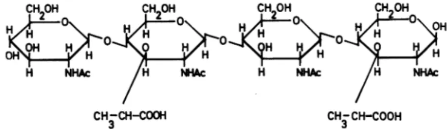
GlycanStrands
In all bacteria so far examined, the glycan strands consist of alternating 83-1,4-linked
N-acetylglucosamine and N-acetylmuramic acid residues (Fig. 2). The only variation so far
en-countered among bacteria is the possible pres-ence ofO-acetyl substituents on C-6 ofsome of the N-acetylmuramic acid residues (vide infra).
PeptideSubunits
The peptide subunits substitute through their N-terminitheD-lactic acidmoiety ofsomeof the
muramic acids in the glycan (Fig. 2). Figure 3
shows several peptide subunits for which the structures have been established. In these sub-units, the y-carboxyl group of theglutamic acid
residue is linked to the next amino acid in the sequences.Dependingupon the bacterialspecies,
the a-carboxyl group of glutamic acid is either
free, orsubstitutedby anamideorbyan amino
acid residue. In Micrococcus lysodeikticus (Fig.
3, typeC),thesubstitutingamino acidisusually Gly (vide infra). This Gly can be replaced by
BACTERIOLYTIC ENZYMES
(a)
(a)
(b)
NHAc CH-CH-CO CH-CH-CO 3 1',,,,,,,
(C)
3|
Peptide peptide Subunit SubunA(a) endo-N-acetylmuramidase; (b):
endo-N-acetylglucosaminidase;
(c):
N-acetylmuramyl-L-alanineamnidase.
FIG.2. Aportionofaglycanstrand. (a),Siteofactionofendo-N-acetylmuramidase;(b),siteofactionof
endo-N-acetylglucosaminidase; (c),siteofactionofN-acetylmuramyl-L-Alaamidase.
D-serine when M. lysodeikticus is grown in a
defined medium in thepresence ofD-Ser (181).
Similarly, the carboxylgroup of meso-DAP not
engagedin apeptide bondmaybesubstituted by
an amide group. Nomenclature involving
meso-DAP-containingpeptidesubunitsmaybe confus-ing. Inorder to specify which of the two asym-metric carbons of meso-DAPhasthe substituted
amino groups, the notation (L) or (D) written immediately beforemeso-DAPhasbeensuggested (9).Ithasalso been proposedtowrite(L) or (D)
immediately after meso-DAP in order to dis-tinguish betweenthecarboxyl-substitutedgroups.
This terminology is used throughout thisreview. The most common peptide subunits are the
peptides A, B, and C (Fig. 3). Thepeptide sub-units,Dand E,(Fig. 3) contain neither L-Lysnor meso-DAP.Moreover, theN-terminalaminoacid
(that is, the one which is joined to the glycan
chains) is not anL-Ala residue. Itis remarkable
that thepeptide subunit D contains no diamino acid residue. Peptide subunits with sequences
L-Ala-'y-D-Glu-diamino acid-D-Ala are known
whichdiffer from peptides A,B,or C (Fig. 3) by thefact thatL-Lys or meso-DAP isreplaced by
another diamino acid. Examples of unusual diamino acids are the following: LL-DAP (in many actinomycetes, vide infra), DD-DAP in a minor part of thepeptidoglycanof B. megaterium and probably in some Micromonospora and
actinomycetes, vide infra; 2,4 diaminobutyric
acid in Corynebacterium tritici (116); 2,6
dia-mino-3-hydroxypimelic acid in several Actino-plannaceae (112); L-ornithine (L-Orn) in M. radiodurans (182), Lactobacillus bifidus var
Pennsylvanicus(175), L. cellobiosus (121-123), and in Treponema reiteri (164); and hydroxylysine,
knowntoreplace L-Lys in Streptococcusfaecalis,
but only under conditions oflysine deprivation
(150). Hydroxylysine was also found as aminor
constituent of thepeptideof S. pyogenes (99).
Finally, variations in thepeptide subunits can also occur in the nature of the dicarboxylic
amino acid residue. The sequence Gly-threo-3-hydroxy-Glu-L-Lys-D-Ala has been characterized
inMicrobacteriumlacticum (144, 145).
Cross-linkingBridges
Several types of cross-linking bridges are
presented inFig. 4-9. The bridges which
cross-link the peptide subunits have identical locations inbacterialpeptidoglycansof types1, II, and III (Fig. 4-7). They extend from the E-amino group of L-Lys or from the amino group on the D-car-bon of meso-DAP (NH2-(D)-meso-DAP) of one
peptidesubunittotheC-terminal D-Alacarboxyl
group of another peptide subunit. According to
thebacterialspecies, however, thebridgespresent great variations in their chemical composition.
IntypeI,thebridgingresults from directpeptide
JEAN-MARIE GHUYSEN Type A GLYCAN L-Ala-D-Glu (L (D) Type B GLYCAN
L-Aa-
D-Glu4
NH2
L*L-Lys-.D-Aba
Type C GLYCANI
L-Ala-D-Glu-Gly
X L.
L-L
4-Aa
Type D GLYCAN I Gly-D-Glut-
HomoSer-.D-AIa
Type E_I
GLYCAN L-SerD-GluL-L-Orn-D-Ala
FIG. 3. Five typesofpeptidesubunits. Glu-NH2 =
isoglutamine. A. Peptidesubunits in E.coli, B. mega-terium, and B. subtilis. In B. subtilis, the carboxyl groups on theDcarbonofmeso-DAPofsomepeptide
subunits are amidated. Vertical bar separating (L)
and (D) represents diaminopimelic acid. B. Peptide
subunits inStaphylococcusaureus,M.roseus, Strepto-coccus pyogenes, A. crystallopoietes, S. faecalis, L.
acidophilus, andL. casei. C. Peptide subunits in
Sar-cinalutea,M.lysodeikticus, M.flavus,andM. citreus.
D.Peptidesubunits in C.poinsettiae,C.flacumfaciens,
and C. betae. E. Peptide subunits in Butyribacterium
rettgeri.
Type I. E.coli B.megaterium B. subtilis
L-A--G---Lu L-Ala--D-Glu CL(L) - D-Ala---SL ()D-AlaI---L. (D) (D) KM endop.
FIG. 4. Peptidoglycan type L. Direct cross-linkage
between twopeptide subunits A (Fig. 3). Arrow: site
ofaction ofthe KMendopeptidase.
bonds between peptide subunits (Fig. 4). This
type of bonding seems to be frequent among meso-DAP-containing peptidoglycans. Thus, the bridges are D-Ala-(D)-meso-DAP. In type II,
the bridgesconsist of several Gly or L-amino acid
residues,or both (Fig. 5), or are composed of one D-amino acid, namely D-isoasparagine (Fig. 6).
Intype III, the bridges are formed by a "head to
tail" assembly of several peptides, each having
thesame sequence of amino acids as the peptide
subunit (Fig. 7). Two types of linkages,
N'(D-alanyl)-L-Lys and D-alanyl-L-Ala, are involved in
thebinding betweenthe peptide subunits. In type IV, in a few bacteria, the bridges which
cross-link the peptide subunits extend from D-Glu of one peptide subunit to the C-terminal D-Ala
carboxylgroup of another peptide subunit. These
peptide bridgesarecomposedof onediamino acid
residue, D-Orn in the case ofC.poinsettiae and some other plant pathogenic Corynebacteria
(115; Fig. 8), D-Lys and D-Orn in the case of Butyribacterium rettgeri (M. Guinandet al.,
Bio-chemistry,in press;Fig.9). As showninFig. 8 and
9, the D-Lys or the D-Orn residues are linked
through their e- or 5-aminogroups, respectively,
to the a-carboxyl group of glutamic acid, and
throughtheir a-aminogroups to thecarboxyl
ter-minalD-Ala. Asimilartypeof bridging probably occursinMicrobacteriumlacticum(145), inwhich case adipeptide Na-(Gly)-Lys wouldextend from
thethreo-3-hydroxyglutamic acid residue ofone
peptide subunit to theD-Ala residue ofanother
peptide subunit.
ENZYMESTHAT DEGRADE BACTERIAL
PEPTIDO-GLYCANS: NATURE OF THEHYDROLYZED LINKAGES
The establishment of thepeptidoglycan struc-turehasnecessitatedthedevelopmentofaccurate
analytical and fractionation techniques and the discovery ofaseriesofdifferenthydrolyticagents
of high specificity. Within the past 10 years, numerous bacteriolytic enzymes have been isolated, whichhave proven tobejustsuchtools. Adescription of these techniques and asurvey of
the lytic enzymes so far discovered and charac-terized have beengivenrecently (41).The follow-ing enzymes, selected for their specific action
uponcriticallinkages,wereparticularlyuseful in
dismantling the bacterial peptidoglycans into meaningfulfragments (Table 1).
Endo-N-Acetylmuramidases
These enzymes, such as egg-white lysozyme
(27),Streptomyces 32 (34, 36, 37) orF1enzymes
(21, 100), and ChalaropsisBenzyme(46,47, 172),
hydrolyze the glycosidic linkages between
N-acetylmuramic acid and N-acetylglucosamine (Fig. 2), releasing fragments with N-acetyl-muramic acid residuesatthereducing end.
BACTERIOLYTIC ENZYMES
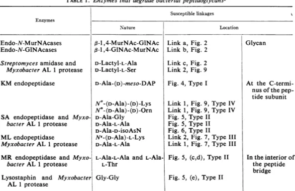
TABLE1. Enzymes that degrade bacterial peptidoglycansa Susceptible linkages Enzymes
Nature Location
Endo-N-MurNAcases 13-1,4-MurNAc-GINAc Link a, Fig. 2 Glycan
Endo-N-GlNAcases 13-1,4-GINAc-MurNAc Link b, Fig.2
Streptomyces amidase and D-Lactyl-L-Ala Link c, Fig. 2 Myxobacter AL 1 protease D-Lactyl-L-Ser Link 2, Fig. 9
KM endopeptidase D-Ala-(D)-meso-DAP Fig. 4, Type I At the C-termi-nusof the pep-tide subunit
N _(D-Ala)-(D)-Lys Link 1, Fig. 9, Type IV
N"-(D-Ala)-(D)-Orn Link 1, Fig. 9, Type IV SA endopeptidase and Myxo- D-Ala-Gly Fig. 5,Type II
bacter AL 1 protease D-Ala-L-Ala Fig. 5, Type II D-Ala-D-isoAsN Fig. 6, Type II
MLendopeptidase NE-(D-Ala)-L-Lys Link 2, Fig. 7,Type III
Myxobacter AL1 protease D-Ala-L-Ala Link 1, Fig. 7,Type III
MR endopeptidase and Myxo- L-Ala-L-Ala and L-Ala- Fig. 5, (c,d), Type II Intheinteriorof
bacter AL1 protease L-Thr thepeptide
bridge Lysostaphin and Myxobacter Gly-Gly Fig. 5, (e), Type II
AL 1 protease
aEndo-N-MurNAcase = endo-N-acetylmuramidase; Endo-N-GlNAcase =
endo-N-acetylglucosami-nidase;MurNAc = N-acetylmuramicacid;GINAc = N-acetylglucosamine.
Type II.
)(a):
A.crystallopoietes;(b): 5 pyogenes;(c):M.roseus
T4
- (d):M.roseus R27;(e).
S. aureus Copenhagen.---G-M-G - -L-Ala-D
-L-Ala-.D-Glu5NH2
(3) XLL-Lys-aD-Ala--(3r (-Gly-Gly- oGly-Gly-Glj~Glu°~NH2 1rL-Ala-L-Ala-L-
Ala-L-Thr(d)
r iLL-ys-D -la-L-Lys-~D-Ala-.~_L-Ala--L-Ala--L-Ala(c)
kL-Ala-L A'a(b)
( 1 ) L-Ala(a)
'L
ttA
t(1) SA endop
(2)
(2): Aminopeptidase (3) Myxobacter ALI
FIG. 5. PeptidoglycantypeII. Peptide bridges composed of glycineorL-aminoacid residuescross-linkingtwo peptidesubunitsB(Fig. 3). (1),Siteofactionofthe SAendopeptidase; (2),siteofactionofaminopeptidase; (3), siteofactionof MyxobacterALIendopeptidase. WallsofStaphylococcus epidermidis belongtothesametypeII. Thebridgesareformed ofGlyandL-Serresidues (D.J.Tipper, Federation Proc.,p.294, 1968). L-Ser is non-randomlylocated. Four kindsofbridgesinthefollowingproportions havebeenshownto occur:
Gly-Gly-Gly-Gly--Gly(20%);Gly-Gly-L-Ser-Gly-Gly (55%); Gly-L-Ser-Gly-Gly-Gly (10%); andL-Ser-Gly-L-Ser-Gly-Gly (15%). 431
JEAN-MARIE GHUYSEN Type II. S. faecalis * L.acidophilus . L.casei
_--G-M-G--
-L-Ala-.D-GluZNH2
gL.L-Lys-.D-AIa---L- Ala .D-OluG.NH2 I,.,..'P...
tLoL-Lys.D-A1a
a.D-Asp.NH2 SA.endop.Myxo. ALI endop.FIG. 6. Peptidoglycan type I. A D-isoasparaginyl
bridge cross-linking two peptide subunits B (Fig. 3). Arrow: site of actiono0 theSA and MyxobacterAL I
endopeptidases.
Endo-N-Acetylglucosaminidases
These enzymes, suchasstreptococcalmuralysin (3) and the glycosidase in lysostaphin (170),
hydrolyze the glycosidic bonds between
N-acetylglucosamine and N-acetylmuramic acid (Fig. 2), releasing fragments with
N-acetyl-glucosamineatthereducingend.
Streptomyces
N-Acetylmuramyl-L-Alanine
Amidase
Thisenzyme(J.M.Ghuysenetal.,Biochemistry,
in press; 34) hydrolyzes the bonds between the D-lactic acid residues of the glycan strands and the N-terminal residues ofthe peptide subunits
(Fig. 2, 7, 9). The N-terminal amino acid is usually L-Ala; thus, the enzymeis designated as
N-acetylmuramyl-L-Ala amidase. However, this
amidase is also able to cleave other
linkages
atidentical locations in thepeptidoglycan, such as
the N-acetylmuramyl-L-Ser linkages in walls of B.rettgeri (Fig. 9).Thisenzymehasnosignificant activity on intact peptidoglycans. To be
func-Type IV. C.poinsettiae;C.flaccumfaciens;C. betae
xGy-'D-Glu---2, ~~~~~~~~~~~~...
D
LHomoSer--D-Ala
-D-Orn:...
Gly-D-Glu °
LHomoSer-D-Ala
-FIG. 8. Peptidoglycan type IV. A D-ornithine bridge
cross-linking two peptide subunits D (Fig. 3). Note the absence of diamino acid in the peptide subunit.
tional, it requires that the glycan first be split.
Thus, this amidase has nobacteriolyticaction. StreptomycesKMEndopeptidase This enzyme(J.M. Ghuysen et al.,Biochemistry
inpress) has been used to hydrolyze the cross-peptide linkages which serve asbridges between
peptide subunits in the meso-DAP-containing peptidoglycansof E. coli and B. megaterium KM
[J.Van Heijenoort et al., Biochemistry, in press; 9 (Peptidoglycan type 1, Fig. 4)]. The bonds specifically hydrolyzed are thus D-alanyl
(D)-meso-DAP linkages. This enzyme also hydro-lyzes the D-Lys and
Na-(D-alanyl)-D-Orn linkages inwalls of B. rettgeri (M. Guin-and et al., Biochemistry, inpress; Peptidoglycan
type IV, Fig. 9). Anendopeptidase whose speci-ficitymustbe identicaltothatof theStreptomyces
KM endopeptidase is present in the autolytic
system of E. coli (177).
Type III. M. lysodeikticus S. lutea M.flavus. M. citreus.
---G-M-G-M-G-M-G-M-G-M-
--(l--or
(3;*-D ---G-M-G ---(1)or (3) -e- ---(1)orL-o-
_---
o()'L
Lee-0-0_-e-£e o'1)
t
~L @-o-
(2)
*-)Q L-Aba--D-Glu-%Gs 1(1 L@-°9
LL-Lys-D-Ala(1): Myxobacter ALI (2): ML endop. (3): Streptomyces Amidase
FIG.7. Peptidoglycantype III.Assemblingofpeptidesubunits C (Fig.3)insomeMicrococcaceae. (1), Siteof
actionof MyxobacterAL Iendopeptidase (hydrolysisof D-alanyl-L-AlalinkagesandofN-acetylmuramyl-L-Ala
linkages); (2),siteofactionofMLendopeptidase(hydrolysisof
Ne-(D-alanyl)-L-Lys
linkages); (3),siteof action-of StreptomycesN-acetylmuramyl-L-Alaamidase.BACTERIOLYTIC ENZYMES Type IV B. rettgeri ---G-M-G--- (2) 1 --L-Ser-D-Glu--- (1)
I
;1 :,D-Lys:
iLKL-Orn-.D-AtaL-.
orD-Ornj
---G-M -G ---L-Ser D-Glu6L.L-Orn-.D-Ala
(1) KM endop. (2) Streptomyces Amidase
FIG. 9. PeptidoglycantypeIV. Twopeptide subunits E(Fig. 3) cross-linkedbya D-Lysor a D-Ornbridge. Note thatthediaminoacidL-Orn in thepeptidesubunit isnot usedfor peptide cross-linking. Arrows: (1) site
ofaction of the KMendopeptidase; (2) siteofaction of Streptomyces N-acetylmuramyl-L-Ala amidase.
In Butyribacterium rettgeripeptidoglycan, D-lys and
iD-Orn bridges occurin the ratio 2:1.
Streptomyces SAEndopeptidase
This enzyme acts on type II peptidoglycans
(32, 33, 39, 99, 117; Fig. 5, 6). Ithydrolyzesthe linkages at the N termini of the peptide bridges
and at the C-terminus of the peptide subunits.
Sensitive linkages are, for example, D-alanyl-Gly, D-alanyl-L-Ala; D-alanyl-D-iso-asparogine.
When opened at their N termini, the peptide bridges which contain glycine or L-amino acid residues, or both (Fig. 5), can be further de-graded (117) byanamino peptidase such asthat secreted by Streptomycesstrains (J. M. Ghuysen
et al., Biochemistry, in press). As a result, the amino acids are sequentially liberated until the
E-amino groupsoflysine in the peptide subunits,
to which the peptide bridges are linked at their C termini, are exposed (Fig. 5).
StreptomycesMLEndopeptidase Thisenzymeis activeontype IIIpeptidoglycans
(Fig. 7;J.Campbelletal.,Biochemistry,inpress; 31, 117). Itspecifically hydrolyzes NE-(D-alanyl)-L-Lysine linkages (link 2, Fig. 7), which serve as
linkinggroupsbetweenpeptidesubunits.
MyxobacterAL I Protease
Incontrast to StreptomycesKM, SA, and ML endopeptidases, all of which have restricted
specificities and are, for example, unable to degrade casein, MyxobacterAL 1enzyme(24) isa
powerful protease, active on casein, and it possesses a broad bacteriolytic spectrum. It has
several sites of action on the cell wall peptide.
(i) Investigations of the mode ofaction ofthis enzyme upon Staphylococcus aureus (59, 17) haverevealed that it catalyzeshydrolysis of two
linkages within the peptide moiety (Fig. 5).
N-terminal Gly and both COOH-terminal D-Ala and COOH-terminal Gly are liberated by this hydrolysis. The pentaglycine bridges are thus
attackedatinternalglycyl-Glylinkagesaswellas at their linkages to the D-alanyl termini of the peptidesubunits (links3,Fig. 5).(ii) Myxobacter
AL 1 enzyme, acting uponwalls of Arthrobacter
crystallopoietes (74, 75), M. lysodeikticus (31), and other Micrococcaceae (J. Campbell et al., Biochemistry, in press), hydrolyzes D-alanyl-L-Alalinkages inbridges oftypeII,asdoes the SA endopeptidase (Fig. 5), and the "head to tail" D-alanyl-L-Ala linkages in the type III bridges (link 1, Fig. 7). (iii) Moreover, Myxobacter AL 1 enzyme is capable of hydrolyzing
D-alanyl-D-isoAsNlinkages (Fig. 6), as does the SA
endo-peptidase, and it has been used to study the
peptidoglycan in L. casei (D. Hungerer,
Feder-ation Proc., p. 294, 1968). (iv) Finally, in all cases so far studied, Myxobacter AL 1 enzyme
also hydrolyzes N-acetylmuramyl-L-Ala linkages
(Fig. 2) ata slow butdetectable rate (31, 71, 75, 171). In this respect, the Myxobacter AL 1 enzyme has a very useful characteristic: it dis-plays amidase activity on substrates with intact glycan chains.Thus, thisenzyme provides a way ofisolatingthepolysaccharide moiety free of its substituent peptide, but retaining its in vivo
degree ofpolymerization.
StreptomycesMR Endopeptidaseand
LysostaphinEndopeptidase
Lysostaphin endopeptidase (11, 170) is known
to split glycyl-Gly linkages at several places within thepentaglycine bridges (type Hie,Fig. 5). Sincethisstructureis limitedtotheStaphylococci,
lysostaphin endopeptidase is specifically
staph-ylolytic. Streptomyces MR endopeptidase
disrupts types I1c and Ild bridges (117; Fig. 5). It hydrolyzes the L-alanyl-L-Thr linkages in M.
roseus R 27and, at a slowerrate, the
L-alanyl-L-Alalinkagesin M. roseus(Thr-).
Peptidase Preparations with Mixed Activities The Streptomyces L3 enzyme preparation,
whenactinguponwalls of C. diphtheriae (J. M.
Ghuysen et al., Federation Proc., p. 410, 1966; 65, 72, 96,97), ismainly abridge-splittingenzyme which catalyzes the hydrolysis of
D-alanyl-meso-DAP.Inthis lattercase,however,itisnotknown which amino group of meso-DAP of onepeptide
subunit is involved in the linkage to D-Ala of 433
JEAN-MARIE GHUYSEN
anotherpeptidesubunit.The L3 enzyme
prepara-tion also contains (i) N-acetylmuramyl-L-Ala amidase
activity,
(ii) an enzyme catalyzing the hydrolysisof anamide that substitutes one of thecarboxylgroups ofmeso-DAP,and (iii) possibly,
aD-Alacarboxypeptidase.
TheFlavobacterium L-11 enzyme (65, 66) and the Staphylococcusepidermidis ALE enzyme(158)
exert their lytic actions upon walls ofS. aureus
through theactivities ofglycyl-Glyendopeptidase, N-acetylmuramyl-L-Ala amidase, and D-alanyl-Gly endopeptidase (in the case of the L-11 enzyme).
As observed with Myxobacter AL 1 enzyme,
the above L-3, L-11, and ALE enzyme
prepara-tions hydrolyze more thanone type of linkage.
From the datapublished to date, itseems likely
that severalenzymes are presentin the L-3,L-11,
andALEpreparationsastheyareobtained.
Summation
The foregoing enzymes permit the specific hydrolysis of many important linkages in the wall peptidoglycans (Table 1).
Endo-N-acetyl-muramidases andendo-N-acetylglucosaminidases
actontheglycanchains.N-acetyl muramyl-L-Ala amidaseshydrolyze linkages at thejunction
be-tween the glycan and the peptide moieties, i.e., linkages thatarelocated at the Ntermini ofthe
peptide subunits. The endopeptidases may be
grouped into three main types: (i) those which hydrolyze various linkages, all of which involve the C-terminal D-Ala of the peptide subunits; (with theexception of the following examples, all
the known endopeptidases fall in this group); (ii) Myxobacter AL 1 which, in some bacterial
walls, hydrolyzes linkages involving both C and N termini of the peptide subunits (Fig. 7); and (iii) lysostaphin and MR endopeptidase, which hydrolyze peptide internal bonds ofthe peptide
bridges involvingneither theC northeNtermini of thepeptide subunits. Withsomebacterial walls (for example, those of S. aureus), Myxobacter
AL 1 also hasthis lattertypeofactivity (Fig.5).
Streptomycessp. appear to bea very interesting
source ofvarious peptidases active on bacterial
peptidoglycans. Streptomyces aminopeptidase (link 2, Fig. 5) and the Streptomyces KM, SA, ML, and MRendopeptidases are basic proteins.
Theyhavebeenpurified andseparated from each
other by chromatography on CM-cellulose, and
theirspecificactivitiesuponsoluble, well-defined, bacterial wall degraded compounds have been studied (J. M. Ghuysen et al., Biochemistry, in
press).
STRUCTURE OF SEVERALBACTERIAL PEPTIDO-GLYCANS AS REVEALED BY ENZYMATIC
DEGRADATIONS:GLYCAN MOIETY Thestudiestobe reported here deal with the
wall peptidoglycan of some gram-positive
bacteria. Intact cell walls were obtained by
mechanical disruption of the cells followed by differential centrifugation. However, these wall preparationswere usually treated with trypsin to remove cytoplasmic contaminants and, in some
instances, proteinconstituents of the walls them-selves. The walls werenotsubjected to any
chem-ical agent such as trichloroacetic acid or hot
formamide. Althoughthesetreatments arewidely used and yield enriched peptidoglycan
prepara-tions by removing nonstructuralwallcomponents
(other polysaccharides and teichoic acids), they also cause random cleavage of some covalent linkages within the peptidoglycan, as has been shownby endgroupanalysis.Inaddition, various chemical modifications, such as formylation of
free aminogroups, can occurwhenhotformamide
is used (114). Moreover, chemically stripped peptidoglycans are not capable of giving any
information about the wayin which the various constitutivepolymersareheld together within the bacterialwalls,aquestionthatultimatelymustbe resolved.
Staphylococcusaureus
Three types of glycan degradation procedure
were carried out with (i) endo-N-acetylmur-amidase (Chalaropsis B enzyme; Streptomyces32 orF1enzymes), (ii) endo-N-acetylglucosaminidase
(from lysostaphin), and (iii) Myxobacter AL 1 enzyme. Inthe firstprocedure,aftersolubilization of the wall by the N-acetylmuramidase (36, 37), teichoic acids were removed on Ecteola
cellulose at pH 5. The peptide substituents
were detached from the glycan fragments by
Streptomyces N-acetylmuramyl-L-Ala amidase
(Fig.2),andtheliberatedpeptideswereremoved
on CM cellulose at pH 6.4. The resulting free
glycan fragments were separated into two com-ponents by preparative paper chromatography followed by gel filtration. They were charac-terized (167) as being Disaccharide 1
(/3-1,4-N-acetylglucosaminyl-N-acetylmuramic acid) and Disaccharide II
[/3-1
,4-N-acetylglucosaminyl-N,6-O-diacetylmuramic acid; (Fig. 10)].Inthesecondprocedure, asimilar degradation
sequence(170) yieldedtwodisaccharides isomeric with Disaccharides I and II, Disaccharide III:
(,B-1,4-N-acetylmuramyl-N-acetylglucosamine)
BACTERIOLYTIC ENZYMES
Disaccharide
I
CH2OH H20H 0 OH OH HHHH H NHCOCH3 H OCH3CH3-CH-C00H
Disaccharide
III
Disaccharide II
CH20H
CHO-CO-CH3
0 ~~~~0 H H OH 0 OHOH H HH H H NHCOCH 0 H NHCOCH 3 1 3 CH3-CH
-COOHDisaccharide IV
3 I 3'A31--CH3-CH-COOH
CH3-CH-COOH
FIG.V10. Structure ofdisaccharides isolated from thepeptidoglycan of Staphylococcus aureus Copenhagen.
Disaccharide I and disaccharide III have also beenobtainedfromM.lysodeikticus. Disaccharide I:
N-acetylglu-cosaminyl-,f-1,4-N-acetylmuramic acid. Disaccharide II: N-acetylglucosaminyl-,3-1,4-N, 6-0-diacetylmuramic
acid. Disaccharide III:N-acetylmuramyl-,6-1,4-N-acetylglucosamine. Disaccharide IV:N, 6-0-diacetylmuramyl-B-I ,4-N-acetylglucosamine.
and Disaccharide IV
[,/-1,4-N-,6-0-diacetyl-muramyl-N-acetylglucosamine (Fig. 10)]. The observed yields of the disaccharides obtained by the various degradation procedures were
con-sistentwith a wall structure in which theglycan
moiety is composed of linear strands of
fl-1,4-linked N-acetylglucosamine pyranoside residues, (i.e., a chitin-like structure). Unlike chitin,
however, every other sugar is substituted by a
3-0-D-lactyl group. In S. aureus, about 50% of
the N-acetylmuramic acid residues have a
6-0-acetylgroup, butwhether thedistribution of this substituentisregular, random, orlocalizedisnot
yet known. Since glycan degradation yields free disaccharide units only if it is followed by
N-acetylmuramyl-L-Ala amidase treatment, all
N-acetylmuramicacidresiduesmust,therefore, be substituted by peptide subunits. For thisreason,
the S.aureuspeptidoglycan is saidtobea"tight"
network.
In the third procedure, Myxobacter AL 1
enzyme wasusedtoachieve,inaone-step
opera-tion, the opening of the pentaglycine bridges
(Fig. 5) and the hydrolysis of the N-acetylmur-amyl-L-Ala amidic linkages, thus yielding free unaltered glycan strands (171). Appropriate
fractionation of the degraded products gaverise
tovarious glycan fractions, some ofwhich were
boundto theteichoic acid polymer. Analyses of the fractions showed the glycan moiety to be
polydisperse with an average chain length of about 12 disaccharide units, but containing
chains withasfewas6and asmany as 50 disac-charide units. These figures were based on the
estimation of theformaldehydogenic end groups that originate from the reducing
N-acetyl-hexosamine termini of the glycan chains on
re-duction withNaBH4. An averagechainlength of 16 disaccharide units can also be deduced from
an estimation of the free N-acetylglucosamine residueswhich areliberated from cell walls after complete hydrolysis of the linkages between
N-435
JEAN-MARIE GHUYSEN
acetylmuramic acid and N-acetylglucosamine by
means of an endo-N-acetylmuramidase (36). This liberation of free N-acetylglucosamine
residues under these conditions (actual data, 32nmoles/mg of wallsorper500nmequivalents of disaccharide units) indicates that N-acetyl-glucosamine residues arelocated at the reducing
ends of the intact glycan chains. These reducing
N-acetylglucosamine termini in the glycan result
veryprobably froman endo-N-acetylglucosamini-dase activity of the staphylococcal autolytic
system(D.J.Tipper, BacteriolProc.,p.48,1968).
Micrococcuslysodeikticus
Similar techniques of degradation, fractiona-tion, and characterization showed the M. lyso-deikticusglycan moietytobeidentical with that of S.aureus,with thefollowing exceptions,however. (i) 0-acetyl substitutionis absent in all butafew
strains. (ii) Only 40% of the N-acetylmuramic
acid residues aresubstituted by peptide subunits (81, 100). (iii) A small amount of the muramic
acid residuesare notN-acetylated (92). (iv)
Split-tingof theglycanwith thehelpofeitherlysozyme
or Streptomyces FK endo-N-acetylmuramidase is
incomplete and free, unsubstituted glycan
frag-ments,fromdi-tooctasaccharides, areproduced. Disaccharide I (60, 81, 110, 139, 147),
disac-charide III) (81; Fig. 10), and a tetrasaccharide (81; Fig. 11) were isolated in good yields, and
theirstructures werethoroughly established.The lysozyme-catalyzed hydrolysis of isolated wall oligosaccharides has been studied in detail (15, 16) and interpreted on thebasis of the
three-dimen-sional model of lysozyme developed by Philips
andco-workers (5,6).Theresultsobtained show thathydrolysisof the isolated tetrasaccharideto
yield disaccharides proceeds chiefly via
trans-glycosilation, leading to theformation of higher
oligosaccharides. Hexa-, octa-, deca- and
dodec-asaccharidesarereadily degraded by
lysozyme
toyield the corresponding di- and tetrasaccharides. Thetetrasaccharide isdegraded at amuch lower
ratethan thehigher oligosaccharidesbecause itis
bound largely to lysozyme in a nonproductive
manner, which does not lead to bond scission. Owing to the partial peptide substitution of its-glycan moiety,the M.lysodeikticus peptidoglycan is referredto as a "loose" networkto contrastit
with that of S. aureus.
Other BacterialPeptidoglycans
Disaccharides have also been isolated after
endo-N-acetylmuramidase degradation of wallsor
envelopes of the following bacteria: M. roseus
(99, 117),Sarcina lutea (J. Campbelletal., Bio-chemistry, in press), Staphylococcus epidermidisr
(D. J. Tipper, Federation Proc., p. 294, 1968),
L. acidophilus (J. Coyette and J. M. Ghuysen,
unpublisheddata), L. casei (D. Hungerer,
Fed-eration Proc., p. 294, 1968), Streptococcus
pyogenes (98, 99), S. faecalis (149, 151), B. megaterium (9), B. licheniformis (93),
Butyri-bacterium rettgeri (M. Guinand etal., Biochem-istry, inpress), and E. coli (J. vanHeijenoortet
al.,Biochemistry, inpress). Noneof these disac-charides was submitted to the same exhaustive structuralinvestigationsas werethose of
Staphy-lococcusaureusand M.lysodeikticus,butanalyses
were made which provided the following data. (i) N-acetyglucosamine and N-acetylmuramic
acidweretheonlytwomonosaccharidespresent.
(ii) Acid hydrolysis, followedby quantitation of glucosamine using the yeast D-glucosamine 6-phosphate N-acetylase, revealedthat halfof the:
hexosamine residues were glucosamine. (iii)!
Reduction of the disaccharide with NaBH4
de-stroyed all of the muramicacid,half ofthetotal hexosamines, andnone of theglucosamine, thus.
establishingthat muramicacid isatthereducing
end of the disaccharide. (iv) Susceptibility to
pig epididymis
exo-j3-N-acetylglucosaminidase,
which is specific for ,B glycosidic linkages,
es-tablished the
(3-anomery
of thelink.(v)
Deter-mination of themolar extinctioncoefficient of thedisaccharide with the Morgan-Elson reaction
(147) established the glycosidic linkage to be 1:4, not 1:6. Hence the linkage
(8-1,4
of thedisaccharide (disaccharide I, Fig. 10) has been wellcharacterizedinseveralcases.Consequently,
CH2OH
CH2OH
CH2OH
CH2OH
OH H 0
H0
H OOH H 0 H
H NHAc H NHAc H NHAM H NHAC
CH-CH-COOH CH-CH-COOH
3
FIG.11. Structure ofa tetrasaccharide isolatedfrom thepeptidoglycan ofM.
lysodeikticus:N-acetylgluco-saminyl-,f-1
,4-N-acetylmuramyl-f-1 ,4-N-acetylglucosaminyl-g3-1
,4-N-acetylmuramicacid.REV.-BACTERIOLYTIC ENZYMES
-the glycosidic linkages, which in the glycan
stands extend from N-acetylglucosamine to
N-acetylmuramic acid, are known to be p3-1,4. Final characterization of the linkages between N-acetylmuramic acid and N-acetylglucosamine requires theisolation andcharacterization of the isomericdisaccharideIII (Fig. 10).This hasbeen done only with the walls of S. aureus and M.
.lysodeikticus.Inall the othercases,the hypothesis
that these latter glycosidic links are also p3-1,4
rests upon the assumption that the
endo-N-acetylmuramidases which hydrolyze these
link-ages withintheglycan haveastrict (3-1,4
speci-ficity, which may not be true. With this one
possible exception, there is good evidence for
-the prevailing hypothesis that the ,8-1,4-linked alternating N-acetylglucosamine and N-acetyl-muramic acid structureis ubiquitous in the bac-terialworld.
Molecular Size oftheGlycan Moiety The estimation ofaveragechain length of the glycan chains hasbeen made foronlyafew
bac-terial walls. As pointed out previously, the S. aureusglycan consists of chains averaging 12 to 16 disaccharide units in length (36, 171). Walls
of Arthrobactercrystallopoietes grown asspheres contain glycan strands composed ofan average
of 17 disaccharide units. Walls from the same
organismgrown as rods contain about65 disac-charidesperchain (74).This marked variation in polymer size between the glycans of rod and of
spherical cells prompted Kolenbrander and
Ensign toinvestigate the spiral-shaped Spirillum serpens (71). The ratio of total hexosamines to
reducing end groupsindicatedan averagelength of about50disaccharide units.However,analyses of the wall glycan of L. casei (D. Hungerer, Federation Proc., p. 294, 1968) and the Porton strain ofB. subtilis (A. D. Warth,personal
com-munication) revealed an average of about 10 disaccharidesperchainfor each.Fromthis small
survey, thereis noreal evidence that a relation-ship exists between cell shape and the average
length of the chains in the wall glycan. Base-Catalyzed Lactyl Eliminationfrom
N-Acetylmuramicacid
Treatment of N-acetylmuramic acid with 10 equivalents ofNaOH for 1 hr at 37C, using a 0.05N solution, gives rise toa neutral, reducing
compound, chromogenic in the Morgan-Elson reaction (without any further alkali treatment), andwithanRF of0.65 inasolution of
1-butanol,
pyridine, and water (6:4:3). It has been shown (32, 166) that under the above conditions, there is aspecificelimination of lactate from
N-acetyl-muramic acid. When the same treatmentis
ap-plied to a
f3-1,4
N-acetylglucosaminyl-N-acetyl-muramyl-peptide subunit, a lactylpeptide and aneutral, reducing disaccharideare produced. The
lactylgroup, when liberated fromthepeptide by acid hydrolysis, was shown to be sensitive to
D-lactic acid dehydrogenase and is thus the D-isomer (166). The modified lactylless
N-acetyl-muramic acid residue can be liberated from the
disaccharidebymeansofthepigepididymis
exo-,B-N-acetylglucosaminidase. In its free form, it has all the properties of the chromogenic
com-pound, which arises directly by alkali treatment
of freeN-acetylmuramic acid. The base-catalyzed elimination of the D-lactylpeptide from the N-acetylmuramic acid residue of the disaccharide-peptide subunitwas interpreted (Fig. 12) as the result of a (-elimination involving the acidic
proton onC-2, inthepositionatothe carbonyl, and theO-lactyl substituent onC-3. Thisresults
in the creation ofadouble bond betweenC-2and
C-3, thus forming a N-acetylglucosaminyl-2,3-dehydro-N-acetylglucosamine disaccharide. This
2,3-dehydro-N-acetylglucosamine, upon libera-tionbyenzyme,wouldcyclize intoaA3 dehydro-furane derivative, a chromogen in the Morgan-Elson reaction. The demonstration of this
(3-elimination castssomelightonthemechanism of thechromogen formationintheMorgan-Elson reaction. It also provides another way of
re-moving the peptide moiety from the glycan. Further examples ofthe use of thistype of
deg-radation will be given later. Attention should also be called to the factthat (-elimination can occur at 37 C under moderately alkaline
condi-tions. Extreme care mustbe taken in the
interpre-tation of the results of enzymatic degradations involving these conditions.
STRUCTURE OF SEVERAL BACTERIAL PEPTIDOGLYCANS As REVEALED BY
ENZYMATIC DEGRADATIONS:
PEPTIDE MOIETY
Theconsistencyofstructurefoundin the glycan moiety isnotreflectedinthepeptide substituents.
It is necessary, therefore, to discuss individual
speciesseparately.
PeptidoglycansofType I
Thebestknownexampleofthistypeof peptido-glycan (Fig. 4) is found in E. coli, which is, in
fact,thefirstpeptidoglycantohave beenanalyzed enzymatically (177).It wasshowntocontaintwo
mainstructural elements, the so-called fragment
C6 or the disaccharide peptide subunit
L-Ala-D-Glu-meso-DAP-D-Ala and fragment C3, a dimer
inwhich twoC6 fragments arelinked through a
437
JEAN-MARIE GHUYSEN NHc -NHc 1 R-0 C-H ,/1 \
~~0
7 0.05 N NaOH K H H-C o HO\Ht |~~~~~370;
lh NHAc H-C-OH A CH20H -O-R + NHAc CH20HH-C
',Occ'
H6
~~~~H-C
OH O -C7 HO \J~H I CHAc H-C-OHI
B CH2OH CH20H 0 OH OH HO X NHAc C K NHAc % 'H-C"~ -.C'I I %H C>sC" + H-C-OH H-C-OH CH20H \D / D NHAc Ik H-CLCK/
I
I_
OHW[C
-O OHI
H-C-OH CH20H Chromogen I NHAc k H-C C-H borale11
I100',
7minI
H-C-OHI
CH20H Chromogen IIIFIG. 12. ,3-Elimination of D-lactyl-peptide from disaccharide peptide. R, D-lactyl peptide residue;A, N-acetyl-glucosaminyl-j9-1,4-N-acetylmuramyl-peptide subunit; B, Prochromogen
glycoside:N-acetylglucosaminyl-,6-1,4-2,3-dehydro-N-acetylglucosamine; Exo-,6-GkcNAcase:exo-13-N-acetylglucosaminidase; C andD,free
N-acetyl-glucosamine andfree2,3-dehydro-N-acetylglucosamine,respectively.Duringthecourseofitsenzymaticliberation, 2,3-dehydro-N-acetylglucosamine would cyclizeintoa A3-dehydrofuran derivativeorchromogenLTransformation
of chromogen Iintoafuranderivative suchaschromogenIIIwould result fromfurthertreatmentwith borate at 100 C(32; figurereprinted by permission).
peptide linkage in which one amino group of meso-DAP is engaged. This pioneering work of Weidel and his colleagues (177) contributed greatlytoourpresentconceptof the wall
peptido-glycan as an enormous, net-like bag-shaped macromolecule. The E. coli autolyticsystem(109), which contains atleast fiveenzymes
(endopepti-dase, N-acetylmuramyl-L-Ala amidase,
endo-N-acetylmuramidase, D-Ala carboxypeptidase, and
exo-13-N-acetylglucosaminidase) specifically
de-grading the wall peptidoglycan into small fragments, as well as the demonstration of the generalstructureassignedtotheoriginal peptido-glycan, have been described in several reviews (38, 41, 85, 156, 177). In this peptidoglycan, virtually all of thedisaccharide units in the glycan chains are substituted by peptide subunits.
Some ofthepeptide subunitsoccur astripeptides
L-Ala-meso-DAP-D-Glu, the last D-Ala residue being lostasaresultofthe action of theautolytic
D-Ala carboxypeptidase. Thecarboxylgroups of
Glu and ofmeso-DAP, which are not engaged inpeptide linkages, have no amide substituents.
About 50% of the peptide subunits are
cross-linked to form peptide dimers, the disaccharide units fromtwoadjacent glycanchains thus being paired (161). It is probable that there is a
ran-domdistributionof thedisaccharidepeptide sub-unitand of its dimer throughoutatleastamajor part of the network (Fig. 13). Only recently (J.
van Heijenoort et al., Biochemistry, inpress; 9,
22) hasacompletestructurebeen assignedtothe
peptidoglycanofE.coli,aswellastoother
meso-DAP peptidoglycans from gram-positive
bac-teria, such as that of B. megaterium KM. The
B. megaterium walls also presentalowdegreeof
peptide cross-linking. About 85% of the total DAP residues are meso. Most of these peptides occur as uncross-linked tetrapeptide
(L-Ala-D-Glu-meso-DAP-D-Ala) and tripeptide (L-Ala-D-Glu-meso-DAP) subunits. About 15% of the
totalDAPresiduesinthesewallsareDD. Noneof
these latter residues exhibits free amino groups
(9).
Characterization of the NH2-(L)-meso-DAP
in the link to glutamic acid. Making use ofthe
exo-q-GlcNAcase
37°,pH 4 a h
REV.-BACrERIOLYTIC ENZYMES M M M M G |
G/
GI
MML..UM
MG/
G G / IMU-t.M
MUz....M
G(
GG/
G/
/ / / / M M M MG|G
| G | G I G I /G/
/G// / MU zMU M M-L..M M-z-M
/ / / / GG(
G GFIG. 13. Schematic representation of a peptido-glycansheet ofE. coli. In this loosenetwork,all ofthe
N-acetylmuramic acid residues are substituted either
bypeptidemonomers (peptide subunits A;Fig. 3) or bypeptide diners (Fig. 4).
factthatdinitrophenylation of amino acidsoften enhances their optical rotation, Diringer and Jusic (22) determined the configuration of that
asymmetric carbon of the meso-DAP residue which, in the E. coil peptide subunit, has afree
amino group. The mono-dinitrophenyl
(DNP)-meso-DAPobtainedafterdinitrophenylation and acidhydrolysisofthepeptidesubunitpresenteda
molar optical rotation, [M]D, in glacial acetic acidequalto + 250deg a 10%.Sincesynthetic
di-DNP-LL-DAP and synthetic
mono-DNP-LL-DAP have [MmD equalto -444 and -231
deg,
respectively
(61),
the above value of+250
deg provided evidence that, in the E. coli peptide subunit, the free aminogroup of themeso-DAPresidueswas ontheasymmetriccarbon
having
theDconfiguration.Thesameconclusionwasreached for the meso-DAP subunits inB.megaterium KM (9).Adisaccharidepeptidesubunitfraction (with
no amidesubstituent, as in the caseofE. coli), representingamajorpartofthe totalmeso-DAP
peptide subunits, was isolated. After dinitro-phenylation and acid hydrolysis, the isolated
monodinitrophenyl derivative of the meso-DAP
residues exhibited an
[MID equal
to +248 deg,i.e., avalueidentical,within the limits of
experi-mental error, to that observed with the
mono-DNP-DAP isolated from E. coil and to that of the synthetic mono-DNP-(D)-meso-DAP (9).
Moreover, both the synthetic
mono-DNP-(D)-meso-DAP and the natural mono-DNP-meso-DAP exhibited the same anomalous rotatory
dispersions with aCottoneffectcentered on 418
nm and, in the region 450 to 600nm, both fol-lowed thesimplified Drudeequation with identical
Kcoefficients. These determinations proved that
in E.coil and in B.megaterium, the aminogroup
engaged in peptide linkage to glutamic acid is
located on the L-carbon of meso-DAP and, consequently, that the amino group used for cross-linking between peptide subunits must be located onthe D-carbonof meso-DAP.
Characterization of the 'y-carboxyl group of glutamic acid in the link to NH2-(L)-meso-DAP.
The tripeptide monomers
L-Ala-D-Glu-(L)-meso-DAP were isolated from walls of both E. coli
and B. megaterium KM. They were chroma-tographically and electrophoretically indis-tinguishable and inseparable from synthetic tripeptide
L-Ala-,y-D-Glu-(L)-meso-DAP
under conditions which readily distinguish betweenthe two synthetic peptide isomers containing either a- or 7-linked glutamic acid (J. van
Heijenoortet al., Biochemistry, inpress). Char-acterization of the-y-monohydrazide of glutamic acid and the absence of thea derivative among
the compounds arising by hydrazinolysis of the peptide subunits ofE. coli and B. megaterium
KM alsoledtothe conclusion thatglutamic acid
is linkedtomeso-DAPviathe y-carboxylgroup. An identical conclusion was independently reached by Diringer (21a) by hydrazinolysis of theE.coll monomer.
Characterization of the link
meso-DAP-(L)-(D)-Ala. The tetrapeptide monomers with the sequence L-Ala-y-D-Glu-(L)-meso-DAP-D-Ala
were also isolated from E. coil and from B. megateriumKM (J. Van Heijenoort et al.,
Bio-chemistry,
inpress). Thelocation of theterminal D-Ala ontheL-carbon of meso-DAPwasproved by an Edman degradation. After one cycle of the degradation, N-terminal alanine wasre-placedby N-terminalglutamic acid,thus demon-strating the sequence Ala-Glu. Concomitantly,
most of the mono-N-terminal DAP groups
dis-appeared, and no free alanine was liberated. Thus, the terminal D-Alacannotbe inaposition relativetothefree aminogroup,which,asshown above, is onthe D-carbon ofmeso-DAP;
conse-quently, the terminal D-Ala must belocated on
the L-carbon of meso-DAP. After the second
cycle of the degradation, N-terminal glutamic
acid disappeared and was not replaced by any
other terminal amino group. The complete se-quence of the peptide subunit in E. coil and B. megaterium is thus
L-Ala-'y-D-Glu-(L)-meso-DAP-(L)-D-Alaasit isshown inFig. 3A. Characterization of the D-Ala-(D)-meso-DAP cross-linkages between peptide subunits. The
bisdisaccharide peptide dimer from E. coli, i.e.,
439
JEAN-MARIE GHUYSEN
fragment C8 (177), was degraded into disac-charide peptide monomers with the help ofthe
purified Streptomyces KM endopeptidase (J. van Heijenoort et al., Biochemistry, in press).
C- and N-terminal group analyses, before and after degradation, together with the aforemen-tioned demonstration that the amino group
engaged in the linkto glutamic acid is ontheL
carbon of meso-DAP, established that D-Ala-(D)-meso-DAP linkages are involved in peptide
bridging (Fig.4).
The demonstration of the existence of the
same type of peptide bridging in walls of B.
megaterium was attempted (J. van
Heijenoort
et al., Biochemistry, in press). Complications
arose, however, dueto the fact that about 15% of the total DAPresidues are DD.DD-DAP resi-dues were identified as follows. A
disaccharide-peptide monomer fraction containing solely meso-DAP residues and a disaccharide-peptide oligomer fractionwereisolated(9).After dinitro-phenylation and acid hydrolysis of the latter fraction, the meso-DAP residues were removed
as mono-DNP-derivatives. The remaining DAP
residues were then bisdinitrophenylated and
characterized as di-DNP-DD-DAP on the basis of the
[M]D
valueequalto +426 deg. In J. vanHeijenoort's more recent study, the walls of B. megaterium were solubilized with the help of a
purified KM endopeptidase. N- and C-terminal
groups analyses strongly suggestedthat the few
meso-DAP-containing peptide subunits which
are cross-linked are actually engaged in
D-Ala-(D)-meso-DAPlinkages, asinthecaseofE. coli. However, the DD-DAP residues appeared to be involved inanother type ofpeptidecross-linkage,
thesignificance of which isnotclear at the
mo-ment. In summary, it has been now well
es-tablished that the structure presented in Fig. 3
and 4 arevalid for theE. coil and for themajor
partof theB. megaterium
peptidoglycans.
Inthe latter case, however, it is probable thatD-Ala-(D)-meso-DAPpeptide cross-linkagesare notthe
onlyimportantones.Thetwotripeptides,
L-Ala-Ty-D-Glu-(?)-meso-DAP
andL-Ala-D-iso-gluta-minyl-(?)-meso-DAP, have been characterized (93) in walls of B. ficheniformis ATCC 9945
(previously designated B. subtilis ATCC 9945).
Finally, it has been reported recently (A. D.
Warth and D. L. Strominger, Bacteriol. Proc.,
p. 64, 1968) that the peptide subunits and the
peptide cross-linkages in thevegetative cell wall andthesporecortexofB.subtilishavestructures
identical to those found in E. coli with the
ex-ception, however, that in
vegetative
cells of B.subtilis, most of the
peptide
subunits have anamide substituent onthe carboxylgrouplocated
on the D-carbon ofmeso-DAP. Finally, in the
C.diphtheriaepeptidoglycan, both the D-Glu and the meso-DAP residues are amide substituted
(67).
Peptidoglycans of Type II with Peptide Bridges of Glycine or L-Amino Acid Residues, or Both Thefollowing sequential degradation has per-mitted thecharacterizationofthe peptide bridges
andthepeptide subunitsofthisgroup ofbacterial peptidoglycans (99, 117). (Degradation of the
walls ofM. roseusis givenas anexample in Fig.
14.) The first step involves solubilization of the
walls by the SA endopeptidase. Equivalent amounts ofC-terminal D-Ala, in all cases, and
of N-terminal amino groups of either Gly or
L-Ala, depending on the bacterium (Fig. 5),are
released as aresultoftheopening ofthepeptide bridges at their N termini (hydrolysis of links 1, Fig. 5, 14). The second stepinvolves amino-peptidase degradation of the opened peptide bridges until the e-amino group oflysine of the
peptide subunits, to which the bridges are
at-tached, are all exposed (hydrolysis of links 2,
Fig. 5, 14). Quantitation of the number offree
amino acids liberated, per lysine residue, at the
end of the process, andkinetics of the degradation
permitted the determination of the bridge se-quences.Inthecaseof M. roseusR27cell walls,
forexample (Fig. 5, 14), first three L-Ala residues and next, one L-Thr residue per lysine residue
weresequentially liberated. It was also observed that theexposureof the e-amino groupoflysine paralleled both the disappearance of N-terminal threoninegroupsandtheliberation of free L-Thr residues. At the end ofthis second step ofthe
degradation, the peptide subunitsarestripped of the peptide bridges but are still attached to
in-tact glycan strands. The third step involves the
degradation of the glycan into N-acetylglu-cosaminyl-N-acetylmuramic acid disaccharides
through the action of an appropriate
endo-N-acetylmuramidase
(hydrolysis
of links 3, Fig. 14). Thedisaccharide peptide subunits can then be readily isolated from the other degradationproductsbygelfiltrationonSephadex.The fourth
step involves cleavage of the isolated
disac-charide-peptide subunit into free disaccharide
and free peptide with the help of the
N-acetyl-muramyl-L-Ala amidase (hydrolysis of links 4,
Fig.
14). Thus, through controlled degradations involvingfoursuccessiveenzymatic hydrolysesofspecific linkages in the peptidoglycans, the
pep-tide subunits can be readily obtained via the
intermediate isolation of disaccharide-peptide
subunits. Again, it should be emphasized that
structural studies of a
peptidoglycan,
on theBACIERIOLYTIC ENZYMES
G
AM
L-ALa-.D-4
G
(3)...
s/1
M A- r% LILA DL-AIa-.U
-LAU-
MM
2Glu-.NH
L
- Lys-eD-Ala-2 GL-Lys
~~...
-D
-Ala-Aa-4L-
Ala-L-ALa-L-ThrM
1
L-Ala
-.D
-GluNH
2~~~~~~~~~L-Aa-.D-Glu-.NH2
p-LysD-Aa-G
L-Lys
-D-Aa-uL-Ala
.L-Ala
_-L-Ala
.L-Thr-J
*.~~~~~~~~~~~~~~~~~~~y ...:OA/,...,...,... G-M.
L-Ala
-.D-Glu
--NH2
I I. A +3
L-
Aa +1
L-Thr
L-LY
-UrL-A'G
+M
*L-Aba
+D-Glu-NH2
LL-
LLYS.D
Ala
FIG.14. Degradationsequencefor thepeptidoglycan of M.roseusR27.A. Disruption of the L-Ala-L-Ala-L-Ala-L-Thr bridges and liberation ofthedisaccharide-peptide subunits. (1),siteofactionof SAendopeptidase; (2), degradation oftheopened bridgeswithaminopeptidase; (3),siteofactionof endo-N-acetylmuramidase. B. Further degradation of thedisaccharide-peptidesubunit. (4),SiteofactionofN-acetylmuramyl-L-alanineamidase; (5),site
ofaction ofaminopeptidase; (6),siteofactionofexo-fl-N-acetylglucosaminidase.
basis of the fragments liberated by enzymatic degradations, can only be attempted after
thor-ough study of the kinetics to ensure that each step is carried tocompletion (156). Ifthe
enzy-matichydrolysisatanystepisstopped before all susceptible linkages are broken, an exceedingly
complex mixture of fragmentscanbeanticipated at the end of the sequential degradation. In
particular, complete wall solubilization doesnot necessarily indicate that all sensitive linkages have beenhydrolyzed. The peptide subunits ob-tained after the sequential degradation described abovehave beenfully characterized(Fig. 3B, 14)
as tetrapeptide amide
AT--(L-alanyl-D-isogluta-minyl)-L-lysyl-D-Ala (99). The characterization
of this structure was effected by the following
G
.I/
441
JEAN-MARIE GHUYSEN
chemical and enzymatic procedures. (i) A de-termination was made of total amino acid
com-position, ammonia amide,and C- andN-terminal amino acids. (ii) The L-alanyl-D-isoglutaminyl
sequence was demonstrated by degrading the
tetrapeptide with aminopeptidase, which
re-sulted in the liberation ofone freeL-Ala residue andtheconcomitant appearance of one
N-termi-nal D-isoglutaminyl residue per molecule of
tetrapeptide (hydrolysis of links 5, Fig. 14).
(For substrate requirements of the Streptomyces
aminopeptidase, see 99). (iii) The residual
tri-peptide (Fig. 14) was isolated in the form ofa
di-DNP derivative which was chromatographi-cally indistinguishable and inseparable from
synthetic di -DNP-isoglutaminyl -lysyl-
ala-nine. Thechromatographicsystem employed for the comparison readily distinguishes between isomerscontaining a- or
y-linked
glutamic acid,orglutaminyl residues (10, 80). (iv) Thepresence
of an amide substituent was confirmed electro-phoretically andtheoccurrenceof the isoglutam-inyl residue was demonstrated by chemical
de-hydration and reduction, followed by acid
hy-drolysis, according to the procedure ofRessler
and Kashelikar (126). This produced y-amino-butyric acid, as one would predict if, indeed,
the glutamic acid is in the iso form in the endo position. No ornithine, which would be
in-dicative of the presence ofglutaminyl residues,
was detected. (v) Theinvolvement ofthe
y-car-boxyl group of theglutamic acid in the peptide bond was shown by Edman degradation (168, 169). The first cycle removed the N-terminal
alanine, and the N-terminal glutamic acid
ap-peared. After the second cycle ofthe
degrada-tion, ammonia was liberated and N-terminal amino acids were no longer detectable, again demonstrating that NH3 was a substituent of the a-carboxyl group ofglutamic acid. The Ed-man degradation was initially carried out on a
carbohydrate free polypeptide fraction (169).
Itprovided the firstproofforthe occurrenceofan
isoglutaminyl residuein thewallpeptide moiety,
which observation was in agreement with the previous demonstration (57) of the involvement
of the y-linkage ofD-GIutothenextamino acid
inthenucleotideprecursorofthewall. Degrada-tionby MyxobacterAL 1 enzymeofthewallsof
S. aureus (59) and of A. crystallopoietes (171) also ledtotheidentification of the samepeptide subunit andestablishedtheexistenceofa
mono-L-Ala cross-linking bridge in the rod-shaped A.
crystallopoietes (75). Similarly, the samepeptide
subunits in Staphylococcus epidermidis strain
Texas 26 were shownto be cross-linkedby
pep-tide bridges ofGly and L-Ser, with an average
composition ofGly4 L-Ser, (J. D. Tipper,
Fede-ration Proc., p. 294, 1968). The location of the L-Ser residue was determined by Edman
de-gradation (legend, Fig. 5).
Peptidoglycans of Type II with D-Isoasparaginyl Bridge
It hasbeenknown for some time that D-aspartic
acid isaconstituent of cell walls ofsomespeciesof
Streptococcusand ofnumerousspecies of
Lacto-bacillus, where it occurs in amounts nearly
equivalentto thatof L-Lys orD-Glu (20, 55, 56, 151, 173).Thehypothesisthat theD-aspartic acid
residuesarelocated in thewall peptidoglycansas
substituents of the e-amino groups of the L-Lys
residues was first proposed by Swallow and Abraham (159). It wasbased on theisolation of
a derivative of aminosuccinimide,
Ne-(amino-succinyl)-lysine,fromwalls ofL.breviswhichhad been submitted to treatment with 11 N HC1 at
80Cfor43 hr. On furthertreatment with dilute sodiumhydroxide, the
NE-(aminosuccinyl)-lysine
was converted into a mixture ofpredominantly
Ne-(,3-aspartyl)-lysine
and a minor amount ofNe-(a-aspartyl)-lysine. Inlater studies, the same
cyclic peptide N'-(aminosuccinyl)-lysine was
found in hydrolysates ofmany other D-aspartic acid-containing bacterial walls (55, 56). How-ever,theabovering formation, as aresult of acid
treatment and the subsequent interconversion of the aspartyllysine, made it impossible to de-termine whether the original sequence in the
wallpeptidoglycanconsisted ofana-or
,3-aspartyl
peptide. Recent investigations (32) conclusively
established that, in S. faecalis ATCC 9790
(reidentified as S. faecium var. durans by 0.
Kandler, personal communication),
D-isoas-paraginyl residues serve as bridges between
peptide monomers type B (Fig. 3, 6). This
con-clusion arose from the results of a sequential
degradation of thewallsofS.faecalisessentially
as described for S. aureus, M. roseus, and S. pyogenes with, however, the two following modifications. (i) Walls of S. faecalis, when
prepared from cells harvested in logarithmic
phase ofgrowth (log walls), contain apowerful
autolysinwhich has thespecificity ofan
endo-N-acetyl-muramidase (151). It was observed that this enzyme can work in conjunction with the SA endopeptidase. Incubation of log walls with the SA endopeptidase thus resulted in the
appearance of terminal amino groups of
iso-asparagine as well as cleavage of glycosidic
linkages (Fig. 6). (ii) Because of its
D-configura-tion,theD-isoasparaginyl residues atthetermini
of thebridgesnowopened couldnotbeliberated
asfree amino acidsbyfurthertreatmentwiththe