The Relative Role of Bacterial Cell Wall and Capsule in the Induction of
Inflammation in Pneumococcal Meningitis
Elaine Tuomanen, Alexander Tomasz, Bruno Hengstler, and Oto Zak
From the Rockefeller University, Laboratory of Microbiology, New York; and the Research Department,
Pharmaceuticals Division, elBA-GEIGY Limited, Basel, Switzerland The relative contribution of bacterial components to the induction of inflammation
dur-ingStreptococcus pneumoniae meningitis is unknown. Several strains of pneumococci with differences in cell surface characteristics (capsule or cell wall) were compared for the effect on the inflammatory response evoked during infection of the cerebrospinal fluid (CSF) in vivo. The presence of bacterial capsular polysaccharide was not necessary for bacterial growth in CSF in vivo but correlated with greater CSF bacterial density. CSF inflammatory changes began to appear when the bacterial concentration exceeded 105 cfu/ml, regardless of the pneumococcal strain. CSF inflammatory changes could be in-voked by cisternal instillation of 105-106cell equivalents of whole, heat-killed unencapsu-lated strains or their isounencapsu-lated cell walls but not by similar concentrations of heat-killed encapsulated strains or isolated capsular polysaccharide. Hypoglycorrhachia was ob-served only during inflammation caused by live bacteria. The inflammatory response characteristic of naturally acquired pneumococcal meningitis can be reproduced by chal-lenge with both encapsulated and unencapsulated bacteria. The bacterial cell wall ap-pears to be the most potent pneumococcal surface component in inducing CSF inflam-mation.
Many steps in the pathogenesis of pneumococcal meningitis remain unclear. This is particularly true in regard to the contribution of bacterial compo-nents to the induction of the host inflammatory re-sponse once bacteria gain access to the cerebro-spinal fluid (CSF). An understanding of which bacterial component(s) incites the host response is important since products of inflammation that are released during infection may injure host tissues and be detrimental to the recovery from disease [1-3]. This information may provide important clues as to why mortality from pneumococcal dis-ease remains at 20070 despite the development of new, highly bactericidal antibiotics [4]. We present evidence that of the major surface components, the cell wall, and not capsular polysaccharide, appears to be a powerful inducer of the meningeal inflam-matory response.
Received for publication June 15, 1984, and in revised form September 10, 1984.
The use of tradenames is for identification only.
This work was supported in part by a ParkerB. Francis Fel-lowship to Dr. Elaine Tuomanen from the Puritan-Bennett Foundation.
Please address requests for reprints to Dr. Elaine Tuomanen, The Rockefeller University, Box 152, 1230 York Avenue, New York, New York 10021.
535
Materials and Methods
Bacterial strains. Streptococcus pneumoniae
strain All is an encapsulated Rockefeller University laboratory strain (Type II). Strains R6 and lyt 4-4
(an autolysin-deficient tolerant strain) are unencap-sulated and derived from strain All [5]. Strain SIll is a clinical isolate (Type III) obtained from Dr. M. Sande (San FranciscoGeneral Hospital Medical Center, San Francisco, Cali!). Strain 8249 (TypeXIX) is a penicillin-tolerant and penicillin-resistant clini-cal isolate obtained from Dr. Koornhof (Johannes-burg, South Africa).
Preparation ofinocula. Bacteria were grown for 6-8 hr in a synthetic medium [6] supplemented with 0.1% yeast extract (Difco, Detroit, Mich) to mid-logarithmic phase. The cells were centrifuged at 2,000
gfor 10 min at 4 C, then washed, resuspended, and diluted in pyrogen-free saline (elBA-GEIGY, Basel, Switzerland), and injected into the cisterna magna. Inoculum was titrated for cfu on trypticase soy broth (TSB) agar plates containing 5% sheep blood.
Meningitis model. Male chinchilla rabbits weigh-ing 2 kg (Thorn Farm, Biebarach an der Riss, Fed-eral Republic of Germany) were prepared according to the method of Dacey and Sande [7]. Unless other-wise noted, four rabbits were tested for each
varia-ble examined. Under anesthesia with 30 mg/kg iv pentobarbital (Abbot AG, Zug, Switzerland), a den-tal acrylic helmet was attached to the crown of the calvarium. A minimum of 24 hr later, the rabbits were again anesthetized with 1.75 g/kg sc ethyl car-bamate (Urethan'"; Fluka, Buchs, Switzerland) and 15mg/kg iv pentobarbital and positioned in a stero-taxic frame. A spinal needle measuring 25 gauge by 3.5 inch (Becton-Dickinson, St. Augustine de Quad-alix, Spain) was introduced into the cisterna magna and fixed in position on the frame; 0.3 ml of CSF was withdrawn and 0.2 ml of bacteria or bacterial component in pyrogen-free diluent was introduced. Serial 0.3-ml samples of CSF were obtained over the subsequent 12-hrof anesthesia. For sampling beyond this time, the rabbits were reanesthetized with pen-tobarbital (as above) as required.
Immediately upon withdrawal, CSF samples were analyzed for number of cfu as above. Red and white blood cell counts were determined in a Sysmex Microcell Counter (CC-I08; TDA, Kobe, Japan). When red blood cells were detected, the number of leukocytes was adjusted to correct for contamina-tion of CSF by blood. The remainder of each CSF sample was centrifuged (10,000g,3 min) with an Ep-pendorff microcentrifuge, and the supernatant was frozen and stored at -70 C for subsequent chemi-cal analysis. Glucose was determined by a micro-adaptation of a commercial assay kit (Test-com-bination glucose; Boehringer Mannheim, GmbH, Federal Republic of Germany), protein by the Lowry method [8], and lactic acid by a microadaptation of a commercial assay kit (Sigma UV test 826-UV; Sigma Chemical, St. Louis, Mo.).
9
8
u,7
(f) uE
<,6
~ u, U O't5
i·
04
3
2
3
6
9
12
15
18
21
24
27
30
48
HOURS
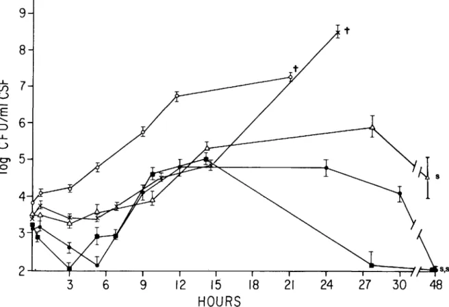
Figure 1. Growth rate of five strains of S. pneumoniae inoculated intracisternally into rabbits. Bacteria were prepared as outlined in Materials and Methods, and the inoculum was adjusted to rv 104
cfu/ml. For each strain, 0.2 ml of this suspension was introduced into the cisterna magna of each of four rabbits. The numbers of cfu/ml in CSF were then observed over time. Strain R6(e);strain 8249 (x); strain SIll(0); strain lyt 4-4 (_); and strain An (.6.). Each
data point is the mean ± standard error of the mean (SEM) of four rabbits.t = all animals in the group died; and s = all animals in the group survived. LDsofor each strain is as follows: R6 = 107; An = 106; SIll = 102; and 8249
= 102 •
Preparation
of
killed bacteria and bacterial com-ponents. S.pneumoniaestrain Au, SUh or R6wasgrown until the bacterial concentration reached108
cfu/ml (verified by cfu titration). A 10-ml culture was placed in a boiling water bath for 10 min and then centrifuged at 2,000gfor 10 min, washed and resuspended in pyrogen-free saline. The recovery of bacterial cells (i.e., cell-sized particles) was deter-mined by a Coulter Counter Model TAll (Coulter Electronics, Harpenden, Hertfordshire, England).
Pneumococcal capsular polysaccharide was pre-pared by dissolving the 14-component pneumococ-cal polysaccharide vaccine (Pneumovax"; Merck, Sharp& Dohme, Rahway, NJ) in pyrogen-free sa-line, and the solution (5 mg of polysaccharide/rnl) was dialyzed extensively against pyrogen-free saline in the cold. (Dialysis membrane was prepared by boil-ing, soaking in 0.5 M EDTA, and then rinsing in pyrogen-free saline.) Purified pneumococcal cell walls were prepared as described previously [5]. The cell wall preparations were weighed and resuspended in pyrogen-free saline at a concentration of 1 mg/rnl. The suspensions were homogenized by a brief (2-3 sec) exposure to sonication (Branson Sonifier, model W 225 R; Heat Systems Ultrasonics, Farmingdale, NY) before inoculation into rabbits.
Statistics. Statistical differences between the
mean values for two groups of rabbits were calcu-lated by using the two-tailed t test.
Results
Growth oj encapsulated and unencapsulated pneumococci in vivo. All pneumonococcal strains tested had the capacity to grow in CSF (figure 1). Encapsulated strains did not have shorter generation times. The lag time before the onset of growth and the growth rates were characteristic of each strain and did not correlate with the presence of bacterial capsule. Lethal meningitis could be induced despite lack of capsular polysaccharide when inocula of high bacterial titer (107cfu/ml) were used. The presence
of capsule was associated with higher and more sus-tained bacterial density in CSF.
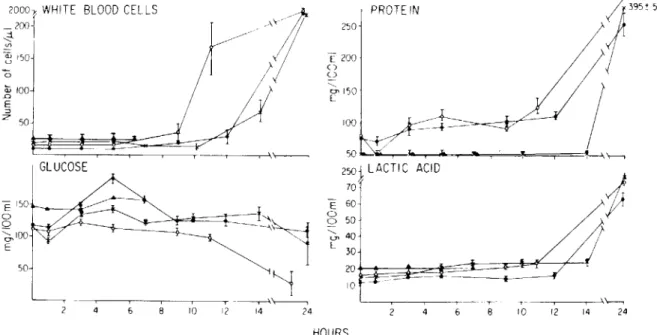
Variation in CSF chemistry in response to encap-sulated and unencapencap-sulated pneumococci. The time course and magnitude of the cellular and chem-ical changes in CSF following inoculation with two encapsulated strains (Types III and XIX), unencap-sulated (R6 ) pneumococci, and pyrogen-free saline
are shown in figure 2. These data were obtained in parallel with the growth curves of figure 1 and can be compared directly. Saline alone did not induce abnormal changes.in CSF cytochemistry. Leukocytes
6
1
PROTE IN~;~'.,;~BO
;,1
250 .> E200 0 Q '< < ,/
ry150 E 100 \\---, 50 L I h " ~\----, 2501
LACTIC ACID 70~
b
l
E 6025
50 b, 40 t E 30/
20~
-10 -\---, j - T T- I i \~ 10 /2 14 24 4 8 10 /2 14 24 HOURS2000):WHITE BLOOD CELLS
_200{
~ ~ 150 a GLUCOSE E 150r---o.---..L.li"----o Q ~IOO E 50 Q:; 100 .c: E :::J Z 50Figure 2. CSF cytochemical profile following intracisternal inoculation of three strains of S. pneumoniae into rabbits. Samples were collected in parallel with those described in figure 1. Each data point represents the mean (± SEM) of samples from four rabbits. Strain R6 (e); strain 8249 (x); strain SIll (0); and saline (A.).
Whi te Blood Cells
24hr 24hrProtein
400 <, 0 ' E E 30 o o 5hrLactic Acid
200 E 150 0 0 100 <, 0 ' E 50 5hr 24hr ~ 4000 <, en Q) 3000 u '0 ~ 2000 Q) ~ E 1000 ~z
5hrGlucose
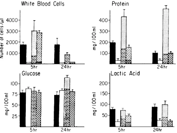
100 E 75 0 0 50 <, 0 ' E 25 5hrFigure 3. Changes in CSF cytochemical profile induced by various concentrations of killed pneumococci, purified cell wall, or capsular polysaccharide. Inocula of 2 x 10⁷heat-killed R
6per 0.2 ml (.) or 2 X 10⁷heat-killed Sm
per 0.2 ml(0)were each introduced into the cisterna magna of four rabbits. Inoculation of 0.2 ml of saline produced results identical to killed SIll (P> .5). Purified cell wall and capsular polysaccharide were each instilled into the cisterna magna of four rabbits at c.oncentrations present on 106cells (cell wall ~,capsule 0),or alternatively at 2 mg/0.2
ml (cell wall 1ITl, capsule 0). CSF was withdrawn at 5 and 24 hr postinoculation and analyzed as indicated (mean ± SEM).
appeared in CSF at different times postinoculation; the striking finding was the correlation of the onset of the influx of leukocytes with the time when the bacterial density surpassed105organisms/ml. After the appearance of leukocytes, the CSF protein and lactic acid concentration began to increase. Both the sequence and magnitude of these changes were simi-lar regardless of the presence or absence of capsule on the bacterium. Glucose concentration decreased only when organisms grew to a density>106cfu/ml. Glucose concentrations decreased to <50 mg/dl dur-ing the inflammatory response to108unencapsulated
organisms.
Difference in the CSF response inducedbykilled pneumococci. The lack of correlation between in-duction of the CSF inflammatory response and the presence of bacterial capsule suggested that bacterial components other than the capsule may be
impor-tant in provoking the host response. The fact that all strains invoked a response at a similar bacterial density in CSF (i.e.,105)indicated that the inciting component had to be present in critical amounts to provoke a response. These two observations sug-gested that killed bacteria without capsule might also induce a CSF inflammatory response if given in the appropriate dose. Indeed, heat-killed preparations of unencapsulated bacteria when administered at
~105cell equivalents per dose produced a CSF re-sponse remarkably similar to that of live bacteria (figure 3). Lower concentrations of killed organisms produced little inflammatory response in the CSF. In contrast to heat-killed unencapsulated bacte-ria, heat-killed encapsulated inocula (106 and 108 cells) evoked only a minimal inflammatory response
(P
<
.01, figure 3). This lack of activity did not ap-pear to be due to alteration of the capsule byheat-ing since heated-isolated capsular polysaccharide retained reactivity equal to that of untreated prepa-rations (data not shown).
One striking difference in the CSF biochemical profile was noted when comparing live and killed inocula. CSF glucose concentrations remained nor-mal during the response initiated by a killed inocu-lum (figure 3). On the other hand, both protein and lactic acid concentrations increased after the inocu-lation of live or killed unencapsulated bacteria. In-oculation of >108killed bacterial cells failed to in-duce hypoglycorrhachia.
Comparison oj isolated cell wall and capsular polysaccharide as inducers oj CSF inflammatory re-sponse. Inoculation of >105killed cells evoked an inflammatory response: this corresponds to t"V0.02
Jigof cell wall material. Cell equivalents of capsular polysaccharide are difficult to calculate since the ma-terial is constantly shed into the medium during bac-terial growth. However, 0.2Jig approximates the amount on 105 cells [9]. Figure 3 details the inflam-matory response produced by inoculation of 106cell equivalents of cell wall or capsular polysaccharide. Capsular polysaccharide produced little response at this concentration (mild increase in 24-hr CSF pro-tein concentration). Cell wall material, on the other hand, evoked an inflammatory response similar to that of whole cells, including pleocytosis and an in-crease in protein and lactic acid concentrations; glu-cose concentration remained normal.
The inflammatory response generated by inocula of 2 mg of these two components is depicted in fig-ure 3 (t"Vl08cell equivalents of cell wall and t"Vl09
cell equivalents of capsular polysaccharide). At these concentrations both preparations induce inflamma-tion although the magnitude of the response to cell wall is significantly greater than that to capsule. Discussion
The CSF inflammatory response to all strains of pneumococci tested was similar, consisting of leu-kocytosis, an increase in protein and lactic acid con-centrations, and hypoglycorrhachia. The fact that pneumococci with widely varying surface character-istics (differing capsular types or absence of capsule) could produce qualitatively and quantitatively simi-lar CSF responses suggests that bacterial compo-nents other than capsule, yet common to all strains, are critical to the induction of CSF inflammatory changes. Potent inflammatory activity was found to
reside in the cell wall of pneumococci. Most impor-tantly, purified wall was active in inducing CSF in-flammation at a concentration of t"Vl06 whole-cell equivalents. This number corresponds closely to the threshold at which CSF inflammation was found to begin with living bacteria (compare figures 1 and 2). In contrast, capsular polysaccharide was inactive at 2Jigper dose, an amount that represents not only t"VI06 whole-cell equivalents but is also 10-100-fold greater than the concentration of capsule detected in rabbit CSF in meningitis [9].
Our experiments do not unequivocally identify the pneumococcal cell walls as the sole agents responsi-ble for the inflammation observed in pneumococ-cal meningitis. A variety of bacterial components (intramembrane or intracellular) can induce inflam-mation in the CSF (authors' unpublished observa-tions). Nevertheless, the pneumococcal cell wall is a plausible candidate for such a role, and our ex-periments clearly show that the specific activity of cell wall material as an inducer of inflammation is high enough to be operative at the bacterial concen-trations observed in pneumococcal meningitis.
The mechanism whereby cell walls induce CSF in-flammation is an yet unclear. The inflammatory ac-tivity of the cell wall has been noted in other ex-perimental systems [10-14]. The pneumococcal cell wall is known to activate the alternative pathway of complement [15, 16] and could in this manner poten-tially generate chemotaxins and damage to the blood-brain barrier early in the course of infection.
Itshould be noted, however,that CSF contains very little complement even during the later stages of meningitis [17, 18]. Chemotactic activity in CSF has been noted to be associated with heat stable sub-stances of low molecular weight, some of which are generated during bacterial growth [19].Itis conceiv-able that cell wall components may be directly responsible for some of this chemotactic activity. Killed encapsulated strains wereinactive, a result sug-gesting the presence of capsular material may mask the inflammatory activity ofthe underlying cell wall.
Itis conceivable that encapsulated bacteria produce inflammation by exposure of the underlying cell wall or by secretion of the cell wall material during growth.
Only actively multiplying organisms at ~106
cfu/ml were found to induce hypoglycorrhachia. These results are similar to those obtained in vitro by Petersdorf et al. [20-22]. The mechanism of CSF hypoglycorrhachia in meningitis is unclear. The
dis-sociability of glucose and lactic acid changes would suggest that increases in the level of lactic acid can-not be taken as evidence of anaerobic glycolysis of decreasing CSF glucose. Our data suggest that the decrease in the level of glucose in CSF is caused by bacterial metabolic products other than the cell wall. The potential clinical relevance of the CSF re-sponse to cell wall material rests on the correlation between the clinical outcome of meningitis and the degree of inflammation [23]. Pneumococcal cell wall components are released into the surrounding medium during stationary-phase autolysis and, per-haps more importantly, during treatment of pneu-mococci with fJ-Iactam antibiotics. Thus, it is con-ceivable that bacterial death, when accompanied by lysis and release of cell wall, could aggravate CSF inflammation and affect the clinical outcome of dis-ease deleteriously. This concept may explain in part the occurrence of deaths despite bacteriologic cure of the patient.
References
1. Scheld WM, Dacey RG, Winn HR, Welsh JE, Jane JA, Sande MA. Cerebrospinal fluid outflow resistance in rab-bits with experimental meningitis. J Clin Invest 1980;66:243-53
2. Feldman WE. Relation of concentrations of bacteria and bacterial antigen in cerebrospinal fluid to prognosis in patients with bacterial meningitis. N Engl J Med 1977; 296:433-6
3. Bornstein DL, Walsh EC. Endogenous mediators of the acute-phase reaction. I. Rabbit granulocytic pyrogen and its chromatographic subfractions. J Lab Clin Med 1978;9:236-45
4. Center for Disease Control. Bacterial meningitis and menigococcemia- United States, 1978. MMWR 1979; 28:277-9
5. Mosser JL, Tomasz A. Choline-containing teichoic acid as a structural component of pneumococcal cell wall and its role in sensitivity to lysis by an autolytic enzyme. J Biol Chern 1970;245:287-98
6. Lacks S, Hotchkiss RD. A study of the genetic material de-termining an enzyme activity inPneumococcus. Biochim
Biophys Acta 1960;39:508-18
7. Dacey RG, Sande MA. Effect of probenecid on cerebrospi-nal fluid concentrations of penicillin and cephalosporin derivatives. Antimicrob Agents Chern other 1974;6: 437-41
8. Lowry OH, Rosebrough NJ, Farr AL, Randall RJ. Protein measurement with folin phenol reagent. J Biol Chern
1951;193:265-75
9. Nolan CM, Ulmer Jr. WC. Enzyme immunoassay of the capsular polysaccharide of Streptococcus pneumoniae
Type III in cerebrospinal fluid in experimental meningi-tis. J Med Microbiol 1980;13:551-60
10. Heymer B, Rietschel ETH. Biological properties of pep-tidoglycans. In: Schlesinger D, ed. Microbiology -1977. Washington, DC: American Society for Microbiology, 1977:344-9
11. Chetty C, Kreger A. Characterization of pneumococcal purpura-producing principle. Infect Immun 1980;29:158-64 12. Reed WP, Jaffee P, Albright EL, Williams RC Jr. Effect of intravenously injected killed pneumococci on leukocytes, complement, and phagocytosis in rabbits. Infect Immun 1980;29:1021-7
13. Goldblum SE, Reed WP, Barton LL. Reduction of circulat-ing granulocytes induced by type 1 pneumococcal cell walls in New Zealand white rabbits. Infect Immun 1981;33:1-6 14. Rasanen L, Arvilommi H. Cell walls, peptidoglycans, and teichoic acids of gram-positive bacteria as polyclonal in-ducers and immunomodulators of proliferative and lym-phokine responses of human Band T lymphocytes. Infect Immun 1981;34:712-7
15. Winkelstein JA, Tomasz A. Activation of the alternative com-plement pathway by pneumococcal cell wall teichoic acid. J Immunol 1978;120:174-8
16. Hummell DS, Berninger RW,Tomasz A, Winkelstein JA. The fixation of C3b to pneumococcal cell wall polymers as a result of activation of the alternative complement path-way. J Immunol 1981;127:1287-9
17. Zwahlen A, Nydegger UE, Vaudaux P, Lambert P-H, Wald-vogel FA. Complement-mediated opsonic activity in nor-mal and infected human cerebrospinal fluid: early response during bacterial meningitis. J Infect Dis 1982;145:635-46 18. Simberkoff MS, Moldover NH, Rahal 11 Jr. Absence of de-tectable bactericidal and opsonic activities in normal and infected human cerebrospinal fluids. J Lab Clin Med 1980;95:362-72
19. Nolan CM, Clark RA, Beaty HN. Experimental pneumo-coccal meningitis: III. Chemotactic activity in cerebrospinal fluid. Proc Soc Exp BioI Med 1975;150:134-6 20. Petersdorf RG, Swarner DR, Garcia M. Synergistic action
of pneumococci and leukocytes in lowering cerebrospinal fluid glucose. Proc Soc Exp Biol Med 1960;103:380-2 21. Petersdorf RG, Harter DH. The fall in cerebrospinal fluid
sugar in meningitis. Arch Neurol 1961;4:21-30 22. Petersdorf RG, Swarner DM, Garcia M. Studies on the
patho-genesis of meningitis. III. Relationship of phagocytosis to the fall in cerebrospinal fluid sugar in experimental pneu-mococcal meningitis. J Lab Clin Med 1963;61:745-54 23. McAllister CK, O'Donoghue JM, Beaty HN. Experimental
pneumococcal meningitis. II. Characterization and quan-titation of the inflammatory process. J Infect Dis 1975;132:355-60