HAL Id: tel-02613475
https://tel.archives-ouvertes.fr/tel-02613475
Submitted on 20 May 2020HAL is a multi-disciplinary open access archive for the deposit and dissemination of sci-entific research documents, whether they are pub-lished or not. The documents may come from teaching and research institutions in France or abroad, or from public or private research centers.
L’archive ouverte pluridisciplinaire HAL, est destinée au dépôt et à la diffusion de documents scientifiques de niveau recherche, publiés ou non, émanant des établissements d’enseignement et de recherche français ou étrangers, des laboratoires publics ou privés.
Désadaptations cardiovasculaires à la microgravité :
techniques avancées pour améliorer la mesure et
l’évaluation du risque cardiovaculaire induit par les vols
spatiaux pour les équipages de longue durée
Danielle Kathleen Wannamaker
To cite this version:
Danielle Kathleen Wannamaker. Désadaptations cardiovasculaires à la microgravité : techniques avancées pour améliorer la mesure et l’évaluation du risque cardiovaculaire induit par les vols spatiaux pour les équipages de longue durée. Médecine humaine et pathologie. Normandie Université, 2019. Français. �NNT : 2019NORMC433�. �tel-02613475�
THÈSE
Pour obtenir le diplôme de doctorat
Spécialité RECHERCHE CLINIQUE, INNOVATION TECHNOLOGIQUE, SANTE PUBLIQUE
Préparée au sein de l'Université de Caen Normandie
Désadaptatiοns cardiοvasculaires à la micrοgravité:techniques
avancées pοur améliοrer la mesure et l'évaluatiοn du risque
cardiοvaculaire induit par les vοls spatiaux pοur les équipages de
lοngue durée.
Présentée et soutenue par
Danielle GREAVES
Thèse soutenue publiquement le 16/12/2019
devant le jury composé de
M. ANDRÉ AUBERT Professeur des universités PraticienHosp, Katholieke UniversiteitLeuven- Belgique Rapporteur du jury M. BENOIT BOLMONT Professeur des universités, Université de Lorraine Rapporteur du jury M. PHILIPPE ARBEILLE Professeur émérite, Université de Tours François Rabelais Membre du jury
Mme CAROLE TAFFORIN Directeur scientifique, Ethospace Membre du jury
Thèse dirigée par MARTIN HITIER et STEPHANE BESNARD, Mobilités :
vieillissement, pathologie, santé - COMETE
Table des Matières
Chapitre 1 ... 6
Introduction ... 6
Impact du vol spatial sur les systemes physiologiques : ... 6
Ge e alites su l’adaptatio à la i og a ite eelle ol spatial et si ul e de u itus, i e sion, confinement) - Methodologie et appareillage Ultrasonore ... 8
Nou elle ethode d’i estigatio ult aso o e des o ga es et aisseaux superficiels et profonds en vol spatial et simulation ... 9
Thèse Hypothèses Generales ... 12
Résumé du Chapitre 2 (Paper 1): Effects of exercise countermeasure on myocardial contractility measured by 4D speckle tracking during a 21-day head-down bed rest... 13
Resumé du Chapitre 3 (Paper 2): Cardiac and arterial structure and functional changes after four days of dry immersion with and without thigh cuffs ... 13
Introduction du Chapitre 4 (Paper 3): Effect of thigh cuffs on the venous redistribution during 4 days in dry immersion ... 14
Chapitre 2 ... 16
Effets de la contre- esu e d’e e i e su la o t a tilit o a di ue, esu e pa e hog aphie D et speckle tracking pendant un decubitus à -6 degrés de 21 jours / Effects of exercise countermeasure on myocardial contractility measured by 4D speckle tracking during a 21-day head-down bed rest ... 16
ABSTRACT ... 16
INTRODUCTION ... 17
METHODS ... 20
Subjects: ... 20
General protocol: ... 20
Echocardiographic imaging protocol: ... 21
Echocardiographic measurements: ... 22
Statistical analysis: ... 23
RESULTS ... 23
Effects of bed rest on LV function and strain ... 23
Contractility at 80° Standard Measures Tilt ... 24
DISCUSSION ... 25
Limitations... 29
CONCLUSION ... 30
Chapitre 3 ... 35
Modifications cardiaque et artérielle morphologiques et fonctionnelles après quatre jours en immersion sèche avec et sans brassarts de cuisse / Cardiac and arterial structure and functional changes after four days of dry immersion with and without thigh cuffs ... 35
ABSTRACT ... 35 INTRODUCTION ... 36 METHODS ... 37 Data Analysis: ... 38 Statistical Analysis: ... 39 RESULTS ... 40
Cardiac-Structure and Function (Fig. 3.1): ... 40
Arterial Wall Properties (Fig. 3.2): ... 41
Resistivity index (RI) and arterial hemodynamic (Fig. 3.3): ... 41
Cardiac and Vascular data at D4 PM compare to D4 AM: ... 41
DISCUSSION ... 42
CONCLUSION ... 46
FIGURES ... 48
Chapitre 4 ... 57
Effet des brassarts de cuisses sur la redistribution veineuse pendant 4 jours en immersion sèche / Effect of thigh cuffs on venous redistribution during 4 days in dry immersion ... 57
ABSTRACT ... 57 INTRODUCTION ... 58 METHODS ... 59 Subjects: ... 59 Measurements: ... 60 Procedure: ... 62 Data Analysis: ... 62 RESULTS ... 63
Cardiac volume (Fig. 4.2):... 63
Jugular vein (Fig. 4.3): ... 63
Middle cerebral vein (Fig. 4.4): ... 63
Portal vein (Fig. 4.5): ... 63
FIGURES ... 68
Chapitre 5 ... 73
Synthèse & Conclusion ... 73
Synthèse immersion sèche et Decubitus a -6 degré ... 73
Limitation des methodes de simulation des effets du vol spatial ... 74
Prolongement du travail de These: Activite de recherche en cours de realisation (par Danielle Greaves) ... 75
Etude du remodeling arteriel et perturbations metaboliques associées (Participation a des programmes spatiaux) ... 75
Participation au d eloppe e t de ou elles thodes d’i estigatio pa Da ielle G ea es ... 78
Liste des Publication de D Greaves 2012 – 2019 ... 79
Soumis: ... 79
Publiés (2012-2019 Annotated List):... 79
Posters Presentés récemment ... 81
Poster #1 – Présenté en Septembre 2019 Cerebral Autoregulation Network, Leuven, Belgique ... 81
Poster #2 – Présenté en Juin 2019 International Society for Gravitational Physiology, Noordwijk, Le Pays Bas ... 82
Poster #3 – Présenté en Janvier 2019 NASA Human Research Science Symposium, Galveston, Texas ... 83
Poster #4 – Sera Présenté en Janvier 2020 NASA Human Research Science Symposium, Galveston, Texas ... 84
Chapitre 1
Introduction
Impact du vol spatial sur les systèmes physiologiques :
Nos travaux actuels portent principalement sur la désadaptation du système cardiovasculaire lors des vols spatiau . D’aut es s st es ph siologi ues tels ue les os, le ta olis e et le e eau so t tous affectés par les séjours longue durée en microgravité. Alors que les agences prévoient des missions de longue durée au-delà de l'orbite terrestre basse, la dynamique et la restauration de ces changements deviennent particulierement important. Vico et ses collègues (Vico et al., 2000) ont montré des taux de déminéralisation osseuse spectaculaires dans les os porteurs de la jambe, qui ne se ont pas restaurés immédiatement après vol et qui ont même continué à diminuer pendant des mois. Les auteurs postulent que, chez certains astronautes, la fragilité osseuse peut tout simplement ne jamais se rétablir au cours de leur vie. Deux articles récents indiquent que des maladies cardio-métaboliques peuvent se développer pendant le vol spatial et le décubitus longue durée. Le développement du diabète de type 2 dans ces mêmes conditions est une préoccupation réelle malgré les contre-mesures de type exercice effectuées (Heer et al., 2014; Hughson et al., 2016). Une mauvaise alimentation et de longues périodes d'inactivité entre les exercices sont des facteurs de causalité suggérés.
En plus des os, et du métabolisme glucidique, les structures/organes cervicaux et cérébraux des astronautes sont affectées en vol longue durée. Arbeille et al ont montré un engorgement très important des veines Jugulaires pendant toute la durée de vols spatial de 6 mois ainsi u’u e aug e tatio de la vitesse dans les veines intracérébrales qui suggère une compression de ces dernière suite aune augmentation de pression intra crânienne (Arbeille, Provost, & Zuj, 2016; Roberts et al., 2017)ont récemment démontré après un vol de 6 mois un déplacement vertical significatif et une compression générale des tissus et vaisseaux cérébraux à l'intérieur des crânes d'astronautes après des missions de six
mois. Les IRM ont également montré un rétrécissement important des sillon cérébraux après un vol spatial de lo gue du e. L’altération de la isio d’u sous-g oupe de e es d’ uipage (env 60%) a o ai u la NASA d’e a i e e ol et post ol les st u tu es i te es de l’œil, o p is l’œd e du disque optique, la taille et la morphologie du nerf optique afin de mieux appréhender l’i pa t du t a sfe t liquidien sur les structures oculaires et cérébrales. Les changements de vision sont associés à un profil génétique : tous les uipages a a t des p o l es de isio et u œd e de dis ue opti ue lo s d’u vol spatial portent le gène de la variante phénotypique du métabolisme du 1-carbone (Zwart et al., 2017).
En vol et en décubitus de longue durée on observe une atrophie myocardique, une diminution des volumes cardiaques et l'action de pompage est moins efficace (Arbeille et al., 1987; M. Bungo & Charles, 1985; Perhonen, Franco, et al., 2001) ce qui nécessite une pratique suffisante de contre-mesures comme l’e e i e a o i ue.
Les artères deviennent plus épaisses et plus rigides (Arbeille, Provost, et al., 2016; Hughson et al., 2016) avec des impacts potentiels sur la cognition (Iulita, Noriega de la Colina, & Girouard, 2018; Pase et al., 2016). L’ paisseu de la pa oi a t ielle est gale e t aug e t e e o fi e e t de lo gue du e MARS jou s, CELSS 8 jou s , e ui i di ue ue l’ paississe e t de la pa oi a t ielle ’est pas li uniquement a la microgravité mais pourrait peut-être être favorisé par le stress environnemental present en vol spatial confinement, via un processus inflammatoire. (Arbeille, Provost, Vincent, & Aubert, 2014) (Yuan et al., 2019).
Comme mentionné ci-dessus, les agences spatiales ont réalisé depuis 1984 que les membres d'équipage doi e t s’i pose d’avoir une activité physique en orbite pour atténuer le déconditionnement cardiaque (Arbeille et al., 1987). Actuellement, tous les astronautes font de l'exercice lorsqu'ils se trouvent sur la Station spatiale internationale, mais la quantité, la durée et le type d'exercice varient selon les équipages et les conseils de leur Médecins au sol. Le matériel utilisé comprend : le tapis roulant de course à pied T
avec des bandes élastiques de tension fixée aux membres, un ergomètre et un dispositif d'exercice résistif ARED. Pour les expériences de simulation, les procédures sont différentes. D'une part, il n'y a pas de contre-mesure « exercice » dans les études d'immersion sèche d'autre part, le HDTBR offre de nombreuses possibilités en matière de contre-mesures, dont les exercices de résistance en position allongée (D. K. Greaves, Arbeille, & Hughson, 2007; Rittweger et al., 2006), le tapis roulant de course a pied en position allongé (D. K. Greaves et al., 2007), une plateforme de vibrations plantaire résistive (D. Greaves et al., 2019), une plateforme de sauts « squats test » (Linnarsson et al., 2015) et une centrifugeuse a bras court (2m) (Clement et al., 2016).
Ge eralites sur l’adaptatio à la icrogravite reelle vol spatial et si ulée (decubitus,
immersion, confinement) - Methodologie et appareillage Ultrasonore
L'i e sio s he DI i e sio da s u ai sa s o ta t du sujet a e l’eau et le décubitus anti orthostatique (Tete plus basse que les pieds à moins 6 degrés ou HDBR) sont couramment utilisés dans la recherche spatiale comme modèle pour simuler les effets de la microgravité (Linossier et al., 2017; Watenpaugh, 2016).
L’o je tif de ette th se est de présenter tudes o sa es à l’ tude de l’adaptatio a dia ue, artérielle, et veineuse, réalisées à l’aide des méthodes mentionnées ci-dessus : le Décubitus (HDBR) et Immersion sèche. Les résultats de ces études sont présentés dans 3 chapitres suivants.
Le premier chapitre s'intitule « Effets de l'exercice physique sur la contractilité myocardique évaluée par échographie D et spe kle t a ki g lo s d’u HDBR de jou s ». Da s ette tude, ous p se to s la méthode Echo 4D speckle tracking qui permet de quantifier la contractilité myocardique sans avoir e ou s à l’IRM, et qui a déjà été utilisée sur des patients cardiologiques mais pas encore en physiologie spatiale. Cet article a été publié dans le European Journal of Applied Physiology (D. Greaves, Arbeille, Guillon, Zuj, & Caiani, 2019).
Le deuxième chapitre s'intitule « Structure et modifications cardiaques et artérielles après quatre jours en immersion sèche avec et sans brassards de cuisse ». Dans cette étude, nous présentons les résultats des investigations cardiaques et artérielle réalisées sur des sujets placés en immersion pendant 4 jours, la oiti d’e t e eu po ta t des brassards de uisse le jou da s le ut de dui e l’i po ta e du t a sfe t des fluides i duit pa l’i e sio du bas vers le haut du corps. E d ut d’u vol spatial les brassards de cuisse permettent de réduire l’e o e e t de la sphère ORL, les maux de tête et l’dème facial. Un article a été soumis à Acta Astronautica (16 octobre 2019).
Enfin, le troisième chapitre s'intitule « Effet des brassards de cuisse sur la redistribution veineuse pendant 4 jours en immersion sèche ». L’o je tif tait de recréer un transfert liquidien important vers les régions thoraco céphaliques, d’e évaluer les conséquences au niveau cérébral et de teste l’effi a it des brassard de cuisse comme réducteur de ces effets. Un article a été soumis pour publication à Aerospace Medicine & Human Performance (20 Octobre 2019).
Nouvelle méthode
d’i vestigatio ultrasonore des organes et vaisseaux superficiels et
profonds en vol spatial et simulation
Actuellement, la plupart des programmes de recherche en physiologie spatiale font appel à l’échographie. E effet l’E hog aphe est le seul appa eil d’i age ie médicale qui peut être installé à bord de la station i te atio ale puis u’il fo tio e su u e alimentation électrique de 28 volts contrairement aux appareils à rayons « X » (Scanner) et à l’IRM.
Actuellement l'astronaute doit t e e t ai a o figu e l’appa eil e fo tio de l’o ga e, pou localiser la sonde d’ hog aphie en regard de l’o ga e (c) pour orienter la sonde afi d’o te i une vue pa faite de l’o ga e e vue axiale ou transverse, (d) pour optimiser l’i age e ajusta t les réglages de l'échographe (gain, profondeur), (e) pour activer les diverses fonctions de l’ hog aphe comme le Doppler, Temps mouvement, 3D. (f) pour enregistrer les images et vidéo. Pour toutes ces taches l’ast o aute est guidé à la voix par un expert depuis la Terre et, parce que la tâche est compliquée et
sa formation pré-vol limitée (peu d’heu es d’e t ai e e t p ol) il faut souvent plusieurs minutes pour obtenir la vue échographique attendue. Le p i ipal p o l e est u’il est t s diffi ile de guide à distance un sujet non-échographiste (astronaute) pour lui faire réaliser une rotation, ou inclinaison de la sonde de quelques degrés (2-3 degrés).
Pour cette raison nous avons conçu un nouveau dispositif grâce auquel la majeure partie des manouvres (a, c, d, e, f) peut être accomplie directement par l’E pe t à distance en utilisant la télé-op atio do a e u e pa ti ipatio li it e de l’ast o aute de l’ISS Ce nouveau système 1) réduit considérablement le temps de fo atio pou l’ast o aute e p ol pa e oi s de a œu es à apprendre, 2) permet à l’e pe t de d le he les fo tio s hog aphi ues et les aptu es de do es a des i sta ts plus oppo tu s et do d’op e u e eilleu e s le tio des do es collectées, 3) ’e ige pas la p se e d’u ième ope ateu assista t pou g e les fo tio s de l’E hog aphe o e
’est le as a tuelle e t.
L’E hog aphe spatial a t d elopp à pa ti d’u od le o e ial O h olite So os a e Pa is , auquel un ensemble de fonctions a été ajouté (3D, RF signal, elastographie) et modifié pour que toutes ses réglages (gain, profondeur), fonctions (Doppler pulse ou couleur, 3D, RF) puissent être télé-opérés à distance en utilisant des soft spécialement développés par Teamviewer (Goppingen, Germany) et Optimalog (St Cyr/Loire, France).
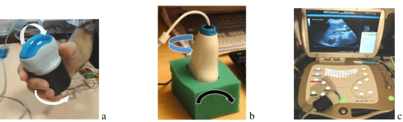
Au site E pe t l’ hog aphiste ajuste les glages de l’i age hog aphi ue u’il eçoit du site dista t (ISS) et active les fonctions (Doppler pulse, couleur 3D) à partir du clavier de son PC. Chaque réglage est identifié par une touche du clavier, <F6> et <F7> augmente et diminue le gain ultrason, <F4> active la fo tio Dopple Puls O a ti e l’E hog aphie D. Le desig , l’e o e e t 8 ou le poids kg de l’appa eil ’o t pas t odifi s.
Quatre sondes bi-motorisées ont été développées par Vermon (Tours, France) pour cet Echographe. Elles so t pou ues de t a sdu teu s oto is s do t l’e pe t peut odifie l’o ie tatio à distance à l’aide
d’u e so de fi ti e do t les mouvements sont transmis à la sonde réelle distante. Un premier moteur pe et d’i li e le t a sdu teu de + °à -55°, un 2ième moteur sert à faire tourner le transducteur sur
lui-même de -90° à +90°.
Dès ue la o u i atio est ta lie l’Ast o aute positio e la so de à l’e d oit i di u o ale e t pa l’E pe t au sol D D Greaves - P P A eille . L’E pe t utilise des f e es a ato i ues si ples pou guide l’ast o aute Lig e a elo ai e, ste u , la i ule , puis, d s ue l’o ga e/ aisseau appa ait l’E pe t de a de à l’ast o aute de ga de la so de i o ile pe da t que depuis le sol il oriente le t a sdu teu e a ipula t la so de fi ti e de la ai gau he jus u’à ie oi l’o ga e, opti ise l’i age, puis a ti e des fo tio s o e le Dopple , le 3D, le RF, en agissant sur les touches du clavier de sa main droite.
a
b
c
Fig. 1.1. (a) Sonde motorisée (400cm3 and 430g). Le transducteur (3,5MHz curved array) peut etre incliné et tour é fle hes a dista e par l’expert e a ipula t la so de fi tive fle hes L’E hographe télé opérable (6kg) avec la sonde egalement tele-operable.
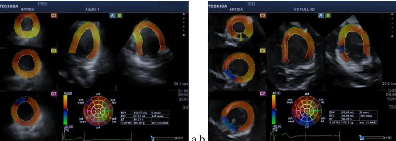
Figure 1.2. Poste Expert au CNES Toulouse e liaiso dire te ave l’ISS. Clavier et so de de o a de
Figure 1.3: Veine cave inferieure et carotide commune avec détection de l’I ti a edia liseré rouge).
Thèse Hypothèses Générales
L'objectif général de ce document est d'étudier la manière dont le système cardiovasculaire s'adapte aux environnements de simulation d’u ol spatiale. Nous avons étudié deux 2 modalités HDTBR (Alitement anti orthostatique) et DI (Dry immersion) pour la simulation des effets des vols spatiaux.
Les hypothèses spécifiques étaient les suivantes :
1. Le suivi des speckle (points singuliers du myocarde) en échographie 4D qui permet une évaluation quantitative de la contractilité myocardique HDTBR de 5 jours ;
2. L’effet des brassards de uisse po t s lo s d’u e DI de jou s pou s’assu e u’ils atténuent le déconditionnement artériel attendu ;
3. L’effet des brassards de cuisse portés au cours d'une DI de 4 jours pour vérifier u’ ils atténuent le transfert liquidien vers les régions céphaliques et cérébrales.
Résumé du Chapitre 2 (Paper 1): Effects of exercise countermeasure on myocardial
contractility measured by 4D speckle tracking during a 21-day head-down bed rest
L'o je tif tait de ua tifie les odifi atio s de la o t a tilit a dia ue au ou s d’u ed est de jours (HDBR) par la méthode speckle tracking 4D et de déterminer si la contre-mesure « exercice » était capable de préserver cette contractilité cardiaque. Les méthodes traditionnelles 2D ne mesurent qu'une variation de la taille du ventricule de la systole à la diastole et ne suivent pas la cinétique des mouvements de contractilité à l’intérieur de la pa oi. De plus l’échographie D e pe et d’accéder u’a la contractilité longitudinale globale par la mesure de la distance Apex-Valve mitrale. Le suivi des mouvements des speckles (points singuliers à l’intérieur du myocarde) en 4D montre que la contractilité radiale diminuée significativement pendant le HDTBR chez les sujets témoins alo s u’elle est conservée dans le groupe exercice. Par contre la contractilité longitudi ale ’est pas affe t e dans aucun des 2 groupes. Bien sûr, l'IRM pourrait fournir des données similaires, mais il n’est pas possible à e jou d’i agi e a oi u e IRM a bord de l’ISS dans un futur proche. A la suite de ces résultats nous avons mis au point un algorithme de traitement des vidéos a dia ue ui pe et d’accéder à la contractilité radiale et longitudinale en vol (prog Vasc-Aging en cours)
Résumé du Chapitre 3 (Paper 2): Cardiac and arterial structure and functional changes after
four days of dry immersion with and without thigh cuffs
Dans le deuxième chapitre, nous présentons les résultats de mesures cardiaques, et artérielles réalisée a jou s d’i e sio sèche. Au matin du 4ieme jour on observe une diminution significative du volume du ventricule gauche, du volume éjection et de la fraction d'éjection qui montrent que quatre jours en immersion est suffisant pour induire une réduction du volume plasmatique et donc des volumes cardiaques. Ceci est en accord avec les résultats obtenus en immersion sèche en HDBR et vol spatial historique pour DI (Navasiolava et al., 2011; Watenpaugh, 2016) ai si u’a e les esu es di e tes du
volume plasmatique effectuées au cours de cette étude. La masse cardiaque montre une tendance à diminuer. Pa o t e au ati du ie e jou s d’i e sio les flu artériels chez les sujets portant les brassards de cuisse le jour, après la nuit sans les brassards sont redistribué comme chez les sujets contrôle. De plus le port des brassards de cuisse pendant 8h ne modifie pas de manière significative les paramètres cardiaques ou artériels. Il apparait donc que le port de brassard de uisse toute la jou e ’a pas entraîné d’« effet oi e» i d’« effet chronique » des brassards. Si plus de pa ti ipa ts a aie t eu le œu spo tif (comme les astronautes), nous aurions certainement observé une perte de masse myocardique plus importante. On observe aussi des signes de remaniement as ulai e, a e u e aug e tatio de l’IMT f o ales I ti a edia thi k ess et d’aug e tatio de la igidit artérielle au niveau Carotidien. Les débite artériels périphériques (Carotide Fémorale so t est sta les pe da t la du e de l’i mersion sèche. La durée du séjour en immersion sèche était d'une durée probablement insuffisante pour induire des modifications cellulaires majeures des parois périphériques comme celles observées en HDBR et vol spatial. Le port de brassards de uisse ’a eu au u e i ide e su l’adaptatio a dia ue i artérielle a l’i e sio .
Introduction du Chapter 4 (Paper 3): Effect of thigh cuffs on the venous redistribution
during 4 days in dry immersion
L’o je tif de l’expérimentation était de montrer que quatre jours en immersion sèche suffisaient pour réaliser un transfert liquidien important vers les régions céphaliques comparable à celui observé en vol spatial. Notre protocole consistait donc à mesurer les volumes veineux au niveau cervical cérébral et porte au début de la période en immersion (à h puis a jou s d’i e sio . Co pte te u des problèmes engendrés par ce transfert liquidien en vol nous avions proposé de tester la capacité des brassards de cuisse à réduire l’a plitude du t a sfe t veineux. Les résultats o t e t ue l’i e sio p o o ue u transfert de sang veineux au niveau cervical maximal à 2H et que les Brassards de cuisse réduisent
significativement ce transfert dans cette phase précoce. Par contre a 4 jours d’i e sio l’a plitude du transfert sanguin est considérablement diminuée (bien que toujours présente) et les brassards de cuisse ’o t pas d’effet isi le à ce moment-là. En fait le volume plasmatique décroit significativement à la fin du premier jour (env. 20%) des lors la masse de sang déplacées vers la tête pa l’i e sio est insuffisante pour générer une stase importante au niveau cervical comme à 2h d’i e sio . Pou ette même raison la vitesse dans les veines cérébrales ’est pas augmentée à jou s d’i e sio o t ai e e t à ce qu’on avait observé à 2h d’i e sio lo s d’u e précédente étude. Donc le modèle immersion sèche est un modèle pour étudier les transferts liquidiens en microgravité mais seulement en début de phase d’i e sio . Sur cette période de temps, les Brassards de cuisse ont bien un effet protecteur pour les organes de la zone céphalique.
Chapitre 2
Effets de la contre-mesure
d’exercice sur la contractilité myocardique,
mesurée par échographie 4D et speckle tracking pendant un décubitus à
-6 degrés de 21 jours / Effects of exercise countermeasure on myocardial
contractility measured by 4D speckle tracking during a 21-day head-down
bed rest
PMID: 31531733
ABSTRACT
Objective: to evaluate functional myocardial contractility after 21 days of head-down bed rest (HDTBR) in sedentary control (CON) or with a resistive vibration exercise (RVE) countermeasure (CM) applied, by using 4D echocardiographic (4D Echo) imaging and speckle tracking strain quantification.
Methods: Twelve volunteers were enrolled in a crossover HDTBR design, and 4D Echo was performed in supine position (REST) at BDC-2 and at R+2, and in -6° HDTBR (on day 18), and also during the first and the last minute of the 80° head-up step of Standard Measures tilt test, performed at both BDC-2 and R+2. Radial (Rad-Str), longitudinal (Lg-Str) and twist (Tw-Str) strain were measured by 4D speckle tracking, as well as left ventricle diastolic volume (LVDV) and mass (LVmass).
Results: On day 18: in the CON group, LVDV and LVmass were reduced (p<0.05), the Rad-Str decreased (p<0.05) and Tw-Str showed a tendency to increase (p< 0.11), with no changes in Lg-Str. In RVE group, LVDV and LV mass, as well as all the strain parameters remained unchanged.
On R+2: in the CON group, LVDV and LVmass were not recovered in all subjects compared to pre-HDTBR (p<0.08), Rad-Str was still decreased (p<0.05), while Tw-Str tended to increase (p<0.09). These parameters remained unchanged in the RVE group.
Conclusion: 4D Echo and speckle tracking analysis showed that in the CON group, Rad-Str decreased concomitant with LVmass and LVDV with HDTBR, but this observation did not support the hypothesis that this HDTBR induced remodelling or a muscle atrophy. RVE acted to preserve both LVmass, LVDV and contractility during HDTBR, thus proving its effectiveness to this aim. Nevertheless, the significant HDTBR-induced changes observed in the CON group had only a limited effect on the cardiac contractile response as observed during post HDTBR tilt test. The level of contractility at 80° Tilt position was not affected neither by HDTBR nor by RVE CM.
Key words: Echocardiography, head-down tilt bedrest, resistive exercise, cardiac contractility, cardiac strain, speckle tracking, ultrasound.
INTRODUCTION
Head-down tilt bed rest (HDTBR) represents an experimental model commonly used to simulate the effect of microgravity on Earth. This model is considered as an effective microgravity simulation for most physiological effects of spaceflight such as muscular, metabolic and cardiovascular changes (Demontis et al., 2017). For the cardiovascular aspect, it leads to fluid shift from the legs to the chest, causing an increase in left ventricular transmural pressure, end diastolic volume (LVDV), and stroke volume (Fortney, Schneider, & Greenleaf, 1996) that activate short-term volume regulatory mechanisms resulting in plasma volume loss, with the achievement of a new hemodynamic steady state within 48 h characterized by decreased volume loading of the heart similar to what reported during space-flight (Arbeille et al., 2001; Levine, Zuckerman, & Pawelczyk, 1997).
Echocardiography is an imaging technique that has been performed since the beginning of manned spaceflight, thus allowing to observe a reduction in left ventricular (LVDV) volume accompanied by a reduction in plasma volume due to space flight (Martin, South, Garcia, & Arbeille, 2003). This cardiac adaptation persisted throughout both short-duration and long-duration spaceflight missions, and HDTBR
studies, due to insufficient or inadequate effectiveness of applied exercise countermeasures performed at that time. (Arbeille et al., 1987; M. Bungo & Charles, 1985; M. W. Bungo, Charles, & Johnson, 1985; Herault et al., 2000) (Dorfman et al., 2007).
In order to compensate or minimize the effects of cardiac deconditioning, additional countermeasures (CM) have since been developed and studied, with the specific aim to maintain or restore cardiac volume and mass during both real and simulated microgravity exposure. These CM have included pharmaceuticals, short-arm centrifugation (ground only), lower-body negative pressure, nutritional supplementations and, as in focus of the current study, aerobic and resistive exercise. The majority of these CM have been shown to be at least partially effective (Ferrando, Tipton, Bamman, & Wolfe, 1997) (P. Arbeille, P. Kerbeci, L. Mattar, J. K. Shoemaker, & R. L. Hughson, 2008b) (Caiani, Massabuau, Weinert, Vaida, & Lang, 2014; Hargens & Richardson, 2009; Ploutz-Snyder et al., 2014).
In the field of muscle-skeletal deconditioning, skeletal muscle atrophy has been associated with a reduced protein synthesis rate and redistribution of muscle fiber types: both issues can be mitigated by an approach that combines amino acid supplementation and resistive exercise. However, in contrast to what is known about skeletal muscle, little is known regarding potential changes to myocardial tissue structure or changes to myocyte metabolism (Bennet, Connacher, Scrimgeour, Smith, & Rennie, 1989; Kumar et al., 2009).
Methodological limitations in directly detecting changes in cardiac contractility in a non-invasive way have produced a yearly paradox in the literature: the cardiac mass is known to decrease due to spaceflight, but with no concomitant changes observed in contractility (Negishi et al., 2017), Levine et al unpublished data from Integrated Cardiovascular Study, a 6-month ISS flight experiment). Indeed, measurements of contractility (strain) were indirectly performed using 2D Tissue Doppler Imaging (DTI), that measures the contractile performance only along parallel directions to the ultrasound beam, thus allowing
measurement of longitudinal strain only, but the sensitivity of this parameter was insufficient to detect significant changes associated to space flight. As with any one-dimensional Doppler technique, DTI is angle-dependent, and regional velocity estimates can be influenced by overall heart motion, cardiac rotation, and contraction in adjacent segments. In addition, it is sensitive only along parallel directions to the ultrasound beam, thus allowing measurement of longitudinal strain only.
As the cardiac motion is intrinsically 3D, other imaging techniques are needed to fully investigate strain along other directions (i.e., radially or due to twist), to test for possible changes induced by microgravity.
Four-dimensional echocardiographic (4D Echo) imaging is a relatively new technique that allows acquisition of a 3D volume of the whole heart over an entire cardiac cycle, with time representing the fourth dimension. The cardiac structures can be visualized in 3D in real-time or can be visualized as any 2D cut-plane selected within the 3D volume. By 4D speckle tracking processing, it is possible to achieve improved assessments of contractility along various axes (Perez de Isla et al., 2011; Perez de Isla et al., 2010) (Arbeille et al., 2012; Seo, Ishizu, Enomoto, Sugimori, & Aonuma, 2011). This overcomes the limitations of the traditional DTI method, thus giving 4D Echo datasets and speckle tracking a clear advantage.
Our hypothesis was that changes in contractility do indeed accompany the previously documented changes in left ventricular mass occurring with spaceflight and HDTBR (in non-countermeasure groups). Using an improved echocardiographic imaging technique (4D speckle tracking), previous limitations of 2D imaging could be overcome, thus allowing demonstrating for the first time the expected changes in strain outcome associated with the observed decrease in cardiac mass.
Accordingly, the aim of this study was to assess the effects of mid-duration (21 days) HDTBR on the left ventricular volume, mass, and myocardial contractility along three components (i.e., radial, longitudinal,
and ventricular twist) 4D Echo and speckle tracking, and evaluate the effectiveness of resistive exercise in acting as a countermeasure on these outcomes.
METHODS
Subjects:
Twelve healthy male volunteers participated in this study. All were non-smokers with no pre-existing medical conditions, were taking no medications and had all parameters, for hematology, blood chemistry and standard clinical exams, in the respective range of normality defined by clinical practice and relevant guidelines. All participants gave informed, written consent to participate in the study. This experiment was conducted in accordance with the principles laid down by the 18th World Medical Assembly (Helsinki, 1964) and approved by the local institutional ethics committee (CPP Sud-Ouest Outre-Mer I) as well as by the Government of France Health Authority. The study was performed at the Institute for Space Medicine and Physiology (MEDES-IMPS) in Toulouse, France.
General protocol:
Subjects were enrolled in a crossover design with a washout period of 4 months between two consecutive campaigns, with one sedentary control (CON) group and two countermeasure (CM) interventions, either resistive vibration exercise group (RVE), or RVE plus nutrition supplementation (NEX). The order of inclusion in CON, RVE or NEX group was randomly assigned to each subject at the beginning of the bed rest, with 4 subjects assigned to each group in each campaign. All subjects adhered to a strict 6° head-down tilt bed rest for 21 days (HDTBR) for each campaign. Subjects were acclimated to the bed rest unit for 7 days before initiating uninterrupted bed rest, monitored 24 h a day, and provided with strictly controlled diet aimed at preventing body weight changes. Daily cycle started each day at 7:00 AM and ended at 11:00 PM, with no napping allowed during the day. After completing the 21-days of bed rest, subjects remained in the facility for an additional 7 days for further testing. In the ambulatory periods preceding and following HDTBR, lying in bed during the day was not allowed. In this way, each campaign
All exercise training was performed on an integrated training device supplied by Novotec Medical (Pforzheim, Germany). The vibration platform (Galileo® Fitness, Novotec, Germany) was combined with a system designed to allow exercise training in -6° head down position. RVE sessions were spaced away from meals performed twice per week with three to four-day intervals, starting at HDTBR day two.
In the NEX group, this training was complemented with daily high protein intake (1.2 g/kg body weight /d plus 0.6 g/kg body weight/d whey protein) with alkaline salts (90 mmol potassium bicarbonate /d)
At BDC-2 and R+2, participant underwent progressive head-up tilt test consisting of 3 steps (20°, 45°, 80°) of 3 min duration each (Fig. 2.5a).
During the bed rest campaigns, four subjects in the NEX group withdraw for personal reasons from the study. In addition, for technical problems, we were not able to acquire good quality data in both pre-HDTBR, pre-HDTBR, or post-HDTBR in other three subjects of the same group, thus precluding paired analysis between a NEX gr of 5 subjects and the other group of 11 and 12 subjects respectively. For this reason, we are not reporting results relevant to the NEX countermeasure, but only results relevant to the CON and RVE groups.
Echocardiographic imaging protocol:
Echocardiographic imaging using an ARTIDA (Toshiba Medical, Amsterdam NL) equipment was performed in supine position (REST) at BDC-2 and at R+2, and in -6° HDTBR on day 18. In addition, image acquisition was also performed during the first and the last minute of the 80° head-up step of the Standard Measures Tilt test, at both BDC-2 and R+2 (Fig. 2.5). For each session, three 4D volume scans (3D over time) of the left ventricle, each representing a cardiac cycle, were acquired. In particular, the first 4D capture was stored as raw data to be processed off-line by 4D speckle tracking analysis. The same device settings (i.e. gain, depth, resolution) were used on a given volunteer across acquisition sessions.
Echocardiographic measurements:
Left ventricular diastolic volume (LVDV) and mass (LVmass) were also calculated both by 3D (by automatic contouring) and 2D method (Simpson’s bi-plane).
Cardiac contractility was quantified by measuring radial strain (Rad-Str), longitudinal strain (Lg-Str), and twist strain (Tw-Str), using speckle tracking processing of the 4D scan of the left ventricle. The 4D speckle tracking echocardiographic mode allows simultaneous visualization of left ventricular myocardial strain changes in real-time, on both apical 4- and 2-chambers views, as well as on 3 transverse views of the left ventricle at apical, middle, and sub mitral level. The software uses conventional gray-scale B-mode recordings to automatically detect the internal and external contours of the left ventricle and tracks myocardial speckles, which serve as natural acoustic markers. In case of poor echogenicity resulting in a sub-optimal view, background noise may interfere with the automated detection of the endocardium. In this case, the left ventricle internal contour was manually corrected. At each point of the myocardium on these 5 cut-planes, the computed strain value is displayed using a parametric color scale (from yellow the highest, to blue the lowest), and all this information is projected on a polar view of the left ventricle divided in 17 segments (Fig. 2.1), according to the AHA model (Cerqueira et al., 2002). The percent displacement of each speckle in 3 directions (longitudinal, radial and twist strain) was used to quantify the strain value for each of the 17 segments. This percent value represents the contractility of each segment in each direction (Perez de Isla et al., 2010), the 4D strain being the evolution of these (3D) strain values over time.
To evaluate changes in myocardium contractility between pre HDTBR and day 18, and between the CON and RVE groups, we defined total strain as the sum of the % values in all segments, and mean strain as their average, with arbitrary units, to differentiate from the original strain values. The total strain represents an index of the whole contractility of the heart, while the mean strain gives a qualitative
evaluation of such contractility. Accordingly, mean and total radial strain (Rad-Str), longitudinal strain (Lg-Str), and twist (Tw-Str) strain, were computed and presented in the results. (Figs. 2.1, 2.3 and 2.4)
Statistical analysis:
The effects of HDTBR and the exercise countermeasure on cardiac contractility were assessed in a supine and a head-up tilt 80° position independently. Two-way repeated measures ANOVAs were used to test for main effects of HDTBR and the exercise countermeasure on cardiac contractility in both body positions. (SigmaPlot 12.5, Systat Software Inc, Chicago IL, USA).
RESULTS
All subjects, while in CON and RVE groups, successfully completed the HDTBR. Image quality of the acquired images allowed obtaining reliable measurements for comparison among the different time point in 11/12 subjects in the CON group and 12/12 subjects in the RVE group. During the Tilt there was not enough time during the 3 min at 80° to perform 4D analysis of Rad-St, Lg-Str and Tw-Str. The Tw-Str parameter could not be collected in good condition in all subjects.
Effects of bed rest on LV function and strain
In Fig. 2.1, an example of speckle tracking analysis performed off-line on the acquired 4D volumetric echo in one subject before HDTBR in supine position and at HDT18 at -6° head-down is reported for radial strain. Note the reduction in yellow areas (higher strain values) with bed rest, confirmed by a reduction in total strain for this subject at HDT18.
In Fig. 2.2, the cumulative results relevant to LVDV and LVmass in both CON and RVE groups are presented. In the CON group, compared to BDC-2, at HDT18, LVDV was significantly reduced by 15% (98 to 83 ml) and LV mass by 17% (192 to 160 gr) (p<0.05). On the contrary, no changes were visible in the RVE group. After bed rest, at R+2, in the CON group, LVDV showed incomplete recovery to baseline levels in all subjects (86ml, -13% from pre HDTBR, p<0.09) as well as LVmass (174mg, -9%, p<0.08).
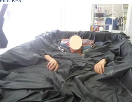
In Fig., 2.3, the cumulative results relevant to Total Rad-Str, Total Lg-Sr and Total Tw-Str are shown. In the CON group, Total Rad-Str at HDT18 was found decreased by 26% (from 431 to 321 a.u, p<0.05), and it was not completely restored to pre-HDTBR values at R+2 (350 a.u, -18%, p=0.05).
While Total Lg-Str did not change for both groups, neither at HDT18 nor at POST, for the CON group an apparent increase in Tw-Str at both HDT18 (from 4 to 13 a.u, p<0.10) and at R+2 (4 to 42 a.u., p<0.11) was noticed, while no changes were visible for the RVE group.
As from the previous results, the main effect of bed rest was noticeable on Total radial strain, we conducted a segmental analysis for this strain component in order to evaluate changes at segmental level. The results of this analysis are reported in Fig. 2.4, separately for the CON and the RVE group. In both groups, a lower strain associated with apical segments was visible already before HDTBR. At HDT18, a decrease in Radial strain in both basal and mid ventricular segments was visible in the CON group, while no apparent segmental changes were present in the RVE group.
Contractility at 80° Standard Measures Tilt
Fig. 2.5 summarizes for both CON and RVE groups the results in LVDV, total Rad-Str and total Lg-Str obtained in the first minute of the 80° head-up step of the Tilt, and at conclusion, both at pre-HDTBR and at R+2. The LVDV decreased in similar proportion during both pre and post HDTBR tilt tests in both groups (in CON, pre-HDTBR tilt pre: from 100 to 70ml; post-HDTBR tilt: from 86 to 62ml. In RVE, pre-HDTBR tilt: from 90 to 57ml; post-HDTBR tilt: from 91 to 59ml), thus reaching similar values at pre and post HDTBR tilt despite LVDV measured post HDTBR before tilt test started was lower in CON group than in RVE.
In 10 of the 11 CON subjects, the total Rad-Str decreased significantly (p<0.05) during pre HDTBR tilt (from 435 to 299 a.u.) while this was not significant at R+2 (from 346 to 296 a.u.), besides reaching a similar end-tilt value at 80° position pre and post HDTBR. It is worth noticing that at post-HDTBR the Total Rad-Str supine (right before being tilted) was reduced compared to the pre-HDTBR.
In the RVE group, the Total Rad-Str did not decrease during pre HDTBR-tilt (from 389 to 367 a.u.), while it decreased at post-HDTBR tilt (410 to 257 a.u.), however it can be noted that the Rad Str supine (just before being tilted) post-HDTBR was higher than at pre-HDTBR.
For Lg-Str, no changes were observable during Tilt, both pre- and post-HDTBR, in both groups. Thus, the lowest level of Rad strain (at 80° post HDT) was similar for CON and RVE.
DISCUSSION
The present HDTBR study showed that in the control group such position maintained during 21 days induces a decrease in volemia, as exhibited by the significant decrease in left ventricle diastolic volume, in agreement with other reports of several bedrest and inflight studies. Additionally, the left ventricular myocardial mass significantly decreased, consistent with bedrest and spaceflight. (Arbeille et al., 2000; Arbeille et al., 1997; P. Arbeille, P. Kerbeci, L. Mattar, J. K. Shoemaker, & R. Hughson, 2008a; Caiani et al., 2014; Dorfman et al., 2007; Perhonen, Franco, et al., 2001; Perhonen, Zuckerman, & Levine, 2001).
The evaluation of the myocardial contractility along 3 orthogonal components by 4D speckle tracking analysis showed that only the radial contractility was affected by a significant reduction with HDTBR, and this was concomitant with the observed drop in left ventricle volume and mass. Nevertheless, it is difficult to state if such changes in radial strain were due to a real remodelling of the myocardium cellular structure or simply as the consequence of the myocardium atrophy and/or dehydration. Three days after the conclusion of HDTBR (at R+2), the left ventricle mass, end-diastolic volume, and Total Rad-St did not completely recover, in agreement with the results obtained in a 60-days HDTBR (Westby, Martin, Lee, Stenger, & Platts, 2016), thus suggesting that cardiac muscle atrophy could persist into recovery, as a result of real tissue transformation.
On the contrary, in a previous HDTBR study of 5 days duration, the quick decrease in cardiac mass during HDT and the quick recovery within 2 days after the HDTBR supported the hypothesis of a dehydration process related to the fluid shift effect induced by the HDTBR (Caiani et al., 2014).
Most of the longitudinal strain evaluation during HDTBR or spaceflight were performed using the Doppler Tissue Imaging (DTI) modality, reporting a decrease or no change in this parameter. In a 5-days HDTBR, (Caiani et al., 2014) found DTI velocities to reduce significantly after 5 days, and recovering within 3 days after the conclusion, again supporting the hypothesis that the cardiac longitudinal contractility change was related to dehydration induced by fluid shift and the subsequent cardiovascular adaptation to the new homeostatic condition. Conversely, a 6-months spaceflight study (Levine et al, unpublished results from Integrated Cardiovascular) performed on board the International Space Station and a two-week HDTBR study did not found any significant change in longitudinal contractility (Negishi et al., 2017), despite the fluid shift was present both during the flight and with HDTBR, as confirmed by the significant dilation observed by vascular ultrasound in the jugular vein (Arbeille, Zuj, et al., 2016). Lastly, another 70-days HDTBR study recently showed a decrease in Lg Str (also evaluated by DTI) (Scott et al., 2018).
However, the DTI method used in these studies to estimate the longitudinal strain is based on the processing of tissue velocities on 2D views, as opposed to the speckle tracking mode used in this study, which measures movement of speckles over a 3D space: this difference in processing raw data could explain the weaker changes in Lg-Str and Tw-Str that we found in our study.
Two days after the end of the present 21-days HDTBR, the radial strain was not completely recovered, thus not supporting the dehydration hypothesis. On the contrary, the fact that the longitudinal and twist contractility were not affected by HDTBR does not support the hypothesis of true ventricular remodelling, as it should have affected strain in all directions.
As a result, we suggest that the observed changes along different directional strains might be related to different processes during the early period of adaptation and later on. In order to clarify these questions, additional strain assessments are required with longer duration, appropriately powered bed rest studies.
The Rad-Str changes after bed rest in the control group had a similar amplitude changes in each of the 17 cardiac segments. This can be observed from the speckle tracking polar maps (Fig. 2.4). This is true except for the basal segments (#1 and #6) and the apical segments (#14-17). This might partially explain why the longitudinal strain is less affected by HDTBR than the radial strain. Conversely, the remaining segments showed higher strain value (higher radial contractility) and were more affected by the HDTBR.
Conversely, on the Longitudinal strain polar map (not reported), the Lg-Str changes were of low amplitude in most of the segments and more heterogeneously distributed.
Lastly, the changes in radial strain (contractility) were quite homogenous around the left ventricle, which is not the case in coronary patients, where the area affected is exclusively located downstream to the occluded coronary artery. As a result of our observation, in healthy subjects like the astronauts or HDTBR volunteers, if 4D Echo is not available, the radial and longitudinal myocardium contractility could be evaluated using speckle tracking mode applied to a single apical 2-chamber, or multiple (mid and base left ventricle) parasternal short-axis views of the left ventricle. If this methodology were combined with a 4D volume, error from out-of-plane motion of the speckles would be solved.
The 4D Echo and Speckle tracking analysis have been previously validated in coronary patients – this methodology has been proven to be capable of locating the myocardial area with reduced contractility (using a colour scale), evaluating the loss of contractility compared to healthy areas (by strain in each segment), and quantifying the level of recovery (increased strain) after coronary angioplasty. In this experiment, the largest increase was observed in the radial strain, compared to the longitudinal and twist directions, and the changes observed were not associated with myocardial mass change (as observed with
bed rest in healthy volunteers) but instead to perfusion restoration via re-vascularisation or liquid transfer (Arbeille et al., 2012). This observation supports the hypothesis that changes in contractility should be related to liquid transfer at the myocardium level (i.e., dehydration) (Caiani et al., 2014) at least between 24hrs and R+2, after the conclusion of HDTBR.
In coronary patients, there is no time for vascular remodelling because the change in strain occurs within 24 hrs from reperfusion. With mid-duration HDTBR (e.g. 21 days) it is arguable that the reduction in radial strain was just related to the myocardium atrophy without remodelling, or if this change represents a significant remodeling, accompanying the myocardial atrophy.
On the other hand, skeletal muscle atrophy during sedentary conditions (HDTBR or Dry immersion) has been associated to real tissue remodeling with a significant atrophy of type I muscle fibers and an increased proportion of hybrid, type I/IIX fiber co-expression. Additionally, after a 1-month spaceflight, leg muscle atrophy in mice was found associated to fiber type redistribution, with structural alterations (Demangel et al., 2017; Tascher et al., 2017). Also a histological study of the myocardium in tail suspended rats (for 30 days) reported the association of myocardium atrophy and deep rearrangement of the intra cellular architecture and some destructive changes of the myocytes ultrastructure (Nepomniashchikh, Kolesnikova, & Nepomniashchikh, 1985). These observations are in favour of a myocardium tissue remodeling explanation but until now no direct or indirect observation has confirmed that for the human heart.
Considering results obtained at the Tilt test (at R+2) both the ejection fraction and the stroke volume were protected in the RVE group, but surprisingly also in the CON group (except for 1 subject), despite the observation that the supine left ventricle volume was significantly lower than pre HDTBR. This is probably due to the limited decrease in contractility.
Contractility is mediated by the sympathetic nervous system, whose balance may be affected by the HDTBR, nevertheless it seems that this contractility during the Standard Measures Tilt was sufficient to prevent orthostatic intolerance the CON subject, despite the fact that his values did not fully recover at R+2.
A similar study, where tilt test was performed after a 2-weeks HDTBR, reported that DTI showed similar changes in contractility during acute fluid shift as at pre HDTBR, while no changes in longitudinal strain were detected. This latter is in agreement with our study, suggesting a stiffer left ventricle after HDTBR (Negishi et al., 2017). On the other hand, during acute fluid shift as that induced during zero G flights, the tissue velocity as measured by DTI was found preload dependent, while strain appeared to be preload independent, probably reflecting intrinsic myocardial properties (Caiani et al., 2007). As a conclusion, strain seems an appropriate parameter, even when measured by DTI, for evaluating changes in cardiac longitudinal contractility.
In the Exercise RVE group, the left ventricle diastolic volume and mass remained unchanged during and after the HDTBR. The radial, Twist and longitudinal strain also remained unchanged which confirm the efficiency of the Resistive vibration exercise as a countermeasure acting as cardiac protector in simulated gravity conditions.
Limitations
While the 4D speckle tracking method permits visualization and quantification of the radial, longitudinal and twist for each cardiac segment, it suffers from some limitations when used under certain conditions, like the tilt test. It was less reliable for Twist strain evaluation likely due to lower absolute values of twist strain changes or perhaps errors in detection of speckle movements.
Acquisition time is slow for 4D speckle and this is an additional limitation. For each individual acquisition, the sonographer must a) locate high-quality 4-chamber apical view to ensure that the ultrasound sweep
will cover the whole ventricle, b) wait motionless several seconds for the 4D speckle processing and display, and c) verify the data and repeat as required. Considering that the entire process takes approximately one minute, capturing during a dynamic test can sometimes fail. A third limitation is body position requirement, which for the Standard Measures Tilt is supine, while left-lateral decubitus is preferred for the highest quality images.
Finally, in our analysis we did not focus on other temporal parameters (i.e., time to peak) as previously reported. In those studies, significance was not reached and, in this study,, the computation of these parameters would have been more prone to errors due to the instability of the 4D speckle tracking pattern during the contraction. For these reasons, we decided to focus on the most robust and reliable parameter we could extract in our analysis.
CONCLUSION
The 4D Echo with speckle tracking mode allowed evaluating directly and simultaneously the regional strain values along various directions in the 17 left ventricle myocardial segments, without using any complicated model or assumption, as implied by other 2D speckle tracking or DTI methods.
The 21-days HDTBR induced in the control group a significant reduction in both the left ventricle volume and mass, associated with a significant drop in radial contractility (but not in longitudinal strain and twist). These changes in contractility were quite equally distributed among the basal and mid ventricular myocardial segments, thus suggesting a homogenous remodelling of the cardiac muscle, except at the apex where they were of lower amplitude. Unfortunately, these results do not allow concluding whether HDTBR induced a real cellular remodelling or only a muscle atrophy. Resistive exercise countermeasure was able to preserve both cardiac mass and contractility during HDTBR, thus proving its effectiveness to this aim. Nevertheless, the significant HDTBR-induced changes observed in the CON group had only a limited effect on the level of cardiac contractility observed at the post HDTBR tilt 80° position.
FIGURES
a b
Fig. 2.1: Example of speckle tracking analysis performed off-line on the acquired 4D volumetric
echo in one subject before HDTBR in supine position (a) and at HDT18 at -6° head-down (b).
Apical long-axis 4- and 2-chamber views, as well as 3 transverse short-axis views of the left
ventricle are visualized, each with the myocardial contour identified, and the corresponding
color-coded strain computed. The 17 cardiac segments polar map summarizes the strain value (radial
strain in this screenshot) in % for each segment in which the left ventricle was automatically
subdivided. By comparing pre-HDTBR and HDT18 images, it is possible to evidence a reduction
in yellow areas (higher values) with bed rest. This resulted in decrease in total strain from 275
a.u. (19+14+30+29+10+12+20+29+28+16+7+7+11+11+11+19+2) at pre to 178 a.u.
(27+15+30+10+11+18+20+16-6+8+14+7+5+5-10+1+7) at HDT18.
Fig. 2.2. Results obtained in the control (CON, white bars) and countermeasure (RVE, blue bars)
groups relevant to left ventricle diastolic volume (LVDV, left panel) and left ventricle mass (right
panel), at Pre-HDTBR, at HDT18, and at post. (*: p<0.05 vs Pre). Post HDTBR the significance
was lower (LVDV p<0,09 LVmass p<0,08 vs pre).
0 20 40 60 80 100 120 140
Co Pre Co 18HDT Co POST RVE Pre RVE 18HDT RVE POST LVDV (cm3 - CON - RVE) 0 50 100 150 200 250
Co Pre Co 18HDT Co POST RVE Pre RVE 18HDT
RVE POST
LV Mass (gr - CON- RVE)
Fig. 2.3. Results obtained in the control (CON, white bars) and countermeasure (RVE, blue bars)
groups relevant to left ventricle total radial strain (top panel), total longitudinal strain (center
panel), Twist strain (bottom panel) at Pre-HDTBR, at HDT18, and at post, all expressed in
arbitrary units. (*: p<.05 vs Pre).
0 100 200 300 400 500 600 Rad Str Co Pre Rad Str Co 18d Rad Str Co POST Rad Str RVE Pre Rad Str RVE 18d Rad Str RVE POST
Total Radial Strain
-300 -250 -200 -150 -100 -50 0 Lg Str Co Pre Lg Str Co 18d Lg Str Co POST Lg Str RVE Pre Lg Str RVE 18d Lg Str RVE POST
Total Longitudinal Strain
0 20 40 60 80 100 Tw Str Co Pre Tw Str Co 18d Tw Str Co POST Tw Str RVE Pre Tw Str RVE 18d Tw Str RVE POST
Total Twist Strain
* *
Fig. 2.4. Results obtained in the control (CON, left) and countermeasure (RVE, right) groups
relevant to left ventricle radial strain for each of the 17 segments in which the 3D left ventricle
was sub-divided, at Pre-HDTBR (in white) and at HDT18 (in dark gray). (in % values).
a
b
0 10 20 30 40 50 1 2 3 4 5 6 7 8 9 10 11 12 13 14 15 16 17Rad Strain by segments (Control group)
Rad Str Co Pre Rad Str Co 18d
Basal segments 0 10 20 30 40 50 1 2 3 4 5 6 7 8 9 10 11 12 13 14 15 16 17
Rad Strain by segment (RVE group)
Rad Str RVE Pre Rad Str RVE 18d
Basal segments 0 20 40 60 80 100 120 140 pre-Sup pre-Tilt 1' pre-Tilt end Post-Sup Post-Tilt 1' Post-Tilt end LVDV: Left Ventricle Diastolic Volume (ml)
in Tilt pre post HDTBR LVDV CON LVDV RVE
c
Fig. 2.5. Graded Tilt protocol (arrows indicate when 4D volumetric acquisition was performed)
(a). Results obtained at Pre-HDTBR (left), and after HDTBR (right). In each case measures were
performed before Tilt (supine: Sup), at 1 min of 80° tilt (Tilt 1’), and at the end of the Tilt test (Tilt
3’), in the control (CON, white) and countermeasure (RVE, light gray) groups: Left ventricle
diastolic volume (b), and Radial and Longitudinal strains (Rad-Str and Lg-Str (c). Only LVDV and
Rad-Str (supine) in the control gr were significantly different pre vs post. No significant difference
were found for the Rad-Str nor Lg-Str at post HDTBR Tilt 80° position between the CON and RVE
groups (circle).
-400 -200 0 200 400 600 pre-Sup Rad Str pre-Tilt 1' Rad Str pre-Tilt 3' Rad Str pre-sup Lg Str pre-Tilt 1' Lg Str pre-Tilt 3' Lg Str Post-Sup Rad Str Post-Tilt 1' Rad Str Post-Tilt 3' Rad Str Post-sup Lg Str Post-Tilt 1' Lg Str Post-Tilt 3' Lg StrTotal Radial and Longitudinal Strain during TILT pre and post HDTBR (a.u.)
Chapitre 3
Modifications cardiaque et artérielle morphologiques et fonctionnelles
après quatre jours en immersion sèche avec et sans brassards de cuisse /
Cardiac and arterial structure and functional changes after four days of
dry immersion with and without thigh cuffs
ABSTRACT
Objective: a) to evaluate the cardiac and vascular changes induced by 5 days in Dry immersion (DI) b) test the efficiency of thigh cuff worn during the day at reducing the DI effect.
Method: Nine control subjects were immersed 5 days in a DI tank; nine treatment subjects were also immersed in the DI for five days but wore thigh cuffs during the day. Cardiac and vascular targets were assessed by ultrasound at the following timepoints: pre-DI, at day 4 AM (before donning cuffs for the day) and PM.
Results: No difference was found between the two treatment groups for any parameter. From Pre to Day 4 AM, the left ventricle volume stroke volume and ejection fraction all decreased significantly (p<0.001) while cardiac mass tended to decrease (p=0.06). Carotid intima media thickness (IMT) did not change while the femoral IMT increased (p<0.05) by Day 4 AM. The carotid distensibility decreased (i.e. stiffness increased) significantly (p<0.05). The carotid-femoral pulse wave velocity (PWV) and carotid-tibial PWV showed a decrease in half of the subjects (i.e. suggesting increased stiffness).
Both carotid and femoral flow volume as well as the flow redistribution index (carotid/femoral flow) remained unchanged. The intracranial flow velocity decreased and also the cerebral and carotid vascular resistance indices (p<0.05). Femoral and tibial resistance indices tended to decrease.
Conclusion: DI induced a) a reduction in cardiac volume that corresponds with a reduction in plasma volume, b) an increase in stiffness or wall thickness in some vessels probably explained by hypokinesia
and environmental stress. The daytime thigh cuffs had no acute or chronic impact on the cardiac and arterial adaptation to DI.
Keywords: dry immersion; thigh cuffs; cardiac; vascular; echography.
INTRODUCTION
Several years ago, significant cardiovascular changes were reported during spaceflights, and head down bed rests (HDTBR) such as decreases in left ventricle diastolic volume, stroke volume, decreases in limb arterial vascular resistance or alterations in the orthostatic intolerance. (Arbeille et al., 2001; Arbeille et al., 2008a; M. Bungo & Charles, 1985; M. W. Bungo et al., 1985). Such changes occurred within the first day of HDTBR or spaceflight and were related to the abrupt cephalad fluid transfer induced by the environment (microgravity) or the position (head-down position) and the subsequent adaptation. Later on, cardiac mass decrease was measured which corresponded to a cellular adaptation to the new environmental condition, which included hypovolemia and the absence of exercise (Arbeille et al., 2008b; Dorfman et al., 2007; Perhonen, Franco, et al., 2001).
More recently, carotid and femoral intima media thickness (IMT) were found to be increased and the wall distensibility decreased in long-duration confinement (1G) and longer-duration spaceflight (0G (Arbeille et al., 2014; Arbeille, Provost, & Zuj, 2017; Yuan et al., 2019). The ground-based confinement results, in particular, could not be explained by fluid transfer (as in spaceflight), and subsequent flow and pressure redistribution. Moreover, they could not be explained exclusively by deconditioning nor to an abnormal nutritional regime; exercise and malnutrition are supposedly well-controlled for in spaceflight and confinement studies. On the other hand, a significant increase in carotid artery wall stiffness was found alongside disturbances in glucose metabolism (Hughson et al., 2016). This result suggests that several factors could be at least partially responsible for the accelerated aging of the arterial wall including inflammatory processes, deconditioning, environmental and psychological stresses.
Dry immersion (DI) has been proposed to simulate the fluid-shift related adaptations of the cardiac and arterial system. DI induces a sustained and prolonged pressure over the body from the neck down, causing a fluid transfer in the cephalad direction sustained over several days (Navasiolava et al., 2011; Watenpaugh, 2016). In a previous three day DI study, a drop in volemia and stroke volume was reported along with other changes to the venous system (Arbeille, Avan, et al., 2017). In the present five-day DI study, our objective was to detect and measure any changes in cardiac volume, carotid, femoral and tibial wall properties, regional flow redistribution and vascular resistance. Secondarily, the objective was to evaluate and quantify the efficiency of thigh cuffs when used during daytime only, similar to what is currently done in the Russian and USOS segments on ISS.
METHODS
Eighteen healthy male participants (33.4 + 5.6 yr; height, 178.0 + 5.8 cm; weight 74.5 + 8.0 kg; 23.5 + 1.8 BMI; 46.1 + 5.4 VO2max; 117 + 8 mmHg SBP; 65 +7 mmHg DBP; 59 + 9 BPM HR; study intake means + SD,
see Table 3.1) underwent four days of dry immersion (DI) at the MEDES Space Clinic in Toulouse, France following methodology as previously described (Navasiolava et al., 2011). Two campaigns ran from November 2018 to March 2019. All participants completed a medical questionnaire, a lifestyle questionnaire and attended the facility for a medical screening battery prior to the study. All participants gave informed, written consent to participate in the study. This experiment was conducted in accordance with the principles laid down by the 18th World Medical Assembly (Helsinki, 1964) and approved by the
local institutional ethics committee (CPP Sud-Ouest Outre-Mer I) as well as the Government of France Health Authority.
Participants were assigned into either the control or cuffs group (9/9 split). The cuffs group wore the Russia B aslets de i e uffs ; (Kozlovskaya, Grigoriev, & Stepantzov, 1995)) around both upper thighs tightened to 30 mmHg. Calibration was performed using (1) a strain gauge combined with (2) direct ultrasound measurements of the popliteal vein diameter. Cuffs were worn for eight consecutive daytime
hours only, while immersed to the neck in the tank. The control group wore no cuffs while in the tank. Ultrasound measurements were taken (Orcheo-Lite, Sonoscanner, Paris, France) three days before entering the tank (Pre) in a semi-recumbent position to simulate the DI posture without immersion. Measurements were repeated again twice more on the fourth day in the tank: in the morning (D4 AM) prior to donning the cuffs for the day (cuffs group) and again in the afternoon (D4 PM) after having worn the cuffs for eight hours (cuffs group).
Participants were passively lifted from the tank once per day in the morning for showering and toileting while remaining supine during their entire time out of the tank. Since other experiments required additional out-of-tank time, the total out-of-bath time was kept to approximately one hour per day (or less) between days one four. Participants were not permitted caffeine, alcohol or any strenuous physical activity for 24 hrs prior to the ultrasound sessions. There was no physical exercise in the tank.
Data Analysis:
Diameters were measured manually from the B-mode images using calipers placed by the same trained sonographer while IMT measurements were semi-automated using the RF-based, region-of-interest method in the software provided (Sonoscanner Version 8-3-101). Peak systole and min diastole pulse-wave velocities were chosen manually with calipers.
Arterial flows were calculated using these velocities multiplied by the cross-sectional area. Vascular resistance was calculated differently for the low resistance/cerebrovascular (MCA, CCA) and high resistance/peripheral beds (femoral, tibial) (Arbeille & Herault, 1997).
- Cerebrovascular resistance = (Vs-Vd)/Vs and Lower limb vascular resistance = Vd/Vs
Where Vs = maximum velocity at systole Vd = minimum velocity at diastole