Identifier et traiter la tendinopathie modérée des
tendons de queue de rat ex vivo
Identifying and treating moderate tendinopathy in rat tail tendons ex
vivo
Faculty of Engineering
Department of Mechanical Engineering PhD thesis
By: Leila Jafari
Presented to Jury: Eve Langelier (director) Fernand Gobeil (co-director)
Pierre Duval (Centre hospitalier Brome-Missisquoi-Perkins) Michel Grandbois (Université de Sherbrooke)
Nathaly Gaudreault (Université de Sherbrooke)
i
Table of Content
List of Figures ...iv
List of Tables ...vi
Abstract... vii
Résumé... ix
Chapter 1. Introduction ... 1
Chapter 2. Literature review ... 5
2.1. Currently available ex vivo models for tendinopathy. ... 5
2.1.1. Ex vivo models of tendinopathy ... 5
2.3. MMPIs and PRPs: The selected healing strategies to apply in ex vivo created tendinopathy models ... 19
2.3.1. MMPIs: general information ... 19
2.3.2. PRP: general information ... 27
2.4. Problematic ... 32
2.5. Research hypotheses ... 33
The hypotheses of this research study are: ... 33
2.6. Objectives ... 33
Chapter 3. Characterization of moderate tendinopathy in ex vivo stress-deprived rat tail tendons ... 35
ii
3.2. INTRODUCTION ... 35
3.3. MATERIALS AND METHODS: ... 42
3.4. RESULTS ... 48
3.5. DISCUSSION ... 55
Chapter 4. Efficacy of combining PRP and MMP inhibitors in treating moderately damaged tendons ex vivo ... 58
4.1. ABSTRACT ... 58
4.2. INTRODUCTION ... 59
4.3. MATERIALS AND METHODS ... 60
4.4. RESULTS ... 67 4.5. DISCUSSION ... 74 Chapter 5. Conclusion ... 77 5.1. Summary ... 77 5.2. Original contribution ... 78 5.3. Future work ... 79
5.3.1 Developing tendinopathy using the ex vivo model with some modifications .... 79
5.3.2 Developing tendinopathy using an in vivo model. ... 80
5.3.3. Developing different stages of tendinopathy ex vivo or in vivo ... 80
5.3.4. Modifying amount and/or time point of SL application on ex vivo tendinopathy model ... 81
iii
Conclusion (en francais) ... 82
Appendix ... 84
A.1 Matrix metalloproteinases ... 84
A.2 Nucleus shape ... 84
Acknowledgement ... 86
iv
List of Figures
Figure 1.1 Schematic structure of a normal tendon49 ... 2
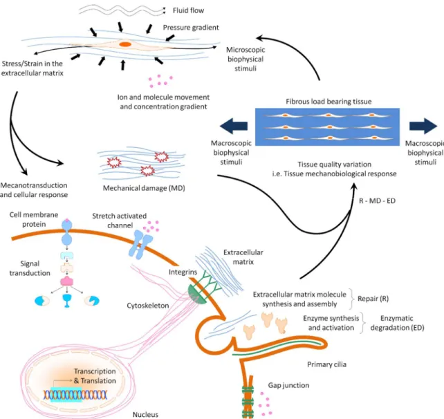
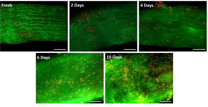
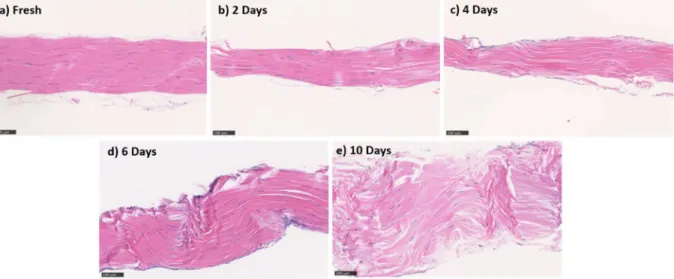
Figure 2.1 Tissue mechanobiological response including cellular response and extracellular matrix mechanical damage. ... 9 Figure 2.2 Increased MMP9 and MMP13 in 6-day SD tendon samples by PCR analysis. .. 27 Figure 3.1 Number and distributions of tendons from each rat. ... 43 Figure 3.2 Typical fluorescence micrographs obtained from freshly isolated tendons experiencing 2, 4, 6, and 10 days of stress deprivation. ... 48 Figure 3.3 Changes in maximum stress at failure (mean and SEM) with stress deprivation. * indicates a significant difference (p<0.05) from day 0 (fresh tendons); ... 49 Figure 3.4 Typical micrographs of longitudinal sections of tendons stained with hematoxylin and eosin. ... 50 Figure 3.5 Quantification of a) space between fibers, b) fiber density, c) cell number, d) nucleus elongation, and e) semiquantification of collagen tortuosity. ... 52 Figure 3.6 Biochemical analysis. ... 54 Figure 4.1 Number and distributions of tendons from each rat. ... 62 Figure 4.2 Representative fluorescence micrographs of tendons in the various treatment groups. ... 68 Figure 4.3 Changes in maximum stress at failure (mean and SEM). ... 69 Figure 4.4 Typical micrographs of longitudinal sections of tendons stained with hematoxylin and eosin. ... 70
v
Figure 4.5 Quantification of the a) space between fibers, b) fiber density, c) cell number, and d) nucleus elongation ... 72 Figure 4.6 Biochemical analysis. ... 73 Figure A.2.1 Typical micrographs of longitudinal sections of fresh and 10 day SD tendons stained with DAPI. ... 85
vi
List of Tables
Table 2.1 The rational and efficacy of available tendinopathy treatments2,43,84,86 ... 11
Table 2.2 Data extracted from MMPI studies. N/A stands for data not available. ... 21
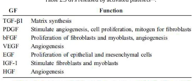
Table 2.3 GFs released by activated platelets86. ... 28
vii
Abstract
Background: Tendinopathy, a general condition of tendon disorder, is common among people specially athletes and workers. Tendinosis, an asymptomatic tendon degeneration without inflammation, is a frequent form of tendinopathy which can be chronic and difficult to successfully treat in long term. The focus of this research study is on this type of tendinopathy. The proper strategy to prevent or treat chronic tendinopathy (CT) is still unclear. Understanding the pathology behind CT may help finding proper treatments. Although significant advances have been made to understand the tendinopathy mechanisms, this condition is still poorly understood.
Studying CT not only is necessary in order to provide further information about etiology and pathogenesis, but also to improve healing strategies. Although many treatment strategies have been suggested for CT disorders, none of them has satisfying results. Subject: Therefore, the main subject of this study was to characterize a CT model ex vivo, which represents the level of degeneration which is not too early to be diagnosed and not too late to be treated. This is a transient stage between early and advanced tendinopathy. We refer to this stage as moderate tendinopathy.
After creating moderate tendinopathy, the efficacy of two existing treatments, i.e. matrix metalloproteinase inhibitor (MMPI) and platelet-rich-plasma (PRP) injections, in combination and alone, on healing moderate tendinopathy model was investigated. Methods: The ex vivo model was characterized by using stress deprivation. Rat tail tendons (RTTs) were cultured without loading for 0 (control), 2, 4, 6, or 10 days. RTTs were subjected to traction testing as well as to histopathological, biochemical, and viability assays at the end of their culture time. Then, the ex vivo moderate tendinopathy model was further cultured for 6 days with or without receiving treatment(s). The treatments included low static loading, alone or in combination with: narrow spectrum MMPI (NI), broad spectrum MMPI (BI), PRP, PRP + NI, PRP + BI. Again, RTTs were subjected to traction
viii
testing as well as histopathological, biochemical, and viability assays at the end of their culture time.
Results: There were moderate degradative changes in the properties of RTTs at day 4 of SD. They included: increases in the space between fibers, cell density, and collagen tortuosity as well as a decrease in collagen density and elongation of cell nuclei. No changes in the stress at failure of RTTs were observed at this time point. Advance degradative changes occurred at later time points, i.e. at days 6 and 10 of SD. They included: more increases in the space between fibers, and collagen tortuosity as well as a decrease in collagen density and elongation of cell nuclei. There was also a significant decrease in stress at failure of RTTs at days 6 and 10 of SD compared to fresh tendons. Moreover, our results showed that PRP + NI added to low static loading, improved mechanical and histological properties of moderately damaged tendons comparing to other treatments, or untreated tendons. Stress at failure of RTTs treated with PRP + NI was improved comparing to 10-day SD tendons (p=0.01). PRP + NI also improved fiber density, nucleus shape, and space between fibers of RTTs when compared to 10-day SD RTTs (p<0.05).
Conclusions: The simple ex vivo model characterized in this study is useful to study the progression of CT, and to investigate the efficacy of potential treatments to stop or reverse the progression of the pathology. Moreover, using this model, we have introduced PRP and NI combination therapy as a potentially efficient treatment for CT.
Key words: tendinopathy, stress deprivation, matrix metalloproteinase inhibitor, platelet-rich-plasma.
ix
Résumé
Contexte: La tendinopathie, une condition générale qui affecte le tendon, est fréquente, spécialement chez les athlètes et les travailleurs. La tendinose, une dégénérescence du tendon sans inflammation, est une forme fréquente de tendinopathie qui peut être chronique et difficile à traiter à long terme. Cette étude est axée sur ce type de tendinopathie. La stratégie appropriée pour prévenir ou traiter la tendinopathie chronique (TC) n'est toujours pas claire. Mieux comprendre la pathologie de la TC contribuera à améliorer les traitements appropriés. Bien que des progrès significatifs aient été réalisés pour comprendre les mécanismes de tendinopathie, cette condition est encore mal comprise.
L'étude de la TC n'est pas seulement nécessaire pour fournir de plus amples informations sur l'étiologie et la pathogenèse, mais aussi pour améliorer les stratégies de guérison. Bien que de nombreuses stratégies de traitement aient été suggérées pour la TC, aucune n’a donné de résultats satisfaisants.
Objet: L'objet principal de cette étude était donc de caractériser un modèle ex vivo de TC, qui représente le niveau de dégénérescence qui n'est pas trop précoce pour être diagnostiqué et pas trop avancé pour être traité. C'est un stade transitoire entre les tendinopathies précoces et avancées. Nous appelons ce stade tendinopathie modérée. Après avoir créé une tendinopathie modérée, l'efficacité de deux traitements existants, à savoir l’ajout d'inhibiteurs de métalloprotéinases de la matrice (IMPMs) et de plasma riche en plaquettes (PRP), en combinaison et individuellement, sur le modèle de tendinopathie modérée a été étudiée.
Méthodes: Le modèle ex vivo créé par la privation de charge mécanique a été caractérisé. Les tendons de queue de rat (TQRs) ont été cultivés en absence de charge mécanique pendant 0 (témoin), 2, 4, 6 ou 10 jours. Les TQRs ont été soumis à des tests de traction ainsi qu'à des analyses histogiques, biochimiques et de viabilité à la fin de leur temps de culture. Ensuite, le modèle de tendinopathie modérée ex vivo a été cultivé pendant 6 jours
x
avec ou sans traitement. Les traitements comprenaient une faible charge statique, seule ou en combinaison avec: IMPM à spectre étroit (IÉ), IMPM à large spectre (IL), PRP, PRP + IÉ, PRP + IL. Encore une fois, les TQRs ont été soumis à des tests de traction ainsi qu'à des analyses histologiques, biochimiques et de viabilité à la fin de leur temps de culture. Résultats: Il y avait des changements modérés dans les propriétés des TQRs au jour 4 de la privation de chargement mécanique. Ils comprenaient : l'augmentation de l'espace entre les fibres, de la densité cellulaire et de la tortuosité du collagène, ainsi qu'une diminution de la densité du collagène et de l'élongation des noyaux cellulaires. Aucun changement dans la contrainte à la rupture des TQRs n'a été observé à ce moment. Les changements de dégradation avancés se sont produits à des moments ultérieurs, à savoir aux jours 6 et 10 de privation de charge mécanique. Ils comprenaient : une augmentation de l'espace entre les fibres et de la tortuosité du collagène, ainsi qu'une diminution de la densité du collagène et de l'élongation des noyaux cellulaires. Il y avait également une diminution significative de la contrainte à la rupture des TQRs aux jours 6 et 10 de privation de chargement par rapport aux tendons frais.
De plus, nos résultats ont montré que le traitement PRP + IÉ, ajoutés à la faible charge statique, améliorait les propriétés mécaniques et histologiques des tendons modérément endommagés par rapport à d'autres traitements ou aux tendons non traités. La contrainte à la rupture des TQRs traités avec PRP + IÉ a été améliorée par rapport aux tendons privés de chargement au jour 10 (p = 0,01). Le traitement PRP + IÉ a également amélioré la densité des fibres, la forme du noyau et l’espace entre les fibres des TQRs par rapport aux TQRs privés de chargement au jour 10 (p <0,05).
Conclusions: Le modèle simple ex vivo caractérisé dans cette étude est utile pour investiguer la progression de la TC et pour étudier l'efficacité des traitements potentiels pour arrêter ou inverser la progression de la pathologie. De plus, en utilisant ce modèle, nous avons introduit la thérapie combinée PRP et IÉ comme traitement potentiellement efficace pour la TC.
xi
Mots clés: tendinopathie, privation de charge mécanique, inhibiteur de métalloprotéinases de la matrice, plasma riche en plaquettes.
1
Chapter 1. Introduction
Tendons are those connective tissues which connect muscle to bone. They consist of two main parts: the extracellular matrix (ECM) and cells (tenocytes) which are, respectively, the inert and active components of tendons. These two components are in a close and bidirectional interaction. However, the mechanical behavior of the tendon is mostly devoted to the ECM, and cells are considered as responsible for tissue remodeling 42.
ECM is mainly made up of water (~ 70%). The dry mass of ECM consists mostly of collagen, elastin, and proteoglycans. Collagen is the most abundant protein in ECM. It provides tendon strength against applied tensile load. Collagen parallel alignment along the tendon enables it to resist tensile load in this direction.
Tenocytes: tenocytes, which are responsible for production of the ECM, have an elongated shape when observed in the tendon’s longitudinal orientation71. Whereas in
cross-sectional view, they appear as star-shaped64. Tenocytes are sparsely distributed between
collagen fibers with their processes making an extensive network inside ECM114. They are
highly responsive to their micro-environmental changes, such as mechanical loads or local stimuli28. For further details regarding tenocytes mechano-responsiveness see section
2.1.2.
Collagen in tendons adopts a hierarchical structure such as displayed in Figure 1.1. Collagen molecules unite into collagen fibrils, collagen fibers, subfascicles (primary
bundles), fascicles (secondary bundles) and tertiary bundles53. Primary, secondary and
tertiary fiber bundles are covered by a thin layer called endotenon and the whole tendon is surrounded by another thin layer called epitenon91.
There have been some misunderstandings in literature regarding using “fiber” and “fibril”. In some texts,
2
Figure 1.1 Schematic structure of a normal tendon49
Tendon disorders are common health problems. For example, 1 of every 10 people and 1 of every 2 runners experience Achilles tendon disorder before the age of 4598. The
consequences of this medical problem include pain, disability, early retirement from sport and work, mental distress, health-care costs, etc.
It should be noted that there is confusion in literature about terminology of tendon disorders. One term which has been used to address tendon problems is “tendinopathy”. Tendinopathy is a general term for tendon disorders with no assumption of underlying pathology. Traditionally, it was thought that underlying pathology of tendon disorders was due to inflammation2,27,83. This condition, i.e. tendon disorder accompanied by
inflammation, is referred to as “tendinitis”. However, later, “non-inflammatory” or “degenerative” theory was considered to be the reason behind CT, or overuse injuries83.
The term used to describe this condition i.e. degenerative condition of tendon without accompanying inflammation is “tendinosis”83,84. These three terms have often been used
interchangeably which is inappropriate. We notify that in this research, the focus is on a tendinopathy model of chronic injury without inflammation.
3
Clinical features: activity-related pain, tenderness, and impaired performance43.
Histological features: hyper-cellularity, loss of cytoplasm elongated shape, rounder nuclei, neovascularization, disruption in collagen organization and orientation, decreasing collagen fibers size and increasing cross sectional area (CSA)43,55.
Biochemical features: changes in gene expression and activity of biomolecules and proteins such as collagens and matrix metalloproteinases (MMPs), enzymes which degrade ECM (for further details, see Appendix). For example, it has been suggested that there is an increase in MMPs level in tendinopathic tendon5. However, these changes are
often model-dependent. In other words, they depend on various factors such as species (human, rat, rabbit, etc), body parts (Achilles tendon, hamstring tendon, tail tendon, etc), and type of pathology (acute, overuse, stress deprivation, etc).
Despite the prevalence of tendon problems, they are difficult to treat successfully84,86.
Historically, treatment strategies were largely focusing on targeting inflammation such as rest, corticosteroid injection, or nonsteroidal anti-inflammatory drugs (NSIADs). However, since there is little or no inflammation in CT2,83, the results of these treatments
on CT are usually unsatisfactory, and there is possibility of recurrence of symptoms43.
Currently new promising treatments (see section 2.2) are being developed, but there is still little evidence to support their use.
To find an optimal healing strategy, understanding the pathology behind tendinopathy is crucial. Using animal models is essential for in-depth studies on this subject. Therefore, in the first part of our literature review (Chapter 2), available models relating to CT are described.
Models are not only beneficial for studying features and causes of CT, but they also enable studying potential treatments. Hence, available treatments for tendinopathy will be reviewed in the second part of our literature review. Thereafter, we will introduce the selected treatments to be studied in this research.
4
Two articles based on the findings of this research are presented in Chapters 3 and 4. In the first article (Chapter 3), a simple ex vivo model to create moderate CT has been characterized. The second article (Chapter 4) presents the results of applying two recent treatments (PRP, and MMPI) in combination and alone on this ex vivo tendinopathic model. Lastly, in Chapter 5, concluding remarks from the results of this project and suggestions for further work are outlined.
5
Chapter 2. Literature review
2.1. Currently available ex vivo models for tendinopathy.
There are numerous pathogenetic mechanisms behind tendinopathy. Moreover, tendons from different body parts, thus subjected to different mechanical and biological conditions, are affected by tendinopathy. Therefore, it is unlikely that only one suitable model exists to represent all tendinopathy conditions.
Tendinopathy models for the study of pathogenesis and the evaluation of potential treatments are developed either in vivo or ex vivo. In vivo models are those inducing tendinopathy in a live animal. Ex vivo models include those using a living tendon kept outside of the body under culture conditions.
In vivo models are essential, because they enable us to have a more complete understanding of tendinopathy27. Ex vivo models are also critical, because they allow
under specific, controlled conditions (e.g. defined media, defined loading protocol, …), the study of the pathological basis as well as of drug efficacy. Since in vivo models are costly and time-consuming, we decided to initially characterize an ex vivo tendinopathy model to verify our hypotheses. In the following sections, the current ex vivo chronic tendinopathy (CT) models will be explained. Finally, we conclude with the method we selected to create CT in this research.
2.1.1. Ex vivo models of tendinopathy
Ex vivo models of tendinopathy allow for great control over experimental conditions27, for
example loading conditions or composition of culture media. Ex vivo models mainly use cyclic loading, and stress deprivation (SD) to induce tendinopathy in harvested tissues. In these models, there is no need to interact recurrently with animals, to make them exercise for example. Therefore, these models may be cheaper and less time consuming.
6 Cyclic loading
This model has been developed to simulate the daily repetitive loading on live tendons. For example, Cousineau and Langelier22 used two loading conditions: i.e. 1.2% strain for
underuse, and 1.8% strain for overuse. For both loading conditions, they have reported a decrease in the peak stress of the cyclically loaded rat tail tendons (RTTs) after 24 hours as well as changes in the ECM after 10 days, mainly for the underuse condition.
In another study26, live avian flexor digitorum tendons were cyclically loaded to either 3
(low), or 12 MPa (high), for either continuously 24 hours (short), or 2h/day for 12 days (long) at 1 Hz. The results revealed a decrease in maximum load at failure (N) of tendons loaded with the following regimens: low/long, high/short and high/long. Maximum stress at failure (MPa) of tendons which were loaded high/short or high/long also decreased significantly.
Cellular deformation as a result of cyclic loading has been reported in a study on rat tail tendons7. The results of this study demonstrated that exposing tendons to 2%, 4% and 6%
strain results in a deformation in cell nuclei.
Cellular response to cyclic loading has also been the subject of some studies. For example, an increase in the level of PGE2 as a result of increasing loading duration of avian flexor
digitorum tendons has been reported26. Moreover, a decrease in MMP-1 levels of cyclically
loaded rat tail tendons has been demonstrated50.
Stress deprivation
Ex vivo SD also could cause tendon degeneration. This model has frequently been used to study tendinopathy. In the following paragraphs, two studies have been explained to introduce the general results. For further studies and results, please see Table 3.1 (chapter 3).
Arnockzky et al 5 developed a stress-deprived model by incubating live tendons without
7
and its pericellular matrix; a reduction in collagen fibril packing; a reduction in ultimate stress, strain at ultimate stress, tensile modulus; and finally, an increased MMPs levels. In a study by Lavagnino et al 49, ex vivo stress deprivation resulted in a reduction in tensile
properties without changes in collagens.
For further details about the effect of stress deprivation on tendon properties ex vivo, please refer to Table 3.1 (chapter 3).
2.1.2. Pathogenesis behind tendinopathy induced by stress deprivation
and cyclic loading
To address the reason of degradative effects of stress deprivation and cyclic loading on tissues, we should first consider that tendon is a mechanosensitive tissue which responds to loading in an adaptive manner45. Mechanical loading on tendons can be sensed by
tendon cells via mechanotransduction mechanisms. Mechanotransduction mechanisms, by which mechanical stimulations are converted into biochemical signals, are mediated by cell deformation, nucleus deformation, cytoskeleton, stretch activated channels, and primary cilium100 (Figure 2.1). According to Arnoczky et al 6, the character of cell response
(catabolic vs. anabolic) to the perceived load depends on its threshold of sensitivity to external load. It has been demonstrated that the preset level of cell sensitivity to the applied load, which is called mechanostat set point of the cell, is regulated by cells through their ability to produce internal tension. This tension may help cells to maintain their homeostatic state of stress6. If the applied load is less than this set point, the response of
cells will be catabolic, while if the load is higher than cell mechanostat set point, cell response will be anabolic.
The degradative effects of stress deprivation, as a result of catabolic response of the cells, could be explained by this theory. Uchida et al 96 reported a simultaneous over-expression
of cytokines such as IL-1b and TNF-a following stress deprivation. These signaling molecules are well-known to stimulate MMPs-1 and -3 in fibroblasts. They addressed these ECM-degradative enzymes as the reason of destructive effects of stress deprivation.
8
However, they did not explain the reason of the catabolic cell response, which seems to be related to mechanostat set point of the cells.
In cyclic loading, beside cell response, mechanical degradation of ECM should also be taken into account. Since tendons have a viscoelastic behavior, the factors which mediate mechanical degradation include: the amount, the frequency and the rate of applied load, and rest periods between loading episodes. Mechanical degradation would lead to tendinopathy if the anabolic response is insufficient to repair tissue damage. This would lead to the accumulation of micro tears in collagen fibers. The partial damages of collagens unable them to properly transfer the external load to cells; as a result, tendon cells undergo understimulation. It means that their perceived load is less than their mechanostat set point. Therefore, their response would be catabolic, and they produce degeneration which may eventually lead to tendinopathy4. Although the etiology is likely
more complicated than this, accumulation of microdamages as a result of fatigue loading has been considered a significant reason for overuse injuries26.
9
Figure 2.1 Tissue mechanobiological response including cellular response and extracellular matrix mechanical damage. Note the mechanotransduction mechanisms by which cell
10
2.1.3. The preferred CT model
Among all ex vivo tendinopathy models, we chose SD to characterize CT in our laboratory. We use SD because it could be achieved by using a simple experimental setup. Therefore, there is no need to use the bioreactor which is not very stable and also has the limitation of number of samples per experiment (4 tendons). Moreover, preparing the samples in this model is less expensive in terms of cost and length of experiment. This is in concordance to the objectives of this research study: because we need to control multiple factors in each experiment, this model allows for having enough samples simultaneously. Moreover, since these samples could all be extracted from the same animal, the risk of inter-animal variability is also reduced, as well as the number of sacrificed animals.
2.2. Current healing strategies available to treat tendinopathy
Despite the prevalence of tendinopathy, the available healing strategies are not completely satisfactory43. Traditional treatments focused on anti-inflammatory drugs such as
corticosteroid injections and non-steroidal anti-inflammatory drugs (NSAID) drugs. However, it may not be the most appropriate target2, because it has been suggested that
there is little or no inflammation in CT2. Even in tendinitis, there is debate on whether
blocking the inflammatory response will be helpful or not84.
Currently, several treatments, (including eccentric exercises, pharmacological treatments, etc) have been introduced to heal tendinopathy. However, there is little high-quality studies to confirm their clinical use21,72,90. A summary of current non-surgical
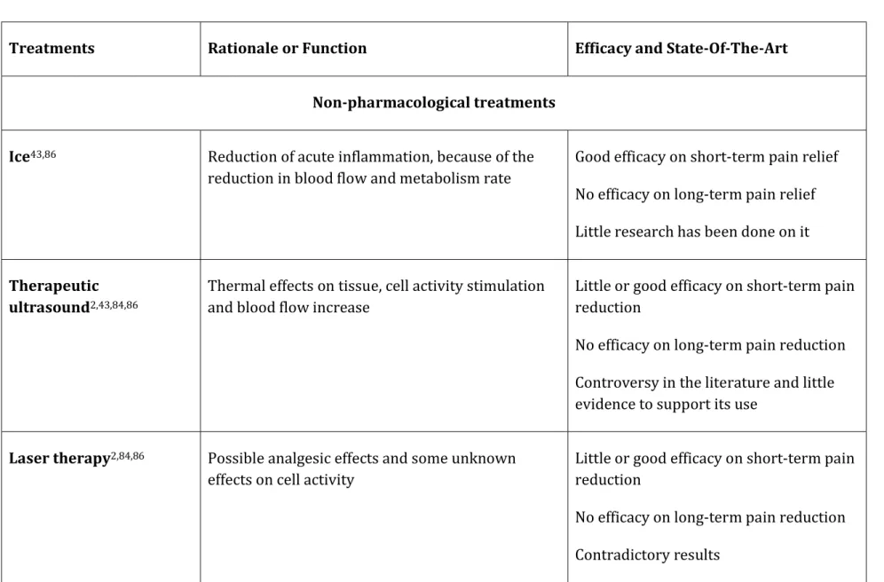
Table 2.1 The rational and efficacy of available tendinopathy treatments2,43,84,86
Treatments Rationale or Function Efficacy and State-Of-The-Art
Non-pharmacological treatments Ice43,86 Reduction of acute inflammation, because of the
reduction in blood flow and metabolism rate
Good efficacy on short-term pain relief No efficacy on long-term pain relief Little research has been done on it Therapeutic
ultrasound2,43,84,86
Thermal effects on tissue, cell activity stimulation and blood flow increase
Little or good efficacy on short-term pain reduction
No efficacy on long-term pain reduction Controversy in the literature and little evidence to support its use
Laser therapy2,84,86 Possible analgesic effects and some unknown
effects on cell activity
Little or good efficacy on short-term pain reduction
No efficacy on long-term pain reduction Contradictory results
Friction massage2,43,84 Cell activity stimulation and blood flow increase Little or good efficacy on short-term pain
reduction
No efficacy on long-term pain reduction Controversy in the literature and little evidence to support its use
Soft tissue mobilization2,84
Cell activity stimulation and blood flow increase Little or good efficacy on short-term pain
reduction
No efficacy on long-term pain reduction Controversy in the literature and little evidence to support its use
Orthotic devices43,84,86 Correcting a static disorder, removal of
precipitating factors and prevention of reinjury
Good efficacy on short-term pain relief No efficacy on long-term pain relief Little evidence to support their effectiveness
Eccentric exercise2,43,84,86 Promote regeneration of normal tissue structure,
probably through an effect on cell activity and matrix remodeling
Good efficacy on short-term pain relief and long-term pain relief
It is currently considered to be the most efficient treatment for tendinopathy, although some studies are contradictory Extracorporeal Shock
Wave Therapy (ESWT)2,43,84,86
Promote tissue healing by stimulation of cell activity, and neovascularization, induction of growth factors (TGF-β1 and IGF-1), increased tenocyte proliferation and collagen synthesis
Very good efficacy on short-term pain relief and long-term pain relief
Some evidence to support its use in calcific tendinopathies of the rotator cuff No clear evidence, or little evidence (controversial information) supporting its use in the treatment of chronic tennis elbow, medial epicondylitis, Achilles and patellar tendinopathies
Pharmacological treatments Nonsteroidal
Anti-inflammatory Drugs (NSAIDs)2,43,84,86
Reduction of inflammation through inhibition of inflammatory factors synthesis
Good efficacy on acute pain
No efficacy on long-term pain relief Researches in acute tendon injury model showed NSAID administration did not
prevent collagen degradation or loss of tensile force in tendons
Its use is still controversy regarding the lack of existence of inflammation in degeneration
Corticosteroid injections2,43,84,86
Reduction of inflammation by inhibition of the synthesis of cytokine genes and proinflammatory factors
Good efficacy on short-term pain relief Little or no efficacy on long-term pain relief
Mixed results
Corticosteroids can cause spontaneous tendon rupture
No evidence to prove its efficiency for chronic tendinopathy
Heparin84,86 Effect on tendon blood flow; possibly results in
improved healing
In a review in 2006, it was mentioned that it has some constructive effects, but more recent animal study found heparin had a degenerative effect
It has not been mentioned in the more recent reviews (2008, and 2011),
therefore may be it is no more an option
Dextrose84 Possibility of releasing growth factors (GFs) and
therefore local tissue proliferation
Since there is no control data nothing can be said
It has not been mentioned in the reviewed literature as a potential treatment since the 2006 review article
MMP inhibitors43,84 Promoting healing by inhibiting the enzymes which
break down tendons, i.e. MMPs.
Because it has been established that using conventional treatments with the effect of inflammatory suppression
May not fully inhibit MMP-based tendon degradation
Very good efficacy on short-term pain relief and long-term pain relief
In clinical trials, aprotinin was used as a broad-spectrum MMP inhibitor
Good clinical improvement mild-Achilles tendinopathy patients were treated more successfully than patellar
Aprotinin may cause anaphylaxis, more studies are needed
Injections of blood or PRP2,43,84,86
Contains growth factors (e.g. transforming growth factor β and platelet-derived growth factor) that promote matrix synthesis and tissue repair
Little or no efficacy on short-term pain relief
Very good efficacy on long-term pain relief
There have been only a few clinical studies in recent years with good results, but in vitro studies have promising results.
Other controlled studies are needed Sclerosant
injections2,43,84,86
Blocks tendon blood flow (targeting
neovascularization and associated pain generating nerve in-growth)
Very good efficacy on short-term pain relief and long-term pain relief
In clinical trials polidocanol was used as sclerosant agent
Some clinical studies report goodshort- and/or long-term result with an increase in strength and a decrease in pain
Other studies needed to evaluate its safety, efficacy, and combination with other drugs
Topical glyceryl trinitrate2,43,84,86
Enhances collagen synthesis Very good efficacy on short-term pain
relief
Excellent efficacy on long-term pain relief Few clinical trials, reported benefit on patient-determined pain, function, and loss of symptoms on Achilles
tendinopathy, chronic supraspinatus tendinopathy and lateral elbow tendinopathy
Polysulphated
glycosaminoglycan84
Inhibition of inflammation, possibly also acting to inhibit metalloproteinase enzyme activity
A number of studies suggesting that injection of glycosaminoglycan polysulphate may lead to an
improvement in disease of the human Achilles and equine superficial digital flexor tendon.
It has not been mentioned in the recent reviews (2008, and 2011), therefore may be it is no more an option
Botulinum toxin A (BTA) injections43
Paralysis caused by BTA involves a reduction in tensile stress on the enthesis
Very good efficacy on short-term pain relief
Good efficacy on long-term pain relief Few articles fromlast years have considered the possibility of making botulinum toxininjections (BTA) to treat epicondylitis
Stem-cell or gene therapy2,43,84,86
Stem cells will be isolated and located in the area of injury or degeneration. Then with local signaling or some exogenous factors, they will be stimulated to differentiate to particular cells
Unknown efficacy on pain relief
There are promising in vitro results for using stem-cell or gene in treatment of tendinopathy.
Animal studies suggest that gene therapy may also improve the capacity of the injured tendon to heal
19
Among all potential treatments, we selected platelet-rich plasma (PRP) and matrix metalloproteinase inhibitors (MMPIs) to apply in our laboratory. To explain this choice, we refer to the following model for tendon mechanobiological response (TMR) (Figure 2.1) as: TMR = Repair – (Mechanical degradation + Enzymatic degradation)22.
This model highlights that the combination of these treatments might be beneficial to simultaneously promote regeneration and inhibit enzymatic degeneration. First, PRP would enhance tendon repair through the release of a variety of autologous growth factors (GFs) playing a key role in tendon repair mechanisms68. Second, MMPIs would inhibit
proteolytic degradation of connective tissues mediated by MMPs. However, in our knowledge the combination of these two treatments has not been studied so far.
In the following section these two healing strategies are explained with more details.
2.3. MMPIs and PRPs: The selected healing strategies to apply
in ex vivo created tendinopathy models
2.3.1. MMPIs: general information
Natural and synthetic MMPIs
MMPIs are enzymes inhibiting the activity and/or expression of MMPs. MMPs are ECM proteolytic enzymes mediating ECM turnover. ECM equilibrium is mediated by, inter alia, a balance between the levels of MMPs and their natural inhibitors, called tissue inhibitors of matrix metalloproteinases (TIMPs).
Dysregulation of the balance between the levels of MMPs and TIMPs interrupts ECM equilibrium. High levels of MMPs could cause tendon degeneration and rupture. Therefore, inhibiting MMPs has been considered as a therapy option for tendinopathy5,73.
MMPIs could be endogenous such as TIMPs, or exogenous such as synthetic MMPIs. TIMPs are divided into four categories: TIMP1, TIMP2, TIMP3, TIMP4. They inhibit all MMPs, however, the ability of some of TIMPs to inhibit some specific MMPs needs to be elucidated
20
in future studies. TIMPs function is not limited to inhibition of MMPs. They also play roles in regulation of angiogenesis and cellular proliferation78.
Although TIMPs have stronger inhibitory effect than synthetic MMPIs, to our knowledge, they have never been used as therapeutic agents to promote tendon healing ex vivo or in vivo. Before further using them as treatment in diverse pathologies, it has been suggested to deepen our understanding of TIMPs’ activities (other than inhibitory ones), since they may cause some unwanted side effects10. Moreover, their broad-spectrum inhibitory effect
makes them undesirable in studies with specific MMPs in target. However, TIMPs could be modified in order to retain their inhibitory effect without their other unwanted biological activities. In this way, they may be applied for therapeutic benefits in future10.
It should be noted that TIMPs have been used as therapeutic agent in some studies of other disease. For instance, Li et al have shown promising results of using recombinant TIMP-2 in cancer treatment52.
Synthetic MMPIs have been used as therapeutic agents in MMP-mediated disease. There are several studies with using synthetic MMPIs to promote tendon healing ex vivo and in vivo. Some of them will be presented in the following sections.
The effect of MMPIs on tendons in ex vivo and in vivo animal studies
Excessive action of MMPs may lead to degeneration and weakening of ECM. Therefore, in this condition, drugs that can decrease the MMPs activity, and therefore ECM degeneration, may play a significant role in promoting tendon healing5. In some ex vivo
and in vivo studies, MMPIs were used to affect MMP activity and prevent or reverse the process of MMP-mediated ECM degeneration in tendons. Table 2.2 summarizes the result of MMPI application on tendons in some studies ex vivo, and in vivo. It should be noted that most of the studies were conducted on surgically-induced tendinopathy, i.e. partially or completely sectioning the tendon using a scalpel and then stitching damaged tendons or leaving them unsutured, models which do not reflect CT.
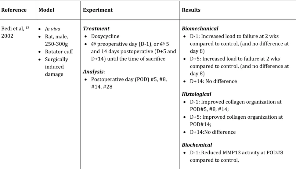
Table 2.2 Data extracted from MMPI studies. N/A stands for data not available.
Reference Model Experiment Results
Bedi et al, 13 2002 In vivo Rat, male, 250-300g Rotator cuff Surgically induced damage Treatment Doxycycline @ preoperative day (D-1), or @ 5 and 14 days postoperative (D+5 and D+14) until the time of sacrifice Analysis:
Postoperative day (POD) #5, #8, #14, #28
Biomechanical
D-1: Increased load to failure at 2 wks compared to control, (and no difference at day 8)
D+5: Increased load to failure at 2 wks compared to control, (and no difference at day 8)
D+14: No difference Histological
D-1: Improved collagen organization at POD#5, #8, #14;
D+5: Improved collagen organization at POD#14;
D+14:No difference Biochemical
D-1: Reduced MMP13 activity at POD#8 compared to control,
Arnoczky et al, 5 2007 Ex vivo Rat, adult Tail tendon Healthy Treatment Doxycycline or Ilomastat @ day 0 Analysis:
7 days after first MMPIs application
Biomechanical
Improvement in ultimate stress, tensile modulus, and strain at ultimate stress with either of MMPIs compared to 7-day SD Histological
Limited the alteration in the pericellular matrix shape, and loss of cell-matrix contact with either of MMPIs;
Biochemical
Reduction in MMP13 activity with either of MMPIs,
No effect on MMP13 protein synthesis with either of MMPIs Demirag et al, 25 2005 In vivo Rabbit, 3.5-4 kg Anterior cruciate ligament Surgically induced damage Treatment A-2-macroglobulin
@ day 0 and 1 after surgery Analysis:
2 and 5 weeks post-surgery
Biomechanical
Greater load to failure at both 2 and 5 weeks
Histological
Dense, more mature, perpendicular collagen fibers
Biochemical
Kessler et al, 44 2014 In vivo Rat, male, 300-400 g Achilles Surgically induced damage Treatment Doxycycline
Starting @ preoperative day, continued up to 4 weeks Analysis:
2 or 4 weeks for histology; 3 weeks for biomechanical properties
Biomechanical
Enhanced biomechanical properties Histological
Improved collagen fiber organization Biochemical Reduced MMP activity Nquyen et al, 70 2017 In vivo Rat, male, 2-3 months old Achilles Surgically induced damage Treatment Doxycycline
Daily administration of MMPI one day after injury
Analysis:
1.5, 3, 6, 9 weeks after injury
Biomechanical
Accelerated recovery in biomechanical properties (i.e., greater equilibrium modulus, higher dynamic modulus and lower creep strain)
Histological
Improved tissue organization, fiber
alignment, and decreased fiber dispersion Biochemical Decreased MMP3 expression at 9 wks Bedi et al, 14 2010 In vivo Rat, male, 250-300 g Treatment A-2-macroglobulin @ day 0 Biomechanical
No change in load to failure, or stiffness Histological
Rotator cuff Surgically
induced damage
Analysis:
2 or 4 weeks after surgery
Greater collagen organization at 4 wks; but not at 2 wks
Reduction in collagen degradation at both 2, and 4 wks Biochemical None Orner et al, 74 2016 In vivo Mouse, female, 8-10 weeks Flexor tendon Surgically induced damage Treatment MMP 9 inhibitor
Daily administration of MMPI starting @ the day of surgery, until day 8 post surgery
Analysis:
7-35 days post surgery
Biomechanical
No difference in biomechanical properties (stiffness, and max load at failure)
Histological
No alteration in tendon morphology Biochemical Decreased MMP9 activity Pasternal et al, 79 2006 In vivo Rat Achilles Surgically induced damage Treatment Doxycucline
Starting @ 1 day before surgery until the time of sacrifice
Analysis:
5, 8, and 14 days after surgery
Biomechanical
Decreased force at failure, and energy uptake
Histological None Biochemical
25
It can be concluded from Table 2.2 that using broad-spectrum MMPIs (BI; e.g., doxycycline and alpha-2-macroglobulin) in surgically induced tendinopathy models results in greater collagen organization14,25,44,70, as well as a reduction in collagen degradation14,70, limited
MMP activity25,44,70, and increased mechanical strength25,44,70.
The effect of MMPIs on tendons in clinical trials
Beside laboratories, MMPIs have also been used in clinical trials to improve tendon healing. Aprotinin is a broad-spectrum MMPI which was initially used for limiting blood loss during surgery and promoting soft tissue healing after surgery73. Years later, it was
introduced as therapy option for chronic tendinopathy. In clinic, aprotinin has been used for treating chronic patellar and Achilles tendinopathies. In one randomized control trial, aprotinin presented superior results than both corticosteroid and saline injections in chronic patellar tendinopathy, 12 months after injection17. The results are mixed about
the effect of aprotinin on Achilles tendinopathy. Some uncontrolled or poorly controlled studies reported high rates of success in management of chronic Achilles tendinopathy with aprotinin9,19. In a study by Brown et al17 on chronic tendinopathy, there was no
significant difference but a trend toward better results by aprotinin than placebo. Another clinical outcome73, demonstrated that in treating chronic tendinopathy, using aprotinin
led to more successful results for treating mid-Achilles tendons than patellar tendons. Although excess activity of MMPs may lead to degeneration and weakening of ECM, normal actions of MMPs are needed for ECM remodeling and repair. It should be noted that applying MMPIs to promote healing in some cases may adversely affect the healing process. In this regard, Pasternak et al79 conducted a test evaluating the effect of
broad-spectrum MMPI, doxycycline, in the prevention or healing of the acute injury of Achilles tendon in rats in vivo. Their results demonstrated negative influence of doxycycline on early stages of tendon healing as measured in tension test by force and energy uptake at failure. These results could be explained by the fact that there was tendon regeneration at the site of injury consisting of a loose collagenous network including mostly proteoglycan and collagen III. MMPs are fundamental to remove this newly formed loose callus to be replaced by more densely organized collagens, mostly collagen I. In fact, this removal is
26
needed for remodeling and consequently building stronger tissue. The negative result from this study and positive result from literature reviewed in the previous section imply that the effect of MMPIs on tendon healing is strongly case-dependent.
Therefore, before applying MMPIs to prevent or heal tendinopathy, this dual activity of MMPs should be taken into account. That is, while in one pathologic model, specific MMP might play a negative role, in another pathologic model, the role of that specific MMP might be positive on tissue quality. In conclusion, the alteration in the presence and activity of MMPs should be studied in each pathologic model, to be able to target MMPs which are destructive to tendon health.
Narrow or broad-spectrum MMPIs
There are some evidence that broad-spectrum MMP inhibition impairs tendon healing, since MMPs play fundamental role in remodeling and healing46,54,75,79. Therefore, more
selective inhibition might lead to positive effects. In fact, it would be beneficial to target specific MMPs which are detrimental to tendon healing, without affecting MMPs which act during tendon healing. This could be achieved by applying narrow-spectrum MMPIs (NI) instead of broad-spectrum MMPIs (BI).
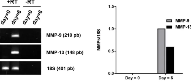
Obviously, choosing appropriate NI, requires identifying MMPs deleterious to tendon healing. Based on the information in the literature regarding destructive MMPs present in tendinopathic rat tendons, MMPs 9 and 13 have first been selected as target MMPs to inhibit by our group. It should be noted that RT-PCR analysis has been performed by our group on 6-day stress-deprived RTTs and an increase in MMP9 and MMP13 has been reported (Figure 2.2). However, for our ex vivo experiment, we focused on MMP13. It is because MMP13 in rats is equivalent to MMP1 (interstitial collagenase) in human and because its expression has been reported to be increased in pathological human tendons5.
27
2.3.2. PRP: general information
PRP therapy is one new alternative in tendon healing. PRP is a blood product obtained from whole blood that contains a high concentration of platelets. The beneficial effect of PRP is probably related to providing an environment full of bioactive molecules and GFs appropriate for tissue regeneration. This environment enables cell migration and proliferation, angiogenesis, and matrix deposition in tendon healing11.
Clinical use of PRP in tendon healing has been expanded, despite the lack of enough randomized control trials with large scales11. However, in order to clinically use PRP safely
and more efficiently, more high-quality in vivo and ex vivo studies should be conducted. In the following section, the PRP biological activity will be explained with more details. Then the current state-of-the-art in using PRP in tendon healing in ex vivo and in vivo animal studies, and in clinical trials will be introduced.
28 PRP biological activity
The beneficial effects of PRP at molecular levels is attributed to the high concentrate content of GFs and proteins of cytokine and chemokine families which are secreted from activated platelets105. Platelets can be activated, i.e. aggregated, endogenously during
injury to the vessel wall, or exogenously by being exposed to bovine thrombin, calcium, or collagen type 1105. Releasing GFs and proteins to the site of injury improves tissue
regeneration. Some GFs identified in PRP include platelet-derived growth factor (PDGF), transforming growth factor (TGF) , basic fibroblast growth factor (bFGF), vascular endothelial growth factor (VEGF), insulin-like growth factor (IGF), hepatocyte growth factor (HGF), and epidermal growth factor (EGF). Table 2.3 demonstrate the roles of these growth factors produced by activated platelets94.
The effect of PRP on tendons in ex vivo and in vivo animal studies
PRP enhances tendon healing by promoting tissue regeneration. The rationale behind using PRP is to have concentrated platelets at the site of tendon injury. These activated, more concentrated platelets release GFs which benefit tissue healing. PRP affects tissue in several aspects:
29
GFs: One of these aspects is increasing GFs such as PDGF and TGF, and VEGF. Increasing PDGF and TGF enhance recruitment of inflammatory cells, which consequently release additional growth factors11,68,89. Moreover, VEGF which plays a key role in signaling
angiogenesis pathway is affected by PRPs. In fact, PRP enhances vascularisation, and consequently tendon healing of surgically induced tendinopathies, in vivo by increasing VEGF expression11.
Tenocyte proliferation: In vitro application of PRP on healthy tendons significantly increases tenocyte proliferation3,68. However, the positive or negative clinically result of
tenocyte proliferation has not yet been investigated11.
Collagen expression: PRP affects collagen expression by increasing total collagen expression. De Mos et al.68 reported a decrease in the gene expression of both collagen I
and collagen III of healthy tendons treated with PRP, while increasing the overall collagen production. This increase in total collagen production is probably attributable to cell proliferation. However, they reported no change in the ratio of Collagen I / Collagen III. In another study, there was a significant increase in mRNA expression of collagen I, collagen III, and ratio of collagen I / collagen III89. More studies are needed to clarify the effect of
PRP on collagen I and collagen III expression.
Rate of repair: Some in vivo animal studies showed that PRP-treated tendons, with surgically induced tendinopathy, healed faster than control tendons56–59. In fact, they
reported enhanced vascularisation in the PRP-treated group leading to accelerated healing process.
Quality of repaired tendon
Histology: Using PRP enhances the histological quality of repaired tendon. More mature, dense and better organized collagen fibers, with more oriented tenocytes have been reported in some in vivo animal studies surgically induced tendinopathy16,56–59.
Strength: PRP enhances the mechanical properties of regenerated tendons which were exposed to surgically induced tendinopathy. Lyras et al, reported an increase in force at
30
failure, ultimate stress, and stiffness of the PRP-treated group compared with control group58. In another study, the repaired tendon in PRP-group featured higher strength at
failure and elastic modulus16.
MMP expression: PRP alters gene expression of matrix metalloproteinases (MMPs). In human tenocyte culture, PRP increases MMP1 and MMP3 slightly. However, MMP13 expression was not affected by PRP68. In another ex vivo study on equine flexor digitorum
superficialis tendon, no change was reported in MMP3 and MMP13 gene expression in response to PRP treatment89. It should be noted that with the current knowledge, it is
difficult to state whether increasing MMP1 and MMP3 expression is beneficial for tendon repair68.
Altogether, based on the literature, there are some evidences of the beneficial effect of using PRP to heal traumatic tendon injury. However more high-quality studies are needed to assess biological activity of PRP and its effect on tissue. Moreover, whether these results are applicable to CT is to be elucidated. In the next section, the results of using PRP to heal CT is presented.
The effect of PRP on tendons in clinical trials
Using autologous blood product such as PRP seems a natural and safe way to improve healing. Clinical trials have shown promising results, however more high-quality clinical trials with long-term follow-up are needed to prove the efficacy of PRP in tendon disorders healing94. The efficacy of PRP in healing of tendinopathies such as lateral elbow
tendinopathy, rotator cuff repair, Achilles tendon repair, and patellar tendinopathy has been investigated in clinical trials. The results are summarized bellow.
Elbow tendinopathy: In a retrospective study by Mautner et al63, patients with different
chronic tendinopathies including elbow tendinopathy, received PRP injection. Ninety three percent of patients with elbow tendinopathy reported moderate improvement to complete resolution of symptoms. In a randomized control trial81, application of PRP
versus corticosteroid resulted in a significant increase in functionality and reduction of pain of tendons with CT. In another control study, patients with chronic elbow tendinosis
31
receiving PRP demonstrated improvement in their visual analog pain scores to 60%, 81% and 93% at 8 weeks, 6 months, and 25.6 month post-treatment, respectively66. A review
study mentions that currently there is more evidence for the efficiency of PRP in treating lateral elbow tendinopathy than other anatomical areas94.
Rotator cuff tendinopathy: In Mautner et al study63, 81% of patients with rotator cuff CT
reported moderate improvement to complete resolution of symptoms at least 6 month after receiving PRP injection. There are also some studies investigating the effect of PRP on rotator cuff acute injury.
Achilles tendinopathy: In Mautner et al study63, 100% of patients with chronic Achilles
tendinopathy reported moderate improvement to complete resolution of symptoms at least 6 month after receiving PRP injection. However, in a randomized control trial study, PRP injection in patients with chronic midportion Achilles tendinopathy combined with an eccentric training program revealed no benefit in clinical and structural properties of tendon over placebo injection at 1 year follow-up41. There is a need for randomized control
trials to clarify the effect of PRP therapy on Achilles tendon disorder.
Patellar tendinopathy: In Mautner et al study63, 59% of patients with chronic patellar
tendinopathy reported moderate improvement to complete resolution of symptoms at least 6 month after receiving PRP injection. In a small non-control study, 20 patients with chronic patellar tendinopathy received PRP injection. There was encouraging results with regard to reducing pain and regaining functionality at 6-month follow-up47. Large-scale
controlled studies with longer follow-up are needed on this subject.
Altogether, conducting further preclinical models are essential to define tendinopathy features and verify the efficacy of treatments. Moreover, it seems that part of treatment strategies should focus on combination of treatments in order to benefit from their strengths and control their weakness.
32
2.4. Problematic
Tendon is a soft tissue which transmits the force generated by muscle to bone. Tendon injury causes marked morbidity and may have a significant impact on many aspects of the patients’ life including work, sport, and even daily needs. Unfortunately, tendon injuries are highly prevalent. By increasing the number of people participating in physical and recreational activities every year, more people will likely be involved with soft tissue disorders resulting in increasing health care cost, and patient morbidity40. Even people
with no intensive activities are prone to tendinopathy. For example, in a study of 58 patients with Achilles tendinopathy, 31% of whom had no vigorous physical activity84.
Although tendon disorder is a common and growing problem, the ideal treatment is still unclear. Traditional treatments may not be the most effective option because they have a high emphasis on anti-inflammatory strategies, while in most cases the pathology underlying tendinopathy is degeneration and not inflammation. There are numerous newer treatments currently available to cure tendinopathy. However, many of these treatments have a poor or no evidence base84.
Defining the aetiology and pathogenesis of tendinopathy may lead to more rational and efficient treatments. The aetiology of tendinopathy seems to be multifactorial, and the pathogenesis of tendinopathy is still unclear101. While there has been some progress in
understanding tendinopathy, the condition remains poorly understood27,111. Moreover,
studying the pathogenesis of tendinopathy in human is difficult, because the available tendinopathy samples come from individuals with advanced pathology27. Therefore, there
is a need to develop animal tendinopathy models, ex vivo or in vivo, to better understand tendinopathy and develop treatments for this disorder. While animal models have been frequently used to study tendinopathy, most of these studies have investigated either early tendinopathy or advanced tendinopathy, and the transient mid-term tendinopathy has been given less attention. We refer to this transient stage between early and advanced tendinopathy, as moderate tendinopathy. Moderate-stage tendinopathy becomes even more important when investigating the potential efficacy of treatments. This is because early-stage human tendinopathy is often asymptomatic48,65 and patients often do not seek
33
medical attention. On the other hand, advanced tendinopathy usually requires invasive surgery for treatment51. Therefore, moderate tendinopathy would be a better time for
studies that aim to estimate the effectiveness of new pharmacotherapies.
To the best of our knowledge, there is no clear clinical translation of different levels of tendinopathy. However, we suggest moderate hiatopathological features in the tendinopathic tendons could probably represent a moderate tendinopathy in clinics. That is, for example, hypercellularity; several partial collagen tears without a complete rupture; changes in the intensity of imaging signals which are typical of well-aligned collagen fibers, at ultrasonography (US) or magnetic resonance imaging (MRI).
This research study aims at creating moderate tendinopathy model ex vivo for the study the pathogenesis of tendinopathy and to investigate the efficacy of promising and new treatments, consisting of selective MMPIs and PRP, applied either alone or in combination.
2.5. Research hypotheses
The hypotheses of this research study are:
1. A rat tail tendon develops moderate tendinopathy after 4 days of stress deprivation in culture conditions ex vivo;
2. The combination of NI and PRP is more efficient for the treatment of moderate tendinopathy ex vivo than each modality alone.
2.6. Objectives
The objectives of this project are:
1. Characterizing moderate tendinopathy model ex vivo.
a. Characterizing rat tail tendinopathy model via stress deprivation ex vivo. b. Verifying the validity of the model by comparing the achieved results in
terms of histological, biochemical, and mechanical features with control tendons and tendinopathy features documented in literature.
34
2. Studying the effect of two treatment modalities (MMP inhibitor and PRP injection), in combination and alone, on the ex vivo tendinopathy model.
a. Applying MMP inhibitor and PRP, in combination and alone, on created model of moderate tendinopathy ex vivo.
b. Evaluating the efficacy of treatments by comparing the changes in histological, biochemical, and mechanical features of treated tendinopathic tissues, untreated tendinopathic tissues, and fresh healthy tendons.
35
Chapter 3. Characterization of moderate
tendinopathy in ex vivo stress-deprived rat tail
tendons
3.1. ABSTRACT
Stress deprivation (SD) has frequently been used as a model to study tendinopathy. Most of these studies have investigated either short-term (early tendinopathy) or long-term SD (advanced tendinopathy), while the transient mid-term SD has been given less attention. Therefore, the main objective of this study was to characterize mid-term SD. To this end, live, healthy rat tail tendons (RTTs) were harvested and cultured without mechanical stress and then were divided into five groups based on their culture time (fresh, 2-day SD, 4-day SD, 6-day SD, and 10-day SD). For each group, the tendons were subjected to traction testing and histopathological, biochemical, and viability assays. Our results showed that 4 days of SD resulted in moderate pathological changes in RTTs. These changes included increases in the space between fibers, cell density, and collagen tortuosity as well as a decrease in collagen density and elongation of cell nuclei. No changes in the stress at failure of tendons were observed at this time point. This simple ex vivo model is expected to be useful for studying the progression of tendinopathy as well as for testing potential mechanobiological or pharmacological strategies to stop or reverse the progression of the pathology.
Keywords: animal, stress deprivation, tendon, mechanical properties, biochemical properties, histology, degradation, MMP, collagen
3.2. INTRODUCTION
Tendinopathy is a frequent health problem that accounts for over 65% of work-related musculoskeletal disorders31. It is important not only to study and understand
36
disease. Many treatment modalities have been proposed for tendinopathy (e.g., nonsteroidal anti-inflammatory drugs, stem cell or gene therapy, eccentric exercises, laser therapy). However, there is very little support of the efficacy of these treatments106. In
vitro, ex vivo and in vivo models of tendinopathy are part of a multiscale approach to understand and develop treatments for tendon disorders15,48. Stress deprivation (SD) of
tendon tissues has been frequently used to develop such models6,30,34. SD can be
categorized as short term or long term based on the investigation time points and the obtained results. For example, studies using short-term ex vivo SD models at 24 h, 48 h, and 72 h6,30,34 observed increases in apoptosis, cell roundness, and MMP 13 gene/protein
expression as well as a decrease in fiber density. In addition to these early changes, long-term studies at 6 days, 12 days102, 1 week, 2 weeks, and 8 weeks1,5,37,49 observed increases
in collagen fiber waviness and disorientation, a decrease in mechanical properties and an increase followed by a decrease in cell density. Table 3.1 summarizes the results from previous ex vivo studies on stress-deprived tendons. These studies allowed characterization of short-term (early) and long-term (advanced) tendinopathy models. However, there is still a need to study different time points within this interval to better understand the transition of the tissue condition during the progression from early to advanced tendinopathy. This transition state shall be referred to as moderate tendinopathy.
In addition to studying early and advanced tendinopathy, studying moderate tendinopathy is crucial to understand all stages of damage that the tendons experience during lesion development. Moderate-stage tendinopathy becomes even more important when investigating the potential efficacy of treatments because early-stage human tendinopathy is often asymptomatic48,65 and patients often do not seek medical attention.
Advanced tendinopathy, however, usually requires invasive surgery for treatment51.
Therefore, moderate tendinopathy would be a better time for studies that are designed to estimate the effectiveness of new pharmacotherapies.
Therefore, in the present study, we designed a moderate SD model consisting of a 10-day SD experiment with measurements taken every 2 days until day 6, and then at day 10 for the histological, biochemical, and mechanical end-point assays.
37
Table 3.1 Data extracted from ex vivo SD studies. N/A stands for data not available.
Reference Model Culture
Duration
Results
Short-term SD (early tendinopathy) Egerbacher et al,29
2008
Rat, Adult, Tail tendon 24 hours Biomechanical
None Histological
Increase in cell apoptosis Biochemical
None Egerbacher et al,30
2006
Rat, Adult, Tail tendon 24 hours Biomechanical
None Histological
38
Biochemical
Increase in MMP13 expression and MMP13 protein synthesis
Arnoczky, S. P. et al,6 2008
Rat, Adult, Tail tendon 1,2,3 days Biomechanical
No change in maximum stress Histological
Cell roundness Biochemical
Increase in MMP13 expression
Gardner et al,34 2008 Rat, 6 months old, Tail tendon 24, 48, 72
hours Biomechanical None Histological None Biochemical Increase in MMP13 expression
39
Long term-SD (advanced tendinopathy)
Wang et al,102 2015 Rabbit, Female, 15 weeks old,
Achilles tendon
6,12 days Biomechanical
None Histological
Increase in cell apoptosis
Day 6: Increase in cell density; Day 12: Decrease in cell density
Cell roundness Loose collagen fibers
Increase in space between collagen fibers
Wavy and disoriented fibers Biochemical
None
Arnoczky et al,5 2007 Rat, Adult, Tail tendon 7 days Biomechanical
Decrease in ultimate stress, tensile modulus and strain at ultimate stress Histological
40
Biochemical
Increase in MMP13 expression and MMP13 protein synthesis
Abreu et al,1 2007 Rat, Male, Adult, Tail tendon 1 week Biomechanical
Decrease in elastic modulus Histological
None Biochemical
Decrease in GAG* content Lavagnino et al,49
2005
Rat, 13 months old, Tail tendon 21 days Biomechanical
None Histological
No change in collagen fiber density Biochemical
41 Hannafin et al,37
1995
Canine, Adult, mixed age and sex, Flexor digitorum tendon
8 weeks Biomechanical
Decrease in tensile modulus Histological
Decrease in cellularity Cell roundness
Mild increase in collagen crimps Biochemical
None