HAL Id: dumas-02968451
https://dumas.ccsd.cnrs.fr/dumas-02968451
Submitted on 15 Oct 2020
HAL is a multi-disciplinary open access archive for the deposit and dissemination of sci-entific research documents, whether they are pub-lished or not. The documents may come from teaching and research institutions in France or abroad, or from public or private research centers.
L’archive ouverte pluridisciplinaire HAL, est destinée au dépôt et à la diffusion de documents scientifiques de niveau recherche, publiés ou non, émanant des établissements d’enseignement et de recherche français ou étrangers, des laboratoires publics ou privés.
predicting the relapse of autoimmune hepatitis in
patients undergoing first line (azathioprine) and third
line (rituximab) treatment
Sabine Tedjini
To cite this version:
Sabine Tedjini. Biological, histological and immunological factors predicting the relapse of autoimmune hepatitis in patients undergoing first line (azathioprine) and third line (rituximab) treatment. Human health and pathology. 2020. �dumas-02968451�
AVERTISSEMENT
Ce document est le fruit d'un long travail approuvé par le
jury de soutenance.
La propriété intellectuelle du document reste entièrement
celle du ou des auteurs. Les utilisateurs doivent respecter le
droit d’auteur selon la législation en vigueur, et sont soumis
aux règles habituelles du bon usage, comme pour les
publications sur papier : respect des travaux originaux,
citation, interdiction du pillage intellectuel, etc.
Il est mis à disposition de toute personne intéressée par
l’intermédiaire de
l’archive ouverte DUMAS
(Dépôt
Universitaire de Mémoires Après Soutenance).
Si vous désirez contacter son ou ses auteurs, nous vous
invitons à consulter la page de DUMAS présentant le
document. Si l’auteur l’a autorisé, son adresse mail
apparaîtra lorsque vous cliquerez sur le bouton « Détails »
(à droite du nom).
Dans le cas contraire, vous pouvez consulter en ligne les
annuaires de l’ordre des médecins, des pharmaciens et des
sages-femmes.
Contact à la Bibliothèque universitaire de Médecine
Pharmacie de Grenoble :
0
UFR DE MÉDECINE DE GRENOBLE
Année 2020
BIOLOGICAL, HISTOLOGICAL AND IMMUNOLOGICAL FACTORS PREDICTING THE RELAPSE OF AUTOIMMUNE HEPATITIS IN PATIENTS UNDERGOING FIRST LINE (AZATHIOPRINE) AND THIRD LINE (RITUXIMAB)
TREATMENT
THÈSE
PRÉSENTÉE POUR L’OBTENTION DU TITRE DE DOCTEUR EN MÉDECINE DIPLÔME D’ÉTAT
Sabine TEDJINI
THÈSE SOUTENUE PUBLIQUEMENT À LA FACULTÉ DE MÉDECINE DE GRENOBLE Le :13/10/2020
DEVANT LE JURY COMPOSÉ DE Président du jury :
Professeur Thomas Decaens Membres :
Professeur Nathalie STURM
Professeur Jean Pierre ZARSKI (directeur de thèse) Dr et MCU Giovanna Clavarino
Dr et MCU Chantal DUMESTRE-PERARD
L’UFR de Médecine de Grenoble n’entend donner aucune approbation ni improbation aux opinions émises dans les thèses ; ces opinions sont considérées comme propres à leurs auteurs.
6
TABLE OF CONTENT:
I. GLOSSARY ... 9
I.1 ENGLISH VERSION... 9
I.2 VERSION FRANÇAISE ... 9
II. RESUME ... 11
III. ABSTRACT... 0
IV. INTRODUCTION ... 1
V. MATERIAL AND METHODS: ... 6
V.1 POPULATIONFEATURE: ... 6
• Inclusion criteria: ... 7
• Exclusion criteria: ... 7
V.2 FIRSTPROTOCOL:HAI-FR-BIO ... 8
V.2.1 Purpose of the study: ... 8
V.3 SECONDPROTOCOL:HAI-FR-BIH ... 9
V.3.1 Purpose of the study: ... 9
• Retrospective end point criteria... 9
• Prospective end point criteria ... 10
V.3.2 Research process ... 10
V.3.3 Projected study milestones ... 12
... 12
... 12
V.4 MEANS : ... 13
V.4.1 Study of Immunological parameters ... 13
• Immunofluorescence staining and flow cytometry analysis methods: ... 13
• Study of Histological parameters: ... 13
V.5 STATISTICALANALYSIS: ... 14
VI. RESULTS OF HAI-FR-BIO:... 15
VI.1 DEMOGRAPHICS: ... 15
VI.2 BIOCHEMICAL PARAMETERS: ... 17
VI.3 IMMUNOLOGICAL PARAMETERS: ... 17
VI.4 HISTOLOGICAL PARAMETERS: ... 19
VII. DISCUSSION : ... 20
VII.1 PREDICTIVE FACTOR OF RELAPSE OF HAI-FR-BIO:... 20
VII.2 RATIONALE OF HAI-FR-BIH: ... 22
• Exploration of circulating lymphocyte CD27 : ... 22
7
VII.2.1 Application of RITUXIMAB in refractory Autoimmune Hepatitis: ... 24
VIII. PROTOCOL HAI-FR-BIH ... 26
VIII.1 RESUMEDELARECHERCHE ... 26
VIII.2 LISTESDESINTERVENANTS: ... 34
VIII.3 VALIDATIONSCIENTIFIQUE ... 35
IX. APPENDICE: ... 36
X. BIBLIOGRAPHY: ... 42
TABLE OF ILLUSTRATION
Figure 1: Flow chart HAI-FR-BIH ... 12Figure 2 : Flow chart HAI-FR-BIO ... 15
Figure 3 : IgG median (vertical scale) box plot in remission and relapse group (horizontal scale) ... 18
Figure 4 : ALT median (vertical scale) box plot in remission and relapse group (horizontal scale) ... 18
Figure 5 : AST median (vertical scale) box plot in remission and relapse group (horizontal scale) ... 18
Figure 6 : Evaluation of presence of PM/ NL and PL in remission and relapse group ... 37
Figure 7: Metavir score activity ... 38
Figure 8 : Physiopathology of B cell implication in AIH ... 38
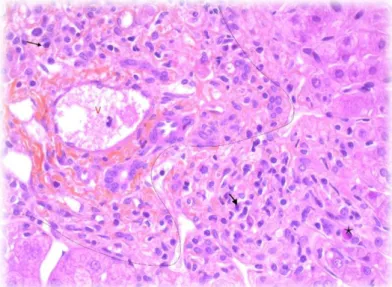
Figure 9 : Liver biopsy with sign of activity of AIH, HES staining . ... 39
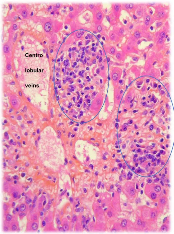
Figure 10 : Liver biopsy of AIH : Mononulcear infiltration in portal space and around the lobular central vein ... 40
Figure 11 : Liver biopsy with sign of activity of AIH, HES staining : lymphoplasmocytic infiltration crossing the bordering lamina, with apoptotic body . ... 41
Table 1 : Characteristic of population study ... 16
Table 2 : Median of ALT/AST/IgG in different group and total population ... 17
Table 3: Evaluation of PM (piecemeal necrosis), LN (lobular necrosis) and PL (presence of plasma cells) in relapse and remission group ... 19
8 Table 5: Scale of plasma cell infiltration ... 36 Table 6 :Panel of antibody used for exploration of leukocyte population in flow cytometry ( first line : stained antibody, second line target on leukocyte population) ... 37
9
I. GLOSSARY
I.1 English version
AASLD : American Association for Study Of Liver Disease AB : Antibody
AIH : Auto immune hepatitis AZA: AZATHIOPRINE
CRID : Clinical Research and Innovation Department EASL : European Association for the Study of the Liver EPC : Endpoint criteria
HBV : Hepatitis B virus
HCC : Hepatocellularcarcinoma HCV: Hepatitis C Virus
IAHG : International Autoimmune Hepatitis Group modified staging System IgG : Type G Immunoglobin
LN : Lobular Necrosis
NAFLD : Non Alcoholic fatty liver disease
NMOSD : Neuromyelitis Optica Spectrum Disorder PBC : Primary Biliary Cholangitis
PEP : primary endpoint
PL : Plasma Cells = Plamocytes PM : Piecemeal Necrosis
PPC : Protection Persons Comity RTX : RITUXIMAB
UGA : University of Grenoble Alpes
UHGA : University Hospital of Grenoble Alpes
I.2 Version Française AC: Anticorps
AZA: AZATHIOPRINE
CBP : Cholangite biliaire primitive CHC: Carcinome hépatocellulaire
10 CHUGA : Centre Hospitalier Universitaire Grenoble Alpes
CJP : Critère de jugement principale CPP : Comité de Protection des Personnes
DRCI : Direction de la Recherche clinique et de l’Innovation HAI : Hépatite auto immune
IgG : Immunolglobuline type G NL : Nécrose lobulaire
NP : Nécrose parcellaire RTX : RITUXIMAB
UGA : Université de Grenoble Alpes VHB : Virus de l’hépatite B
11
II. RESUME
FACTEURS BIOLOGIQUES, HISTOLOGIQUES ET IMMUNOLOGIQUES PREDISANT LA RECHUTE DE L’HEPATITE AUTO IMMUNE CHEZ LES PATIENTS SOUS TRAITEMENT DE PREMIERE LIGNE (AZATHIOPRINE) ET DE TROISIEME LIGNE (RITUXIMAB). S. Tedjini, JP Zarski, G. Clavarino, C. Dumestre-Perard, N. Sturm, JL Bosson.
Introduction : L'hépatite auto-immune est une maladie inflammatoire chronique du foie, rare, aux mécanismes physiopathologiques complexes, avec un taux élevé de récidive à l’arrêt des traitements : 46 % chez l'adulte et 80% chez l’enfant dont 50% à 3 mois, 90% à 28 mois, selon l’AASLD (21-23)). Les principaux facteurs de risque documentés sont le taux d'ALAT, d'ASAT, d'IgG sérique, ou encore la présence d'une inflammation persistante avec plasmocytes peri-portaux sur les biopsies hépatiques, avant arrêt du traitement. Ces éléments ont été étudiés chez des patients sous traitement standard de première ligne par corticoïdes ou Azathioprine. Les marqueurs immunologiques impliqués dans les mécanismes de la dysfonction immunitaire favorisant l'apparition et l'entretien des lésions hépatiques sont peu connus. En utilisant le rationnel d'études portant sur d'autres maladies dysimmunitaires (myasthénie et la neuromyélite optique), nous nous intéresserons à un nouveau marqueur: le taux de lymphocytes B mémoires CD27+ comme facteur prédictif de la récidive après l’arrêt des traitements. Objectif: Explorer les facteurs prédisant la récidive de l'hépatite auto-immune après arrêt des traitements, chez des patients sous traitement de 1ère ligne par AZATHIOPRINE (groupe contrôle) et de 3ème ligne par RITUXIMAB (hépatite auto immune réfractaire).
Matériels et méthodes: Création de deux protocoles d'études monocentriques sur le CHU de Grenoble, incluant 24 patients sur une cohorte initiale de 173, après vérification des critères d’inclusion et d’exclusion:
-HAI FR BIO : étude rétrospective avec comme CJP l’association entre le taux d’ALAT (mesuré en UI/L) avant arrêt du traitement et risque de récidive de l’HAI. L’étude a débuté en Juillet 2020 sur 2 mois, ses résultats sont présentés dans ce travail.
-HAI FR BIH : étude prospective permettant l’analyse de l’association entre le taux de lymphocytes B mémoires CD27+ (mesuré en % des GB), après l'arrêt des traitements tous les 3 mois et le risque de récidive de l’HAI. Ce protocole débutera en Janvier 2021 après validation du Comité de protection des personnes.
Résultats HAI-FR-BIO: Il n’est pas mis en évidence d’ association statistiquement significative entre le taux d'ALAT avant arrêt du traitement et le risque de récidive de l'hépatite auto immune ( p value:
0,148 ) Concernant les critères de jugement secondaires, nous n’avons pas mis en évidence de différence
statistiquement significative entre le taux d’ASAT , la taux IgG ou la présence de nécrose lobulaire, parcellaire, de plasmocytes sur la biopsie hépatique et le risque de récidive de l’hépatite auto immune . Conclusion : Les analyses de HAI-FR-BIO ne mettent pas en évidence d'association statistiquement significative entre le taux d'ALAT avant arrêt du traitement et le risque de récidive. Le principal facteur limitant est le faible nombre de sujets inclus s'agissant d'une maladie rare, avec réalisation d’une étude monocentrique. HAI-FR-BIH est une étude pilote qui permettra l’exploration du taux de lymphocytes B mémoires CD27+ jamais étudié dans l’HAI. L'analyse de ce nouveau biomarqueur permettrait une meilleure compréhension de la physiopathologique immunitaire complexe de cette maladie, avec l'objectif dans un futur proche de réduire le risque de récidive de l'hépatite auto immune en standardisant le suivi du patient.
MOTS CLÉS : Hépatite auto immune, Récidive, AZATHIOPRINE, RITUXIMAB,
Transaminases sériques, Immunoglobuline type G, Lymphocyte B mémoire CD27+.
0
BIOLOGICAL, HISTOLOGICAL AND IMMUNOLOGICAL FACTORS PREDICTING THE RELAPSE OF AUTOIMMUNE HEPATITIS IN PATIENTS UNDERGOING F1RST LINE (AZATHIOPRINE) AND THRID LINE (RITUXIMAB) TREATMENT.
S. Tedjini, JP Zarski, G. Clavarino, C. Dumestre-Perard, N. Sturm, JL Bosson.
Background:Autoimmune hepatitis is a rare chronic inflammatory disease of the liver with complex pathophysiological mechanisms, and a high rate of recurrence when treatment is stopped: 46% in adults and 80% in children, of which 50% at 3 months, 90% at 28 months, according to AASLD (21-23)). The main documented risk factors are ALT, AST, serum IgG, or the presence of persistent inflammation with periportal plasma cells on liver biopsy prior to discontinuation of treatment. These factors have been studied in patients receiving standard first-line corticosteroid or AZATHIOPRINE therapy. Little is known about the immunological markers involved in the mechanism of immune dysfunction that promote the onset and maintenance of liver injury. Using the rationale of studies on other dysimmune diseases (myasthenia gravis and optic neuromyelitis), we will focus on a new marker: the CD27+ memory B lymphocyte level as a predictor of recurrence after stopping treatment.
Objective:To explore factors predicting recurrence of autoimmune hepatitis after discontinuation of treatment, in patients undergoing 1st line treatment with AZATHIOPRINE (control group) and 3rd line treatment with RITUXIMAB (refractory autoimmune hepatitis).
Material and Methods: Creation of two monocentric study protocols at the Grenoble University Hospital, including 24 patients out of an initial cohort of 173, after verification of the inclusion and exclusion criteria:
-HAI FR BIO: retrospective study with as CJP the association between the ALT level (measured in IU/L) before stopping treatment and the risk of HAI recurrence. The study started in July 2020 over 2 months, these results are presented in this paper.
-HAI FR BIH: prospective study allowing the analysis of the association between the CD27+ memory B lymphocyte rate (measured in % of GB), after stopping treatment every 3 months and the risk of HAI recurrence. This protocol will begin in January 2021 after validation by the Committee for the Protection of Persons.
Results HAI-FR-BIO: There was no statistically significant association between the ALT level before discontinuation of treatment and the risk of recurrence of autoimmune hepatitis (p value: 0.148). Regarding secondary endpoints, no statistically significant difference was found between the AST level, the IgG level or the presence of lobular, fragmentary or plasma cells necrosis on liver biopsy and the risk of recurrence of autoimmune hepatitis.
Conclusion: Analyses of HAI-FR-BIO did not show a statistically significant association between the ALT rate before treatment discontinuation and the risk of recurrence. The main limiting factor is the small number of subjects included as this is a rare disease; the study being monocentric moreover. HAI-FR-BIH is a pilot study that will explore the level of CD27+ memory B lymphocytes never studied in HAI. The analysis of this new biomarker would allow a better understanding of the complex immune pathophysiology of this disease, with the goal in a close future of reducing the risk of recurrence of autoimmune hepatitis, by standardizing patient follow-up.
KEY WORD : Autoimmune hepatitis, Recurrence, AZATHIOPRINE, RITUXIMAB, Serum transaminases, Immunoglobulin type G, CD27+ memory B lymphocyte
1
IV. INTRODUCTION
Auto immune hepatitis or AIH is a chronic inflammatory disease of liver occuring at any age, affecting preferentially women and girls (71%-95% , 60%-76%) with no regards to ethnicities
(1). The first description was made in 1950 by Waldenström as a disease affecting young
women characterized by jaundice, high serum gammaglobulin level, and amenorrhea causing liver cirrhosis. It has been known by many different labels including “lupoid” hepatitis, but AIH has been accepted as the most appropriate term. The wide heterogeneity of clinical presentation and relatively rare incidence of this disease, from 16 to 18 cases per 100000 persons in Europe (2), has limited the advancement in clinical trials. Therefore, more than 50 years after its original description, AIH remains a therapeutic challenge.
The physiopathology is very complex and link to dysfunction of immune homeostasis with an increase in the Th2 and Th1 response and a decrease in circulating regulatory T cells (3,4). The immune models currently described imply activation of naive T lymphocytes by antigen presenting cells, in the presence of an autoantigen (HLA class II), releasing cascade of activation of the TH1 pathway (via interleukin 2) with a pro-inflammatory effect and the TH2 pathway (mainly mediated by interleukin 4) with a pro-fibrosis effect (5,6). Activation of the TH 17 pathway induces an increase in pro-inflammatory and pro-fibrotic cytokines while stimulating hepatocyte survival. At the same time, we can constate a qualitative and quantitative deficit of the regulatory T lymphocytes making it impossible to stop production of adenosine, inflammation, hepatocyte destruction and pro-fibrosis effect on liver.
There are currently several types of autoimmune hepatitis characterized by different serological profiles. The validated detection technic for autoantibodies is indirect immunofluorescence (ILF) for the vast majority of antibodies: antinuclear antibodies, anti-smooth muscle antibodies, anti-LKM antibodies (anti-kidney microsome antibody) with the exception of anti-SLA / P (Soluble Liver Antigen) detected only by the Elisa or Western Blot methods. 20% of AIH are seronegative.
According to the latest guidelines of European Association for the Study of the Liver (EASL guidelines 2015), the most common is type 1, representing 90% of all AIHs. It is characterized by the presence of anti-nuclear antibodies (65%), anti-smooth muscle anti-actin F type AB, anti-SLA AB. These are mainly women, of all ages with HLA DR3 and DR4 or DR13. This form of autoimmune hepatitis usually occurs in adulthood and is often cortico-sensitive (7,8).
2 Autoimmune hepatitis type 2 accounts for 10% of AIHs and is characterized by the presence of antibodies directed against the endosplasmic reticulum: anti-liver microsome or LKM-1 antibody, rarely LKM-3 and anti-liver cytosol or anti LC1 antibody. It occurs mainly in childhood with a positive HLA DR3 and DR7 profile. The disease is generally more severe and responds badly to the administration of corticosteroids and immunosuppressive drugs (2). Finally, autoimmune hepatitis type 3 is very similar to type 1 and characterized by presence of anti SLA / P (the only antibody specific of AIH and a poor prognosis factor) and often Ro S52 antibody (2).
Other autoantibodies are currently described to help with the diagnosis of seronegative forms: atypical anti-neutrophil cytoplasmic antibody p ANCA and anti-mitochondrial antibody AMA (present in primary biliary cholangitis, described in 8-12% of cases in AIH with or without overlap syndrome) (2,9).
These auto antibodies also have a prognosis value. Indeed their blood levels are correlated in children and adults to the severity of the disease and to treatment response. However they are not correlated to AIH activity along the follow-up. Presence of anti SLA antibody (only specific AIH antibody) is associated to a severe disease and a higher risk of relapse after stopping treatment (9) .
Note that presence of anti-nuclear antibodies and anti-smooth muscle antibodies is also described in chronic liver disease of: PBC, HBV, HCV, NAFLD and even in alcoholic cirrhosis. However, presences of both antibodies increase the diagnostic sensitivity of AIH from 58 to 74 %. (9)
Other immunological features of this disease include the presence of hypergammaglobulinemia (IgG) in 90% of patients.
Histological analysis is essential for positive diagnosis, evaluation of AIH activity and liver fibrosis gradation with or without cirrhosis at diagnostic time (30% of adults and 38% of children). In particular, it eliminates alternative diagnosis. There are no specific sign of autoimmune hepatitis on liver biopsy, however 4 lesions are characteristic (10):
1-The most typical lesion is the interface hepatitis (66% of case), also called piecemeal necrosis, which denotes inflammation of hepatocytes at the junction of the portal tract and hepatic parenchyma.
3 When interface hepatitis is absent or mild, AIH is unlikely.(11).
2- Irregular distribution and intense portal infiltrate with either periportal or (in cases with bridging fibrosis or cirrhosis) paraseptal interface hepatitis is typical. This infiltrate is composed of plasma cells, eosinophils and lymphocytes. The plasma cells are characteristic, but they are not always the dominant cells in 1/3 of AIH, and in the appropriate clinical setting, the diagnosis can be made with only modest numbers. In some cases, lymphocytes are dominant instead. When this occurs, plasma cells in small clusters at the interface support the diagnosis
(12).
3-Rosette formation (33% of AIH) is seen with significant lobular involvement and regenerative activity.
4- Emperipolesis (65 % of AIH ) caracterised by penetration of an intact cell within a single intact cell difference from phagocytosis (12,13).
Portal-to-portal or portal-to central fibrosis and cirrhosis are seen. Other features also help to establish the diagnosis. For example, AIH differs from chronic hepatitis C in having more severe lobular inflammation and necrosis as well as greater numbers of plasma cells, more marked interface hepatitis, and broad areas of parenchymal collapse.
Centrilobular necrosis is well described in AIH : hepatocyte necrosis, inflammation and centrolobular haemorrhage are described in 17 % of AIH (14,15). These lesions are rarely isolated, mostly associated with interface hepatitis.
Biliary changes are uncommon in AIH and are almost always indicative of some other disorders. However isolated bile duct injury as destructive lymphocytic cholangitis, can be seen in 10-15% of AIH and does not exclude the diagnosis.
Regarding the severe or fulminant autoimmune hepatitis 4 lesions are described (16) : -Extensive necrosis with centrolobular haemorrhage
-Centrovenular endothelitis
-Lymphocyte follicle absent in case of chronic AIH -Infiltration mainly composed by plasma cell
Key histological features contributing positively to a score allowing diagnosis of AIH, according to the revised International Autoimmune Hepatitis Group modified staging System (IAHG) of 2008, are interface hepatitis (+3), which is the most important; a lymphoplasmacytic
4 infiltrate (+1); and rosette formation (+1) with a specificity diagnosis of 89 %. This simplified score has a good specificity (90% vs 73%) but lower sensitivity (100% vs 95%) compare to the original of 1990. These scores are a diagnostic aid whose use is limited in children , acute and
fulminant AIH or even overlap syndrome (12).
The current recommendation of the learned societies (2,8,17) is to treat all patients with active or advanced disease at the fibrosis / cirrhosis stage. Asymptomatic patients require individual management according to their risk factors (age, comorbidity, etc.)
Finally, decompensated cirrhotic patients or those without signs of inflammatory activity on the biopsy do not benefit from the treatment.
First-line treatment is based on the administration of corticosteroids alone or in combination with AZATHIOPRINE (IMUREL®).
The second line of treatment is composed, without a high level of evidence, of mTor inhibitors and calcineurin inhibitors.
Finally, in the event of the failure of the two first lines therapy with contraindication to liver transplantation, a therapeutic alternative could be RITUXIMAB (MABTHERA® : anti-CD20 monoclonal antibody) for which is currently not licensed therapy in AIH.(13,14) . It is a chimeric (murine / human) monoclonal antibody directed against B cells. RITUXIMAB is currently licensed for positive B cell lymphoma, rheumatoid arthritis, or vasculitis associated with anti-neutrophil cytoplasmic antibodies.
Biological remission is defined as the normalization of ALAT, ASAT levels and IgG concentration. Approximately 80 to 90% of patients present an improvement in transaminase levels quickly with the institution of these immunosuppressive drugs; 20% obtained prolonged remission after stopping treatment according to the EASL repository, with a median follow-up of more than 6 years. (8)
Histological remission, defined as minimal hepatitis (AIH score less than or equal to 4 on biopsy), is rarer and lags behind normalization of laboratory values by at least 6-12 months (2). Indeed around 45% of the patients who maintain a ALT level and gamma globulin concentration in the limit of the normal value present a persisting histological activity and therefore as shown in the study of Dhaliwal HK of 2015 a higher need for liver
5 transplantation :« failure to attain remission histologically despite doing so biochemically is
associated with >2-fold increased risk of progression to transplantation and all-cause mortality »(20)
After obtaining remission, treatment should be continued for three years and at least 24 years months after the normalization of transaminase and IgG levels (biological remission).
However, the recurrence of the disease after stopping treatment is very frequent and is defined by the raise in ALT to 3 times upper the limit of normal value or moderate elevation in ALT and IgG> 20ng/mL according to IAHIG criterion. Relapse rate is estimated at 46 % in adults to 80% in children in the largest American cohorts according to AASLD. In 50% of case it occurred in the first three months and in 90% of case in the first 28months after stopping treatment (21–23). In Europe, the Dutch AIH working Group have reported a relapse rate of 59%, 81% and 88% at 1, 3 and 5 years, respectively, even when treatment was tapered and/or discontinued after clinical and biochemical remission had been sustained for >2 years (21).
The risk of relapse appears to be higher in young patient (< 18 years old), and in HLADRB1*04:01 positive patient (24).
If relapse occurred, the recommendation of AASLD guidelines 2010 is re-introduction of the first line of treatment with corticosteroids more or less associated with AZATHIOPRINE. After two recurrences of HAI, subsequent discontinuation of treatment after remission is contraindicated (2)
The predictive factors for relapse described in the literature in patients receiving conventional first line treatment are:
-On the biological level, we describe a rate of ALT at 50 % of the normal upper limit and IgG level > 12g/L measured before stopping treatment (25) as predictive factor of relapse.
- Histologically, the predictors of recurrence identified in the literature are the persistence of
inflammation with lobular necrosis and piece meal necrosis (interface hepatitis). More recently, one study of 2004 introduced the persistence of periportal plasma cells on liver biopsy and before treatment stopped, as a predictive factor of relapse.
-In immunological terms: Currently, there are no validated markers for monitoring
6 gravis and optic neuromyelitis (NMOSD) , which also are auto immune disease treated with RTX, explorate a new biomarker for monitoring inflammation and relaspe of disease after stopping treatment : the level of CD27 memory B lymphocytes +. These type of lymphocytes B allow in physiological condition to increase and prolonge the immune response against antigen that triggers its differentiation. For all we know, patient undergoing treatment by RTX can be follow by measurement of lymphocyte B CD20+ in the blood, but with a delay of re-emergence of at least 6 months but some patients will relapse before. These memory B cell seems to be more sensitive to relapse in NMOSD and myasthenia that usual B lymphocyte. Level of CD27+ memory B lymphocyte has never been tested in patients with autoimmune hepatitis treated with RTX. Based on the same rationale as the studies cited above, the CD27 + memory B cells rate could be a witness to RTX-induced "therapeutic" immunosuppression and its rise at a distance from treatment would predict recurrence of AIH.
The aim of our study was to explore in refractory autoimmune hepatitis patients on RITUXIMAB (a new third-line treatment) the already validated predictive factors for relapse in patients on standard first-line therapy (i.e. AST, ALT, type G immunoglobulin levels and histological features) and a new immunological marker used in the follow-up of other autoimmune diseases (such as myasthenia gravis or optic neuromyelitis): blood level of CD27+ memory B lymphocyte after cessation of treatment.
V. MATERIAL AND METHODS:
In order to explore predictive factors of autoimmune hepatitis recurrence we have created two study protocols.
The first protocol is named HAI-FR-BIO, it allows the study of serum transaminases, immunoglobulins and liver biopsy slides, measured before the cessation of treatment. It is a retrospective study without patient intervention (research not involving the human person). The second protocol, named HAI-FR-BIH, is prospective and will mainly allow the study of new blood immunological markers after the cessation of treatments (research involving the human person).
7 Each study takes place in France in one center, University Hospital of Grenoble Alpes (UHGA). The population was composed of male and female, aging between 18 to 75 years old, with seronegative or seropositive AIH all histologically proven and followed in the UHGA. We retrospectively assessed the number of patients followed in UHGA for AIH from 2007 to 2020 i.e. 173 patients. From this database, only 24 patients meet protocol inclusion and exclusion criteria presented below. We could therefore estimate a number of subjects including at 22 subjects considering that 90 % gave their non-opposition.
Patients were separated into 2 groups, one under standard therapy use as a group control and this other under new therapy by RITUXIMAB:
-Group 1 (control): patients who received a first line therapy by AZATHIOPRINE in monotherapy and currently in remission (A) or in relapse (B).
-Group 2: patients with refractory AIH who received a third line of treatment by RITUXIMAB and currently in remission (C) or in relapse (D).
• Inclusion criteria:
- Male or female from 18 to 75 years old followed at UHGA
- Seronegative autoimmune hepatitis (negative Actin F smooth muscle AB , negative SLA AB, negative LKM1 AB , negative LC1 AB) or seropositive (positive Anti-Actin F smooth muscle antibodies, positive Anti-SLA AB, positive Anti-LKM1 AB or positive Anti-LC1 AB )
- Autoimmune hepatitis was histologically proven (liver biopsy in favor). - Having had a treatment interruption for AIH remission in the last 3 months - Not having objected to the study after clear, fair and intelligible information
• Exclusion criteria:
-Patients with Overlap syndrome (an overlap syndrome between autoimmune hepatitis and cholestatic liver disease).
-Refractory AIH under alternative treatment other than RITUXIMAB. -Liver transplantation in group 1.
8 V.2 FIRST PROTOCOL: HAI-FR- BIO
We began our analysis by creation of this small assay as a preliminary analysis. It only focused
on retrospective computorized data and paper medical records available in the
Hepato-gastro-enterology Department. Validation of this protocol was obtained in July 2020 by the
Department of Clinical Research and Innovation. Data were collected between July to August
2020 and data analysis was performed in August 2020.
V.2.1 Purpose of the study:
The primary end point criteria is assessed retrospectively: description of the relation between ALT rate measured in UI/L within one month before stopping treatment and the risk of AIH recurrence after treatment stopped in group 1 and 2.
The second end point criterias are multiple, also assessed retrospectively in the two groups: -Description of the report between AST level in UI/L, IgG in g/L measured within one month before stopping treatment, and the risk of AIH recurrence after treatment stopped in group 1 and 2.
-Description of the relationship between the presence of parcel, lobular necrosis according to Metavir score (cotation of NP from 0 to 3, NL from 1 to 2), plasma cell on a liver biopsy performed before stopping treatment, and the risk of AIH recurrence after treatment stopped in group 1 and 2.
This first protocol is a pilot study, allowing a retrospective description and analysis of
well-known predictive factor (transaminases, immunoglobulin and liver biopsy before treatment stopped) of AIH relapse.
The study of innovative immunological factor in serum (CD27+ memory B cells serum level) and on liver biopsy (immuno-labelling in formalin of CD27+ memory B cells, plasmocytes and CD20+ memory B cells) of patients monitored for autoimmune hepatitis requires the creation of a prospective study concerning human being called "HAI-FR-BIH".
9 V.3 SECOND PROTOCOL: HAI-FR-BIH
The protocol was created between 2019 and 2020, at Grenoble University Hospital with involvement of Hepatologists, Immnologists, Pathologists and Statisticians. This is a type 3 study of the Jardé law, involving the human being. It was submitted to the Department of Clinical Research and Innovation in June 2020 and will be deposed at Protection of Persons Committee in November 2020 (awaiting budget approval)
HAI-FR-BIH is an explorative monocentric, observational and prospective study (with retrospective data collection also) succeeding to HAI-FR-BIO that will take place in the same center: Grenoble University Hospital, and will debute in January 2021.
V.3.1 Purpose of the study:
In this prospective and retrospective research, we differentiate between two types of judging criteria. Retrospective judgment criteria already studied in preliminary analysis by HAI-FR-BIO, and prospective criteria justifying the creation of this new protocol.
• Retrospective end point criteria
The primary end point is the same that HAI-FR-BIO and based on strong guidelines of
AASLD (2):
Study of the relationship between the ALT level measured in IU / L, 1 month before stopping treatment and the recurrence of AIH after treatment stopped, with determination of an ALT threshold in absolute value, in two groups of patients (group 1 and 2).
The secondary end points are:
1a / Description of the relationship between AST level value measured in IU / L, within one month before stopping treatment, and the risk of recurrence of AIH after treatment stopped, in the 2 groups (group 1 and 2).
1b/ Description of the relationship between IgG concentration measured in g /, L within one month before stopping treatment, and the risk of recurrence of AIH after treatment stopped in the 2 groups (group 1 and 2).
10 • Prospective end point criteria
2/ Study of liver biopsy performed before stopping treatment with RITUXIMAB (group 2) and AZATHIOPRINE (group 1) to analyze the relationship between:
-a) The inflammatory activity of AIH, defined by the presence of piece meal necrosis and lobular necrosis, and the risk of recurrence of AIH after treatment stopped
-b) The presence of plasma cells, CD 20 +, CD27 + memory B cells and the risk of recurrent AIH after treatment stopped.
3 / Description of the relationship between the rate of blood CD27 + memory B cells (measured in% relative to leukocyte count) at 3, 6 ,9 and 12 months after stopping treatment, and the risk of AIH recurrence after treatment stopped, only in group 2.
V.3.2 Research process
After signing the consents, participants will be seen again by the principal investigator at the inclusion visit and every 3 months for 12 months (M3-M6-M9-M12, +/- 15 days), as part of routine care, with an interview, a standard clinical examination and subsequent biological sampling:
1) The main samples for routine monitoring of autoimmune hepatitis will be taken every 3 months as part of routine care: serum ASAT/ALAT transaminases in IU/L, and a weight assay of IgG in g/L.
2) A sample consisting of 2 EDTA tubes of 6 ml with 1 dry tube of 7 ml containing a serum separator will be taken to perform the following assays:
o Routine care:
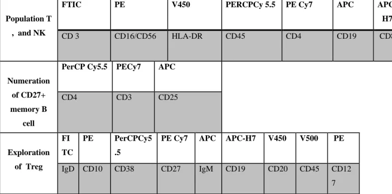
-Immunophenotyping by flow cytometry for the study of T, NK, and B lymphocyte subpopulations, in particular CD27+ memory B lymphocytes (1 EDTA tube of 6 ml).
o Excluding routine care:
Immunophenotyping by flow cytometry for exploration and characterization of a lymphocyte sub-population: T reg (T regulatory lymphocyte).
11 o Excluding routine care and with storage carried out in the gastroenterology department : -Serum storage (1 dry tube with 7ml serum separator) for analysis of cytokines and chemokines of interest, such as IP10(8), IFN-gamma, IL-12, IL-6, IL-10 and galectin-9 (16).
-Storage of isolated PBMCs (from 1 6 ml EDTA tube) and frozen at -80° for exploration of the above cytokine and chemokine genes by quantitative PCR. These analyses will be carried out in a second phase subject to budget and human resources availability.
3) At inclusion and in routine care, a search for anti-tissue antibodies by indirect immunofluorescence, the search for liver marker antibodies by immunodot will be carried out for patients with an unknown serological profile.
Finally, we will review the liver biopsy slides taken before stopping treatment in certain patients. These slides are currently stored in the pathology laboratory of UHGA and will be reviewed with the supervision of Pr N. Sturm in charge of UF ACP.
On these slides, we will analyze the following criteria: - Quantification of parcel and lobular necrosis
- Description of the presence of interface hepatitis
- Semi-quantitative evaluation by immuno-labelling of plasma cells (CD38+ and MUM1), memory B lymphocytes CD 20+ and CD27+. Note that the labelling of CD27+ memory B lymphocytes requires the use of a new antibody produced by Abcam (Anti-CD27 antibody - Monoclonal rabbit clone [EPR8569] - ref. ab131254 -(dilution between 1/100 and 1/250)).
12
V.3.3 Projected study milestones
-“Filfoie” network call for tenders filed on July 31, 2020. -PPC Protocol filings expected November 2020
-Start of the protocol January 2021 according to the following schedule: -Start of inclusions and end of inclusions: January 2021 - June 2021 (6 months) -Duration of participation of each subject: 12 months
-End of data collection : June 2022
-Expected date of basic freeze: November 2022 -End date of statistical analysis: February 2023 Total research duration: 26 months
Inclusion visit by investigator
-Information formulary and collection of the non-opposition -Interrogation - clinical examination - collection of biologies of current practice
-Validation of eligibility criteria
Follow-up consultation every 3 months (M3-M6-M9) : Clinical examination and blood
test.
End of study consultation at 12 months Clinical examination and blood test.
Consultation / hospitalization in case of recurrence ( R ): Clinical examination and blood
13 V.4 MEANS :
V.4.1 Study of Immunological parameters
• Immunofluorescence staining and flow cytometry analysis methods:
Phenotypic analysis of lymphocyte (T, B, Treg and NK) suspensions will be performed by flow cytometry. Cells will be stained with a different panel of colored antibody combinations. The following monoclonal antibodies will be used as a “backbone” in all combinations: CD3-FITC, PE-CD16/CD56 , PerCP-Cy5.5- CD45, PECy7-CD4, APC-IgM, APC-CD25, APC-H7-CD19, PeCy7-CD3 , PECy7-CD4, CD10-PE, CD20-V450, CD127-PE, HLA-DR V450, CD38-PerCP Cy5.5, PerCP Cy5.5-CD4, CD19-APC, APC-H7-CD8, CD45-V500 (BD Biosciences) , IgD FTIC (Agilent/Dako). Clone and isotype specificity of these antibodies are detailed in Table 6. The antibodies will be used at the dilution recommended by the manufacturers. Samples containing 106 cells in a volume of 100 μl will be incubated with the combination of antibodies at the supplier recommended concentration during 20 minutes in the dark, at 4°C. After erythrocytes lysis with NH4Cl at 4°C for 5 minutes, samples will be washed with HBSS medium (Gibco).
Analysis will be then performed using 3-laser, 8-colour BD FACSCanto II flow cytometer (BD Bio- sciences) and FACSDiva software version 6 (BD Biosciences). BD CompBeads (BD Biosciences) is used for compensation settings. Cytometer performances will be checked daily using CST beads (BD Biosciences).
• Study of Histological parameters:
Finally, we have reviewed liver biopsy slides available in several patients. These slides were produced before the decision to stop treatment and the analysis of the histological activity with parcel and lobular necrosis, semi quantitative evaluation of plasma cells (using CD38+ AB and MUM labeling) ,CD 20 + and CD27 + memory B cells on biopsy.
This evaluation is performed under the supervision of a senior Pathologist Pr N Sturm, in the pathology department using old biopsies of patients stored in IBD.
14 Graduation of lobular and parcellar necrosis is based on the Metavir scoring system which evaluated necrotico-inflammatory activity without taking into account portal inflammation contrary to Ishak Score (Figure 6).
The proofreading of liver biopsy is realized under optical microscope, slide are already marked with HES staining and for some of them MUM immunostaining of the plasma cells is available. Immunostaining by CD38+ AB for plasma cells and anti-CD27+ memory B cells antibody will be performed in the second protocol after getting patient and PPC consent.
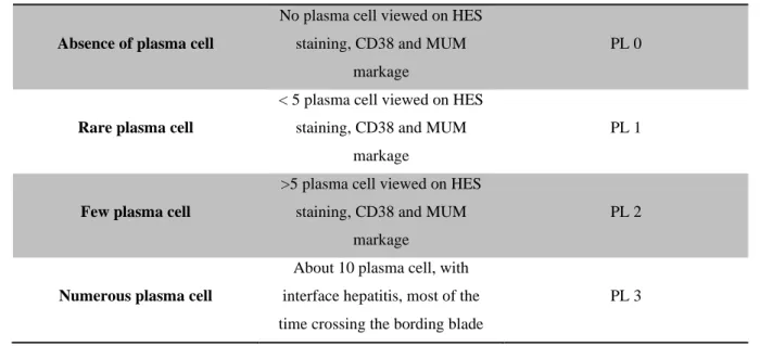
The labeling of CD27 + memory B cells involves utilization of a new antibody produced by Abcam (Anti-CD27 antibody - Monoclonal rabbit clone (dilution between 1/100 and 1/250.) Semi quantitative evaluation of plasma cell is performed using a specific classification describe in Table 4.
V.5 STATISTICAL ANALYSIS:
As a pilot, monocentric and descriptive study, the descriptive analysis initially consists of measurement of medians, interquartile concerning continuous variables of no normal distribution.
Concerning dichotomous variables, we will express them in percentage and effective.
The first and second end point criteria of the two protocols analyze continuous variables with application of a non -parametric “Wilcoxon-Mann-Whitney” test in the absence of all conditions of validity for the Student test.
Concerning the second end point criteria exploiting biopsy, the analysis is performed on dichotomous variables with the use of a Chi2 or Fischer test when the Cochran criteria are not met.
The condition for applying the Student test is the normality of the analyzed parameters. The application conditions according to Cochran's criteria for the Chi2 test are a workforce greater than 20 individuals, and a theoretical workforce in the comparison of variables greater than 5 individuals for each subclass.
15
VI. RESULTS OF HAI-FR-BIO:
VI.1 Demographics:
Data were collected retrospectively in the collection of 173 biopsies established from 2007 to 2020 in University Hospital of Grenoble. A total of 24 patients were recruited for HAI-FR-BIO protocols according to inclusion and exclusion criteria, without opposition. Twenty patients were under AZATHIOPRINE therapy (group 1) and only four under RITUXIMAB therapy (group 2).
All patients were in sustainable remission for at least 2 years in the AZA group as recommended in EASL guidelines 2019. However in RTX group only one patient respected a delay of 2 years in remission before stopping treatment. A total of 6 patients relapsed, 4 in group 1 and 2 in group 2. Mean delay of relapse was 20.5 months (range: from 3 to 48 months) after treatment stop; only one patient relapsed in the first 3 months.
Most patients were female (62,5%), with a median age of 56 years-old (range 26-75). In this population 33% patient had an associated auto immune disease such as hypothyroid, Sjogren syndrom, connectivitis and even 1 vasularitis (knotty periarteritis). Cirrhosis was present in 5 patients (20 %) and always well compensed (Child Pugh A). Analysis of immunological profile showed a majority of type 1 AIH (58%), equality of type 2 AIH (20%) and seronegative AIH (20%). Primary analysis was performed on biochemical, immunological and histological parameters using the first protocol (HAI-FR-BIO) as indicated in material and methods.
POPULATION OF UHGA :
173 patients labeled HAI on liver biopsy between 2007 and 2020
GROUP 1 : AZATHIOPRINE GROUP 2 : RITUXIMAB Remission 1A Relapse 1B n = 4 Remission 2C Relapse 2D n = 2 Included: N =24 Excluded -Overlap sd : n= 54
-Relapse before treatement stop n= 25 -Alternative therapy: n= 20 -LT in group 1 : n= 13 -Lost to follow up : n= 12 -Diagnosis error: n= 12 -Death: n= 10 -Extreme age: n= 3
16
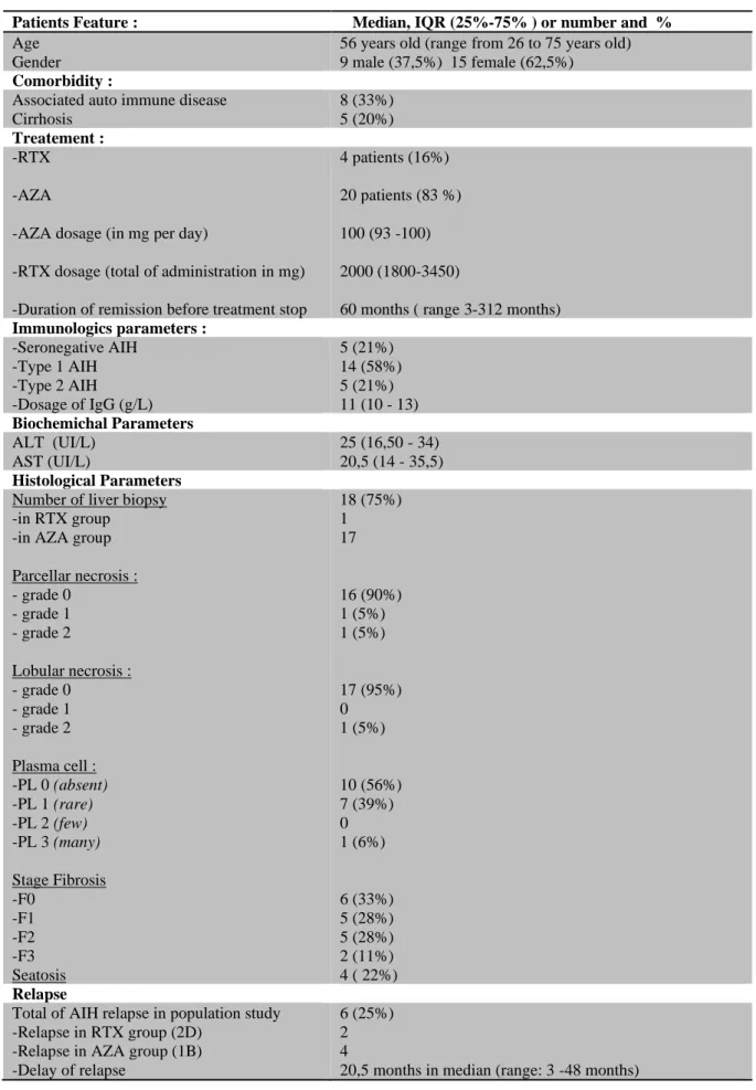
Table 1 : Characteristic of population study
Patients Feature : Median, IQR (25%-75% ) or number and % Age
Gender
56 years old (range from 26 to 75 years old) 9 male (37,5%) 15 female (62,5%)
Comorbidity :
Associated auto immune disease Cirrhosis 8 (33%) 5 (20%) Treatement : -RTX -AZA
-AZA dosage (in mg per day)
-RTX dosage (total of administration in mg) -Duration of remission before treatment stop
4 patients (16%) 20 patients (83 %) 100 (93 -100) 2000 (1800-3450)
60 months ( range 3-312 months) Immunologics parameters : -Seronegative AIH -Type 1 AIH -Type 2 AIH -Dosage of IgG (g/L) 5 (21%) 14 (58%) 5 (21%) 11 (10 - 13) Biochemichal Parameters ALT (UI/L) AST (UI/L) 25 (16,50 - 34) 20,5 (14 - 35,5) Histological Parameters
Number of liver biopsy -in RTX group
-in AZA group Parcellar necrosis : - grade 0 - grade 1 - grade 2 Lobular necrosis : - grade 0 - grade 1 - grade 2 Plasma cell : -PL 0 (absent) -PL 1 (rare) -PL 2 (few) -PL 3 (many) Stage Fibrosis -F0 -F1 -F2 -F3 Seatosis 18 (75%) 1 17 16 (90%) 1 (5%) 1 (5%) 17 (95%) 0 1 (5%) 10 (56%) 7 (39%) 0 1 (6%) 6 (33%) 5 (28%) 5 (28%) 2 (11%) 4 ( 22%) Relapse
Total of AIH relapse in population study -Relapse in RTX group (2D)
-Relapse in AZA group (1B) -Delay of relapse
6 (25%) 2 4
17 VI.2 Biochemical Parameters:
Analysis of aminotransferase levels: ALT and AST levels measured one month before
treatment stopped in total population show a median of 25 UI/L (IQR 25%-75%: 16,50 - 34)
for ALT and 20.5 UI/L (IQR 25%-75%: 14 - 35,5) for AST with no normal distribution.
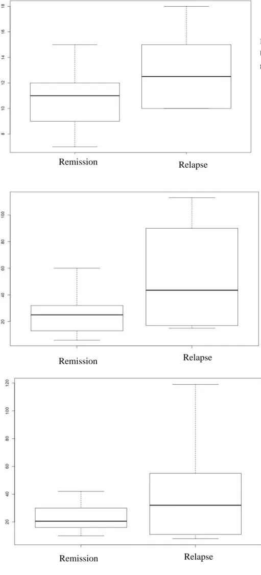
-Median of ALT measured one month before treatment stopped is superior in relapse group 1B and 2D: 43,5 UI/L (IQR 25%-75% :17 - 85) compared to remission group 1A and 2C: 26,5 UI/L (IQR 25%-75%: 14,5 - 31,5) (Figure 3). Nevertheless, this tendency, there is no significant statistical difference between the 2 groups after treatment stopped (p value of 0,148 by using
the Mann Withney test).
-Median of AST measured one month before treatment stopped, is also superior in relapse group 1B and 2D: 32 UI/L (IQR 25%-75%: 11,75– 53,75) compared to the remission group 1A and 2C: 20,5 UI/L (IQR 25%-75%: 16,25– 29). (Figure 4). However, no significant correlation is observed between AST rate and relapse of AIH after treatment stopped (Mann Withney test: p
value 0,333).
VI.3 Immunological Parameters:
Analysis of IgG concentration measured one month before treatment stopped in total
population show a median of 11g/L (IQR 25%-75%: 10-13) with no normal distribution. Median of IgG concentration measured one month before treatment stopped is a higher in relapse group 1B and 2D: 12,5 g/L (IQR 25%-75%: 10,5– 14,5) compared to remission group 1A and 2C: 11g/L (IQR 25%-75%: 9,25–12). However, there is no statistical difference between the 2 groups (Mann Withney: p-value 0. 191) (Figure 2).
All these results are presented in Table 3 below.
Table 2 : Median of ALT/AST/IgG in different group and total population
Relapse group Remission group Population
study
1B 2D total 1A 2C total total
ALT in UI/L 17 50 43,5 24 20,5 26,5 25
AST in UI/L 14 87 32 22 22,5 20,5 20,5
18
Figure 4 : ALT median (vertical scale) box plot in remission and relapse group (horizontal scale) Figure 3 : IgG median (vertical scale) box plot in remission and relapse group (horizontal scale)
Figure 5 : AST median (vertical scale) box plot in remission and relapse group (horizontal scale)
Relapse
Remission Relapse
Remission Relapse
19 VI.4 Histological Parameters:
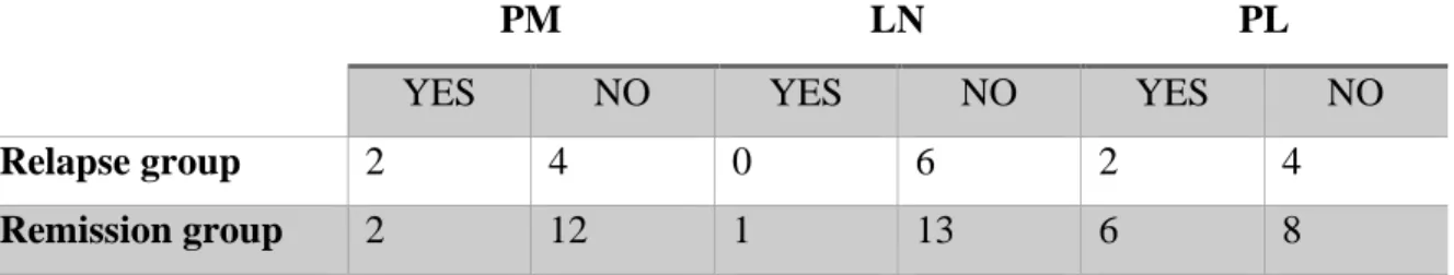
Liver biopsy analysis was performed in 18 patients (17 in AZA group and only 1 in RTX group). -Piecemeal necrosis (PM) is present in only 2 patients, but only in remission group (table 3 and
figure 6). Evaluation of relationship between PM and relapse after treatment stop is not
statistically significant (p-value : 1 at Fischer test).
-Lobular necrosis (LN) is present in only 1 patient with remission (table 3 and figure 6). Evaluation of association between LN and relapse is not statistically significant (p value: 1 at
Fischer test).
-Plasma cells (PL) are present in 8 on 18 biopsy (45%) most of time in rare number (i.e. grade
PL1 : 39% of the biopsy ) and in only one liver biopsy we observed many plama cells (i.e. grade PL3). Evaluation of relationship between presence of plasma cells and relapse after treatment
stop was not statistically significant (p-value : 1 at Fischer test). Results are displayed in table 3 and Figure 6.
Table 3: Evaluation of PM (piecemeal necrosis), LN (lobular necrosis) and PL (presence of plasma cells) in relapse and remission group
PM LN PL
YES NO YES NO YES NO
Relapse group 2 4 0 6 2 4
20
VII. DISCUSSION :
VII.1 Predictive factor of relapse of HAI-FR-BIO:
This first protocol HAI-FR-BIO dos not highlight significant association between the known biological and histological markers quoted earlier and relapse, with a principal limitation linked with the low effective of studied patient.
Concerning the primary end-point criteria, we observe a tendency to a higher ALT level in relapse group (43.5 UI/L) as compared to remission group (26.5 UI/L) with a wide dispersion of value (IQR 25% :17 UI/L – IQR 75% 85 UI/L) sometimes above the normal value related to the fact that most city laboratories do not have the same standards. To overcome this variability, these rates are generally expressed qualitatively (% of normal value) in the literature which was not possible in our study due to the lack of data in the medical records at the time of collection (normal range of city laboratories not recorded). Note also that the population study is very heterogeneous with potential uncontrolled recurrence factors such as age, autoantibodies, etc... Another bias in RTX group could be the delay of remission before treatment stopped, which is inferior of 2 years of sustainable biological remission in 3 on 4 patient, as normally recommend by EASL guidelines 2015(8). Of course, these recommendations are difficult to apply to the patient on RITUXIMAB in view of the treatment modalities and its pathophysiological mechanism that is discuss later. A recent study of Harlt and al. in J.Hepatol 2015 (25) demonstrates that a rate of ALT level below the upper limit combined with an IgG level of less than 12g/L before treatment stopped, could be a reliable predictive factors of sustained remission after treatment cessation (25). In this study ALT level is analyzed as predictive factor of relapse on 28 patients treated by AZATHIOPRINE or CORTICOSTEROID, with a sustainable remission of at least 2 years before treatment stopped. On these 28 patients, 54% patient belong to remission group and 46% to relapse group, with a median of follow up of 28 months after treatment cessation. They observe a median of ALT before treatment cessation at 20.1 UI/L (IQ 95%: 14-34) in relapse group VS 14.7 UI/L (IQ 95%: 8-17) in remission group, with a significant statistical difference (p value 0.008 with Man Withney test). Based on these results and using the Nquery software, to detect a statistically significant relationship in our study between the ALT level before treatment cessation and the risk of autoimmune hepatitis recurrence (with alfa risk of 5% and beta-risk of 20%) we should have included 68 patients. Despite the size of the initial population of approximately 150 patients, this target of 68 patients
21 enrolled is not achieved because most patients had exclusion factors such as overlap syndrome or a progressive discontinuation of AZATHIOPRINE with recurrence before treatment was totally stopped. Note also that the population study is very heterogeneous with potential uncontrolled recurrence factors such as age, autoantibodies, etc...
Moreover in the study of Harlt and al in J.Hepatol 2015 (25), we observed 62% of relapse before 6 months ,77% in 10 months and 92% at 1 year . In our study the delay of relapse after treatment cessation seem to be longer with a median of 20.5 months (range 3-48 months).
Finally, the study of the association between IgG concentration before stopping treatment and recurrence of AIH after treatment stopped is also not statistically significant, but we could observe a tendency toward a higher level in the relapse group.
Regarding analysis of histological markers, we also do not observe statistically significant correlation between the presence of plasma cells before treatment stopped and the risk of recurrence of autoimmune hepatitis after treatment cessation, mainly related to the low number of liver biopsies available in relapse group (4 biopsies) compare to remission group (14 biopsies). In recent publication, we do no longer consider persistence of inflammation on liver biopsies before treatment discontinuation (defined by AIH score greater than 3) as a predictive factor of relapse, but we rather focus on the type of persistent inflammation. Two recent studies of Dhaliwal HK and al JHepatol 2015 (20) and Czaja AJ and al Liver 2003 (28), concentrate mainly on the presence of periportal plasma cells before stopping treatment, as a stigma of insufficient impregnation of corticosteroids to inhibit IL-2 promoting the cytotoxic reaction. The sensitivity of periportal plasma cells for recurrence is 31% with a specificity of 93%, predictability rate of relapse 50% (20,28).
Neverthless in our protocol, evaluation of plasma cells was only semi quantitative using a HES staining which decrease accuracy and facility to identify these cells inside the mononlucear infiltrate. For this reason the next protocol involve an immunostaining using CD38+ AB, MUM staining to simplify plasma cells counting.
Regarding the other histological markers of inflammation in autoimmune hepatitis such as parcellar necrosis and lobular necrosis, none of them are linked to relapse in our study. The same observation is made in the study of Czaja AJ and al Liver 2003, with most of the time a presence of interface hepatitis before treatment stopped in patients who relapse but without any significant objective difference with the remission group (36 VS 20 %, p 0.1)(28) . According
22 to the latest recommendation, histological remission before stopping treatment reduces the risk of recurrence by 28% (2). Therefore, in AASLD guidelines of 2019 (2) realization of liver biopsy before treatment cessation is no longer mandatory in adults but only advised.
VII.2 Rationale of HAI-FR-BIH :
The future study HAI-FR-BIH start in January 2020 after consent of the PPC. We use the same primary end-point criteria because of is reproducibility and is statistical relevant in the literature. However same limitations are likely to be observed because the low number of patients include in the study. According to the consort statement of 2010, we emphasize on the explorer nature of this pilot study which investigate new immunological predictive factor of AIH relapse:
• Exploration of circulating lymphocyte CD27 :
Two recent study show a correlation between the level of CD27+ memory B cells post-treatment with RITUXIMAB and the risk of recurrence of the myasthenia or optica neuromylitis, making it possible to determine the timing of resumption of RITUXIMAB injections. The positivity threshold of CD27+ memory B cells is defined at 0,05% of the total leukocyte count , with indication to carry on or retreat above this rate (27) .The mean time of detection for re-emerging CD27+ memory B cells was 7.5 months (extreme value : 3-12 months ) in myasthenia (26) and 8.9 months (range 6 to 16 months) in optica neuromyelitis(27). After 1 year under the limit of 0,05% of total leukocyte, patients are considered as prolonged responders. Another important point is the absence of significant correlation between detection of CD27+ memory B cells and CD19+ memory B cells which is not a good marker for monitoring time of retreatment. Note also that RTX level is not correlate with CD27+ memory B cells re-emergence, and can be still detected about 3-6 months after last injection.
Our protocol will explore CD27+ memory B cells only in RITUXIMAB group, every 3 months during 1 a year, with a delay of at least 3 months after treatment cessation. We rely our analysis on the previous positive threshold of 0.05% of total leukocyte count to affirm the resurgence of CD27+ memory B cells.
23 Another ambition of our study is to search for CD27+ memory B cells on a liver biopsy realized before treatment stopped using a new formalin-compatible CD27+ antibody. Therefore, we could evaluate comportment of CD27+ memory B cells before treatment stopped in hepatic tissue, to improve knowledge of liver injury mechanisms in autoimmune hepatitis; especially the type of cells involved which may represent a marker of disease activity.
• Cytokine storage :
One of them is the GALECTINE 9 (Gal-9) which is study in a recent paper published by
Matsuoka N and al in Medecine 2019 (29). This cytokine appear to have a role in liver
inflammation, fibrosis and even tumorgenesis. Physiological hypothesis relies on the fact that Gal-9 has a immunomodulatory property through interaction with T cells. The cellular origin of serum Gal-9 in AIH patients is unknown, but is presumed to be hepatic stellate cells, macrophages, and/or Kupffer cells, all of which have known roles in liver fibrosis (30,31). During this study (29) they compare serum Gal-9 levels in AIH patients, HCC patients, and healthy subjects. Thirst observation is a serum Gal-9 levels significantly higher in AIH patients compared with CHC patients (13.8±4.9 ng/mL vs 8.9±3.0 ng/mL, P<.001) or healthy subjects (13.8±4.9 ng/mL vs 5.0±1.3 ng/mL, P<.001). Second observation is a weak but significant correlations between Gal-9 and alanine aminotransferase (ALT) ( r:0,280 with p : 0,013) or TB levels (r:0,356 with p :0,001).
Concerning correlation between Gal-9 and necro-inflammatory grades on histological analysis, they could observe an increase in Gal-9 levels proportional to the necro-inflammatory grade ( 11.1±4.5ng/mL for A1, 15.1±4.9ng/mL for A2, and 14.3±6.2ng/mL for A3) but the differences are not statistically significant .
Finally, they study circulating levels of Gal-9 before and after corticosteroid therapy in 57 AIH patients. Serum levels of Gal-9 is decreased by corticosteroid therapy and we can observe a significant difference in serum levels of Gal-9 before and after corticosteroid therapy in AIH patients (14.1±4.9ng/mL vs 8.3±3.8 ng/mL, P<.001).
Other cytokines such as Mac-2 binding protein glycan isomer (M2BPGi) and C-X-C motif chemokine 10 (CXCL10) also show promoting use (32). CXCL10 also call IP10 seems to be a potentially useful serum marker of hepatic inflammation, strongly expressed by hepatocyte in AIH with correlation to ALT serum level. Indeed two studies show a correlation between their
24
serum level and AIH remission after treatment by AZATHIOPRINE or PREDNISONE (29,33).
Moreover as observed in the previous study (29) M2BPGi and CXCL10 seems to be significantly correlated with Gal-9 level in AIH patient (r = 0.593, p<0.0000001 for M2BPGi and r = 0.516 , p<0.00001 for CXCL10 ).
Yet the play of these cytokines in AIH follows up need to be more investigated. In our future study only a storage of serum and mononuclear cell will be performed along patient follow up and kept for future analysis. Mononuclear cell storage will allow us to study the genes of the cytokines and chemokines quoted above by quantitative PCR. Founding will be needed to perform futher analysis of this cytokin in our population of AIH patient in remission or relapse after treatment stopped.
VII.2.1 Application of RITUXIMAB in refractory Autoimmune Hepatitis:
The exact pathophysiological mechanisms by which RITUXIMAB acts in autoimmune hepatitis are not well known. The current hypothesis is that hepatic autoantigens are presented to B cells which triggers a dysregulated T cells response against them. This lead to activation of cytokines cascade involving TNF and Interferon source of hepatocyte injury and death
(Figure 5 ) (34). Also we could observe a correlation between the elevation of intra-portal B
cells level and the serum IgG rate in these patients. The biding of RITUXIMAB to CD20+ results in depletion of B cells and Th1 response increasing INF and TNF. It could also increase the activity of T-reg lymphocytes allowing the restoration of immune tolerance. Mouse models have shown that a decrease in B cells numbers affects CD4 activity in the immune response. Another control route is the blocking of the FcgRIIIa receptor present on the surface of monocytes, macrophages and Natural killer (CD8) implicated in hepatocyte lesions of AIH.
In our study, only 4 patients have been treated for refractory AIH, all of them responded with only one who experiment an important side effect (a urinary septic shock). Despite the small number of patients treated by RTX in our study, we observe a rate of 50% relapse with 2 patients relapse on 4, the first at 6 months and the second at 24 months after the last injection of RTX. Concerning the two patients in remission, last injection is realized 26 months and 12 months ago (last-minute update). There are a dozen studies in the literature describing the effectiveness of RITUXIMAB in achieving remission. One of the most relevant published by Burak KW and
25 small population of patients with refractory AIH. The number of patients was 6, with the primary endpoint being the reduction in the level of ASAT and IgG concentration evaluated at the 42nd and 72nd week of treatment. The results show a significant reduction in ASAT levels (p 0.003) and IgG concentration (p = 0.056) in 4/6 patients. No serious adverse reactions, defined as opportunistic infections or injection reactions, have been shown.
Another study published by Than NN and al in JHEP report of 2019 (34)also prove the efficacy
and security of RITUXIMAB in a population of 22 patients with refractory AIH treated during 42 months. This study is an international multicentric study. 71% patients were free from AIH flare-up during 24 months and 23% developed flare-up at 4, 21,22,23 and 24 months. This suggests that RITUXIMAB may have a potential to settle biochemical response in refractory AIH up to 18 months after last injection, allowing physician to establish next treatment strategy to maintain remission. Concerning side effects, one patient developed recurrent urinary tract infection and one papilloma virus related tongue cancer. The strong point of this study is it multicentric and international design. Indeed, other studies are mainly "case reports" or small trials not easily reproductible due to heterogeneous population feature with different definition of refractory hepatitis, different treatment history or multiple associated comorbidity (most of them having hemopathy with a marketing authorization for RITUXIMAB). Nowadays there is no multicentric larged-controlled trial to validate this observation on security and safety of RITUXIMAB for AIH, but this would be needed. There is also no specific protocol for RITUXIMAB administration in AIH, nor for follow-up during and after cessation of treatment. Way of using RTX is variable in published data, and even in our 4 patients treated in Grenoble University Hospital. Indeed 3 patients received 2 injections using modality of administration indicated in hemopathy, while 1 patient was treated during 5 years with a total of 13 injections every 4 months. We hope that this new protocol will help to standardize patient follow-up based on CD27+ memory B cells as a criterion for recurrence and therefore for re-introduction of treatment.
26
VIII. PROTOCOL HAI-FR-BIH
VIII.1 RESUME DE LA RECHERCHE
TITRE
Etude des facteurs biologiques, histologiques et immunologiques prédisant la récidive de l’hépatite auto immune chez les patients
sous traitement de 1 er ligne (IMUREL®) et de 3ème ligne
(MABTHERA®)
TITRE COURT HAI-FR-BIH
INVESTIGATEUR COORDONNATEUR / PRINCIPAL
Pr ZARSKI JEAN PIERRE
PROMOTEUR CHU GRENOBLE ALPES
JUSTIFICATION / CONTEXTE
L’hépatite auto-immune (HAI) est une maladie inflammatoire chronique du foie pouvant survenir à tout âge. La physiopathologie est complexe et liée au dysfonctionnement de l’homéostasie immunitaire avec une augmentation de la réponse Th2 et Th1 (5,6) et une diminution des lymphocytes T régulateurs circulants (3) (4) . Cette pathologie affecte les personnes de tout âge, principalement les sujets jeunes et les femmes, de toutes origines ethniques. Il s’agit d’une maladie rare dont l’incidence est de 16 à 18 cas pour 100 000 personnes en Europe et augmente autant chez l’homme que chez la femme. Il existe 3 sous classes d’hépatites auto immunes en fonction du profil sérologique :
Selon l’EASL (European Association for the Study of Liver) la plus fréquente est l’HAI type 1 représentant 90% de l’ensemble des HAI. Elle se caractérise par la présence d’anticorps (Ac) antinucléaires (65%), anticorps anti muscle lisse type anti actine F, anticorps anti-SLA. Il s’agit principalement de femmes, de tout âge. Cette forme d’hépatite auto-immune survient généralement à l’âge adulte et est souvent cortico-sensible(35).
L’hépatite auto immune de type 2 représente 10% des HAI ; elle se caractérise par la présence d’anti-liver microsome ou LKM-1, rarement LKM-3 et anticorps anti-liver cytosol ou anti LC1. Elle survient principalement dans l’enfance. La