HAL Id: hal-01731874
https://hal.univ-lorraine.fr/hal-01731874
Submitted on 14 Mar 2018
HAL is a multi-disciplinary open access
archive for the deposit and dissemination of sci-entific research documents, whether they are pub-lished or not. The documents may come from teaching and research institutions in France or abroad, or from public or private research centers.
L’archive ouverte pluridisciplinaire HAL, est destinée au dépôt et à la diffusion de documents scientifiques de niveau recherche, publiés ou non, émanant des établissements d’enseignement et de recherche français ou étrangers, des laboratoires publics ou privés.
Impact of a strictly defined nutrition protocol on very
premature infants’ growth and morbidity
Apolline Wittwer
To cite this version:
Apolline Wittwer. Impact of a strictly defined nutrition protocol on very premature infants’ growth and morbidity. Sciences du Vivant [q-bio]. 2015. �hal-01731874�
AVERTISSEMENT
Ce document est le fruit d'un long travail approuvé par le jury de
soutenance et mis à disposition de l'ensemble de la
communauté universitaire élargie.
Il est soumis à la propriété intellectuelle de l'auteur. Ceci
implique une obligation de citation et de référencement lors de
l’utilisation de ce document.
D'autre part, toute contrefaçon, plagiat, reproduction illicite
encourt une poursuite pénale.
Contact : [email protected]
LIENS
Code de la Propriété Intellectuelle. articles L 122. 4
Code de la Propriété Intellectuelle. articles L 335.2- L 335.10
http://www.cfcopies.com/V2/leg/leg_droi.php
2
UNIVERSITE DE LORRAINE FACULTE DE MEDECINE DE NANCY
2015 N°
THÈSE
pour obtenir le grade de
DOCTEUR EN MÉDECINE
Présentée et soutenue publiquement
dans le cadre du troisième cycle de Médecine Spécialisée Par
Apolline WITTWER
le 27 Octobre 2015
IMPACT OF A STRICTLY DEFINED NUTRITION PROTOCOL ON
VERY PREMATURE INFANTS’ GROWTH AND MORBIDITY
Examinateurs de la thèse :
Monsieur le Professeur HASCOËT, Président Monsieur le Professeur SCHWEITZER, Juge Monsieur le Professeur FEILLET, Juge
3
Président de l’Université de Lorraine :
Professeur Pierre MUTZENHARDT Doyen de la Faculté de Médecine Professeur Marc BRAUN
Vice-doyens
Pr Karine ANGIOI-DUPREZ, Vice-Doyen Pr Marc DEBOUVERIE, Vice-Doyen
Assesseurs :
Premier cycle : Dr Guillaume GAUCHOTTE
Deuxième cycle : Pr Marie-Reine LOSSER Troisième cycle : Pr Marc DEBOUVERIE Innovations pédagogiques : Pr Bruno CHENUEL Formation à la recherche : Dr Nelly AGRINIER
Animation de la recherche clinique : Pr François ALLA
Affaires juridiques et Relations extérieures : Dr Frédérique CLAUDOT Vie Facultaire et SIDES : Dr Laure JOLY
Relations Grande Région : Pr Thomas FUCHS-BUDER Etudiant : M. Lucas SALVATI
Chargés de mission
Bureau de docimologie : Dr Guillaume GAUCHOTTE
Commission de prospective facultaire : Pr Pierre-Edouard BOLLAERT Universitarisation des professions paramédicales : Pr Annick BARBAUD Orthophonie : Pr Cécile PARIETTI-WINKLER
PACES : Dr Chantal KOHLER Plan Campus : Pr Bruno LEHEUP International : Pr Jacques HUBERT
==========
DOYENS HONORAIRES
Professeur Jean-Bernard DUREUX - Professeur Jacques ROLAND - Professeur Patrick NETTER Professeur Henry COUDANE
==========
PROFESSEURS HONORAIRES
Jean-Marie ANDRE - Daniel ANTHOINE - Alain AUBREGE - Gérard BARROCHE - Alain BERTRAND - Pierre BEY
Marc-André BIGARD - Patrick BOISSEL – Pierre BORDIGONI - Jacques BORRELLY - Michel BOULANGE Jean-Louis BOUTROY - Jean-Claude BURDIN - Claude BURLET - Daniel BURNEL - Claude CHARDOT - François CHERRIER Jean-Pierre CRANCE - Gérard DEBRY - Jean-Pierre DELAGOUTTE - Emile de LAVERGNE - Jean-Pierre DESCHAMPS
Jean-Bernard DUREUX - Gérard FIEVE - Jean FLOQUET - Robert FRISCH - Alain GAUCHER - Pierre GAUCHER
Hubert GERARD - Jean-Marie GILGENKRANTZ - Simone GILGENKRANTZ - Oliéro GUERCI - Claude HURIET Christian JANOT - Michèle KESSLER – François KOHLER - Jacques LACOSTE - Henri LAMBERT - Pierre LANDES
Marie-Claire LAXENAIRE - Michel LAXENAIRE - Jacques LECLERE - Pierre LEDERLIN - Bernard LEGRAS Jean-Pierre MALLIÉ - Michel MANCIAUX - Philippe MANGIN - Pierre MATHIEU - Michel MERLE - Denise MONERET-VAUTRIN Pierre MONIN - Pierre NABET - Jean-Pierre NICOLAS - Pierre PAYSANT - Francis PENIN - Gilbert PERCEBOIS
Claude PERRIN - Guy PETIET - Luc PICARD - Michel PIERSON – François PLENAT - Jean-Marie POLU - Jacques POUREL Jean PREVOT - Francis RAPHAEL - Antoine RASPILLER – Denis REGENT - Michel RENARD - Jacques ROLAND
René-Jean ROYER - Daniel SCHMITT - Michel SCHMITT - Michel SCHWEITZER - Daniel SIBERTIN-BLANC - Claude SIMON Danièle SOMMELET - Jean-François STOLTZ - Michel STRICKER - Gilbert THIBAUT - Hubert UFFHOLTZ - Gérard VAILLANT Paul VERT - Colette VIDAILHET - Michel VIDAILHET – Jean-Pierre VILLEMOT - Michel WAYOFF - Michel WEBER
4
==========
PROFESSEURS ÉMÉRITES
Professeur Pierre BEY - Professeur Marc-André BIGARD – Professeur Jean-Pierre CRANCE
Professeur Jean-Pierre DELAGOUTTE – Professeur Jean-Marie GILGENKRANTZ – Professeure Simone GILGENKRANTZ Professeur Philippe HARTEMANN - Professeure Michèle KESSLER - Professeur Jacques LECLÈRE
Professeur Alain LE FAOU – Professeure Denise MONERET-VAUTRIN - Professeur Pierre MONIN Professeur Jean-Pierre NICOLAS - Professeur Luc PICARD – Professeur François PLENAT - Professeur Jacques POUREL Professeur Michel SCHMITT – Professeur Daniel SIBERTIN-BLANC - Professeur Paul VERT - Professeur Michel VIDAILHET
PROFESSEURS DES UNIVERSITÉS - PRATICIENS HOSPITALIERS
(Disciplines du Conseil National des Universités)
42ème Section : MORPHOLOGIE ET MORPHOGENÈSE
1ère sous-section : (Anatomie)
Professeur Gilles GROSDIDIER - Professeur Marc BRAUN
2ème sous-section : (Cytologie et histologie)
Professeur Bernard FOLIGUET – Professeur Christo CHRISTOV
3ème sous-section : (Anatomie et cytologie pathologiques)
Professeur Jean-Michel VIGNAUD
43ème Section : BIOPHYSIQUE ET IMAGERIE MÉDECINE
1ère sous-section : (Biophysique et médecine nucléaire)
Professeur Gilles KARCHER – Professeur Pierre-Yves MARIE – Professeur Pierre OLIVIER
2ème sous-section : (Radiologie et imagerie médecine)
Professeur Michel CLAUDON – Professeure Valérie CROISÉ-LAURENT
Professeur Serge BRACARD – Professeur Alain BLUM – Professeur Jacques FELBLINGER - Professeur René ANXIONNAT
44ème Section : BIOCHIMIE, BIOLOGIE CELLULAIRE ET MOLÉCULAIRE, PHYSIOLOGIE ET
NUTRITION
1ère sous-section : (Biochimie et biologie moléculaire)
Professeur Jean-Louis GUÉANT – Professeur Jean-Luc OLIVIER – Professeur Bernard NAMOUR
2ème sous-section : (Physiologie)
Professeur François MARCHAL – Professeur Bruno CHENUEL – Professeur Christian BEYAERT
4ème sous-section : (Nutrition)
Professeur Olivier ZIEGLER – Professeur Didier QUILLIOT - Professeure Rosa-Maria RODRIGUEZ-GUEANT
45ème Section : MICROBIOLOGIE, MALADIES TRANSMISSIBLES ET HYGIÈNE
1ère sous-section : (Bactériologie – virologie ; hygiène hospitalière)
Professeur Alain LE FAOU - Professeur Alain LOZNIEWSKI – Professeure Evelyne SCHVOERER
2ème sous-section : (Parasitologie et Mycologie)
Professeure Marie MACHOUART
3ème sous-section : (Maladies infectieuses ; maladies tropicales)
Professeur Thierry MAY – Professeur Christian RABAUD – Professeure Céline PULCINI
46ème Section : SANTÉ PUBLIQUE, ENVIRONNEMENT ET SOCIÉTÉ
1ère sous-section : (Épidémiologie, économie de la santé et prévention)
Professeur Philippe HARTEMANN – Professeur Serge BRIANÇON - Professeur Francis GUILLEMIN Professeur Denis ZMIROU-NAVIER – Professeur François ALLA
2ème sous-section : (Médecine et santé au travail)
Professeur Christophe PARIS
3ème sous-section : (Médecine légale et droit de la santé)
Professeur Henry COUDANE
4ème sous-section : (Biostatistiques, informatique médicale et technologies de communication)
Professeure Eliane ALBUISSON – Professeur Nicolas JAY
47ème Section : CANCÉROLOGIE, GÉNÉTIQUE, HÉMATOLOGIE, IMMUNOLOGIE
1ère sous-section : (Hématologie ; transfusion)
Professeur Pierre FEUGIER
2ème sous-section : (Cancérologie ; radiothérapie)
Professeur François GUILLEMIN – Professeur Thierry CONROY - Professeur Didier PEIFFERT Professeur Frédéric MARCHAL
3ème sous-section : (Immunologie)
5
4ème sous-section : (Génétique)
Professeur Philippe JONVEAUX – Professeur Bruno LEHEUP
48ème Section : ANESTHÉSIOLOGIE, RÉANIMATION, MÉDECINE D’URGENCE,
PHARMACOLOGIE ET THÉRAPEUTIQUE
1ère sous-section : (Anesthésiologie - réanimation ; médecine d’urgence)
Professeur Claude MEISTELMAN – Professeur Hervé BOUAZIZ - Professeur Gérard AUDIBERT Professeur Thomas FUCHS-BUDER – Professeure Marie-Reine LOSSER
2ème sous-section : (Réanimation ; médecine d’urgence)
Professeur Alain GERARD - Professeur Pierre-Édouard BOLLAERT - Professeur Bruno LÉVY – Professeur Sébastien GIBOT
3ème sous-section : (Pharmacologie fondamentale ; pharmacologie clinique ; addictologie)
Professeur Patrick NETTER – Professeur Pierre GILLET – Professeur J.Y. JOUZEAU (pharmacien)
4ème sous-section : (Thérapeutique ; médecine d’urgence ; addictologie)
Professeur François PAILLE – Professeur Faiez ZANNAD - Professeur Patrick ROSSIGNOL
49ème Section : PATHOLOGIE NERVEUSE ET MUSCULAIRE, PATHOLOGIE MENTALE,
HANDICAP ET RÉÉDUCATION
1ère sous-section : (Neurologie)
Professeur Hervé VESPIGNANI - Professeur Xavier DUCROCQ – Professeur Marc DEBOUVERIE Professeur Luc TAILLANDIER - Professeur Louis MAILLARD – Professeure Louise TYVAERT
2ème sous-section : (Neurochirurgie)
Professeur Jean-Claude MARCHAL – Professeur Jean AUQUE – Professeur Olivier KLEIN Professeur Thierry CIVIT - Professeure Sophie COLNAT-COULBOIS
3ème sous-section : (Psychiatrie d'adultes ; addictologie)
Professeur Jean-Pierre KAHN – Professeur Raymund SCHWAN
4ème sous-section : (Pédopsychiatrie ; addictologie)
Professeur Bernard KABUTH
5ème sous-section : (Médecine physique et de réadaptation)
Professeur Jean PAYSANT
50ème Section : PATHOLOGIE OSTÉO-ARTICULAIRE, DERMATOLOGIE ET CHIRURGIE
PLASTIQUE
1ère sous-section : (Rhumatologie)
Professeure Isabelle CHARY-VALCKENAERE – Professeur Damien LOEUILLE
2ème sous-section : (Chirurgie orthopédique et traumatologique)
Professeur Daniel MOLE - Professeur Didier MAINARD - Professeur François SIRVEAUX – Professeur Laurent GALOIS
3ème sous-section : (Dermato-vénéréologie)
Professeur Jean-Luc SCHMUTZ – Professeure Annick BARBAUD
4ème sous-section : (Chirurgie plastique, reconstructrice et esthétique ; brûlologie)
Professeur François DAP - Professeur Gilles DAUTEL - Professeur Etienne SIMON
51ème Section : PATHOLOGIE CARDIO-RESPIRATOIRE ET VASCULAIRE
1ère sous-section : (Pneumologie ; addictologie)
Professeur Yves MARTINET – Professeur Jean-François CHABOT – Professeur Ari CHAOUAT
2ème sous-section : (Cardiologie)
Professeur Etienne ALIOT – Professeur Yves JUILLIERE
Professeur Nicolas SADOUL - Professeur Christian de CHILLOU DE CHURET – Professeur Edoardo CAMENZIND
3ème sous-section : (Chirurgie thoracique et cardiovasculaire)
Professeur Thierry FOLLIGUET – Professeur Juan-Pablo MAUREIRA
4ème sous-section : (Chirurgie vasculaire ; médecine vasculaire)
Professeur Denis WAHL – Professeur Sergueï MALIKOV
52ème Section : MALADIES DES APPAREILS DIGESTIF ET URINAIRE
1ère sous-section : (Gastroentérologie ; hépatologie ; addictologie)
Professeur Jean-Pierre BRONOWICKI – Professeur Laurent PEYRIN-BIROULET
3ème sous-section : (Néphrologie)
Professeure Dominique HESTIN – Professeur Luc FRIMAT
4ème sous-section : (Urologie)
Professeur Jacques HUBERT – Professeur Pascal ESCHWEGE
53ème Section : MÉDECINE INTERNE, GÉRIATRIE ET CHIRURGIE GÉNÉRALE
1ère sous-section : (Médecine interne ; gériatrie et biologie du vieillissement ; médecine générale ;
addictologie)
Professeur Jean-Dominique DE KORWIN - Professeur Athanase BENETOS Professeure Gisèle KANNY – Professeure Christine PERRET-GUILLAUME
6
2ème sous-section : (Chirurgie générale)
Professeur Laurent BRESLER - Professeur Laurent BRUNAUD – Professeur Ahmet AYAV
54ème Section : DÉVELOPPEMENT ET PATHOLOGIE DE L'ENFANT,
GYNÉCOLOGIE-OBSTÉTRIQUE, ENDOCRINOLOGIE ET REPRODUCTION
1ère sous-section : (Pédiatrie)
Professeur Jean-Michel HASCOET - Professeur Pascal CHASTAGNER - Professeur François FEILLET Professeur Cyril SCHWEITZER – Professeur Emmanuel RAFFO – Professeure Rachel VIEUX
2ème sous-section : (Chirurgie infantile)
Professeur Pierre JOURNEAU – Professeur Jean-Louis LEMELLE
3ème sous-section : (Gynécologie-obstétrique ; gynécologie médicale)
Professeur Philippe JUDLIN – Professeur Olivier MOREL
4ème sous-section : (Endocrinologie, diabète et maladies métaboliques ; gynécologie médicale)
Professeur Georges WERYHA – Professeur Marc KLEIN – Professeur Bruno GUERCI
55ème Section : PATHOLOGIE DE LA TÊTE ET DU COU
1ère sous-section : (Oto-rhino-laryngologie)
Professeur Roger JANKOWSKI – Professeure Cécile PARIETTI-WINKLER
2ème sous-section : (Ophtalmologie)
Professeur Jean-Luc GEORGE – Professeur Jean-Paul BERROD – Professeure Karine ANGIOI
3ème sous-section : (Chirurgie maxillo-faciale et stomatologie)
Professeur Jean-François CHASSAGNE – Professeure Muriel BRIX
==========
PROFESSEURS DES UNIVERSITÉS
61ème Section : GÉNIE INFORMATIQUE, AUTOMATIQUE ET TRAITEMENT DU SIGNAL
Professeur Walter BLONDEL
64ème Section : BIOCHIMIE ET BIOLOGIE MOLÉCULAIRE
Professeure Sandrine BOSCHI-MULLER
==========
PROFESSEURS DES UNIVERSITÉS DE MÉDECINE GÉNÉRALE
Professeur Jean-Marc BOIVIN
PROFESSEUR ASSOCIÉ DE MÉDECINE GÉNÉRALE
Professeur associé Paolo DI PATRIZIO
==========
MAÎTRES DE CONFÉRENCES DES UNIV ERSITÉS - PRATICIENS HOSPITALIERS 42ème Section : MORPHOLOGIE ET MORPHOGENÈSE
1ère sous-section : (Anatomie)
Docteur Bruno GRIGNON – Docteure Manuela PEREZ
2ème sous-section : (Cytologie et histologie)
Docteur Edouard BARRAT - Docteure Françoise TOUATI – Docteure Chantal KOHLER
3ème sous-section : (Anatomie et cytologie pathologiques)
Docteure Aude MARCHAL – Docteur Guillaume GAUCHOTTE
43ème Section : BIOPHYSIQUE ET IMAGERIE MÉDECINE
1ère sous-section : (Biophysique et médecine nucléaire)
Docteur Jean-Claude MAYER - Docteur Jean-Marie ESCANYE
2ème sous-section : (Radiologie et imagerie médecine)
Docteur Damien MANDRY – Docteur Pedro TEIXEIRA
44ème Section : BIOCHIMIE, BIOLOGIE CELLULAIRE ET MOLÉCULAIRE, PHYSIOLOGIE ET
NUTRITION
1ère sous-section : (Biochimie et biologie moléculaire)
Docteure Sophie FREMONT - Docteure Isabelle GASTIN – Docteur Marc MERTEN
Docteure Catherine MALAPLATE-ARMAND - Docteure Shyue-Fang BATTAGLIA – Docteur Abderrahim OUSSALAH
2ème sous-section : (Physiologie)
Docteur Mathias POUSSEL – Docteure Silvia VARECHOVA
3ème sous-section : (Biologie Cellulaire)
Docteure Véronique DECOT-MAILLERET
45ème Section : MICROBIOLOGIE, MALADIES TRANSMISSIBLES ET HYGIÈNE
1ère sous-section : (Bactériologie – Virologie ; hygiène hospitalière)
Docteure Véronique VENARD – Docteure Hélène JEULIN – Docteure Corentine ALAUZET
2ème sous-section : (Parasitologie et mycologie (type mixte : biologique)
7
3ème sous-section : (Maladies Infectieuses ; Maladies Tropicales)
Docteure Sandrine HENARD
46ème Section : SANTÉ PUBLIQUE, ENVIRONNEMENT ET SOCIÉTÉ
1ère sous-section : (Epidémiologie, économie de la santé et prévention)
Docteur Alexis HAUTEMANIÈRE – Docteure Frédérique CLAUDOT – Docteur Cédric BAUMANN Docteure Nelly AGRINIER
2ème sous-section (Médecine et Santé au Travail)
Docteure Isabelle THAON
3ème sous-section (Médecine légale et droit de la santé)
Docteur Laurent MARTRILLE
47ème Section : CANCÉROLOGIE, GÉNÉTIQUE, HÉMATOLOGIE, IMMUNOLOGIE
1ère sous-section : (Hématologie ; transfusion : option hématologique (type mixte : clinique)
Docteur Aurore PERROT
2ème sous-section : (Cancérologie ; radiothérapie : cancérologie (type mixte : biologique)
Docteure Lina BOLOTINE
4ème sous-section : (Génétique)
Docteur Christophe PHILIPPE – Docteure Céline BONNET
48ème Section : ANESTHÉSIOLOGIE, RÉANIMATION, MÉDECINE D’URGENCE,
PHARMACOLOGIE ET THÉRAPEUTIQUE
2ème sous-section : (Réanimation ; Médecine d’Urgence)
Docteur Antoine KIMMOUN (stagiaire)
3ème sous-section : (Pharmacologie fondamentale ; pharmacologie clinique)
Docteure Françoise LAPICQUE – Docteur Nicolas GAMBIER – Docteur Julien SCALA-BERTOLA
4ème sous-section : (Thérapeutique ; Médecine d’Urgence ; Addictologie)
Docteur Nicolas GIRERD (stagiaire)
50ème Section : PATHOLOGIE OSTÉO-ARTICULAIRE, DERMATOLOGIE ET CHIRURGIE
PLASTIQUE
1ère sous-section : (Rhumatologie)
Docteure Anne-Christine RAT
3ème sous-section : (Dermato-vénéréologie)
Docteure Anne-Claire BURSZTEJN
4ème sous-section : (Chirurgie plastique, reconstructrice et esthétique ; brûlologie)
Docteure Laetitia GOFFINET-PLEUTRET
51ème Section : PATHOLOGIE CARDIO-RESPIRATOIRE ET VASCULAIRE
3ème sous-section : (Chirurgie thoracique et cardio-vasculaire)
Docteur Fabrice VANHUYSE
4ème sous-section : (Chirurgie vasculaire ; médecine vasculaire)
Docteur Stéphane ZUILY
52ème Section : MALADIES DES APPAREILS DIGESTIF ET URINAIRE
1ère sous-section : (Gastroentérologie ; hépatologie ; addictologie)
Docteur Jean-Baptiste CHEVAUX
53ème Section : MÉDECINE INTERNE, GÉRIATRIE et CHIRURGIE GÉNÉRALE
1ère sous-section : (Médecine interne ; gériatrie et biologie du vieillissement ; médecine générale ;
addictologie)
Docteure Laure JOLY
55ème Section : OTO-RHINO-LARYNGOLOGIE
1ère sous-section : (Oto-Rhino-Laryngologie)
Docteur Patrice GALLET (stagiaire)
==========
MAÎTRE DE CONFÉRENCE DES UNIVERSITÉS DE MÉDECINE GÉNÉRALE
Docteure Elisabeth STEYER
==========
MAÎTRES DE CONFÉRENCES 5ème Section : SCIENCES ÉCONOMIQUES
Monsieur Vincent LHUILLIER
19ème Section : SOCIOLOGIE, DÉMOGRAPHIE
Madame Joëlle KIVITS
60ème Section : MÉCANIQUE, GÉNIE MÉCANIQUE, GÉNIE CIVIL
Monsieur Alain DURAND
61ème Section : GÉNIE INFORMATIQUE, AUTOMATIQUE ET TRAITEMENT DU SIGNAL
8
64ème Section : BIOCHIMIE ET BIOLOGIE MOLÉCULAIRE
Madame Marie-Claire LANHERS – Monsieur Pascal REBOUL – Monsieur Nick RAMALANJAONA
65ème Section : BIOLOGIE CELLULAIRE
Monsieur Jean-Louis GELLY - Madame Ketsia HESS – Monsieur Hervé MEMBRE
Monsieur Christophe NEMOS - Madame Natalia DE ISLA - Madame Nathalie MERCIER – Madame Céline HUSELSTEIN
66ème Section : PHYSIOLOGIE
Monsieur Nguyen TRAN
==========
MAÎTRES DE CONFÉRENCES ASSOCIÉS Médecine Générale
Docteure Sophie SIEGRIST - Docteur Arnaud MASSON - Docteur Pascal BOUCHE
==========
DOCTEURS HONORIS C AUSA
Professeur Charles A. BERRY (1982)
Centre de Médecine Préventive, Houston (U.S.A)
Professeur Pierre-Marie GALETTI (1982)
Brown University, Providence (U.S.A)
Professeure Mildred T. STAHLMAN (1982)
Vanderbilt University, Nashville (U.S.A)
Professeur Théodore H. SCHIEBLER (1989)
Institut d'Anatomie de Würtzburg (R.F.A) Université de Pennsylvanie (U.S.A)
Professeur Mashaki KASHIWARA (1996)
Research Institute for Mathematical Sciences de Kyoto (JAPON)
Professeure Maria DELIVORIA-PAPADOPOULOS (1996)
Professeur Ralph GRÄSBECK (1996)
Université d'Helsinki (FINLANDE)
Professeur Duong Quang TRUNG (1997)
Université d'Hô Chi Minh-Ville (VIÊTNAM)
Professeur Daniel G. BICHET (2001)
Université de Montréal (Canada)
Professeur Marc LEVENSTON (2005)
Institute of Technology, Atlanta (USA)
Professeur Brian BURCHELL (2007)
Université de Dundee (Royaume-Uni)
Professeur Yunfeng ZHOU (2009)
Université de Wuhan (CHINE)
Professeur David ALPERS (2011)
Université de Washington (U.S.A)
Professeur Martin EXNER (2012)
9
A notre président du jury et directeur de thèse,
Monsieur le Professeur Jean-Michel HASCOET,
Professeur de pédiatrie,
Vous nous faites l’honneur de présider et diriger ce travail de thèse. Nous vous sommes très reconnaissante de nous avoir confié ce sujet et nous espérons avoir répondu à vos attentes à travers ce travail.
Vous nous avez encadrée dans la rédaction de ce manuscrit avec rigueur et bienveillance. Nous vous remercions pour votre écoute et votre disponibilité.
Nous nous réjouissons de poursuivre notre formation au sein de votre équipe, et de bénéficier de votre savoir et de votre expérience dans l’apprentissage de la néonatologie.
10
A notre juge,
Monsieur le Professeur Cyril SCHWEITZER,
Professeur de pédiatrie,
Nous sommes touchée que vous ayez accepté de juger notre thèse.
Nous vous remercions pour vos conseils, votre présence et votre soutien tout au long de notre cursus, ainsi que pour l’enseignement et le partage de votre savoir dans le domaine de la pédiatrie durant notre formation.
Nous espérons que ce travail sera digne de votre intérêt.
11
A notre juge,
Monsieur le Professeur François FEILLET,
Professeur de pédiatrie,
Vous nous faites un grand honneur en acceptant de juger notre thèse. Nous connaissons vos compétences dans le domaine de la pédiatrie et de la nutrition, et nous vous remercions de nous faire partager votre savoir.
Nous espérons que ce travail aura su vous intéresser.
12
A notre juge,
Monsieur de Docteur Sébastien SKWERES,
Docteur en pédiatrie,
Nous vous remercions pour votre aide dans la rédaction de ce travail ainsi que pour vos encouragements.
Nous nous réjouissons de travailler à vos côtés.
13
A toutes les personnes qui ont permis la réalisation de ce travail,
Et en particulier,
A Sabine Guignon, pour son aide précieuse dans le recueil des données et ses conseils avisés. A Anne-Fleur André, pour son efficacité dans la recherche des dossiers, ainsi que pour sa patience lors de la saisie dans EpiData …
14
A toutes les personnes rencontrées au cours de mon internat et avec qui j’ai eu le plaisir de travailler.
Et en particulier,
Les docteurs Nedjma Cheurfa, et Marie-Françoise Deblay pour m’avoir confortée vers la voie de la pédiatrie.
L’équipe de pédiatrie de Metz, et toutes les infirmières, pour m’avoir guidée lors de mes débuts.
Les médecins de l’équipe de néonatologie de Metz, les Docteurs Billaud, Juif-Elmerich, Kneib, Mpeti, et Pinaud, pour m’avoir fait découvrir la néonatologie et donné l’envie d’exercer dans cette discipline. A toutes les infirmières pour leur bonne humeur, et leur accueil durant mon semestre.
Monsieur le Professeur Leheup, pour le partage de son immense savoir et de sa passion pour la génétique clinique.
Le Docteur François Guerin, pour son calme et sa bonne humeur. Les six mois passés à tes côtés ont été très agréables.
L’ensemble du service de médecine infantile III, pour sa gentillesse. Le Docteur Carole Legagneur, pour sa disponibilité et ses apprentissages. Le Docteur Crystèle Bonnemains, pour ses enseignements, sa bonne humeur et ses chocolats.
Le service de médecine infantile II, pour votre travail remarquable auprès des enfants et de leur famille. Merci particulièrement aux Docteurs Contet, Mansuy, et Phulpin pour leurs connaissances et leurs qualités humaines.
15 Le service de réanimation pédiatrique, en particulier les Docteurs Le Tacon, Maria, Girard, Roignot, ainsi que les anesthésistes, j’ai beaucoup appris à vos côtés.
Le service des urgences pédiatriques, pour leur excellent accueil durant les gardes. Je vous rejoins bientôt !
Le service de la Maternité régionale de Nancy, pour m’avoir confortée dans cette voie. Mon semestre parmi vous a été très enrichissant. Merci aux Docteurs Barthelemy, Gérard, Greze, Hamon, Hubert, Jellimann, Maciejewski, Rouabah, Todosi, Skweres, Valdes, et Vieux, pour leur gentillesse, leur disponibilité, et la qualité de leur enseignement. Je serai heureuse de travailler à vos côtés.
Aux petits poussins devenus grands, les Docteurs Lauria et Coquelet, j’ai hâte de vous rejoindre !
Un grand merci à toute l’équipe para médicale de la maternité, pour la qualité de leur travail, et leur bonne humeur à toute épreuve !
Le Docteur Laetitia Lambert, pour ces six riches mois passés à tes côtés. Merci de m’avoir fait partager tes multiples talents interdisciplinaires, ce fut très, très enrichissant, à tout point de vue ! Je te remercie également de m’avoir confié un sujet de mémoire de DES et de m’avoir guidée dans sa réalisation.
Le service de réanimation néonatale de l’hôpital Antoine Béclère à Clamart, j’ai énormément appris à vos côtés durant ces six mois très formateurs. Au Pr De Luca, qui entraine son service dans des projets novateurs. Merci particulièrement aux chefs de clinique, Aurélie, Yohan et Guillaume, et à Rafik et Valentina, vous êtes tous un peu mes modèles. A toute l’équipe médicale et paramédicale pour sa bonne humeur et son accueil, mais Pénélope vous quitte !!! Petit clin d’œil à Nadine, pour nos papotages.
16 A tous mes co-internes, pour tous les bons moments passés en stage à vos côtés : Aurélie, Aurore, Arnaud, Antonin, Anne-Charlotte, Aris, Audrey, Clémence, Eléonore, Emeline R., Fidelia, Françoise, Isabelle J, Isabelle M, Kaci, Lorraine, Mathilde Q, Mathilde H, Maelle, Marie, Prisca, Roseline, Sébastien S. et Tara.
A Aurore, pour ces 18 mois passés côte à côte à l’hôpital en se soutenant mutuellement, on a commencé et clôturé notre internat ensemble !
A Eléonore, Sam et leur petite Rose, quelle belle famille !
Et à la fine équipe de réanimation pédiatrique : Flo (et le gang des petites frisées), Seb, Huguette (et son Tanon d’or), Emmeline L., et Erika (pour toutes nos galères partagées ensemble).
17
A ma famille,
A mes parents, mes repères indispensables. Merci d’avoir été et d’être toujours là pour moi, en toute circonstance. J’ai franchi toutes ces étapes grâce à vous et je vous suis très reconnaissante pour votre soutien inconditionnel. Votre présence et vos encouragements sont pour moi les piliers fondateurs de ce que je suis et de ce que je fais. Je vous dédie ce travail, je vous aime.
Maman, ton épanouissement dans ton métier et ton amour pour les enfants m’ont montré la voie de la pédiatrie, merci d’avoir cru en moi.
A mon frère, Basile, je suis fière de toi et de ton parcours. J’espère que notre complicité restera toujours intacte.
A ma belle-sœur, Marjorie, tu es comme une sœur pour moi.
A mon oncle et ma tante, Agnès et Christian, merci de votre présence et de votre soutien tout au long de ma vie. Votre force et votre courage malgré les épreuves me rendent admirative. A ma cousine, Raphaëlle, je serai toujours là pour toi.
A ma cousine Anne-Bérangère, et à Antoine, pour votre amour et votre bonne humeur. Malgré la distance, nous restons unis.
A ma filleule, Marie-Capucine, te voir grandir chaque jour est un pur bonheur. A ma tante Michèle, que je regrette de ne pas voir assez.
A la mémoire de mes grands-parents, André, Betty, Marie-Thérèse et Paul, vous me manquez, j’aurais tant aimé que vous soyez présents pour partager ma réussite.
18
A mes amis,
A Anne-Sophie, merci de ta présence, ton écoute, et d’être toujours là pour moi. A Brice, pour sa gentillesse, et au merveilleux couple que vous formez.
A Mélanie, merci pour ton soutien en toute circonstance.
A Marine, Céline et Delphine, mes amies d’enfance, quand on se retrouve c’est comme si on ne s’était jamais quittées !
A mon groupe d’amis : Arnaud, Adrien, Bertrand, Clément, Christelle, Delphine, Lauriane, Laura, Marine, Marc, Mélanie, Marion, Matthieu, Nicolas, Paul, Pierre D, Pierre G., Séverine, Tara. Vous m’êtes très chers ! Merci pour tous ces moments passés ensemble, nos fous rires, nos soirées, nos vacances, nos discussions, nos phrases cultes et tous nos moments à venir ... Ce sont tous ces petits instants qui paraissent banals qui créent les meilleurs souvenirs lorsque l’on est avec les bonnes personnes.
A Pepsi, cette pauvre bête et Foxy, partis si vite. Et à Ictus, qui m’accompagne chaque jour. A Laura et Lauriane pour nos confidences. Merci Lauriane et Alex pour les virées parisiennes et pour votre accueil chaleureux !
A Mimi et Fred, Benjamin et Pierre, et Camille S pour leur amitié sincère.
A Benjamain, Emeric, Diane, et Romain pour notre complicité infaillible malgré la distance. Aux sous-colleurs, Anne-Sophie, Benjamain, Benjamin, sans vous je n’en serais pas là aujourd’hui.
19
SERMENT
«
A
u moment d'être admis à exercer la médecine, je promets et je
jure d'être fidèle aux lois de l'honneur et de la probité. Mon premier souci
sera de rétablir, de préserver ou de promouvoir la santé dans tous ses
éléments, physiques et mentaux, individuels et sociaux. Je respecterai
toutes les personnes, leur autonomie et leur volonté, sans aucune
discrimination selon leur état ou leurs convictions. J’interviendrai pour les
protéger si elles sont affaiblies, vulnérables ou menacées dans leur
intégrité ou leur dignité. Même sous la contrainte, je ne ferai pas usage
de mes connaissances contre les lois de l'humanité. J'informerai les
patients des décisions envisagées, de leurs raisons et de leurs
conséquences. Je ne tromperai jamais leur confiance et n'exploiterai pas
le pouvoir hérité des circonstances pour forcer les consciences. Je
donnerai mes soins à l'indigent et à quiconque me les demandera. Je ne
me laisserai pas influencer par la soif du gain ou la recherche de la
gloire.
Admis dans l'intimité des personnes, je tairai les secrets qui me sont
confiés. Reçu à l'intérieur des maisons, je respecterai les secrets des
foyers et ma conduite ne servira pas à corrompre les mœurs. Je ferai
tout pour soulager les souffrances. Je ne prolongerai pas abusivement
les agonies. Je ne provoquerai jamais la mort délibérément.
Je préserverai l'indépendance nécessaire à l'accomplissement de ma
mission. Je n'entreprendrai rien qui dépasse mes compétences. Je les
entretiendrai et les perfectionnerai pour assurer au mieux les services
qui me seront demandés.
J'apporterai mon aide à mes confrères ainsi qu'à leurs familles dans
l'adversité.
Que les hommes et mes confrères m'accordent leur estime si je suis
fidèle à mes promesses ; que je sois déshonoré et méprisé si j'y
manque ».
19
SUMMARY
I.
ABSTRACT
20
II.
INTRODUCTION
22
III.
PATIENTS AND METHODS
25
1.
Objective
25
2.
Study design
25
3.
Subjects
26
4.
Methods
26
5.
Statistics
27
IV.
RESULTS
28
1.
Population
28
2.
Primary outcome: impact on weight and Z-score
30
V.
DISCUSSION
40
VI.
CONCLUSION
45
VII.
BIBLIOGRAPHY
46
20
I. ABSTRACT
La nutrition chez le grand prématuré est un enjeu majeur dans leur prise en charge avec pour objectif d’atteindre la croissance post natale des fœtus de même âge gestationnel. La malnutrition durant les premières semaines de vie est la première cause de retard de croissance extra-utérin, associé à des effets secondaires majeurs à long terme.
Nous avons introduit en 2014 dans le service de réanimation néonatale du CHU de Nancy un protocole strict de nutrition entérale et parentérale alors qu’auparavant seules de simples recommandations étaient mises en place.
L’objectif principal de notre étude était d’évaluer l’impact de ce protocole strict sur la croissance des enfants nés très grands prématurés de 26 à 32 semaines d’aménorrhée (SA) sur leur croissance évaluée par Z-score corrélé à l’âge gestationnel. L’objectif secondaire était d’évaluer la tolérance de ce protocole.
Nous avons réalisé une étude de cohorte sur deux périodes de six mois, avant et après introduction de ce protocole, séparées par une période de 6 mois pour éviter un biais lié à la période d’apprentissage du nouveau protocole.
Soixante-douze enfants prématurés de 26 à 32 SA ont été inclus durant la période « avant » et 86 durant la période « après ». Les deux groupes étaient parfaitement comparables. Nous avons mis en évidence une amélioration significative de la croissance rapportée au Z-score entre les deux groupes, à J42, J63 et J70, avec une amélioration de façon significative chez les enfants de sexe masculin. Chez les enfants avec un retard de croissance modéré (<-1DS), nous avons noté une tendance au rattrapage de leur retard à partir de J49, devenant significative à partir de J70.
21 Concernant nos critères d’évaluation secondaires, il n’y avait pas de différence significative sur le taux d’entérocolite ulcéro-nécrosante, la durée d’hospitalisation, l’âge à la sortie, ni la durée de nutrition parentérale ou de voie centrale.
En conclusion, notre protocole nutritionnel strict et optimisé permet une amélioration de la croissance chez les enfants grands prématurés de 26 à 32 SA, notamment après 6 semaines de vie, et sans impact sur la morbidité à court terme. Des améliorations dans le premier mois de vie sont encore à apporter afin d’optimiser la croissance extra-utérine de ces enfants. Notre étude va se prolonger par une étude de compliance au protocole afin d’évaluer nos pratiques.
22
IMPACT OF A STRICTLY DEFINED NUTRITION
PROTOCOL ON VERY PREMATURE INFANTS’ GROWTH
AND MORBIDITY
II. INTRODUCTION
Nutrition is a major issue in neonatal intensive care units. Many studies have demonstrated that premature infants may develop severe cumulative nutritional deficits during the first weeks of life. Embleton et al. (1) demonstrated that 55% of the variation in postnatal growth restriction (PGR) was related to the cumulative energy and protein deficits. These findings are similar to those of others (2–4), indicating that PGR seems inevitable in preterm infants, regardless of the level of the intensive care unit. Many preterm infants cumulate nutritional deficits and fall progressively below the standard growth references during the initial hospital stay, even when they had appropriate weight for gestational age at birth. PGR is defined as a body weight below the tenth percentile of intrauterine growth expectation at the time of discharge from the hospital (5).
Malnutrition in the first weeks of life is the primary cause of PGR which is associated with impaired neurodevelopmental outcome, short stature and adverse long-term outcome in adulthood such as metabolic disorders, cardiovascular disease risk and consequences on morbidity (6–9).
In human studies, the importance of nutrition in the early period of life is well described, and the term “programming” has been proposed to emphasize that early nutrition should be considered not simply in terms of meeting immediate nutritional needs but also for the potential long-lasting or life-long biological effects (10).
23 The goal of nutrition in preterm infants is to achieve a postnatal growth rate approximating that of normal fetuses of the same gestational age. This should allow fetal body composition and functional outcome similar to those of infants born at term (1,2).
This is a complicated task because several confounding factors may influence postnatal growth quality and outcomes in preterm infants. Firstly, perinatal factors related to the initial status of the infant; secondly, factors related to the post-natal conditions of the infants; lastly, inadequate nutrition related to the absence of appropriate nutritional protocol or guidelines considered of secondary importance with regards to respiratory or other factors (11–13). For instance, nutritional deficit and inappropriate postnatal growth may impair healing processes and facilitate chronic lung disease development whereas infants presenting with respiratory distress have more nutrition intolerance. In any case, optimizing nutrition will significantly contribute to improve the outcome of premature infants. This optimization need to start from birth as it appears that the window for PGR prevention and catch-up growth is rather narrow (13). In addition, it has been well demonstrated that nutritional restriction followed by overfeeding may lead to significant morbidity in adulthood (7,8).
The prevention of severe nutrient deficits during hospital stay may be achieved through the implementation of optimized nutritional policies relying upon improvement in the knowledge of macro and micronutrients needs.
Indeed, there is a great heterogeneity in parenteral nutrition practices among neonatal units. The recommendations for nutrition in preterm infants are mainly based on historical data and experts recommendations; global studies scientifically well conducted are scarce. As feeding regimens have proven to be inadequate with regards to infants’ growth, improved feeding strategies are needed to promote growth, based on strict standardized protocols.
In the neonatal intensive care unit (NICU) of Nancy, parenteral and enteral nutrition, used to be implemented and adjusted according to 2004 guidelines, which defined global nutritional
24 intakes with reference to recommendations of the time (14,15). From May 1st, 2014, we
decided to update the recommendations according to more recent ESPGHAN (European Society of Paediatric Gastroenterology, Hepatology and Nutrition Committee) recommendations (16), to develop and follow a strict nutritional protocol rather than only global guidelines. This protocol is based on ESPGHAN recommendations and recent
available publications (16–19). All physicians and caregivers working in the Unit have been
trained to use this new protocol.
The objective of this study is to evaluate the impact of the implementation of this strict
defined nutrition protocol on very premature infants’ growth and morbidity using a before / after comparison design, with a 6 months wash-out period to waive a learning phase bias.
25
III. PATIENTS AND METHODS
1. Objective
The primary outcome was to evaluate the impact on growth of the introduction of a new enteral and parenteral nutrition protocol in preterm infants born between 26 to 32 weeks of gestational age (GA), until hospitalization discharge. To take the variations in gestational and postnatal age into account, body weight was converted into Z-score using Olsen preterm infants reference growth chart(20).
Secondary outcomes were to evaluate digestive tolerance by the incidence of necrotizing enterocolitis (NEC), and its influence on the duration of hospitalization. We also evaluated the impact of the new policy on parenteral nutrition total duration, and on the incidence of late onset infections.
2. Study design
In this cohort study, all infants born in the Maternity hospital and hospitalized in our NICU were studied retrospectively during two periods of six months, before and after the introduction of the new parenteral and enteral optimized nutrition protocol. These two periods were separated by a washout period of six months. We performed a comparative analysis between these two independents groups. Data were collected prospectively and routinely from the two different periods as part of the infants’ files: respectively from the 1st of May to the
26
3. Subjects
All infants with GA ≥ 26 weeks and < 32 weeks of GA, born within the two periods and admitted in our NICU were included in the study. Final evaluation was completed either at discharge from the NICU or at 42 weeks of postmenstrual age (PMA).
Infants dead before discharge or with any congenital malformation were excluded from the study.
4. Methods
Enteral and parenteral nutrition were initiated and modified according to the given protocol detailed in Annex 1 and based on ESPGHAN recommendations (Annex 2). Early enteral nutrition with human milk (HM) was promoted with infant’s own mother’s milk or banked pool donor’s milk. Pasteurized banked donor milk was provided until the mothers were able to supply their infants with their own breast milk. Storage and handling of human milk followed strict hygienic routines. Mean HM content was assumed to be about 70 kcal/100 mL with 1.2g of protein, 4.2g of fat and 7 g/100 mL of carbohydrate. HM fortification and preterm infant formulas were also used and their formula compositions were based on the product’s labelled nutritional content. The parenteral solutions were prepared by the hospital pharmacy according to medical prescription.
For the purpose of this study, data pertaining to date of birth, birth weight, gestational age at birth, sex, secondary outcomes, date of hospital discharge, were collected in infants’ charts. Body weight was measured at birth, and collected weekly afterward using standard unit scales accurate to +/- 5 grams (g). Body weight was converted to standard deviation score (Z-scores) using the Olsen preterm infants reference growth chart (20), according to GA.
27 Secondary outcomes were the incidence of NEC, the interruption of nutrition and its duration, the duration of parenteral nutrition, the incidence of late onset sepsis, bronchopulmonary dysplasia (BPD) and chronic lung disease (CLD) rates, antenatal steroids rate, ibuprofen, caffeine and doxapram treatments. Chronic lung disease was defined by the use of supplemental oxygen at 36 weeks PMA and bronchopulmonary dysplasia by the use of supplemental oxygen at 28 days of life.
NEC was reported as Bell stages. Patent ductus arteriosus (PDA) requiring treatment was diagnosed by echocardiography. Late onset sepsis was defined as the occurrence > 4 days post-natal age, and rated confirmed if there was bacteria growth in the blood culture with clinical signs of septicemia.
Those data were collected and reported in a data sheet (Annex 3).
5. Statistics
Normally distributed data, assessed by a Shapiro-Wilk test of normality, are presented as mean values with SD; non-normally distributed data are presented as median value with range. To evaluate differences between groups, we used the Student t test for continuous variables and the Chi2-test or Fisher exact test when appropriate, for categorical variables. For continuous variables not normally distributed, we used the Mann-Whitney U test.
Categorical data are presented as actual numbers or percentages.
Postnatal evolution of growth variables were assessed longitudinally by z-score after correction for gestational age.
Observed differences were considered statistically significant if P < 0.05. All analyses were performed with SYSTAT 12 software (2007, Systat Software Inc. ® San Jose CA, USA).
This study was registered by the CNIL (Commission Nationale de l’Informatique et des
28
IV. RESULTS
1. Population
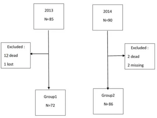
From May to October 2013, 85 infants were born between 26 and 32 weeks of GA and involved in the study. During the study period, 13 were excluded (12 died and 1 file lost). The remaining 72 infants (group 1) had a birthweight (BW) of 1154 +/- 276 g, and a GA of 29 +/- 1.42 weeks.
From May to October 2014, 90 infants were born between 26 and 32 weeks GA and involved in the study. During the study period, 2 were excluded (died) and 2 had missing data for nutritional intakes. The remaining 86 infants (group 2) had a BW of 1215 +/- 332 g, and a GA of 28.9 +/- 1.7 weeks. (Figure 1).
Figure 1: Flow chart of the population
The clinical characteristics of the study population are described in Table 1. The groups were similar with no significant difference for sex, birth weight, birthweight z-scores (-0.494 +/-
29 1.139 in group 1 versus -0.426 +/- 1.096 in group 2, p=0.705), GA, prenatal steroids, PDA, or BPD.
We observed a significant difference between the two groups for two characteristics: first, there was statistically more treatment of PDA by surgery in the second group (0 in group 1 versus 5 in group 2, p = 0.008), and second the duration of treatment by Doxapram was longer in group 2 than in group 1 (10.8+/-11.54 days in group 1 versus 22.89+/-11.73 days in group
Table 1 : Clinical characteristics of thestudy population
Group 1 (n=72) Group 2 (n=86) All population p (n=158) Boys, n (%) 37 (51,39) 44 (51,16) 81 (51,27) 0,977 Birth weight (g) 1154 (+/- 276) 1215 (+/-332) 0,21
Birth weight z-score -0,494 (+/- 0,140) -0.426 (+/- 1.096) 0.705
Gestational age , weeks 29,0 (+/- 1,4) 28,9 (+/- 1,7) 0,69
Any prenatal steroids, n (%) 66 (91,67) 76 (88,4) 142 (89,9) 0,632
1 course, n (%) 49 (68,01) 41 (47,7) 90 (56,7) 0,434
2 courses, n (%) 17 (23,6) 10 (11,6) 27 (17,1) 0,434
Complete course, n (%) 29 (40,3) 38 (44,2) 67 (42,4) 0,266
Partial course n (%) 35 (48,6) 32 (37,2) 67 (42,4) 0,266
PDA, n (%) 17 (23,6) 13 (15,1) 30 (19) 0,187
PDA surgically closed, n (%) 0 5 (5,8) 5 (3,16) 0,008
PDA medically closed, n (%) 15 (20,8) 13 (15,1) 28 (17,7) 0,705
Doxapram, n (%) 21 (29,2) 28 (32,6) 49 (31) 0,611
Doxapram duration (days) 10,8 (+/- 11,54) 22,89 (+/- 11,73) 0,001
BPD, n (%) 35 (48,6) 35 (40,7) 70 (44,3) 0,7
CLD, n (%) 18 (25) 20 (23,3) 38(24,1) 0,622
30 2). Those differences are explained by a best availability of pediatrics surgeons in the year 2014, and by a modification of our protocol for Doxapram treatment between the two periods.
2. Primary outcome: impact on weight and Z-score
A postnatal weight loss occurred in the two groups during the first week of life, with no difference on the minimal global weight on day 7 (1121 +/- 233 g in group 1 versus 1159 +/- 305 g in group 2; p = 0.385). Mean weight loss was 2.8% in group 1 and 4.9% in group 2 (NS).
There was no significant difference on raw weight between the two groups from birth to day 133 even if we observed a tendency on day 70: 2289 +/- 338.4 g in group 1 versus 2542 +/- 457 in group 2, p = 0.063. It seemed to be a moderate higher weight gain velocity in group 2 from day 56 to day 133, with no statistically significant difference (Figure 2).
31 Figure 2 : Evolution on raw weight between two groups; D= days; p>0.05 for all values.
During the first weeks of life, growth failure was observed in weight z-scores. From birth to day 7, weight z-score decreased significantly from -0.494+/-1.139 to -1.402 +/- 0.834 in group 1 (p<0.001) and from -0.426+/-1.096 to -1.233+/-0.907 in group 2 (p<0.001) with no significant difference between the two groups (p=0.705).
The negative changes in weight z score from birth up to 19 weeks of life, were lower in group 2 as compared to group 1: weight z-score decreased during the first 70 days of life in group 1 (-2.001 +/- 0.747 at day 70) whereas it stopped decreasing from day 35 in group 2 (-1.435 +/- 0.791). Thus, the minimal z-score was significantly more important and delayed in group 1 than in group 2. We observed a less important decrease in the z-score in group 2, which became statistically different on day 42. Weight z-score longitudinal evolution is shown in Figure 3.
32 Finally, weight z score stabilized in group 1 after 10 weeks of life, between -1.5 and -2 when the stabilization was obtained earlier, after 5 weeks of life, remaining between -1 and -1.5 in group 2.
Figure 3: Longitudinal weight z-score evolution from birth to day 84. (D=day, *p < 0.05.)
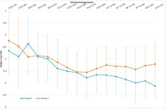
When we analyzed weight z-score changes with regards to PMA, we noticed a higher weight z-score in group 2, with statistically significant differences between the two groups from 36 to 39 weeks GA. This significant difference was not sustained because of a smaller size in the study population due to early discharge: at 40 weeks PMA 1.914+/0.783 in group 1 versus -1.277+/-0.877 in group 2 (p=0.081), and at 41 weeks PMA -2.133 +/-0.522 in group 1 versus -1.217+/-0.931 in group 2 (p=0.066).
In addition, the lowest weight z-score was noticed at 33 weeks PMA in group 2 (-1.545+/-0.788) but was delayed and lower in group 1 (-2.133+/-0.522 at 41 weeks PMA). We noticed
33 that weight z-score in group 1 exceeded -2 at 41 weeks PMA, defining a severe EUGR at term, which was not observed in group 2. Indeed, in group 2 we observed a catching-up effect from 33 weeks PMA and a weight z-score improving up to -1.217 +/-0.931 at 41 weeks PMA. In group 1, there was no catch-up at all, and weight z-score was still decreasing, leading to a EUGR at 41 weeks PMA (Figure 4).
Figure 4: Weight z-scores evolution with PMA; *p<0.05.
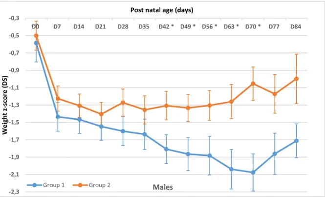
Looking at sex differences, we observed a difference on the effect of the new protocol between males and females infants. Indeed, there was a significant difference in males from group 1 and from group 2 from day 42 to day 70, with a catching up effect in weight Z score with the new nutrition protocol: on day 70 weight z-score was -2,077 +/- 0,642 in group 1 and
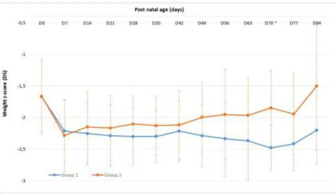
-1,054 +/- 0,637 in group 2 (p<0,002) (Figure 5A). Conversely, there was no significant difference in females between the two periods (Figure
34 -2,3 -2,1 -1,9 -1,7 -1,5 -1,3 -1,1 -0,9 -0,7 -0,5 -0,3 D0 D7 D14 D21 D28 D35 D42 * D49 * D56 * D63 * D70 * D77 D84 Wei gh t z-sco re ( D S)
Post natal age (days)
Males
Group 1 Group 2
Figure 5A : Longitudinal weight z-score evolution from birth to day 91 in males. D=day, *p < 0.05.
Figure 5B : Longitudinal weight z-score evolution from birth to day 84 in females. D=day, p > 0.05 for all values.
35 In addition, we wanted to evaluate the effects of the new protocol for intra-uterine growth restriction (IUGR) infants. We stratified infants in three subgroups: no IUGR if birthweight Z-score was >-1, moderate IUGR if birthweight Z-score was within [-2;-1] range and severe IUGR if birthweight Z-score was <-2. The two populations were comparable for stratification: in group 1 there were 37,14 % of infants with moderate IUGR, and 10% with severe IUGR versus 32,19% and 8% respectively in group 2 (p>0,05 for all values, Table 3).
Table 3 : IUGR in the two groups
Group 1 (n=70) Group 2 (n=87)
No IUGR (%) 37 (52,8) 52 (59,8)
Moderate IUGR (%) 26 (37,1) 28 (32,2)
Severe IUGR (%) 7 (10) 7 (8)
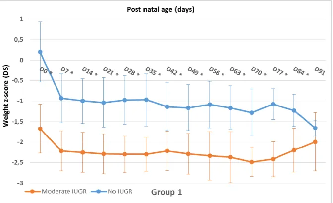
There was no significant difference between the two periods in infants with no IUGR or severe IUGR. However, in infants with moderate IUGR, we observed a tendency to catch up from day 49 in group 2 as compared to group 1, which became significant on day 70 (-2,482 +/- 0,359 in group 1 versus -1,848 +/- 0,593 in group 2, p= 0,049) (Figure 5).
Figure 6: Longitudinal weight z-score evolution from birth to day 84 in infants with moderate IUGR. D=day, * p < 0.05.
36 We confirmed this significant difference when we compared infants with moderate IUGR with infants with no IUGR in each group. There was a better increase in weight z-score with the new nutrition protocol for infants with moderate IUGR, who tend to catch up. The difference between moderate IUGR and no IUGR decrease progressively to become not significant from day 77 whereas there was no catch up in group 1 (Figure 7A and 7B).
37 Figure 7A: Longitudinal weight z-score evolution from birth to day 91 in infants with moderate IUGR compared with no IUGR in the group 1. D=day, * p < 0.05.
Figure 7B: Longitudinal weight z-score evolution from birth to day 91 in infants with moderate IUGR compared with no IUGR in group 2. D=day, * p < 0.05.
38
3. Secondary outcomes
The secondary outcomes evaluated the potential impact of our new nutritional protocol on the population morbidity (Table 3).
Because of a faster increase of enteral feeding, we evaluated its tolerance by the rate of NEC in the two groups, and found no difference between the two groups. We even noticed a lower rate of NEC in group 2, but this was not statistically significant (25% in group 1 versus 17.6% in group 2, p = 0.23).
While our new protocol lead to a faster achievement of enteral nutrition, there was no significant difference for the duration of hospitalization, central venous line duration, parenteral nutrition duration, alimentation withdrawals and their duration or the rate of infection (Table 3).
39 Table 3 : Comparison of secondary outcomes between the two groups
Group 1 (n=72) Group 2 (n=86) All population (n=158) p NEC 18 (25) 15 (17,44) 33 (20,9) 0,23 Episodes (n) 1,11 (+/-0,076) 1,071 (+/-0,267) 0,702 1 episode, n (%) 16 (22,22) 13 (15,11) 29 (18,3) 0,702 2 episodes, n (%) 2 (2,78) 1 (1,16) 3 (1,9) 0,702 Stage 1 of Bell, n (%) 4 (5,55) 1 (1,16) 5 (3,2) 0,13 Stage 1B of Bell, n (%) 1 (1,38) 2 (2,32) 3 (1,9) 0,13 Stage 2 of Bell, n (%) 13 (18) 8 (9,3) 21 (13,3) 0,13 Stage 2B of Bell, n (%) 0 3 (3,5) 3 (1,9) 0,13 Stage 3 of Bell, n (%) 0 1 (1,16) 1 (0,6) 0,13 Alimentation withdrawal, n (%) 43 (59,72) 42 (48,8) 85 (53,8) 0,196 Duration of alimentation stop (days) 6,44 +/- 6,14 5,86 +/- 5,38 0,642
CVL , n (%) 70 (97,2) 74 (86) 144 (91,1) 0,051
CVL duration (days) 15,72 +/- 10,48 15,3 +/- 9,4 0,803
Infection, n (%) 32 (44,4) 40 (46,5) 72 (45,6) 0,743
Confirmed, n (%) 25 (34,7) 36 (41,9) 61 (38,6) 0,087
Parenteral alimentation duration
(days) 15,75 (+/- 9,13) 14,13 (+/- 8,34) 0,254
Hospitalization duration (days) 54,9 (+/-27,1) 51,52 (+/-31,73) 0,314
Age at discharge (WGA) 36,82 (+/-3,53) 36,09 (+/-3,72) 0,166 NEC : necrotizing enterocolitis, CVL : central veinous line, PDA : patent ductus arteriosus
40
V. DISCUSSION
Growth rate in the NICU is influenced by many factors and the role of each independent factor is difficult to isolate from others because most of them are correlated.
In the present study, we demonstrated that the introduction of a strict nutritional protocol strategy, directed to optimize and individualize the nutritional regimen, according to the most recent recommendations, led to growth improvement with partial limitation of postnatal growth restriction in very preterm infants. Indeed, the infants receiving the new nutritional policy seemed to show a higher weight rate velocity during both enteral and parenteral nutrition. Also, the drop in weight z-score from the sixth to the 19th week of life was
significantly lower in the second group as compared to the first group. In addition, we noticed that weight z-scores correlated to PMA increased significantly from 36 to 39 weeks PMA after the introduction of our new protocol, whereas it did not stop to decrease in group 1, leading to severe EUGR at 41 weeks PMA. The difference between the two groups was not statistically significant from 40 weeks PMA because of a lack of power, the sample size becoming too small related to infants’ discharged from the hospital.
Our results showed that the minimal z-score was higher and earlier in group 2 than in group 1, with a catching up effect after day 35 which was not observed before the introduction of our new protocol. Our results suggest that a strict implemented nutritional protocol may improve weight z-score in preterm infants born between 26 and 32 weeks of gestation. This policy limited the apparition of EUGR (z-score <-2) before discharge. This is a very promising result demonstrated that this kind of protocol may be efficient in that population.
Of note, there was no difference between the two groups concerning raw weight. It demonstrates that weight z-score is a better predictor to show post-natal growth restriction, as this was already known in the literature (21).
41 There was a significant difference between males and females, with a significant growth improvement between the two periods for males only. This difference may be due to the fact that males had lower weight z-score at birth than females in our population.
In addition, we showed that preterm infants with moderate IUGR are able to catch up progressively and joined growth rate similar to that of non IUGR infants from day 70. That may be explained by the fact that those infants have increased nutrition needs, and that a strictly optimized protocol help to prevent the addition of antenatal deficits with postnatal deficits. Those data are consistent with the literature (22,23).
Senterre T et al. (18) reported almost same findings. Indeed, the authors found that optimization of nutritional policies, based on the most recent recommendations, during parenteral and enteral nutrition resulted in improved growth and limitation of postnatal growth restriction of a cohort of 102 preterm infants, as we observed in our study. In their study, weight score at 5 weeks of life was -1.14+/-0.6 whereas our results showed a lower z-score, even after the introduction of our new regimen.
Another study by Senterre and Rigo (4) showed a similar weight z-score between 3 days and 3 weeks of postnatal age with respectively for infants born <28 weeks GA and infants between 28 and 30 weeks GA, - 0.65+/-0.70 and -0.6+/-0.53 of z-score change at 30 days PMA. In our study weight z-score change at 35 days was - 1.103 in group 1 and - 1.009 in group 2, meaning a more important early drop in z-score. In our study, the growth rate started to improve after one month postnatally. Therefore, our data suggest that we need to improve the nutritional regimen for the first month of life in order to minimize the initial growth restriction
Moltu et Al. (24) aimed to determine whether an increased supply of energy, protein, essential fatty acids, and vitamin A reduced postnatal growth failure in very low birth weight infants.
42 They showed a significant difference in the median growth velocity of 17.4 g/kg/j in the intervention group and 13,8 g/kg/j in the control group. Their z score change at 36 weeks was -1,24 +/- 1,07 in the intervention group and -1,46 +/- 0,5 in the control group, with no significant difference. In comparison, our study showed a z score change at 36 weeks of - 1,18 in group 1 versus - 0,83 in group 2 with a significant difference (p=0,009), meaning that our intervention was indeed efficient.
A significant part of postnatal growth restriction in preterm infants has been linked to insufficient nutrition, which is mainly caused by fears of metabolic intolerance of post-natal, or fears of NEC (2,3). Wilson et al (25) introduced the concept of early “aggressive nutrition” that has been demonstrated safely support protein accretion and growth (26). Recent recommendations have included this policy (27) but a wide variability in nutritional practices still exists with frequent insufficient intakes, especially during the first weeks of life (3,17,28,29) .
A recent meta-analysis (30) found that common denominators in the five studies they
analyzed were the introduction of enhanced parenteral nutrition immediately after birth including the start of intravenous lipids on the first postnatal days. Although there was still an heterogeneity regarding the advance of parenteral macronutrient supply, a mean total energy supply from 70 kcal/kg/day to 113 kcal/kg/d with a total protein supply of 3 to 3.8 g/kg/d was achieved by one week of age. Four studies reported cumulative mean energy and protein supply during the first 4 weeks ranging from 112 to 139 kcal/kg/d and 3.7 to 4 g/kg/d protein. Among these studies, the smallest decline in weight z score during the first 4 weeks was
observed in the group with the highest energy and protein supply (24).
Obviously, early nutritional deficits were not regained before hospital discharge in our study. Infants were fed with nutrient intakes designed to meet current reference daily intakes. The situation is further complicated when oral feedings have to be interrupted for clinical reasons.
43
In that situation, additional transient deficit may occur. Therefore, complex interactions between other factors as clinical morbidity must be taken into account.
We evaluated the secondary effects or the benefits on other clinical outcomes of our new protocol. We did not find any difference after the introduction of our new strategy. The rate of NEC was not different between the two groups, even if enteral nutrition was increased faster. We noted a tendency to decrease NEC between group 2 (17.65%) and group 1 (25%), but this was not significant (p = 0.23). It confirms that early “aggressive” nutrition for preterm do not imply an excess of risk for NEC, and this value is consistent with the literature (2). Enteral
feeding was introduced early and parenteral feeds were stopped in infants by day 15,75 +/-
9,13 in group 1, and by day 14,13 +/- 8,34 in group 2 (p=0,254).
The two groups were similar for the duration of central venous line and parenteral nutrition. We expected a shorter duration with the introduction of the new protocol because enteral feeding was increasing faster and the new protocol stated that central venous line should be removed earlier than before (140 ml/kg/day of enteral feeding versus 160 ml/kg/day before). This part of the protocol was obviously not respected. We speculate that it may be for fear to modify this practice which may need more time than six months to be implemented. Our study recognized this problem and this point will be emphasized in the future. It proves that when a new protocol is introduced in a unit, medical caregivers must be trained and a time for adaptation is needed. This probably explains also why we did not find a decrease in late onset sepsis which are mostly caused by prosthetic material such as central venous line.
The study has much strength, such as the extensive retrospective standardized collection of data as well as regularly routinely measured values of weight throughout the study period.
44 Conversely, our study has limitations. We did not study the compliance to our new protocol, in order to verify if there was a direct relationship between nutrients intake and growth in our population. This part of the study is still ongoing to evaluate our practice in the unit and to update our nutrition protocol accordingly.
Another limitation of the study is that it is not a randomized trial; however, it is not ethical to voluntary limit nutritional support in some infants.
Confounding factors, such as medical treatment, mechanical ventilation, could have affected the results, but were not investigated in this study. Other limitations include limited reliability of growth measurements as well as the retrospective collection of nutritional data.
There is also a large variation in protein, and fat composition of human milk (31) that we were not able to determine at the time of the study, and this variation of the nutrient supply between the children is another limitation of our study.