HAL Id: hal-02324253
https://hal.archives-ouvertes.fr/hal-02324253
Submitted on 5 Nov 2020
HAL is a multi-disciplinary open access
archive for the deposit and dissemination of
sci-entific research documents, whether they are
pub-lished or not. The documents may come from
teaching and research institutions in France or
abroad, or from public or private research centers.
L’archive ouverte pluridisciplinaire HAL, est
destinée au dépôt et à la diffusion de documents
scientifiques de niveau recherche, publiés ou non,
émanant des établissements d’enseignement et de
recherche français ou étrangers, des laboratoires
publics ou privés.
Hydrocarbon Receptor and impacts skin homeostasis
Laura Lozza, Pedro Moura-Alves, Teresa Domaszewska, Carolina Lage
Crespo, Ioana Streata, Annika Kreuchwig, Andreas Puyskens, Marina Bechtle,
Marion Klemm, Ulrike Zedler, et al.
To cite this version:
Laura Lozza, Pedro Moura-Alves, Teresa Domaszewska, Carolina Lage Crespo, Ioana Streata, et al..
The Henna pigment Lawsone activates the Aryl Hydrocarbon Receptor and impacts skin
homeosta-sis. Scientific Reports, Nature Publishing Group, 2019, 9 (1), �10.1038/s41598-019-47350-x�.
�hal-02324253�
the Henna pigment Lawsone
activates the Aryl Hydrocarbon
Receptor and impacts skin
homeostasis
Laura Lozza
1, pedro Moura-Alves
1,2, teresa Domaszewska
1, Carolina Lage Crespo
3,
Ioana streata
4, Annika Kreuchwig
5, Andreas puyskens
1, Marina Bechtle
1, Marion Klemm
1,
Ulrike Zedler
1, Bogdan silviu Ungureanu
6, Ute Guhlich-Bornhof
1, Anne-Britta Koehler
1,
Manuela stäber
1, Hans-Joachim Mollenkopf
7, Robert Hurwitz
8, Jens Furkert
5,
Gerd Krause
5, January Weiner 3rd
1, António Jacinto
3, Ioana Mihai
4, Maria Leite-de-Moraes
9,
Frank siebenhaar
10, Marcus Maurer
10& stefan H. e. Kaufmann
1,11As a first host barrier, the skin is constantly exposed to environmental insults that perturb its integrity. tight regulation of skin homeostasis is largely controlled by the aryl hydrocarbon receptor (AhR). Here, we demonstrate that Henna and its major pigment, the naphthoquinone Lawsone activate AhR, both
in vitro and in vivo. In human keratinocytes and epidermis equivalents, Lawsone exposure enhances
the production of late epidermal proteins, impacts keratinocyte differentiation and proliferation, and regulates skin inflammation. To determine the potential use of Lawsone for therapeutic application, we harnessed human, murine and zebrafish models. In skin regeneration models, Lawsone interferes with physiological tissue regeneration and inhibits wound healing. Conversely, in a human acute dermatitis model, topical application of a Lawsone-containing cream ameliorates skin irritation. Altogether, our study reveals how a widely used natural plant pigment is sensed by the host receptor AhR, and how the physiopathological context determines beneficial and detrimental outcomes.
The skin acts as an important first barrier of the body, which is constantly exposed to diverse environmental and
mechanical insults, such as pollution, infection, injury and radiation, amongst others1. Additionally, the
applica-tion of cosmetics and other agents can have a major impact on skin homeostasis1. Among the most widely used
skin dyes, are the extracts of Lawsonia inermis, commonly known as Henna2. In traditional medicine, Henna has
been widely used to treat bacterial and fungal infections, inflammation, cancer and various skin pathologies3,
but the underlying mechanisms remain insufficiently understood. Major side effects of Henna preparations are
caused by the additive para-phenylenediamine (PPD) that has been associated with allergic contact dermatitis4,5.
As natural product, Henna comprises a mixture of numerous compounds most of which are poorly characterized
1Department of Immunology, Max Planck Institute for Infection Biology, Charitéplatz 1, D-10117, Berlin, Germany. 2Nuffield Department of Clinical Medicine, Ludwig Institute for Cancer Research, University of Oxford, Oxford,
UK. 3CEDOC, NOVA Medical School, NOVA University of Lisbon, Lisbon, 1169-056, Portugal. 4Human Genomics
Laboratory - University of Medicine and Pharmacy of Craiova, Craiova, Romania. 5Leibniz-Forschungsinstitut
fuer Molekulare Pharmakologie (FMP), Robert-Rössle-Strasse 10, 13125, Berlin, Germany. 6Research Center of
Gastroenterology and Hepatology, University of Medicine and Pharmacy of Craiova, Craiova, Romania. 7Microarray
Core Facility, Max Planck Institute for Infection Biology, Charitéplatz 1, D-10117, Berlin, Germany. 8Biochemistry
and Protein Purification Core Facility, Max Planck Institute for Infection Biology. Charitéplatz 1, D-10117, Berlin, Germany. 9Laboratory of Immunoregulation and Immunopathology, INEM (Institut Necker-Enfants Malades),
CNRS UMR8253, INSERM UMR1151 and Paris Descartes University, Paris, France. 10Department of Dermatology
and Allergy, Charité-Universitätsmedizin Berlin, Berlin, Germany. 11Hagler Institute for Advanced Study, Texas A&M
University, College Station, TX, USA. Laura Lozza and Pedro Moura-Alves contributed equally. Correspondence and requests for materials should be addressed to L.L. (email: lozza@mpiib-berlin.mpg.de) or P.M.-A. (email: pedro. mouraalves@ludwig.ox.ac.uk) or S.H.E.K. (email: kaufmann@mpiib-berlin.mpg.de)
Received: 11 October 2018 Accepted: 15 July 2019 Published online: 26 July 2019
opeN
both chemically and functionally. The responsible pigment for the red colour after Henna application on skin, is
the 1,4-naphthoquinone Lawsone, constituting 1–2% of the leaves6,7.
Recently, we unveiled that bacterial pigmented virulence factors, such as phenazines produced by
Pseudomonas aeruginosa and the 1,4-naphthoquinone Phthiocol (Pht) from Mycobacterium tuberculosis, bind
to and activate the Aryl Hydrocarbon Receptor (AhR), leading to AhR mediated immune defenses and
detoxi-fication of these virulence factors8. AhR is an evolutionarily conserved transcription factor widely expressed by
almost all types of cells9–11. In its inactive state AhR resides in the cytoplasm in association with various
chap-erones. Upon activation, AhR binds to the AhR nuclear translocator (ARNT), and the resulting heterodimer induces the transcriptional regulation of multiple target genes, notably cytochrome P450 monooxygenases
(CYP1A1 and CYP1B1) and its own repressor, the AhR repressor (AHRR)11.
Earlier studies of AhR functions focused on detoxification of xenobiotic ligands such as benzo[a]pyrene, an
ingredient of tobacco smoke12 and the highly toxic 2,3,7,8-tetrachlorodibenzo-p-dioxin (TCDD)13. The list of
ligands is continuously expanding, encompassing endogenous molecules (e.g. tryptophan (Trp), kynurenine or formylindolo[3,2-b] carbazole (FICZ)), dietary compounds and bacteria-derived ligands, and others (e.g.
Itraconazole, Lipoxin A4, Prostaglandin G2 and Quercetin)8,14–18. In parallel with the increasing number of
lig-ands, the biological functions attributed to this receptor are constantly growing rendering this receptor a ‘moving target’ of intense research14,19–21.
In the skin, AhR-mediated signals are critical in tissue regeneration, pathogenesis, inflammation and
home-ostasis9,22,23 and AhR emerged as crucial player in the maintenance of skin integrity and immunity9,11. However,
the outcome of AhR activation varies profoundly according to ligand properties, target cells and interactions with
other signaling cascades22–25.
Here, we aimed to better characterize the effects of Lawsone, defining its mechanisms with an emphasis on skin, the central target tissue of Henna. We demonstrate that the main pigment of Henna, Lawsone, activates the AhR-transcriptional program and modulates skin homeostasis and recovery after external insult. We show that Lawsone inhibits proliferation, and accelerates differentiation of keratinocytes. Specifically, experiments with human skin equivalents, zebrafish and mice, reveal that Lawsone modulates tissue homeostasis and tissue regen-eration, thereby interfering with the physiological process of wound healing. Despite its detrimental effect on wound healing, Lawsone’s capacity to reduce proliferation and promote keratinocyte differentiation, in parallel to modulation of skin inflammation, renders it a promising candidate for therapy of hyperproliferative skin diseases.
Results
Henna and lawsone activate the AhR pathway in keratinocytes.
AhR triggering depends on thequality and quantity of the activators as well as the intrinsic characteristics of the cell types11. Due to its similarity
with known AhR ligands (Fig. 1A), such as TCDD and the mycobacterial pigment Pht8,24, we hypothesized that
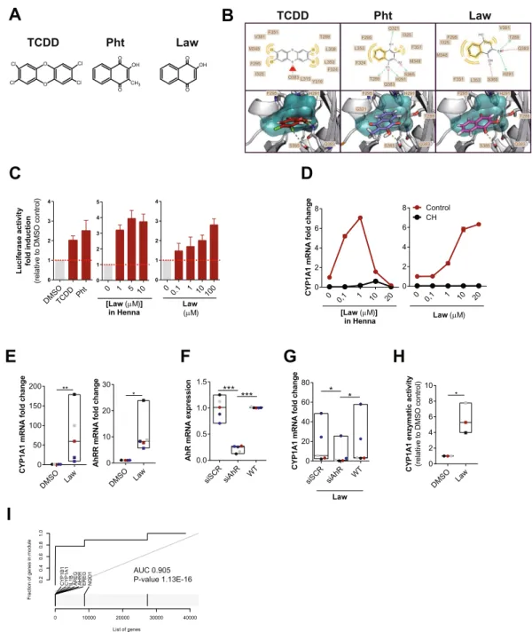
Lawsone, the main pigment from Henna, modulates AhR activity. In silico modeling studies predicted that all three
molecules fit into the AhR binding pocket, albeit with different affinities (Figs 1B and Supplement 1A). The key
res-idues Thr289, His291, Phe295, Ser365 and Gln383 are involved in forming hydrogen bonds with each of the three ligands. Lawsone has similar interactions as Pht. After rescoring, the free binding energy was as follows: TCDD (ΔG Bind −47.568 kcal/mol), Pht (ΔG Bind −42.850 kcal/mol) and Lawsone (ΔG Bind −38.591 kcal/mol), with the lower value indicating a stronger binding in the ligand-receptor complex. Binding to AhR was confirmed
in a previously established competition assay8, where Lawsone was able to displace radioactively labeled TCDD
bound to AhR (Fig. Supplement 1B).
Keratinocytes are the most prominent cell type in the epidermis23, which constitute the first contact with
exter-nal agents, including Henna7. We developed an AhR-luciferase reporter HaCaT (immortal human keratinocyte)
cell line and measured AhR activation as readout of luciferase activity after stimulation. As can be seen in Fig. 1C,
both TCDD and Pht induced AhR activation in keratinocytes. Similarly, Henna and the 1,4-naphthoquinone
Lawsone also activated AhR (Fig. 1C). Dose-dependent AhR activation was further confirmed in other cell types,
using the AhR-luciferase reporter THP-1 (human macrophage) cell line8 (Fig. Supplement 1C). Extending our
analysis to human primary keratinocytes (HEK cells), we evaluated whether the expression of AhR target genes
was differentially regulated. CYP1A1 was induced upon stimulation with both Henna and Lawsone (Fig. 1D).
AhR dependency was confirmed using the specific AhR inhibitor, CH22319126 (CH, Fig. 1D). CYP1A1
tran-scription increased after stimulation with Henna containing 1 μM of Lawsone, while it decreased at higher
con-centrations (Fig. 1D, left). Henna preparations contain several components, aside from Lawsone2, which would
interfere with the kinetics of AhR activation. When cells were stimulated with Lawsone, CYP1A1 was upregulated
in a dose-dependent manner (Fig. 1D, right), without affecting cell viability (Fig. Supplement 1D–F). In
kerat-inocytes obtained from different donors, CYP1A1 and AHRR were consistently induced by Lawsone (Fig. 1E),
Pht and TCDD (Fig. Supplement 1G). Notably, TLR2 stimulation (Pam2CSK4) did not activate AhR (Fig. Supplement 1G). Silencing of AhR in these cells by RNA interference (RNAi) was validated by Western Blotting (Fig. Supplement 1H), and led to reduced CYP1A1 expression, further confirming that Lawsone induced CYP1A1
in an AhR dependent manner (Figs 1F,G and Supplement 1I). Inhibition of CYP1A1 can lead to indirect AhR
activation in a mechanism involving Trp19,27. Using the EROD assay28, CYP1A1 enzymatic activity was increased
by Lawsone in HEK cells (Fig. 1H), as well as by the other ligands tested (Fig. Supplement 1J), thus excluding an
indirect role of CYP1A1 in AhR induction in this context.
To further validate our findings, we performed microarray analysis of HEK cells stimulated with Lawsone.
We identified a set of AhR dependent genes (Table 1) and visualized the gene enrichment using receiver
oper-ating characteristic (ROC) curves29. A high score of the area under the curve and low q value indicate a
signifi-cant and specific enrichment of AhR target genes upon stimulation with Lawsone (Figs 1I and Supplement 1K).
Consistently, Ingenuity Pathway Analysis predicted the AhR canonical pathway amongst the top differentially
A
DMSOTCDD Ph t 0 1 2 3 4 Luciferase ac tivity fo ld inductio n (rel ativ et oD MS Oc ontrol ) 0 1 5 10 0 1 2 3 4 5 [Law (µM)] in Henna 0 0.1 1 10 100 0 1 2 3 4 Law (µM)B
D
0 0,1 1 10 20 0 2 4 6 8 CYP1A1 mRNA fo ld chan ge [Law (µM)] in Henna 0 0,1 1 10 20 0 2 4 6 8 CH Control Law (µM)E
DMSO Law 0 50 100 150 200 CY P1A 1m RN Af ol dc hang e ** DMSO Law 0 10 20 30 AhRR mR NA fol dc hang e * TCDD Pht Law Cl Cl Cl Cl O O OH O O CH3 OH O O TCDD Pht LawI
F
G
DMSO Law 0 2 4 6 8 10 CY P1 A1 enzymati ca ctivity (relati ve toD MS Oc ontrol ) *H
siSCRsiAhR WT 0.0 0.5 1.0 1.5 Ah Rm R NA ex pr es si on *** *** siSCR siAhR WT 0 20 40 60 80 CYP1A1 mR NA fo ld chan ge Law * * 0 10000 20000 30000 40000 List of genes Fraction of genes in module
0. 20 .4 0. 60 .8 1. 0
CYP1B1 CYP1A1 IL1B AREG AHRR EREG NQO1
AUC 0.905 P-value 1.13E-16
C
F295 H291 T289 Q383 S365 F295 I325 M348 F351 L353 S365 H291 Q383 T289 V381 F295 H291 Q383 S365 V381 Q383 L353 F351 M348 F295 I325 F324 L315 Y310 T289 L308 F295 H291 T289 Q383 S365 G321 F324 L353 T289 Q383 F351 I325 F295 M348 S365 H291 G321Figure 1. Henna and Lawsone activate AhR in HaCaT and human primary keratinocytes. (A) Chemical structures of TCDD, Phthiocol (Pht) and Lawsone (Law) and (B) in silico modeling studies predicting binding of these molecules in the AhR binding pocket. Upper panel: 2D-interaction plot (LigandScout 4.1), hydrogen-donor (green dashed), -acceptor (red dashed), hydrophobic (orange); lower panel: 3D-interaction models, hydrogen bonds (yellow dashed), potential halogen bond (green dashed). (C) Luciferase activity of AhR reporter HaCaT cells stimulated for 4 hours (h) with TCDD, Phthiocol (Pht), Henna or Lawsone (Law). (D) Dose dependent CYP1A1 expression of HEK cells stimulated for 4 h, in the presence (black dots) or absence (red
dots) of the AhR inhibitor CH223191 (CH, 12 µM) normalized to control DMSO in the absence of Lawsone.
(E) CYP1A1 and AHRR expression after 4 h Lawsone (10 µM) stimulation of HEK cells normalized to DMSO.
Each dot represents one individual. (F-G) HEK cells were transiently transfected with AhR-siRNA (siAhR) or Scramble control (siScr) in different individuals (dots). Each color depicts results of the same individual. (F) AhR knockdown validation relative to non-transfected wild type (WT) cells. (G) CYP1A1 expression after 4 h stimulation with Lawsone normalized to DMSO. (H) 48 h CYP1A1 enzymatic activity in HEK cells treated with
Lawsone (10 µM) normalized to DMSO. (I) AhR-target gene enrichment after Lawsone stimulation (10 µM)
relative to TLR2 stimulation (Pam2CSK4, 300 ng/mL). Area under the curve (AUC), q-values and highly enriched genes are indicated. (C,E–H) Data from at least 3 independent experiments are shown. (D) Data from 1 representative experiment out of 2 is shown. (C) Mean + S.E.M., (D) Mean, (E–H) Floating bars, Mean Min to
Name Gene Reference
glutamate-cysteine ligase, catalytic subunit GCLC
Baird L., Arch Toxicol (2011) 85:241–272
NAD(P)H dehydrogenase, quinone 1 NQO1
ferritin, light polypeptide FTL
glutathione S-transferase alpha 1 GSTA1
glutathione S-transferase alpha 2 GSTA2
glutathione S-transferase alpha 3 GSTA3
glutathione S-transferase alpha 4 GSTA4
glutathione S-transferase alpha 5 GSTA5
glutathione S-transferase alpha 6, pseudogene GSTA6P
glutathione S-transferase alpha 7, pseudogene GSTA7P
glutathione S-transferase mu 1 GSTM1
glutathione S-transferase mu 2 (muscle) GSTM2
glutathione S-transferase mu 3 (brain) GSTM3
glutathione S-transferase mu 4 GSTM4
glutathione S-transferase mu 5 GSTM5
glutathione S-transferase omega 1 GSTO1
glutathione S-transferase omega 2 GSTO2
glutathione S-transferase omega 3, pseudogene GSTO3P
glutathione S-transferase pi 1 GSTP1
glutathione S-transferase theta 1 GSTT1
glutathione S-transferase theta 2 (gene/pseudogene) GSTT2
glutathione S-transferase theta 2B (gene/pseudogene) GSTT2B
glutathione S-transferase zeta 1 GSTZ1
hematopoietic prostaglandin D synthase HPGDS
aldo-keto reductase family 1, member A1 (aldehyde reductase) AKR1A1
aldo-keto reductase family 1, member B1 (aldose reductase) AKR1B1
aldo-keto reductase family 1, member B10 (aldose reductase) AKR1B10
aldo-keto reductase family 1, member B15 AKR1B15
aldo-keto reductase family 1, member C1 AKR1C1
aldo-keto reductase family 1, member C2 AKR1C2
aldo-keto reductase family 1, member C3 AKR1C3
aldo-keto reductase family 1, member C4 AKR1C4
aldo-keto reductase family 1, member D1 AKR1D1
aldo-keto reductase family 1, member E2 AKR1E2
aldo-keto reductase family 7, member A2 AKR7A2
aldo-keto reductase family 7, member A3 (aflatoxin aldehyde reductase) AKR7A3
potassium channel, voltage gated subfamily A regulatory beta subunit 1 KCNAB1
potassium channel, voltage gated subfamily A regulatory beta subunit 2 KCNAB2
potassium channel, voltage gated subfamily A regulatory beta subunit 3 KCNAB3
ATP-binding cassette, sub-family C (CFTR/MRP), member 1 ABCC1
Continued
Name Gene References
Cytochrome P450, family 1, member A1 CYP1A1 Hankinson, 1995; Katiyar et al., 2000; Mukhtar et al., 1986
Cytochrome P450, family 1, member B1 CYP1B1 Hankinson, 1995; Katiyar et al., 2000; Mukhtar et al., 1986
Aryl hydrocarbon receptor repressor AHRR Baba et al., 2001; Frericks et al., 2007
TCDD-inducible poly(ADP-ribose) polymerase TIPARP Lo and Matthews, 2012; Frericks et al., 2007
Interleukin-1β IL-1β Sutter et al., 1991
plasminogen activator inhibitor-2 PAI-2 Sutter et al., 1991
epiregulin EREG Patel et al., 2006
amphiregulin AREG Du et al., 2005
insulin-like growth factor 1 receptor IGFR1 Lo and Matthews, 2012
NADP(H):quinone oxidoreductase 1 NQO1 Wang et al., 2013
Table 1. AhR dependent genes. The table includes AhR target genes containing the xenobiotic-responsive element (XRE) in the promoter region and genes described to be induced by AhR activation.
extended our analysis to the enrichment of genes associated with this pathway (Table 2). The area under the curve indicates that Nrf2-related genes were less enriched compared to AhR-related genes (Fig. Supplement 1M,N), pointing to a preferential activation of AhR. In summary, our results demonstrate that the 1,4-naphtoquinone Lawsone, the critical pigment in Henna, binds and activates the AhR pathway in keratinocytes. While the effects of Henna may be confounded by other components in the extract, Lawsone specifically activates AhR without causing cell toxicity, at least at the conditions tested.
Lawsone stimulation modulates keratinocyte proliferation and differentiation.
The AhRpath-way impacts on epidermal differentiation, and the consequences of AhR activation considerably depend on the
properties of the ligands and the target cells22,25,31,32. As demonstrated in Fig. 2A, Lawsone inhibited
keratino-cyte proliferation. Furthermore, microarray analysis of HEK cells stimulated with Lawsone pointed to a skew-ing towards differentiation (Fig. Supplement 2A). ROC curve analysis of genes of the epidermal differentiation
Name Gene Reference
ATP-binding cassette, sub-family C (CFTR/MRP), member 2 ABCC2
ATP-binding cassette, sub-family C (CFTR/MRP), member 3 ABCC3
ATP-binding cassette, sub-family C (CFTR/MRP), member 4 ABCC4
ATP-binding cassette, sub-family C (CFTR/MRP), member 5 ABCC5
ATP-binding cassette, sub-family C (CFTR/MRP), member 6 ABCC6
ATP-binding cassette, sub-family C (CFTR/MRP), member 8 ABCC8
ATP-binding cassette, sub-family C (CFTR/MRP), member 9 ABCC9
ATP-binding cassette, sub-family C (CFTR/MRP), member 10 ABCC10
ATP-binding cassette, sub-family C (CFTR/MRP), member 11 ABCC11
ATP-binding cassette, sub-family C (CFTR/MRP), member 12 ABCC12
ATP-binding cassette, sub-family C (CFTR/MRP), member 13, pseudogene ABCC13
cystic fibrosis transmembrane conductance regulator (ATP-binding cassette
sub-family C, member 7) CFTR
UDP glucuronosyltransferase 1 family, polypeptide A complex locus UGT1A
UDP glucuronosyltransferase 1 family, polypeptide A1 UGT1A1
UDP glucuronosyltransferase 1 family, polypeptide A2 pseudogene UGT1A2P
UDP glucuronosyltransferase 1 family, polypeptide A3 UGT1A3
UDP glucuronosyltransferase 1 family, polypeptide A4 UGT1A4
UDP glucuronosyltransferase 1 family, polypeptide A5 UGT1A5
UDP glucuronosyltransferase 1 family, polypeptide A6 UGT1A6
UDP glucuronosyltransferase 1 family, polypeptide A7 UGT1A7
UDP glucuronosyltransferase 1 family, polypeptide A8 UGT1A8
UDP glucuronosyltransferase 1 family, polypeptide A9 UGT1A9
UDP glucuronosyltransferase 1 family, polypeptide A10 UGT1A10
UDP glucuronosyltransferase 1 family, polypeptide A11 pseudogene UGT1A11P
UDP glucuronosyltransferase 1 family, polypeptide A12 pseudogene UGT1A12P
UDP glucuronosyltransferase 1 family, polypeptide A13 pseudogene UGT1A13P
UDP glucuronosyltransferase 2 family, polypeptide A1, complex locus UGT2A1
UDP glucuronosyltransferase 2 family, polypeptide A2 UGT2A2
UDP glucuronosyltransferase 2 family, polypeptide A3 UGT2A3
UDP glucuronosyltransferase 2 family, polypeptide B4 UGT2B4
UDP glucuronosyltransferase 2 family, polypeptide B7 UGT2B7
UDP glucuronosyltransferase 2 family, polypeptide B10 UGT2B10
UDP glucuronosyltransferase 2 family, polypeptide B11 UGT2B11
UDP glucuronosyltransferase 2 family, polypeptide B15 UGT2B15
UDP glucuronosyltransferase 2 family, polypeptide B17 UGT2B17
UDP glucuronosyltransferase 2 family, polypeptide B24 pseudogene UGT2B24P
UDP glucuronosyltransferase 2 family, polypeptide B25 pseudogene UGT2B25P
UDP glucuronosyltransferase 2 family, polypeptide B26 pseudogene UGT2B26P
UDP glucuronosyltransferase 2 family, polypeptide B27 pseudogene UGT2B27P
UDP glucuronosyltransferase 2 family, polypeptide B28 UGT2B28
UDP glucuronosyltransferase 2 family, polypeptide B29 pseudogene UGT2B29P
UDP glycosyltransferase 3 family, polypeptide A1 UGT3A1
UDP glycosyltransferase 3 family, polypeptide A2 UGT3A2
UDP glycosyltransferase 8 UGT8
complex (EDC), and family I and II keratins (Table 3) revealed a significant enrichment upon Lawsone
stimula-tion (Fig. 2B). This was mainly due to upregulation of the genes involved in formation of the cornified envelope
(Supplementary Dataset File 1). Cornifelin (CNFN), hornerin (HRNR), late cornified envelope 3D (LCE3D),
keratin 2 (KRT2) and filaggrin 2 (FLG2) are critical for epidermal differentiation33,34. qRTPCR analysis confirmed
the induction of these genes in HEK cells upon Lawsone exposure (Fig. 2C). Thus, Lawsone modulates the
expres-sion of genes involved in cornified envelope generation.
A
24 48 72 96 0 2000 4000 6000 Time (h) Cell number DMSO Pht Law **** **** ********B
C
0 10000 20000 30000 40000 List of genes Fraction of genes in modul
e 0. 00 .2 0. 40 .6 0. 81 .0 FLG2 LCE3D CNF N KR T2 HRNR AUC 0.63 P-value 3.59E-07 0 10000 20000 30000 40000 List of genes
Fraction of genes in modul
e 0. 00 .2 0. 40 .6 0. 81 .0 LCE3D KRT 2 HRNR CNF N FLG2 AUC 0.68 P-value 1.69E-33
E
DMSO Law 0 20 40 60 80 100 Ki67/DAP I( % ) n.s. DMSO Law DMSO Law 10 µM 10 0 µM 5 d 10 dF
β-actin Cornifelin Loricrin Filaggrin Law (µM) - + - + - + - + 100 10 100 10 Culture (d) 5 10G
0 20 40 60 80 KRT2 CNF N HRNR LC3 D FLG2 mRNA fold induction(normalized to DMSO control
)
D
LCE3D SiScr SiAh R 0 0.4 0.8 1.2 1 mRNA fold induction(normalized to SiScr control)
SiScr SiAh R HRNR KRT2 SiScr SiAh R CNFN SiScr SiAh R
Figure 2. Lawsone stimulation modulates keratinocyte proliferation and differentiation. (A) Nuc red Live 647 positive HEK cells at different time points after stimulation with Lawsone (Law, 10 µM) and Phthiocol (Pht, 50 µM), compared to DMSO. (B) Epidermal differentiation complex and keratin gene enrichment of HEK cells after Lawsone stimulation (10 μM) and relative to TLR2 stimulation (Pam2CSK4, 0.236 μM) at (left) 4 h and (right) 24 h. Area under the curve (AUC), q-value and highly enriched genes are indicated. (C) KRT2, CNFN,
HRNR, LCE3D and FLG2 expression of HEK cells after 24 h stimulation with Lawsone (10 µM) normalized to
DMSO. Each color depicts results of the same individual. (D) LCE3D, KRT2, HRNR and CNFN expression on HEK cells transfected with AhR-siRNA (siAhR) or Scramble control (siScr) and further stimulated for 24 h with Lawsone (10 μM). Values are relative to siScr. Each color depicts results of the same individual. (E, top) Epidermal skin equivalents were stimulated for 5d with Lawsone (10 µM) or DMSO and stained with DAPI (blue) and the proliferation marker KI67 (purple). (E, bottom) Percentage of KI67 positive cells normalized to the total number of cells (DAPI). (F) Representative of an in vitro epidermis model experiment stained for Cornifelin (red) and Loricrin (green) and (G) protein expression of Filaggrin, Cornifelin and Loricrin at day 5 or 10 of culture after stimulation with 10 or 100 µM of Lawsone (blots were cropped from the same gel. Full unedited gels are provided in Supplementary Data). (A,C) Data from 3 independent experiments are shown. (D) Data from 2 independent donors. (E top, F,G) One representative experiment out of 2 is shown. (E) Pooled data from 2 different experiments is shown. (A) Mean + S.E.M., (C–E bottom) Floating bars, Mean Min to Max. (A) Two-way ANOVA with Fisher’s test, (C) One-way ANOVA with Dunn’s test. (E, bottom) Student’s t-test. *P < 0.05; **P < 0.01, ***P < 0.001, ****P < 0.0001.
approved
symbol approved name categories References
Keratin type I
KRT9 keratin 9, type I Human type I epithelial keratins
Schweizer et al., 2006;
http://www.genecards. org/
KRT10 keratin 10, type I Human type I epithelial keratins
KRT12 keratin 12, type I Human type I epithelial keratins
KRT13 keratin 13, type I Human type I epithelial keratins
KRT14 keratin 14, type I Human type I epithelial keratins
KRT15 keratin 15, type I Human type I epithelial keratins
KRT16 keratin 16, type I Human type I epithelial keratins
KRT17 keratin 17, type I Human type I epithelial keratins
KRT18 keratin 18, type I Human type I epithelial keratins
KRT19 keratin 19, type I Human type I epithelial keratins
KRT20 keratin 20, type I Human type I epithelial keratins
KRT23 keratin 23, type I Human type I epithelial keratins
KRT24 keratin 24, type I Human type I epithelial keratins
KRT25 keratin 25, type I Human type I epithelial keratins
KRT26 keratin 26, type I Human type I epithelial keratins
KRT27 keratin 27, type I Human type I epithelial keratins
KRT28 keratin 28, type I Human type I epithelial keratins
keratin type II
KRT1 keratin 1, type II Human type II epithelial keratins
Schweizer et al., 2006;
http://www.genecards. org/
KRT2 keratin 2, type II Human type II epithelial keratins
KRT3 keratin 3, type II Human type II epithelial keratins
KRT4 keratin 4, type II Human type II epithelial keratins
KRT5 keratin 5, type II Human type II epithelial keratins
KRT6A keratin 6A, type II Human type II epithelial keratins
KRT6B keratin 6B, type II Human type II epithelial keratins
KRT6C keratin 6C, type II Human type II epithelial keratins
KRT7 keratin 7, type II Human type II epithelial keratins
KRT8 keratin 8, type II Human type II epithelial keratins
KRT71 keratin 71, type II Human type II epithelial keratins
KRT72 keratin 72, type II Human type II epithelial keratins
KRT73 keratin 73, type II Human type II epithelial keratins
KRT74 keratin 74, type II Human type II epithelial keratins
KRT75 keratin 75, type II Human type II epithelial keratins
KRT76 keratin 76, type II Human type II epithelial keratins
KRT77 keratin 77, type II Human type II epithelial keratins
KRT78 keratin 78, type II Human type II epithelial keratins
KRT79 keratin 79, type II Human type II epithelial keratins
KRT80 keratin 80, type II Human type II epithelial keratins
non epidermal differentiation
complex-associated CNFN Cornifelin Kennedy et al., 2013
epidermal differentiation complex CRNN Cornulin Mischke et al., 1996; Kypriotou et al., 2012 FLG Filaggrin
FLG2 Filaggrin Family Member 2
HRNR Hornerin
IVL Involucrin
LCE1A Late Cornified Envelope 1A
LCE1B Late Cornified Envelope 1B
LCE1C Late Cornified Envelope 1C
LCE1D Late Cornified Envelope 1D
LCE1E Late Cornified Envelope 1E
LCE1F Late Cornified Envelope 1F
LCE2A Late Cornified Envelope 2A
LCE2B Late Cornified Envelope 2B
LCE2C Late Cornified Envelope 2C
LCE2D Late Cornified Envelope 2D
LCE3A Late Cornified Envelope 3A
LCE3B Late Cornified Envelope 3B
LCE3C Late Cornified Envelope 3C
LCE3D Late Cornified Envelope 3D
Epidermal differentiation occurs after activation of the AP-1 transcription factor35. To interrogate whether
epidermal differentiation requires AP-1 activity, keratinocytes were stimulated with Lawsone in the presence of
the AP-1 inhibitor tanshinone IIA (TIIA)36. Efficient blocking of AP-1 activity was shown by inhibition of CSF3
expression (Fig. Supplement 2B)36. Lawsone induced upregulation of CNFN, HRNR, LCE3D and KRT2 (Fig.
Supplement 2C), and of the AhR-target genes CYP1A1 and AHRR even in presence of TIIA indicating an AP-1 independent activation. Moreover, inhibiting AhR by RNAi reduced expression of these genes upon Lawsone
approved
symbol approved name categories References
LCE3E Late Cornified Envelope 3E
LCE4A Late Cornified Envelope 4A
LCE5A Late Cornified Envelope 5A
LCE6A Late Cornified Envelope 6A
LEP7 Late Envelope Protein 7
LOR Loricrin
NICE-1 Cysteine-Rich C-Terminal 1
RPTN Repetin
S100A1 S100 Calcium Binding Protein A1 S100A2 S100 Calcium Binding Protein A2 S100A3 S100 Calcium Binding Protein A3 S100A4 S100 Calcium Binding Protein A4 S100A5 S100 Calcium Binding Protein A5 S100A6 S100 Calcium Binding Protein A6 S100A7 S100 Calcium Binding Protein A7 S100A8 S100 Calcium Binding Protein A8 S100A9 S100 Calcium Binding Protein A9 S100A10 S100 Calcium Binding Protein A10 S100A11 S100 Calcium Binding Protein A11 S100A12 S100 Calcium Binding Protein A12 S100A13 S100 Calcium Binding Protein A13 S100A14 S100 Calcium Binding Protein A14 S100A15 S100 Calcium Binding Protein A15 S100A16 S100 Calcium Binding Protein A16 S100A7L2 S100 Calcium Binding Protein A7-Like 2
SPRR1A small proline-rich proteins 1A
SPRR1B small proline-rich proteins 1B
SPRR2A small proline-rich proteins 2A
SPRR2B small proline-rich proteins 2B
SPRR2C small proline-rich proteins 2C
SPRR2D small proline-rich proteins 2D
SPRR2E small proline-rich proteins 2E
SPRR2F small proline-rich proteins 2F
SPRR2G small proline-rich proteins 2G
SPRR3 small proline-rich proteins 3
SPRR4 small proline-rich proteins 4
THH Trichohyalin
THHL1 Trichohyalin-Like 1
Table 3. Epidermal differentiation complex and keratin genes. The table includes genes of the epidermal differentiation complex and keratins.
A
B
C
D
DMSO + CHBrightfield EROD Merge
Henna DMSO TCDD TCDD + CH Henna + CH
Brightfield EROD Merge
Law Law+ CH DMSO DMSO + CHHenna Henn a +CH 0 1×10⁷ 2×10⁷ 3×10⁷ 4×10⁷ 5×10⁷ **** **** DMSO DMSO + CHLaw Law + CH 0 1×10⁷ 2×10⁷ 3×10⁷ 4×10⁷ **** **** DMSO DMSO + CHTCDD TCDD + CH 0 1×10⁷ 2×10⁷ 3×10⁷ 4×10⁷ 5×10⁷ 6×10⁷ C YP 1A ac tivi ty (total in tensity a. u. ) **** ****
E
DMSO Henn a 0 200 400 600 CY P1 A mR N Af ol d ch an ge **** *** DMSO Henna 0 50 100 150 AhRR am RN Af ol d change ** ** DMSO Henna 0 10 20 30 40 50 Ah R R b m RNA fold ch an ge Control CH **** **** DMSO La w 0 10 20 30 40 50 CY P1A mR NA fo ldc hang e **** **** DMSO La w 0 1 2 3 4 AhRR am RN Af ol d ch ang e **** **** DMSO La w 0 1 2 3 4 5 Ah R R b m RN A fo ld ch ange Control CH **** **** A B C D E F G H 1 2 3 4 5 6 7 8 9 10 11 12 Stimulate (4 h) Wash 0 dpf Decorionate 1 dpf 2 dpf EROD (10min) Wash Analysis ImageFigure 3. Henna and Lawsone activate AhR in zebrafish larvae. (A,B) Fold induction of CYP1A, AhRRa and
AhRRb transcripts from zebrafish larvae (2 days post-fertilization, dpf) treated (red squares) or not (black
circles) for 2 h with 5 µM of AhR inhibitor CH223191, followed by further 4 h stimulation with (A) Henna (equivalent to 10 μM Lawsone), (B) Lawsone (10 μM) or DMSO vehicle control. Triplicates of 12 larvae depicted in each data point. (C) Scheme of the semi-high throughput experimental design developed to measure zebrafish larvae CYP1A enzymatic activity. (D) Representative images obtained upon CYP1A activity measurements using an Array Scan TM XTI Live High Content Platform. (E) CYP1A enzymatic activity expressed as total intensity of resorufin detected per larva (each dot represents one larva). 1 representative experiment out of 3 are shown (n = 36 larvae per condition). (A,B) Data from 1 representative experiment out of 3 is shown. (A,B) Floating bars, Mean Min to Max. (A,B) way ANOVA with Bonferroni’s test. (E) Two-way ANOVA with Fisher’s test. **P < 0.01, ***P < 0.001; ****P < 0.0001.
exposure (Fig. 2D). Thus, Lawsone requires AhR activation to induce the expression of genes involved in the formation of the cornified envelope independently of AP-1 activity.
To validate our findings, we treated fresh skin biopsies from individuals after skin surgical excision with Lawsone and confirmed the upregulation of CYP1A1 and AHRR (Fig. Supplement 2E), but not of KRT2, CNFN,
FLG and LCE3D (Fig. Supplement 2E). We reasoned that fully differentiated skin obtained in biopsies may mask
subtle differences of Lawsone on epidermal layers containing proliferating keratinocytes. Hence, we visualized
epidermal differentiation over time in human epidermis equivalent models23,34. Keratinocytes were treated daily
with Lawsone, and tissue differentiation was analyzed after 5 or 10 days of culture (Fig. Supplement 2F). As shown
in Fig. 2E, the percentage of Ki67 positive cells after 5 days of treatment was slightly reduced, although not
signif-icantly, pointing to inhibition of proliferation, as observed in vitro (Fig. 2A). Importantly, treatment with 10 μM
Lawsone increased the thickness of the stratum corneum after 5 and 10 days (Fig. 2F) and correlated with higher
expression of loricrin (at 5 days), cornifelin (at 10 days) and filaggrin (at 10 days) measured by
immunofluores-cence and Western blotting (Fig. 2F,G). At higher concentrations, Lawsone further boosted the differentiation of
the stratum corneum resulting in a disorganized epidermal structure (Fig. 2F). Hence, Lawsone impacts
epider-mal differentiation in human skin.
Lawsone activates the AhR pathway in zebrafish larvae and modulates tissue regeneration.
Inorder to further evaluate consequences of Lawsone exposure during tissue regeneration in vivo, we took
advan-tage of a previously established zebrafish model37–39. This model organism has been extensively used in toxicology,
including studies with AhR37, as well as in skin wound healing and re-epithelization studies38,40. The epidermis
and dermis layers occur in zebrafish larvae as early as 1 day post fertilization (dpf)40. 2dpf larvae were exposed
to Henna and Lawsone for 4 hours and AhR dependent gene expression was evaluated (Fig. Supplement 3A).
DMSO x [µ m] y [µm] -320 320 600 -100 Wound stimuli Law x [µ m] -32 0 320 600 -100
D
DMSO Law 0.0 0.2 0.4 0.6 0.8 1.0 Di re ctionality **E
DMSO Law 0 2 4 6 8 10 Spee d (µ m/ mi n) n.s. DMSO Law 0 200 400 600 800 A ccumulate dD is tanc e (µ m ) * DMSO Law 0.0 0.2 0.4 0.6 0.8 1.0 FM I **A
TCDD (10 nM) DMSO Law (5 µM) Law (1 µM) Non-cut controlB
DMSOTCDD Law (5µM ) Law (1µM ) 0 50 100 %a rea re ge ner at ed (nor ma lize dD MS Oc ontrol ) ****C
DMSO Law0 min 60 min 120 min 180 min 240 min 299 min
Scale bar= 50µm
Figure 4. Lawsone inhibits wound healing and skin regeneration in vivo. (A) Representative images of zebrafish fin regeneration 3 days post amputation (dpa) and exposure to different stimuli. Regenerated area depicted in red. (B) Quantification of the zebrafish tail fin area regenerated, normalized to DMSO treated larvae. (C) Neutrophil migration to zebrafish tailfin wounds visualized in DMSO or Lawsone-treated transgenic larvae Tg(mpeg.mCherryCAAX SH378 mpx:GFP i114). Frames from representative movies of migrating leukocytes in the wounded tail fin are shown. The lines indicate tracking of individual neutrophils over the indicated time point of the experiment. Wound is represented with a white dashed line. (D) 2D tracks of individual neutrophils migrating in the tail fin of wounded neutrophil-GFP zebrafish 3dpf larvae exposed to 10 µM Lawsone (n = 8) or DMSO (n = 23). (E) Quantification of 2D directionality, Forward migration index (FMI), accumulated distance and speed of individual leukocytes in the wounded tailfin. (B) Pooled data from 4 independent experiments with at least 24 larvae per condition per experiment, Mean + S.E.M., (E) Data from 2 pooled experiments, Mean + S.E.M. (B) One-way ANOVA with Fisher’s test, (E) Student’s t-test. *P < 0.05; **P < 0.01; ***P < 0.001; n.s.-not significant.
Zebrafish express three isoforms of AhR (AHR1a, AHR1b and AHR2)37,39 and 2 isoforms of AhRR (AHRRa and
AHRRb)40. As in humans, the expression of CYP1A, as well as the repressors AHRRa and AHRRb, are regulated
in an AhR dependent manner39. The expression of the three genes was increased upon stimulation with Henna,
Lawsone (Fig. 3A,B) or TCDD (Fig. Supplement 3B). Gene induction was reversed by the AhR inhibitor,
vali-dating AhR dependency. Similar to human cells (Figs 1H and Supplement 1J), larvae exposed to TCDD, Henna
or Lawsone increased CYP1A enzymatic activity (Fig. 3C–E), which was reversed by CH223191 (Figs 3D,E and
Supplement 3C). Under these conditions, no toxicity was observed (Fig. Supplement 3D). Thus, these in vivo results further substantiate our in vitro findings demonstrating that Lawsone activates AhR signaling.
We then performed tail fin regeneration assays and found that fin regeneration was inhibited in the presence
of Lawsone (Figs 4A,B, Supplement 4A) as observed previously with Dioxin41,42. Tissue damage induces the early
recruitment of leukocytes to restore barrier integrity and tissue homeostasis, which critically determines the
regenerative outcome43. Using a transgenic zebrafish line expressing GFP-labeled neutrophils
(mpeg.mCherry-CAAX SH378 mpx:GFP i114)44,45 we observed that upon exposure of the tailfin wound to Lawsone, neutrophils
moved (i) more randomly, (ii) for longer distances and (iii) with decreased directionality, as compared to controls
(Figs 4C–E, Supplement 4B and Movie Supplement 1). Moreover, neutrophils continued to patrol around in a
“zig-zag” fashion and were not arrested at the wound (Fig. 4C,D and Movie Supplement 1). Notably, Lawsone
exposure did not affect the speed of mobilizing cells (Fig. 4E). We conclude that Lawsone inhibits early steps of
tissue regeneration by affecting physiological leukocyte attraction.
We extended our studies to a mouse wound healing model46. Application of 10 μM of Lawsone on the wound
for 5 consecutive days delayed wound healing (Fig. Supplement 4C,D). In sum, Lawsone interferes with the nat-ural process of wound healing in different models.
Lawsone ameliorates skin recovery in a model of contact skin irritation.
Besides the inductionof genes of epidermal differentiation, the analysis of keratinocytes stimulated with Lawsone revealed that genes
related to psoriasis, dermatitis and inflammation were also affected (Table 4). Accordingly, we evaluated whether
Lawsone ameliorates skin disorders characterized by irritation, inflammation and epidermal hyper-proliferation,
in a human model of acute irritant contact dermatitis47. Skin irritation was induced by a single application of
30 µL of 5% sodium dodecyl sulfate (SDS) using self-adhesive patches which had been identified as reliable dose
to induce an irritant contact dermatitis47. Lawsone was dissolved in base cream at different concentrations (0.5%,
1%, and 3%) and topically applied on the skin of the forearm of healthy volunteers 24 h upon exposure to SDS. Images of the irritation spot and blood flux were taken daily. Decreased intensity of the flux was detected upon
exposure to Lawsone, with slight differences between the concentrations and individuals tested (Fig. 5A,B). Time
dependent resolution of irritation was observed in all individuals, but a strikingly faster reduction in blood flux
was detected upon Lawsone exposure (Fig. 5C). Thus, Lawsone dose dependently inhibits human skin responses
to irritation suggesting that detrimental or beneficial effects of Lawsone on the skin depend not only on its intrin-sic nature but also on the context of skin (dys)function.
Discussion
Despite the widespread use of the Henna plant Lawsonia inermis as a cosmetic dye for hair and skin, and its broad exploitation in traditional medicine due to assumed beneficial effects, little is known about the underlying
mech-anisms and role of its essential pigment, Lawsone2.
In our study, Lawsone emerged as an AhR ligand, directly binding to this receptor and eliciting AhR depend-ent responses in differdepend-ent in vitro and in vivo models. Moreover, we demonstrated that Lawsone interferes with the physiological skin regeneration processes. Lawsone modulated epidermal cell proliferation and differentiation in the skin, profoundly affecting wound healing. Nevertheless, in acute irritant contact dermatitis, Lawsone ame-liorated irritation and accelerated healing.
In the skin, AhR plays a fundamental role in the maintenance of skin integrity in face of continuous
environ-mental insults25 and the outcome of its activation is fine-tuned by the interplay of the individual ligand properties
and the physiological state of the skin25. Exposure to Lawsone induced the expression of AhR dependent genes
not only in human primary keratinocytes and keratinocytic cell lines, but also in zebrafish larvae and human skin biopsies. AhR dependency was validated by RNAi and by using the pharmacologic AhR inhibitor CH223191. Activation of AhR can be related to inhibition of CYP1A1 activity, increasing expression of Trp metabolites
acti-vating AhR19. Here, Lawsone did not inhibit the enzymatic activity of CYP1A, neither in zebrafish nor in human
keratinocytes.
AhR has been shown to affect epidermal differentiation22,34. Under homeostatic conditions, AhR KO mice
suffer from impaired barrier formation with enhanced transepidermal water loss and reduced expression of
pro-teins involved in epidermal differentiation22,32. Similar results were obtained after exposure of keratinocytes to
AhR antagonists22, pointing to an essential role of the AhR in the physiological development of the skin barrier.
Accordingly, endogenous Trp metabolites (e.g. FICZ) modulate keratinocyte functions and differentiation15,
while exogenous AhR-activators such as TCDD upregulate genes of epidermal differentiation33,48,49. Although
FICZ and TCDD are both high-affinity AhR ligands, TCDD resists Cyp1-mediated degradation13, while FICZ is
efficiently degraded50, suggesting that both ligand affinity and stability, shape the action on target cells. Consistent
with this, TCDD favors keratinocyte differentiation but also gives rise to chloracne in overexposed humans51,
characterized by the appearance of pustules and cysts in the skin52. Constitutive AhR activation in keratinocytes
also causes inflammed skin lesions53. Hence, depending on ligand and context, AhR modulation can act as a
“double-edged sword”, leading to beneficial or detrimental outcomes on skin regeneration.
In our studies, Lawsone differentially regulated distinct genes and proteins involved in keratinocyte dif-ferentiation. In agreement, proliferation of primary keratinocytes in a human organotypic skin model was decreased. Notably, the expression of specific keratinocyte differentiation genes upon Lawsone exposure was
AhR dependent. Although Lawsone did not affect survival of keratinocytes, high concentrations profoundly shuf-fled the epidermal layers, giving rise to a thick and fragile cornified structure. Cell proliferation in regenerating
zebrafish larval caudal fins in response to Dioxin has been shown to decrease42. Similarly, here we showed that
Lawsone impairs zebrafish larval fin regeneration. Moreover, wound healing experiments in zebrafish and mouse models revealed a delay in this process caused by Lawsone. In sum, different in vitro (cell lines and human skin model) and in vivo (mouse and zebrafish) approaches conclusively demonstrate that Lawsone impacts tissue proliferation, differentiation and regeneration.
AhR mediated effects can result from different interactions between this receptor and other intracellular
sig-naling pathways, such as Nrf211,54. Here, we showed that Lawsone upregulated the expression of the
antioxi-dant enzyme NQO1, a gene also regulated by Nrf2. Nrf2 is known to protect against reactive oxygen species30,54
and AhR and Nrf2 interactions were found crucial for the cytoprotective effects of the fungicide ketoconazole
in keratinocytes55. In our microarray analyses, AhR dependent responses were induced more profoundly, and
occurred earlier, than Nrf2 responses suggesting an important role of AhR in initiating cell responses. Yet, it is tempting to speculate that some of the elicited effects on skin may involve AhR and other molecules, such as Nrf2. Chronic inflammatory skin disorders emerge as outcome of diverse environmental and immune factors, and dis-eases such as psoriasis and atopic dermatitis are characterized by dysbalanced AhR signaling. Accordingly, therapeutic
interventions by AhR-targeting strategies have been suggested25,33,36. For example, coal tar has been widely used for
treatment of atopic dermatitis and was shown to induce AhR dependent responses in the skin34. Coal tar is composed of
A
48h
72h
96h
7 days
contr ol 0.5% 1% 3% 0 50 100 150 200 250 Lawsone %flux reduction * * ** * * contr ol 0.5% 1% 3% 0 50 100 150 200 250 Lawsone %flux reductio n Lawsone %flux reduction contr ol 0.5% 1% 3% 0 50 100 150 200 250 * *C
B
72h
96h
7days
Contro l 0.5% 1% 3% LawsoneFigure 5. Lawsone ameliorates skin recovery in a model of human contact skin irritation. (A) Representative images of blood flux measured using the MoorFLPI-2 Full_Field Laser Perfusion Imager V1.1 software at 48-72-96 h and 7 days upon application of 0.5% SDS. Cream containing increasing concentration of Lawsone (% of Lawsone = weight of Lawsone (g) per 100 g of cream) was applied 24 h after SDS treatment. (B) Example of (top) irritation spots and (bottom) blood flux quantification. After SDS applicationall individuals were treated as follow: far left: control cream, left: 0.5%; right 1%; far right 3% Lawsone cream. (C) Percentage of flux reduction at different time points normalized to the respective average flux intensity measured at 48 h post-SDS application. (A) Representative responses of 2 out of 9 volunteers are shown. (C) Data from 9 individuals are shown. One-way ANOVA with Fisher’s test. *P < 0.05; **P < 0.01; ***P < 0.001.
Symbol logFc. (Law vs DMSO at 24 h) p-value IFIT1 −2,58 5,53E-21 MX1 −2,31 9,39E-17 ISG15 −2,20 3,06E-17 ISG15 −2,19 3,06E-17 IFIT3 −1,69 3,19E-13 IFI6 −1,66 7,69E-17 IFI44 −1,54 1,29E-12 EPSTI1 −1,44 1,36E-11 IFNK −1,24 4,32E-10 TOP2A −1,22 2,13E-10 IFIH1 −1,20 3,84E-11 PPP1R3C −1,20 1,44E-10 SAMD9L −1,12 3,91E-07 IGFBP3 −1,10 3,71E-13 PARP9 −1,10 6,15E-09 PARP9 −1,10 6,15E-09 MKI67 −1,08 5,65E-10 OAS2 −1,07 1,70E-05 SOCS1 −1,02 3,30E-10 EFNB2 −1,01 1,65E-07 OAS1 −0,99 3,67E-07 CTSL2 −0,98 1,34E-07 DDX58 −0,97 9,52E-05 IRF9 −0,97 2,64E-07 PDK4 −0,95 0,000375155 SYNE2 −0,95 6,10E-06 CSPG4 −0,92 3,75E-09 SGK1 −0,92 2,15E-09 IFI44L −0,90 3,19E-05 EIF2AK2 −0,89 2,34E-05 RTP4 −0,88 2,53E-06 KRT15 −0,87 9,02E-05 SPC25 −0,87 9,74E-08 ANXA1 −0,85 1,61E-08 LAMP3 −0,85 2,63E-09 CAV1 −0,84 0,00298468 CCL27 −0,82 1,16E-06 DSG1 −0,81 4,72E-08 SP100 −0,81 4,12E-06 STAT1 −0,79 2,37E-09 TAGLN −0,78 1,02E-05 GJB2 −0,78 2,33E-05 PBK −0,78 4,38E-07 CCNA2 −0,77 3,25E-08 TIMP3 −0,77 4,81E-07 ANXA2 −0,75 0,000797819 GBP2 −0,73 3,67E-06 IL15 −0,72 2,30E-05 AHNAK −0,71 0,005674451 JUN −0,70 9,43E-08 ID4 −0,70 0,000169748 IL33 −0,68 0,000121647 TLR3 −0,68 2,94E-05 OPTN −0,67 8,05E-07 SLC6A2 −0,67 8,47E-06 JAK2 −0,66 0,009480402 NR3C1 −0,65 9,22E-05 PTRF −0,64 1,47E-07 BLNK −0,64 0,000567571 Continued
Symbol logFc. (Law vs DMSO at 24 h) p-value CAMK2N1 −0,63 5,82E-06 P4HA2 −0,63 0,019848721 FGF7 −0,63 0,00122151 IFIT5 −0,60 4,69E-06 MX2 −0,58 0,003413461 USP18 −0,53 4,04E-05 TRIM21 −0,53 0,012172344 OAS3 −0,52 0,002218834 ITSN2 −0,47 0,010777607 IFI35 −0,39 0,039516673 BATF2 −0,39 0,041688705 PNPT1 −0,29 0,037644415 PML −0,22 0,030117951 MAP3K9 0,34 0,010663036 FIGF 0,60 0,000923682 DUSP2 0,65 0,000736741 S100A8 0,65 7,22E-08 ALOX12B 0,66 6,43E-06 EGR1 0,66 3,18E-06 MANF 0,66 1,07E-07 CST6 0,67 5,62E-06 CPNE7 0,67 2,13E-06 POMC 0,68 0,000202198 FSCN1 0,69 4,83E-07 PPIF 0,70 7,83E-07 PGD 0,71 2,20E-05 CSK 0,73 0,016210156 MPHOSPH6 0,74 1,41E-06 FABP5 0,77 1,47E-09 CBR1 0,82 1,16E-05 CHRM1 0,85 4,90E-06 TNXB 0,86 5,07E-06 S100A9 0,86 1,09E-09 WNT5A 0,86 1,11E-08 LCN2 0,88 1,41E-06 AhRR 0,90 2,04E-05 AREG 0,93 4,67E-07 IFI30 1,00 4,21E-11 HMOX1 1,05 1,50E-11 MMP1 1,05 4,94E-12 GAL 1,12 3,20E-11 IL1A 1,20 4,85E-12 SPRR1A 1,20 2,65E-13 IL36G 1,23 7,89E-11 EPHX1 1,24 0,000139207 ARG1 1,29 2,74E-13 SERPINB3 1,33 1,13E-10 EREG 1,35 5,67E-11 SERPINB4 1,46 3,19E-08 ALDH1A3 1,48 8,05E-11 TGM3 1,49 1,42E-07 SLC45A4 1,59 1,85E-11 SECTM1 1,70 1,96E-13 SPRR2C 1,98 1,08E-14 IL1B 2,50 1,11E-19 CYP1A1 4,70 4,98E-23 CYP1B1 5,56 1,51E-22
Table 4. Psoriasis and dermatitis differentialy regulated genes. The table includes the genes involved in psoriasis and dermatitis that are differentialy regulated upon stimulation with Lawsone.
a mixture of organic compounds, and their safety and carcinogenicity have not been completely elucidated56. Similarly,
Henna extracts contain hundreds of different components, including phenolic compounds, terpenes, steroids and
alkaloids2, but a comprehensive investigation validating the biological activities of these compounds is still missing.
The effects of Henna can result from synergistic and antagonistic properties of numerous active substances. In fact,
adverse events of Henna have been described, for example after ingestion and mucosal contact57, although it appears
nontoxic when applied to the skin2. Henna has been used for treating radiation-induced dermatitis, as well as for
anti-carcinogenic, anti-microbial and anti-inflammatory purposes, although underlying mechanisms and molecules
involved remain elusive2,3,58. Given its low cell toxicity, Lawsone has clinical potential for treatment of skin disorders
characterized by hyperproliferation and inflammation. Indeed, our results demonstrate that topical administration of a cream containing small amounts of Lawsone ameliorates the irritation by a chemical insult. Similarly, topical
application of FICZ ointment reduces the inflammation in a mouse model of chronic mite-induced dermatitis59 while
intraperitoneal injection of FICZ reduces inflammation in a psoriasis-like skin model31. Current strategies to
ame-liorate psoriasis explore potential therapies by modulating expression of inflammatory cytokines, including IL-1760.
Curiously, in an Imiquimod-induced psoriasis model in mice, we observed a consistent reduction of IL-17 expression upon Lawsone topical exposure (unpublished data), pointing to potential therapeutic applications of Lawsone in skin disorders involving IL-17. Therefore, as an alternative to treatments using an undefined mixture of compounds (e.g. coal tar or Henna), we propose the Henna pigment Lawsone, and other naturally occurring naphthoquinones, as prom-ising therapeutic candidate medicines for skin diseases. The 1,4-naphthoquinones form a family of natural pigments
isolated from plants and fungi, widely used for staining food, clothing, skin and hair and in traditional medicine61.
These include Vitamin K, Shikonin from the Chinese herb Lithospermum erythorhizon62 and Juglone from the Black
Walnut tree63 that also activate AhR (unpublished data).
In conclusion, we demonstrate that the worldwide used natural product Henna and its pigment Lawsone, are sensed by AhR thereby impacting skin homeostasis. Therefore, although different AhR ligands may act as “double-edged sword” and pose harm or benefit depending on the structure and pathophysiological context, such features should be explored as future treatment options for specific dermatologic pathologies.
Materials and Methods
1,4-naphthoquinone compounds and AhR agonists/antagonist.
Lawsone(2-hydroxy-1,4-naphtho-quinone), Dioxin (TCDD, 2,3,7,8-tetrachlorodibenzo-p-dioxin), Phthiocol (Pht, 2-hydroxy-3-methyl-1,4-naph-thoquinone) were obtained from Sigma-Aldrich, and CH223191 from Santa Cruz Biotech. All compounds were solubilized in DMSO. Henna was acquired in a conventional shop and dissolved in water. To ensure that the concentration of Lawsone in the Henna preparation was comparable to that of the purified pigment employed in our experiments, we quantified the amount of Lawsone contained in the commercial Henna powder preparation by thin-layer chromatography (TLC).
In silico homology modeling.
A BLAST search with the sequence of hAhR PASB as a template revealed 58 hits in the Protein Data Bank (PDB) of experimental crystal structures. Based on sequence alignment, simi-larities, as well as bound ligands, 7 crystal structures were selected for a multiple sequence alignment and used to build a multiple template-based homology model of hAhR PASB. Apart from X-ray complex of HIF2α/ARNT,previously used as single template64–66, we additionally downloaded HIF2α complexed with agonists and
antag-onists (PDB ID: 3F1O, 4GHI, 4GS9, 4H6J, 5TBM (chainA)), homologous complexes of HIF1α (4ZPR (chain B)) and of Clock/BMAL1 (4F3L) from the PDB and isolated the respective chains. Modeller 9.17 was used to create the multiple template-based homology model of hAhR. The resulting models were ranked by DOPE scoring. The best scoring model was selected for all subsequent modeling activities. Subsequently, model quality was checked, and the Protein Preparation wizard included in Maestro11v0 software (Schrödinger, LLC, New York, NY, 2018) was used to adjust structural defects using default values. All ligands were downloaded from Pubchem and there-after analyzed by the Ligand Preparation Wizard to correct improper connectivity.
In silico docking studies.
Molecular docking was performed using Glide included in Maestro 11v0 soft-ware. Glide docking methodologies use hierarchical filters searching for possible ligand positions in the receptor binding-site region. Initially we set up the receptor grid defining the shape and properties of the receptor binding site important for scoring the ligand poses in later steps. Ligand flexibility was accounted by exhaustive sampling of ligand torsions during the docking process and suitable poses selected for further refinement of torsional space in the field of the receptor. Finally, in a post-docking minimization the selected poses were minimized with full ligand flexibility. The docking results were ranked by GlideScore.The receptor grid for the hAhR homology models was set up using default parameters. Flexible ligand docking was carried out in a standard precision (SP) approach. The resulting GlideScore is an estimate of the binding affin-ity. Molecular mechanics application Prime MM-GBSA was used for rescoring the docking poses. MM-GBSA binding energies (MMGBSA ΔG Bind) are approximate free binding energies of protein-ligand complexes, with a more negative value indicating stronger binding.
AhR binding studies.
AhR binding experiments were performed as described previously67. Briefly, livers fromWT mice were collected and minced in MDEG buffer (25 mM MOPS, 1 mM DTT, 1 mM EDTA and 10% Glycerol, pH 7.5). Lysates were further homogenized, ultracentrifuged (100,000 g, 1 h) and the cytosolic fraction collected. Protein concentration was determined and diluted to a final concentration of 5 mg of cytosol protein/mL. Binding studies were performed upon overnight 4 °C incubation with [3 H] TCDD, in the presence or absence of an excess of unlabeled TCDD. After incubation, charcoal Norit A suspension was added into the reaction mixture and incubated on ice. After centrifugation (25,000 g, 15 min at 4 °C), radioactivity was measured in a scintillation counter.
Cell culture and stimulation.
Human epidermal keratinocytes (HEK) (Life Technologies) were grown in Epilife medium containing human keratinocyte growth supplement (Life technologies) and 1% (v/v) penicil-lin–streptomycin-gentamycin (GIBCO). Cells were used between 50–70% of confluence to avoid spontaneous differentiation due to dense cultures and up to three passages. Cells were trypsinized 15 minutes (min) at 37 °C, washed with blocking buffer (PBS + 1% FCS) at 180 g for 7 min, counted and plated overnight. HEK cells were then incubated with Lawsone or positive controls as indicated in the text, in the absence of epidermal growth factor, and analyzed at different time points. For AhR inhibition 12 µM of the AhR inhibitor CH223191 was added to HEK cells 1 hour (h) before stimulation with the AhR activators. Alternatively, HEK cells were treated for 24 h with ON-TARGET plus siRNA AHR (NM_001621) and ON-TARGETplus Non-targeting Pool (Table S1, Dharmacon), according to manufacturer’s instructions. Cells were then stimulated with ligands, and CYP1A1 transcripts analyzed after 4 or 24 h. CYP1A1 expression was normalized to glyceraldehyde-3-phosphatedehy-drogenase (GAPDH) and results shown as fold induction (2−δδCt) against non-transfected cells treated with the
vehicle control (DMSO).
In some experiments, cells were pretreated for 15 min with 1 µM of the the AP-1 inhibitor TIIA (Sigma-Aldrich) before stimulation with AhR activators. The time was selected by measuring the inhibition of
CSF3 expression (target of AP1)36.
HaCaT cells (Human keratinocyte cell line provided by DKFZ, Heidelberg and CLS)68 and THP1 cells (human
monocytes, ATCCTIB-202, Wesel, Germany) were grown in DMEM and RPMI 1640, respectively. Both media were supplemented with 10% fetal calf serum (FCS), 1% (v/v) penicillin–streptomycin, 1% (v/v) gentamycin, 1% (v/v) sodium pyruvate, 1% (v/v) L-glutamine, 1% (v/v) non-essential amino acids, 1% (v/v) HEPES buffer
and 0.05% M2-mercaptoethanol (all reagents provided by GIBCO). Cells were kept at 37 °C in 5% CO2. THP-1
cells were differentiated into macrophages by treatment with 200 nM of phorbol-12-myristate-13-acetate (PMA, Sigma-Aldrich).
Lentiviral infection and reporter cell line development.
The construct for generation of the AhRreporter cell lines was obtained from SABiosciences (http://www.sabiosciences.com/reporter_assay_product/
HTML/CLS-9045L.html). Briefly, the Cignal
™
Lenti XRE Reporter is a replication incompetent, VSV-gpseu-dotype lentivirus expressing the firefly luciferase gene under the control of a minimal (m) CMV promoter and tandem repeats of the dioxin-responsive element (DRE). Upon stimulation of the AhR pathway, induction of luciferase expression can be used as readout of activation. Lentiviral infection was performed according to the
protocols available at RNAi Consortium website (
https://www.broadinstitute.org/genome_bio/trc/publicProto-cols.html). 2.2 × 10⁴ cells per well in a 96 well plate (NUNC) were plated overnight. Following day, medium was
removed and lentiviruses were added to the cells in medium containing 8 mg/ml of polybrene (Sigma-Aldrich). Plates were spun down for 90 min at 2200 rpm at 37 °C. Transduced cells were further selected using puromycin (Calbiochem; 5 mg/ml) 2 days (d) after infection.
Luciferase assay.
AhR reporter cell lines were stimulated for specified time and concentration of the ligand. Cells were harvested in reporter lysis buffer (Promega) and supernatant used to determine luciferase activity using Dual-Glo Luciferase Assay System (Promega) according to manufacturer’s instructions. Luciferase activity was normalized to the amount of protein determined by Bradford reaction (Protein Assay Kit, Pierce). Results are shown as fold induction by normalizing the activation of the different compounds against non-stimulated or vehicle control.Ex vivo stimulation of skin biopsies.
Skin was cut in small pieces (1 cm2) and treated 24 h with vehiclecontrol (DMSO) or 10 μM of Lawsone followed by cell disruption and lysis in Trizol.
stimulation and development of human epidermal skin equivalents.
Undifferentiated humanepi-dermal skin equivalents (EpiDerm model, EPI-201, MatTek Corporation) were cultured at the air–liquid interface for 10d. Cells were daily treated with 10 μM or 100 μM of Lawsone or DMSO.
Immunostaining of HeK cells or human skin equivalents and image analysis.
Cytotoxicity ofLawsone was measured by phosphorylation at Ser139 residues of the H2A.X histone. Phosphorylation of the H2A.X histone occurs at the site of DNA damage after exposure to polyaromatic hydrocarbons, hydroxyl radicals
or ionizing radiation69. 4 h after Lawsone exposure, HEK cells were fixed with 2% paraformaldeyde for 20 min
at room temperature (RT) and permeabilized with 0.1% Triton for 5 min at RT. After 30 minutes in blocking buffer, cells were stained with α-phospho-Histone H2A.X (Millipore) for 1 h at RT, followed by staining with the α-rabbit IgG AlexaFluor488 (Dianova,) for 1 h at RT. Nuclei were stained using Nuc red Live 647 (Life tech-nologies). Cell image acquisition and analysis was perfomed using Arrayscan XTI Live High Content Platform (ThermoFisher Scientific).
Formalin-fixed paraffin-embedded skin equivalents were stained either with hematoxylin and eosin (Sigma-Aldrich) or anti-human cornifelin (Sigma) and loricrin (Abcam), followed by anti-rabbit AlexaFluor555 and 488 respectively. Nuclei were counterstained with DAPI. Images were acquired using a Leica DMRB
fluores-cent microscope and analyzed with ImageJ (https://imagej.nih.gov/ij/).
RT–PCR and RT-PCR multiplex gene expression profiling.
Total RNA was extracted using 500 µLof trizol (Life technologies), followed by chloroform (1:5) and isopropanol (1:2) phase separation. RNA was washed with ethanol and resuspended in RNase free water. RNA quality and concentration were determined by spectrophotometry (Nanodrop 2000c, ThermoFischer Scientific). Complementary DNA (cDNA) synthesis was generated using Superscript III Reverse Transcriptase (Invitrogen), according to manufacturer’s instructions.