HAL Id: hal-00287606
https://hal.archives-ouvertes.fr/hal-00287606
Submitted on 12 Jun 2008HAL is a multi-disciplinary open access archive for the deposit and dissemination of sci-entific research documents, whether they are pub-lished or not. The documents may come from teaching and research institutions in France or abroad, or from public or private research centers.
L’archive ouverte pluridisciplinaire HAL, est destinée au dépôt et à la diffusion de documents scientifiques de niveau recherche, publiés ou non, émanant des établissements d’enseignement et de recherche français ou étrangers, des laboratoires publics ou privés.
The estrogen effects on endothelial repair and
mitogen-activated protein kinase activation are
abolished in endothelial nitric-oxide (NO) synthase
knockout mice, but not by NO synthase inhibition by
N-nitro-L-arginine methyl ester.
Audrey Billon, Stéphanie Lehoux, Laetitia Lam Shang Leen, Henrik Laurell,
Cédric Filipe, Vincent Benouaich, Laurent Brouchet, Chantal Dessy, Pierre
Gourdy, Alain-Pierre Gadeau, et al.
To cite this version:
Audrey Billon, Stéphanie Lehoux, Laetitia Lam Shang Leen, Henrik Laurell, Cédric Filipe, et al.. The estrogen effects on endothelial repair and mitogen-activated protein kinase activation are abolished in endothelial nitric-oxide (NO) synthase knockout mice, but not by NO synthase inhibition by N-nitro-L-arginine methyl ester.. American Journal of Pathology, American Society for Investigative Pathology, 2008, 172 (3), pp.830-8. �10.2353/ajpath.2008.070439�. �hal-00287606�
Title: The estrogen effects on endothelial repair and mitogen-activated protein kinase
activation are abolished in endothelial NO synthase knockout mice, but not by NO synthase inhibition by N-nitro-L-arginine methyl ester (L-NAME)
Short title: Endothelial NO synthase and estradiol effects
Audrey Billon*, Stéphanie Lehoux†, Laetitia Lam Shang Leen‡, Henrik Laurell*, Cédric Filipe*, Vincent Benouaich*, Laurent Brouchet*, Chantal Dessy§, Pierre Gourdy*, Alain-Pierre Gadeau‡, Alain Tedgui†, Jean-Luc Balligand§, Jean-François Arnal*.
From * INSERM U858, I2MR, CHU Rangueil, BP 84225, 31432 Toulouse Cedex 4, France, † INSERM U689 Centre de Recherche Cardiovasculaire Inserm Lariboisière, Paris, France,
‡ INSERM U828, Pessac, France,
§ Unité de Pharmacothérapie, FATH 5349, Bruxelles, Belgique.
Corresponding author : Jean-François Arnal INSERM U858
I2MR, CHU Rangueil – BP 84225 31432 Toulouse Cedex 4, France
Tel : 33 5 61 32 36 83; Fax : 33 5 61 32 21 41
arnal@toulouse.inserm.fr
Number of text pages: 24 Number of figures: 5+1(suppl.) This work was supported by INSERM, Université Paul Sabatier and Faculté de Médecine Toulouse-Rangueil, the European Vascular Genomics Network (the European Community's sixth Framework Programme for Research, Contract N° LSHM-CT-2003-503254), the Fondation de France, the Fondation de l'Avenir, and the Conseil Régional Midi-Pyrénées and Aquitaine. A.B. was supported by a grant from the Société Française d’Hypertension Artérielle.
Abstract
We have previously shown that estrogen exert a vasoprotective effect by accelerating reendothelialization after perivascular artery injury, through the activation of the estrogen receptor alpha (ERα). Since E2 is known to increase the bioavailability of nitric oxide, we
used the same model in this study to characterize the role of the eNOS pathway in this process.
Surprisingly, we found that the effect of 17β-estradiol (E2) on reendothelialization was not
altered after pharmacological inhibition of NOS enzymatic activity by N-nitro-L-arginine methyl ester (L-NAME), whereas it was abolished in eNOS deficient (eNOS-/-) mice. This discrepancy between eNOS gene inactivation and the pharmacological inhibition of eNOS was confirmed in the more classical model of endovascular injury. When assessing the involvement of eNOS protein in short-term membrane-associated signaling events induced by E2, we found that E2 stimulates the phosphorylation of ERK1/2 in isolated perfused carotid arteries from wild-type mice, in the absence or in presence of L-NAME, whereas this effect was abolished in carotid artery from eNOS-/- mice. Similar results were obtained on mouse aortic endothelial cells in primary culture.
These data reveal an original and unexpected role of eNOS, where its presence but not its enzymatic activity appears determinant for estrogen signaling in the endothelium. The consequences of this novel function of eNOS in vascular diseases should now be explored.
Introduction
The endothelium is a major regulator of vascular homeostasis, influencing vascular tone and exerting anticoagulant, anti-platelet adhesion/aggregation, and fibrinolytic activities. Loss of endothelial monolayer integrity represents an important early step in atherosclerotic plaque development, and likewise endothelial destruction provoked by endoluminal angioplasty is a major effector of the subsequent restenosis1.
17β-estradiol (E2) exerts several vasoprotective effects, such as vascular healing. We previously demonstrated that E2 accelerates reendothelialization2 and E2 was also shown to prevent neointimal hyperplasia in rats3 and medial hyperplasia in mice4. In addition, E2 promotes vasorelaxation and inhibits platelet aggregation through potentiation of endothelial NO and prostacyclin production. Indeed, E2 stimulates endothelial NO synthase (eNOS) activity acutely through a membrane-derived non-genomic effect5, 6. E2 also increases NO bioactivity chronically through a decreased breakdown of NO, as a consequence of a reduced production of reactive oxygen species7, 8.
The effects of E2 can be mediated by estrogen receptor alpha (ERα) or beta (ERβ), two members of the nuclear receptor superfamily that are encoded by 2 distinct genes9. We and others previously showed that ERα, but not ERβ, is responsible for important vascular
effects of E2 such as NO production10-12 and arterial healing2, 13. Although ERs are classically defined as ligand-activated transcription factors14, it has become clear that short-term “extragenomic” responses play an important role in cultured endothelial cells, leading to PI3kinase-AKT and eNOS activation6, 10, 15. Both eNOS and a fraction of ERα are present in caveolae. Even though, to date, no direct interaction between the two proteins have been reported, they appear to be organized into a functional signaling complex in caveolae16.
Losordo and colleagues17 previously showed that the accelerating effect of E2 on vascular reendothelialization is abolished in eNOS-/- mice, demonstrating the implication of eNOS. Concomitantly, we observed a persistence of the E2 effect on reendothelialization in a perivascular injury model using a pharmacological approach where eNOS was inhibited by
L-NAME (N-nitro-L-arginine methyl ester). We have pursued this initial and contradictory observation through more thorough studies using both pharmacological inhibition of NOS activity by L-NAME and mice deficient in eNOS in both endovascular and perivascular injury models. In both models, we confirmed that the accelerating effect of E2 was abolished in eNOS-/- mice, but also unexpectedly that the effect of E2 was unaltered after pharmacological inhibition of NOS activity. A similar discrepancy was also observed on the phosphorylation status of MAP kinases, ERK1 and ERK2, after E2 treatment in an ex vivo model of isolated perfused carotid artery and in vitro in primary culture of mouse aortic endothelial cells. Thus, these data argue for a crucial role of eNOS which is independent of its nitric oxide synthase activity in the mediation of the estrogen effect in mouse carotid artery.
Materials and Methods
Animals
The investigation was in agreement with the Guide for the Care and Use of Laboratory Animals published by the US National Institutes of Health. The mice were housed in stainless steel cages in groups of 5, kept in a specific pathogen free facility. C57Bl/6J wild type mice were purchased from Charles River (France). eNOS-/- mice18 and ERα-/- mice19 were maintained in our animal facility.
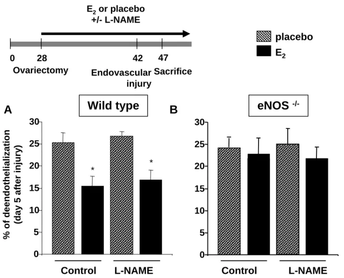
For all surgical procedures, female mice were anesthetized by intraperitoneal injection of a mixture of ketamine and xylasine and allowed to recover on a 37°C heat pack. They were ovariectomized at 4 weeks of age and given or not 60-day time-release E2 pellets (0.1mg E2, releasing 80µg/kg/d) (Innovative Research of America, Sarasota, FL) implanted subcutaneously into the backs of the animals with a sterile trochar. The NOS inhibitor, nitro-L-arginine methyl ester (L-NAME) (Sigma) (50 mg/kg/d) was given in the drinking water after ovariectomy. Electric carotid artery injury was performed 2 weeks later, in 6-week-old mice.
cGMP measurements
cGMP (the second messenger of NO) content in the thoracic aorta, the lung and the cerebellum was measured to assess the level of inhibition of NO synthase activity. Mice given L-NAME (0 or 50mg/kg/d) (8 mice in each group) were killed with an overdose of ketalar and used for determination of cGMP (the second messenger of NO) content in the thoracic aorta, in the lung and in the cerebellum. Thoracic aortae, lung and cerebella were removed and frozen in liquid nitrogen, and the tissues were stored frozen (-80°C) until measurement of the cGMP level. Frozen samples were thawed in glass potter in the presence of IBMX (10-4 M) and sonicated 10 times using ten 20 sec bursts. The homogenates were centrifuged (4000 rpm at 4°C during 15 min) and the supernatant fraction was taken for radioimmunoassay of cGMP using a commercial kit (Amersham). The samples were acetylated to increase the sensitivity of the assay. Data are the average of values from triplicate incubations. cGMP content was expressed as pmol per mg protein.
Perivascular injury model
The perivascular injury model was performed as previously described2. Briefly, the left common carotid artery was exposed via an anterior incision of the neck. The electric injury was applied to the distal part of the common carotid artery. The optimal conditions were determined as follows: electric power of 2 W applied for 2 seconds to each millimeter of carotid artery over a total length of 4 mm with the help of a size marker placed parallel to the long axis of the carotid.
Endovascular injury model
The endovascular injury model was adapted from Lindner et al.20. The right common carotid artery was exposed from the bifurcation, via an anterior incision of the neck. The external carotid was ligated distally and looped proximally with 8-0 silk suture. Silk loops were placed around the internal and common carotids to temporally restrict blood flow in the area of surgical manipulation. The ligature on the common carotid was precisely positioned against the sternocleidomastoid muscle 4 mm from the carotid bifurcation. This distance was very uniform from one animal to another. The external carotid artery was incised. The injury was performed using a 0.3 mm diameter swab built by loosely winding and sticking an 8-0 silk suture around a 0.16 mm diameter blunted guide wire. The swab was introduced, advanced through the common carotid artery and withdrawn 3 times. A 4 mm length denudation from the bifurcation of the common carotid artery was performed using the silk ligature on the common carotid as injury limit. The device was removed and the external carotid ligated proximally. Blood flow was then restored by loosening the loops around the internal and common carotids. The skin incision was closed and animals were allowed to wake up under warm conditions.
Quantification of endothelial regeneration
Three or five days after injury, the endothelial regeneration process was evaluated by staining the denuded areas with Evans blue dye (Sigma, Saint Quentin Fallavier, France) as
previously described2. Briefly, the common carotid artery was dissected with an adjacent portion of the aortic arch and carotid bifurcation. The artery was then opened longitudinally and placed between slides with Fluoprep (BioMérieux, Marcy l’Etoile, France). The ratio between the area stained in blue and the total carotid artery area was calculated. The surface of the area that remained deendothelialized was indexed to the total carotid artery area to take into account the changes in vessel area due to both elasticity of the carotid artery and the flattening of the vessel between slides.
Organ culture
Female mice between 8 and 10 weeks of age were sacrificed by a lethal injection of sodium pentobarbital (50 mg/kg IP). Carotid arteries were immersed in an organ culture bath filled with Dulbecco’s modified Eagle’s medium containing antibiotics (100UI/L penicillin, 100 mg/L streptomycin and 10 µg/L fungizone) and supplemented with 5% steroid-free fetal calf serum. Each arterial segment was maintained for 72h in a closed perfusion circuit described previously21, exposed to an intraluminal hydrostatic pressure of 80 mmHg with continuous renewal of medium within the intraluminal space at minimal shear stress (0.5 dynes/cm2). Organ culture of the carotid segments was carried out under sterile conditions in an incubator containing 5% CO2 at 37°C. At the end of the organ culture period, 10nM E2 (Sigma) or vehicle (DMSO) was injected in the intraluminal compartment for 15-60 min, in the presence (100µM) or absence of L-NAME. Thereafter vessels were quickly removed and processed as described below.
Cultured endothelial cells.
Endothelial cells (EC) were obtained from aorta according to the primary explant procedure adapted from a previously described protocol22, allowing the isolation of limited but pure cell preparations. Serum-starved EC were exposed for theindicated periods of time to E2 (10nM) in the presence or absence of L-NAME (100µM) or NG-Monomethyl-L-arginine acetate
(
L-NMMA) (Sigma)(100 and 300µM).Western Blot
Cultured vessels were ground in ice-cold lysis buffer containing 20mmol/L Tris-HCl (pH 7.5), 5 mmol/L EGTA, 150 mmol/L NaCl, 20 mmol/L glycerophosphate, 10mmol/L NaF, 1mmol/L sodium orthovanadate, 1% Triton X-100, 0.1% Tween 20 and protease inhibitors (Roche, Neuilly-sur-Seine, France). Detergent-soluble fractions were retained, and protein concentrations in samples were measured using a Bradford protein assay (Bio-Rad, Marnes-la-Coquette). For western blot analysis, lysates containing 30µg of protein were electrophoresed on polyacrylamide gels and transferred to PVDF membranes (Amersham, Piscataway, NJ). Membranes were incubated with anti-phospho-ERK1/2, anti ERK1/2 (Cell Signaling Technology, Beverly, MA) or anti-ERK2 antibodies (Santa Cruz, Heidelberg, Germany). An enhanced chemiluminescence system was used as the detection method (ECL+, Amersham).
Immunohistochemical analysis
Arteries were embedded in paraffin and sections perpendicular to the long axis of the carotid were cut at the level of the reendothelialized area of the injured carotid artery. Sections were subjected to standard hematoxylin/eosin staining and to eNOS immunodetection. Detection of eNOS expression was determined with rabbit polyclonal anti-eNOS (1:200; Santa Cruz Biotechnology, Cat# sc-654). The specificity of anti-eNOS antibody was tested on carotid artery cross sections from eNOS-/- mice.
Statistics
Data are expressed as mean±SEM. For cultured vessels, western blot band density
was quantified using Image Gauge software (Fuji). One-way ANOVA (or two-way) was used to compare data for different genotypes or inhibitors. When ANOVA analyses yielded significant results, comparisons were done using Bonferroni’s test.
Results
eNOS, but not NO production, is required for the effect of E2 on reendothelialization.
To define the role of NO production in the stimulating effect of E2 on reendothelialization in the perivascular injury model, we first used a pharmacological approach; ovariectomized wild type C57Bl/6J mice were treated chronically with L-NAME (50mg/kg/d). As shown in Figure 1A, pharmacological inhibition of eNOS activity did not alter the accelerating effect of E2 on reendothelialization 5 days post-injury. Similar results were obtained at day 3 injury (not shown). Similar results were also obtained at day 5 post-injury in C57Bl/6J mice treated chronically with a two-fold higher dose of L-NAME (100mg/kg/d; not shown).
As these results were inconsistent with a previous study by Losordo and colleagues17, implicating eNOS in the E2 effect on reendothelialization, we decided to repeat the study in the endovascular injury model. As observed in the perivascular injury model, treatment with L-NAME did not influence the capacity of E2 to speed up reendothelialization in the endovascular injury model (Figure 2A). In order to verify that the dose of L-NAME used (50mg/kg/d) efficiently blocked NO production in our experiments, the cGMP content in the thoracic aorta of C57Bl/6J mice treated with L-NAME (0 or 50mg/kg/d) for 8 days were 1.06 ± 0.05 and 0.35±0.03 pmol/mg protein respectively (P<0.0001), demonstrating a 3-fold drop in the second messenger of NO in L-NAME treated mice.
The cGMP content in the lung of C57Bl/6J mice given placebo or L-NAME 50mg/kg/d were 0.22±0.04 and 0.11±0.02 pmol/mg protein, respectively (P<0.001). These decreased levels of vascular cGMP were not further altered by doubling the dose of L-NAME (i.e. 100 mg/kg/d for 8 days), strongly suggesting that the dose of 50mg/kg/d used else in this study as well as in previous works23, 24 maximally blocks the eNOS activity. Moreover, we observed a 4.5-fold drop in cGMP in cerebellum preparations from L-NAME treated mice, demonstrating an inhibition of neuronal NOS (not shown).
Taken together, our data show that inhibiting NO production with L-NAME did not influence the effect of E2 on endothelial regrowth in vivo, irrespective of the arterial injury model.
The divergence between our findings and those previously published17 prompted us to reassess the role of eNOS five days after injury using a transgenic approach. Indeed, in agreement with the reported data, the accelerating effect of E2 on reendothelialization was abolished in eNOS-/- mice, both in the perivascular injury model (Figure 1B) as in the endovascular injury model (Figure 2B). Again, similar results were obtained at day 3 post-injury (not shown).
As shown in Figure 1B and 2B, the absence of E2 effect on reendothelialization was confirmed in these animals, and no “additional” effect of L-NAME was observed in either vascular injury model.
Altogether, these data showed that in vivo, eNOS, but not NO production, is required for the accelerating effect of E2 on reendothelialization.
eNOS expression is enhanced in the reendothelialized intimal area.
Given the importance of the eNOS protein in the reendothelialization process, we next performed immunohistochemical analyses to compare its expression levels in injured and uninjured carotid arteries. Compared with the endothelium of an intact (uninjured) artery (Figure 3A), eNOS expression was clearly enhanced in the regenerated endothelium of healing carotid artery, day 5 post-injury in wild type mice (Figure 3B), in agreement with previous observations made in regenerated endothelium in rat25 and in proliferating cultured endothelial cells26. In this electric injury model, the regenerated endothelial area can be easily recognized by the characteristic absence of underlying smooth muscle cells. The expression of eNOS was restricted to the intima in both intact and injured vessels, suggesting that it was mainly expressed at the level of the endothelium even after injury. As anticipated, no eNOS specific staining was detected in arteries of eNOS-/- mice (Figure 3C and 3D).
eNOS, but not NO production, is required for E2-induced ERK1/2 phosphorylation in
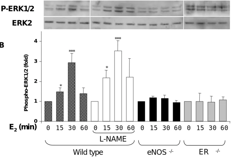
isolated perfused carotid artery and primary culture of mouse aortic endothelial cells. We then sought to explore the respective roles of eNOS deficiency and eNOS enzyme inhibition in E2-induced ERK1/2 activation, using an ex vivo model. As shown in Figure 4, ERK1/2 phosphorylation was enhanced in wild type carotid arteries exposed to E2, reaching an activation peak at 30 min and declining at 60 min. A delayed ERK1/2 activation have previously been reported in cultured endothelial cells exposed to E227. We also found that ERK1/2 phosphorylation by E2 was preserved in carotid arteries from wild type mice treated with 100 µM L-NAME, demonstrating that E2 acted independently of NO production. In contrast, E2-induced ERK1/2 activation was abolished in carotid arteries from eNOS -/-mice. Similarly, E2 failed to induce ERK1/2 phosphorylation in carotid arteries from ERα -/-mice.
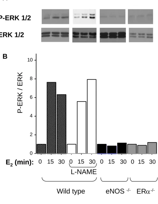
We then reassessed the respective roles of eNOS deficiency and eNOS enzyme inhibition in cultured endothelial cells from mouse aortae. As shown in Figure 5, we found that E2 stimulated the phosphorylation of ERK1/2 equally in untreated and in L-NAME- treated cells from WT mice. L-NMMA, another NOS inhibitor, did not alter the effect of E2 stimulated the phosphorylation of ERK1/2 in endothelial cells from WT mice (not shown). Moreover, pretreatment with 0.5 mM DPTA NONOate, a NO donor, did not restore the effect of E2 in endothelial cells from eNOS-/- mice (not shown). On the other hand, activation of ERK1/2 by E2 was abolished in eNOS-/- and ERα-/- cells. A deficiency in intracellular BH4 levels does not appear to explain these differences, since neither the supplementation of BH4 (3µM) nor of the BH4 synthesis substrate sepiapterin (50 µM) to cultured aortic endothelial cells from C57BL/6 wild-type mice altered the phosphorylation of ERK1/2 in response to E2 (not shown).
As gene inactivation is known to favor gene expression remodeling, in particular “gene compensations”, we tried to restore eNOS expression in eNOS-deficient endothelial cells through transfection. However, we did not succeed with this approach due to the difficulty of transfecting these fragile cells, which underwent apoptosis soon after transfection.
We therefore sought to abolish eNOS expression in eNOS-expressing endothelial cells using siRNA against eNOS. We first performed this experiment in endothelial cells from eNOS +/+ mice, but again these cells did not tolerate transfection. We next tried a more classical cell model of endothelial cells: HUVECs. Unfortunately, we were unable to elicit ERK1/2 activation in response to E2 in HUVEC, even in freshly harvested cell at very early passages. This was likely due to lack of ER gene expression (assessed by RT-PCR and Western blotting) at variance with previous work28, but in agreement with other29.
Finally, we tried a HUVEC derived cell line (EA.hy926) that has been shown to express ERα30. In this model, we were able to elicit MAPK activation in response to E
2, and a clear decrease of this signal after eNOS siRNA treatment (Fig I, supplementary data). These data indicate that the level of eNOS protein expression influence the capacity of E2 to activate ERK1/2 in vitro. However, we were struck by the poor reproducibility of this model, as in two out of four experiments, we did not observe an increase in ERK1/2 phosphorylation in response to E2 in control cells.
Altogether, these data suggest that the E2 effect leading to ERK1/2 phosphorylation is mediated by ERα and appears to be strictly dependent on the presence of eNOS protein, but not its activity, in both ex vivo and in vitro models. Noteworthy, the reproducibility of the effects observed in vivo or ex vivo was much more robust than the ones observed in cultured endothelial cells.
Discussion
Altogether, our results reveal an original and unexpected role for eNOS in endothelial regeneration and in plasma membrane E2 signaling, acting as a signaling protein independently of its NO-producing function. Indeed, our finding that accelerated reendothelialization induced by E2 was abolished in eNOS-/- mice agreed with a previous report17, but we were surprised to observe that the pharmacological inhibition of NOS with L-NAME had no influence on the beneficial effect of E2 in two models of vascular injury.
Our finding that the in vivo effects of E2 differ between L-NAME-treated and eNOS -/-mice appears robust for the following reasons: 1) it was established in two different experimental models, endovascular and electric perivascular carotid artery injury; 2) pharmacological inhibition of eNOS using L-NAME was previously shown to maximally block NO synthase activity23, 24, 31, and its efficacy was verified in the present work; 3) abrogation of the effect of E2 by eNOS gene inactivation has now been demonstrated independently, since the eNOS-/- model used in the present work18 was different from that used by Losordo and colleagues32.
To better understand the interaction between E2 and eNOS, we explored the respective influences of eNOS gene inactivation and eNOS activity inhibition on the short-term effects of E2 in an ex vivo artery model. This model was used to specifically study endothelium in its native matrix and mechanical (submitted to physiologic pressure) environment, in the absence of confounding hormonal or circulating factors encountered in vivo33. As previously described in cultured human, bovine and porcine endothelial cells27, 34, 35, we found that E
2 induced ERK1/2 activation in isolated perfused carotid arteries. Interestingly, and in agreement with our reendothelialization data, the plasma membrane signaling of E2 was strictly dependent on the presence of eNOS protein, but not eNOS activity in isolated perfused carotid arteries. The limitation of this model in interpreting ERK1/2 activation is that both endothelial and smooth muscle cells express ERK1/2. However, the exclusive expression of eNOS in the endothelium (Fig 3) on the one hand, and the similarity in results between in vivo and ex vivo models on the other hand, reinforce the
concept that plasma membrane signaling of E2 is strictly dependent on the presence of eNOS protein, but not eNOS activity, in the endothelial cells of carotid arteries.
eNOS gene disruption and NOS pharmacological inhibition are often viewed as comparable strategies, as they in some aspects generate similar results, such as the induction of hypertension and endothelial dysfunction18, 23, 24, 31, 32. However, discrepancies between these two models have already been suggested, but never directly demonstrated. For instance, eNOS-/-apoE-/- mice develop two-fold more fatty streak lesions than apoE -/-mice36 whereas inhibition of NO production by L-NAME does not influence fatty streak deposit in apoE-/- mice24, 36. Similarly, de Mey and colleagues previously showed an abrogation of shear stress induced vascular remodeling in eNOS-/- mice37 , but not in L-NAME treated rats38, suggesting that eNOS, but not eNOS activity, could play a role in vascular remodeling. This kind of dissociation between protein expression and enzymatic activity was nicely demonstrated for PI3Kγ39 which participates in two distinct signaling
cascades: a dependent pathway that controls PKB/AKT activity, and a kinase-independent pathway that relies on protein interactions with a phosphodiesterase to negatively modulate cardiac contractility.
Since eNOS is largely present in caveolae and a fraction of ERα is also localized in
this membrane compartment, these two molecules could associate in a functional molecular complex16 30. Moreover, both ERα and eNOS were shown to interact with HSP9040 and caveolin 141, 42, which reinforces the probability of a caveolar interaction of these proteins and supports of the hypothesis that eNOS acts in a multi-protein assembly, allowing ERK1/2 activation in response to E2.
Even though L-NAME is not a specific inhibitor of eNOS, but also inhibits other NOS isozymes, such as neuronal NOS (nNOS) and to some extent inducible NOS (iNOS), the lack of specificity could hardly be influencing our conclusions concerning the effects on the carotid artery. Indeed, the inhibition of iNOS by L-NAME is only partial in vivo at the dose used in the present study43, 44. However, L-NAME did not alter the observed loss of E2 effect in eNOS-/- mice, ruling out an inference of the other NOS isoforms in the abrogation of the E2
effects in eNOS -/- mice. Finally, a significant contribution of superoxide production by eNOS uncoupling is unlikely since, 1) BH4 supplementation did not influence the result observed in wild-type cultured cells; 2) L-NAME inhibits superoxide production, as demonstrated in previous in vivo45 and ex vivo46 studies; 3) L-NAME inhibits superoxide production of purified nNOS 47, 48.
The in vivo effects of E2 probably involve both genomic and non-genomic mechanisms. Although ERs are classically defined as ligand-activated transcription factors14, it has become clear that short-term “extragenomic-to-genomic” responses play an important role in cultured endothelial cells6, but also in osteoblasts and in cortical neurons (as for instance the activation of PI3kinase-AKT pathways as well as MAP kinase pathways)49, 50. The characterization of the respective roles of these “membrane” and “classical” effects represents an important but difficult challenge for the next years15, 51, 52.
Beyond this unexpected role of eNOS in E2 signaling, the clinical implications of our observations could be significant. First, the strong correlation between our in vivo and ex vivo findings support an implication of plasma membrane estrogen signaling in its protective effects. Second, given that deficiency in E2 has been recognized to modulate endothelial function53, altered eNOS expression or trafficking in the context of endothelial dysfunction could hinder the beneficial effects of E2, thus sustaining a vicious circle. This could for instance help to explain the highly negative relationship reported between smoking and estrogen atheroprotection54. Third, the proposed role of eNOS, acting within a molecular complex independently of its NO producing activity, may not be restricted to E2 signaling but may also influence endothelial response to other vasoactive compounds. These specific roles of eNOS should be more precisely explored in future work.
Acknowledgments:
We thank Alexia Schambourg, Marie-José Fouque and Hortense Berges for invaluable technical assistance. eNOS deficient mice were kindly provided by Pr Godecke (Dusseldorf,
Germany) and ERα deficient mice were kindly provided by Pr Chambon and Dr A. Krust
References:
1. Bennett MR, O'Sullivan M: Mechanisms of angioplasty and stent restenosis: implications for
design of rational therapy, Pharmacol Ther 2001, 91:149-166.
2. Brouchet L, Krust A, Dupont S, Chambon P, Bayard F, Arnal JF: Estradiol accelerates
reendothelialization in mouse carotid artery through estrogen receptor-alpha but not estrogen
receptor-beta, Circulation 2001, 103:423-428.
3. Krasinski K, Spyridopoulos I, Asahara T, van der Zee R, Isner JM, Losordo DW: Estradiol
accelerates functional endothelial recovery after arterial injury, Circulation 1997, 95:1768-1772
4. Sullivan TR, Jr., Karas RH, Aronovitz M, Faller GT, Ziar JP, Smith JJ, O'Donnell TF, Jr.,
Mendelsohn ME: Estrogen inhibits the response-to-injury in a mouse carotid artery model, J
Clin Invest 1995, 96:2482-2488
5. Lantin-Hermoso RL, Rosenfeld CR, Yuhanna IS, German Z, Chen Z, Shaul PW: Estrogen
acutely stimulates nitric oxide synthase activity in fetal pulmonary artery endothelium, Am J
Physiol 1997, 273:L119-126
6. Mendelsohn ME: Nongenomic, ER-mediated activation of endothelial nitric oxide synthase:
how does it work? What does it mean? Circ Res 2000, 87:956-960.
7. Arnal JF, Clamens S, Pechet C, Nègre-Salvayre A, Allera C, Girolami JP, Salvayre R, Bayard
F: Ethinylestradiol does not enhance the expression of nitric oxide synthase in bovine aortic
endothelial cells but increases the release of bioactive nitric oxide by inhibiting superoxide
anion production., Proc. Natl. Acad. Sci. U. S. A. 1996, 93:4108-4113
8. Wagner AH, Schroeter MR, Hecker M: 17beta-estradiol inhibition of NADPH oxidase
expression in human endothelial cells, Faseb J 2001, 15:2121-2130
9. Couse JF, Korach KS: Estrogen receptor null mice: what have we learned and where will they
lead us? Endocr Rev 1999, 20:358-417.
10. Chen Z, Yuhanna IS, Galcheva-Gargova Z, Karas RH, Mendelsohn ME, Shaul PW: Estrogen
receptor alpha mediates the nongenomic activation of endothelial nitric oxide synthase by
estrogen, J Clin Invest 1999, 103:401-406
11. Darblade B, Pendaries C, Krust A, Dupont S, Fouque MJ, Rami J, Chambon P, Bayard F,
Arnal JF: Estradiol alters nitric oxide production in the mouse aorta through the alpha-, but not
12. Pendaries C, Darblade B, Rochaix P, Krust A, Chambon P, Korach KS, Bayard F, Arnal JF:
The AF-1 activation-function of ERalpha may be dispensable to mediate the effect of estradiol
on endothelial NO production in mice, Proc Natl Acad Sci U S A 2002, 99:2205-2210.
13. Pare G, Krust A, Karas RH, Dupont S, Aronovitz M, Chambon P, Mendelsohn ME: Estrogen
receptor-alpha mediates the protective effects of estrogen against vascular injury, Circ Res
2002, 90:1087-1092
14. Reid G, Denger S, Kos M, Gannon F: Human estrogen receptor-alpha: regulation by
synthesis, modification and degradation, Cell Mol Life Sci 2002, 59:821-831
15. Haynes MP, Li L, Russell KS, Bender JR: Rapid vascular cell responses to estrogen and
membrane receptors, Vascul Pharmacol 2002, 38:99-108
16. Chambliss KL, Yuhanna IS, Mineo C, Liu P, German Z, Sherman TS, Mendelsohn ME,
Anderson RG, Shaul PW: Estrogen receptor alpha and endothelial nitric oxide synthase are
organized into a functional signaling module in caveolae, Circ Res 2000, 87:E44-52
17. Iwakura A, Luedemann C, Shastry S, Hanley A, Kearney M, Aikawa R, Isner JM, Asahara T,
Losordo DW: Estrogen-mediated, endothelial nitric oxide synthase-dependent mobilization of
bone marrow-derived endothelial progenitor cells contributes to reendothelialization after
arterial injury, Circulation 2003, 108:3115-3121
18. Godecke A, Decking UK, Ding Z, Hirchenhain J, Bidmon HJ, Godecke S, Schrader J:
Coronary hemodynamics in endothelial NO synthase knockout mice, Circ Res 1998,
82:186-194
19. Dupont S, Krust A, Gansmuller A, Dierich A, Chambon P, Mark M: Effect of single and
compound knockouts of estrogen receptors alpha (ERalpha) and beta (ERbeta) on mouse
reproductive phenotypes, Development 2000, 127:4277-4291.
20. Lindner V, Fingerle J, Reidy MA: Mouse model of arterial injury, Circ Res 1993, 73:792-796
21. Lemarie CA, Esposito B, Tedgui A, Lehoux S: Pressure-induced vascular activation of nuclear
factor-kappaB: role in cell survival, Circ Res 2003, 93:207-212
22. Sonveaux P, Martinive P, DeWever J, Batova Z, Daneau G, Pelat M, Ghisdal P, Gregoire V,
Dessy C, Balligand JL, Feron O: Caveolin-1 expression is critical for vascular endothelial
growth factor-induced ischemic hindlimb collateralization and nitric oxide-mediated
23. Arnal JF, Warin L, Michel JB: Determinants of aortic cyclic guanosine monophosphate in
hypertension induced by chronic inhibition of nitric oxide synthase, J. Clin. Invest. 1992,
90:647-652
24. Elhage R, Bayard F, Richard V, Holvoet P, Duverger N, Fievet C, Arnal JF: Prevention of fatty
streak formation of 17beta-estradiol is not mediated by the production of nitric oxide in
apolipoprotein E-deficient mice, Circulation 1997, 96:3048-3052
25. Poppa V, Miyashiro JK, Corson MA, Berk BC: Endothelial NO synthase is increased in
regenerating endothelium after denuding injury of the rat aorta, Arterioscler Thromb Vasc Biol
1998, 18:1312-1321
26. Arnal JF, Yamin J, Dockery S, Harrison DG: Regulation of endothelial nitric oxide synthase
mRNA, protein, and activity during cell growth, Am J Physiol 1994, 267:C1381-1388
27. Klinge CM, Blankenship KA, Risinger KE, Bhatnagar S, Noisin EL, Sumanasekera WK, Zhao
L, Brey DM, Keynton RS: Resveratrol and estradiol rapidly activate MAPK signaling through
estrogen receptors alpha and beta in endothelial cells, J Biol Chem 2005, 280:7460-7468
28. Li L, Haynes MP, Bender JR: Plasma membrane localization and function of the estrogen
receptor alpha variant (ER46) in human endothelial cells, Proc Natl Acad Sci U S A 2003,
100:4807-4812
29. Baker VL, Chao VA, Murai JT, Zaloudek CJ, Taylor RN: Human umbilical vessels and cultured
umbilical vein endothelial and smooth muscle cells lack detectable protein and mRNA
encoding estrogen receptors, J Soc Gynecol Investig 1997, 4:316-324
30. Figtree GA, McDonald D, Watkins H, Channon KM: Truncated estrogen receptor alpha 46-kDa
isoform in human endothelial cells: relationship to acute activation of nitric oxide synthase,
Circulation 2003, 107:120-126
31. Chatziantoniou C, Boffa J, Ardaillou R, Dussaule J: Nitric oxide inhibition induces early
activation of type I collagen gene in renal resistance vessels and glomeruli in transgenic mice.,
J. Clin. Invest. 1998, 101:2780-2789
32. Shesely EG, Maeda N, Kim HS, Desai KM, Krege JH, Laubach VE, Sherman PA, Sessa WC,
Smithies O: Elevated blood pressures in mice lacking endothelial nitric oxide synthase, Proc
Natl Acad Sci U S A 1996, 93:13176-13181
33. Lehoux S, Esposito B, Merval R, Tedgui A: Differential regulation of vascular focal adhesion
34. Geraldes P, Sirois MG, Bernatchez PN, Tanguay JF: Estrogen regulation of endothelial and
smooth muscle cell migration and proliferation: role of p38 and p42/44 mitogen-activated
protein kinase, Arterioscler Thromb Vasc Biol 2002, 22:1585-1590
35. Razandi M, Pedram A, Park ST, Levin ER: Proximal events in signaling by plasma membrane
estrogen receptors, J Biol Chem 2003, 278:2701-2712
36. Knowles JW, Reddick RL, Jennette JC, Shesely EG, Smithies O, Maeda N: Enhanced
atherosclerosis and kidney dysfunction in eNOS(-/-)Apoe(-/-) mice are ameliorated by enalapril
treatment, J Clin Invest 2000, 105:451-458
37. van der Heijden OW, Essers YP, Spaanderman ME, De Mey JG, van Eys GJ, Peeters LL:
Uterine artery remodeling in pseudopregnancy is comparable to that in early pregnancy, Biol
Reprod 2005, 73:1289-1293
38. Ceiler DL, De Mey JG: Chronic N(G)-nitro-L-arginine methyl ester treatment does not prevent
flow-induced remodeling in mesenteric feed arteries and arcading arterioles, Arterioscler
Thromb Vasc Biol 2000, 20:2057-2063
39. Patrucco E, Notte A, Barberis L, Selvetella G, Maffei A, Brancaccio M, Marengo S, Russo G,
Azzolino O, Rybalkin SD, Silengo L, Altruda F, Wetzker R, Wymann MP, Lembo G, Hirsch E:
PI3Kgamma modulates the cardiac response to chronic pressure overload by distinct
kinase-dependent and -inkinase-dependent effects, Cell 2004, 118:375-387
40. Russell KS, Haynes MP, Caulin-Glaser T, Rosneck J, Sessa WC, Bender JR: Estrogen
stimulates heat shock protein 90 binding to endothelial nitric oxide synthase in human vascular
endothelial cells. Effects on calcium sensitivity and NO release, J Biol Chem 2000,
275:5026-5030
41. Garcia-Cardena G, Fan R, Stern DF, Liu J, Sessa WC: Endothelial nitric oxide synthase is
regulated by tyrosine phosphorylation and interacts with caveolin-1, J Biol Chem 1996,
271:27237-27240
42. Razandi M, Oh P, Pedram A, Schnitzer J, Levin ER: ERs associate with and regulate the
production of caveolin: implications for signaling and cellular actions, Mol Endocrinol 2002,
16:100-115
43. Darblade B, Batkai S, Causse E, Gourdy P, Fouque MJ, Rami J, Arnal JF: Failure of
L-nitroarginine to inhibit the activity of aortic inducible nitric oxide synthase, J Vasc Res 2001,
44. Miller M, Thompson J, Lui X, Eboly-Childress S, Sadowska-Krowicka H, Zhang X, CLark D:
Failure of L-NAME to cause inhibition of nitric oxide synthesis: role of inducible nitric oxide
synthase., Inflamm Res 1996, 45:272-276
45. van Deel ED, Merkus D, van Haperen R, de Waard MC, de Crom R, Duncker DJ: Vasomotor
control in mice overexpressing human endothelial nitric oxide synthase, Am J Physiol Heart
Circ Physiol 2007, 293:H1144-1153
46. Bendall JK, Alp NJ, Warrick N, Cai S, Adlam D, Rockett K, Yokoyama M, Kawashima S,
Channon KM: Stoichiometric relationships between endothelial tetrahydrobiopterin, endothelial
NO synthase (eNOS) activity, and eNOS coupling in vivo: insights from transgenic mice with
endothelial-targeted GTP cyclohydrolase 1 and eNOS overexpression, Circ Res 2005,
97:864-871
47. Cardounel AJ, Xia Y, Zweier JL: Endogenous methylarginines modulate superoxide as well as
nitric oxide generation from neuronal nitric-oxide synthase: differences in the effects of
monomethyl- and dimethylarginines in the presence and absence of tetrahydrobiopterin, J Biol
Chem 2005, 280:7540-7549
48. Pou S, Keaton L, Surichamorn W, Rosen GM: Mechanism of superoxide generation by
neuronal nitric-oxide synthase, J Biol Chem 1999, 274:9573-9580
49. Kousteni S, Bellido T, Plotkin LI, O'Brien CA, Bodenner DL, Han L, Han K, DiGregorio GB,
Katzenellenbogen JA, Katzenellenbogen BS, Roberson PK, Weinstein RS, Jilka RL,
Manolagas SC: Nongenotropic, sex-nonspecific signaling through the estrogen or androgen
receptors: dissociation from transcriptional activity, Cell 2001, 104:719-730
50. Mannella P, Brinton RD: Estrogen receptor protein interaction with phosphatidylinositol
3-kinase leads to activation of phosphorylated Akt and extracellular signal-regulated 3-kinase 1/2
in the same population of cortical neurons: a unified mechanism of estrogen action, J Neurosci
2006, 26:9439-9447
51. Mendelsohn ME, Karas RH: Molecular and cellular basis of cardiovascular gender differences,
Science 2005, 308:1583-1587
52. Warner M, Gustafsson JA: Nongenomic effects of estrogen: why all the uncertainty? Steroids
2006, 71:91-95
53. Sader MA, Celermajer DS: Endothelial function, vascular reactivity and gender differences in
the cardiovascular system, Cardiovasc Res 2002, 53:597-604
55. Edgell CJ, McDonald CC, Graham JB: Permanent cell line expressing human factor
Figures legends:
Figure 1. Perivascular injury model: effect of E2 on the reendothelialization process in
ovariectomized wild type (A) and eNOS -/- (B) mice treated or not (placebo) with E2 (80 µg/kg/d) and receiving or not L-NAME (50 mg/kg/d) in the drinking water. Results show
percent deendothelialization calculated as ratio of remaining deendothelialized area to total carotid artery area, at day 5. *P<0.05 vs respective placebo group.
Figure 2. Endovascular injury model: effect of E2 on the reendothelialization process in
ovariectomized wild type (A) and eNOS -/- (B) mice treated or not (placebo) with E2 (80 µg/kg/d) and receiving or not L-NAME (50 mg/kg/d) in the drinking water. Results show
percent deendothelialization calculated as ratio of remaining deendothelialized area to total carotid artery area, at day 5. *P<0.05 vs respective placebo group.
Figure 3. Immunostaining of eNOS in non injured (A, C) and reendothelialized (B, D) carotid arteries of wild type (A, B) and eNOS-/- mice (C, D) at day 5 after perivascular injury.
Figure 4. Acute phosphorylation of ERK1/2 induced by E2 in carotid arteries from wild
type mice is insensitive to L-NAME but is abolished in eNOS-/- carotid arteries. A, Representative Western blot showing short-term phosphorylation kinetics of ERK1/2 induced by E2 (10nM), administered in the intraluminal compartment of carotid arteries after 3 days in organ culture. ERK1/2 phosphorylation is equivalent in untreated and L-NAME-treated (100µM) carotid arteries from WT mice, but abolished in carotid arteries from eNOS-/- and ERα-/- mice. B, Bar graph represents the quantification of the phospho-ERK1/2 / ERK2 ratio,
normalized to the control within each group. *P<0.05 and ***P<0.001 vs respective control group.
Figure 5. eNOS deficiency prevents E2-induced ERK1/2 activation in MAECs. Isolated
mouse endothelial cells were treated with E2 (10nM) for different times. A, Representative immunoblotting experiments show that phosphorylation of ERK1/2 is enhanced by E2 in endothelial cells from WT but not eNOS–/– or ERα–/– mice. B, Bar graph represents the quantification of the P-ERK/ ERK ratio, normalized to the control within each group (n=2 experiments).
Supplementary data.
Figure I. Effect of siRNA-mediated eNOS knockdown on E2-induced ERK1/2 activation
in EA.hy926 cells. Immunoblots from E2-treated EA.hy926 endothelial cells transfected with scramble or eNOS siRNA. The cell lysates were resolved by SDS-PAGE and analyzed in immunoblots probed as indicated with specific antibodies directed against eNOS, phospho-ERK1/2, total ERK1/2.
Materials and Methods
siRNA transfection. Small interference RNA duplex oligonucleotides (ON-TARGET Plus) were from Dharmacon, Inc. (Lafayette, CO). EA.hy926 cells (courtesy of Edgell C. J.) were cultured as previously described 55 and transfected with siRNA (100nM) using LipofectAMINE 2000 (Invitrogen) following the instruction provided by the supplier. Forty-eight hours later, cells were harvested and subjected to Western blot analysis as described in the main Material and methods section. The antibodies used were from Santa Cruz (anti-eNOS) and Cell Signaling Technology (anti-phospho-ERK1/2 and anti ERK1/2).