HAL Id: inserm-00452350
https://www.hal.inserm.fr/inserm-00452350
Submitted on 2 Feb 2010HAL is a multi-disciplinary open access archive for the deposit and dissemination of sci-entific research documents, whether they are pub-lished or not. The documents may come from teaching and research institutions in France or abroad, or from public or private research centers.
L’archive ouverte pluridisciplinaire HAL, est destinée au dépôt et à la diffusion de documents scientifiques de niveau recherche, publiés ou non, émanant des établissements d’enseignement et de recherche français ou étrangers, des laboratoires publics ou privés.
Protein arginine methylation in estrogen signaling and
estrogen-related cancers.
Catherine Teyssier, Muriel Le Romancer, Stéphanie Sentis, Stéphan Jalaguier,
Laura Corbo, Vincent Cavaillès
To cite this version:
Catherine Teyssier, Muriel Le Romancer, Stéphanie Sentis, Stéphan Jalaguier, Laura Corbo, et al.. Protein arginine methylation in estrogen signaling and estrogen-related cancers.. Trends in Endocrinol-ogy and Metabolism = Trends in EndocrinolEndocrinol-ogy & Metabolism , Elsevier, 2010, 21 (3), pp.181-9. �10.1016/j.tem.2009.11.002�. �inserm-00452350�
REVIEW ARTICLE 1 2 3 4 5
Protein arginine methylation in estrogen signaling and estrogen-related cancers
6 7 8 9 10
Catherine Teyssier1, Muriel Le Romancer2, Stéphanie Sentis2, Stéphan Jalaguier3, Laura 11
Corbo2 and Vincent Cavaillès3 12
13 14 15
1
INSERM, U554, Montpellier, F-34090, France ; CNRS, UMR5048, Centre de Biochimie
16
Structurale, Universités Montpellier 1 & 2, Montpellier, F-34090, France
17
2
Université de Lyon 1, ISPB and IFR62, Lyon F-69003, France ; Equipe labellisée “La 18
Ligue,” ; U590 INSERM, Centre Léon Bérard, 28 rue Laennec, Lyon, F-69003, France 19
3
IRCM, Institut de Recherche en Cancérologie de Montpellier, Montpellier, F-34298, France;
20
INSERM, U896, Montpellier, 34298, France ; Université Montpellier1, Montpellier,
F-21
34298, France ; CRLC Val d’Aurelle Paul Lamarque, Montpellier, F-34298, France
22 23
Corresponding author: Dr Vincent Cavaillès (v.cavailles@valdorel.fnclcc.fr) 24
Abstract:
26 27
Estrogen signaling pathways regulate cellular processes such as proliferation and 28
differentiation, and if deregulated, are involved in several human pathologies. Post-29
translational modifications (PTMs) play important roles in estrogen signaling pathways. This 30
review focuses on recent findings pertinent to arginine methylation of non-histone proteins 31
and their implications in estrogen signaling. We describe protein arginine methyltransferases 32
and demethylases, the role of methylarginine proteins in estrogen action and cross-talk with 33
other PTMs such as phosphorylation and lysine methylation. The relationships between 34
various PTMs form a specific code which might play an important role in hormone signaling. 35
In addition, deregulations of arginine methylation or of enzymes responsible for these 36
modifications could be key events in estrogen-dependent cancers such as breast cancer. 37
Arginine methylation was first described as a post-translational modification (PTM) of 39
histones in the late 1960s. However, only recently the responsible enzymes (Box 1) and wide 40
variety of substrates of this modification have been identified. While the addition of each 41
methyl group does not modify the charge of the residue, it does increase its bulkiness and 42
hydrophobicity. Interactions of a methylated protein with its binding partners can therefore be 43
affected by this modification and impact the physiological functions of the substrate protein. 44
Methylarginine substrates include transcription factors, nucleic acid-binding factors, signal 45
transducers, splicing factors and histones. Because of the large number of substrates, protein 46
arginine methyltransferases (PRMTs) and arginine methylation regulate various cellular 47
processes such as cell differentiation, DNA repair, RNA processing, signal transduction, 48
cellular localization, and apoptosis [1-3]. In this review, we describe the recent studies 49
implicating arginine methylation in estrogen transcriptional regulation and likewise, in 50
estrogen-related diseases. 51
52 53
Role of PRMTs and methylarginine proteins in estrogen signaling
54
Estrogen action – Genomic and non-genomic effects
55
Nuclear estrogen receptors act mainly as ligand-activated transcription factors [4]. The 56
binding of estrogens such as 17 -estradiol (E2) induces a protein conformation change in the 57
receptor that allows recruitment of coactivator complexes with chromatin-remodeling or 58
histone-modifying activities [5] (Box 2). Steroid-regulated promoters recruit the ligand-bound 59
receptors and regulatory proteins in an ordered, cyclical manner with multiple rounds of 60
coactivator assembly and disassembly [6]. Histone acetylases and some methyltransferases 61
lead to a more open chromatin and increase gene transcription. P300, an acetyltransferase, and 62
PRMT1 and CARM1, two well-characterized PRMTs, cooperate synergistically to regulate 63
hormone target genes [7]. The activated estrogen receptor can also bind corepressors, such as 64
RIP140 [8], which recruit enzymes with histone deacetylase activity (HDAC) to repress 65
transcription. Anti-estrogen compounds like tamoxifen prevent steroid action by inducing a 66
conformation in the ligand binding pocket of the receptor that fails to bind coactivators and 67
allows the recruitment of corepressor proteins (such as NCoR and SMRT) together with 68
HDACs [9]. 69
In addition to transcriptional regulation, ERα also mediates events through its association with 70
signaling molecules outside the nuclei and independent of its direct influence on the genome 71
[10]. For example, estradiol triggers cell proliferation and cell survival through activation of 72
MAPK kinases and Akt pathways [11]. These non-genomic actions of estrogens occur rapidly 73
and independently of protein synthesis. At the molecular level, ERα palmitoylation anchors a 74
pool of ERα at the plasma membrane [12] where it interacts with Src, PI3K and other scaffold 75
proteins as MNAR (modulator of non-genomic activity of ER). This complex can therefore 76
activate the downstream pathways [13]. Recently, novel non-genomic action of estrogens in 77
breast cancer cells has been described, involving the association of membrane ERα with 78
HDAC6. This association induces tubulin deacetylation, potentially contributing to estrogen-79
induced cell migration [14]. 80
81
Methylarginine proteins involved in estrogen action
82
Arginine methylation affects estrogen-mediated transcription by modifying both histone and 83
non-histone proteins. Since histone methylation has been widely described in a various 84
number of reviews [15-17], this section focuses on methylation of non-histone proteins and 85
their role in estrogen action (Table 1 and Figure 1). 86
Estrogen receptor
Because PRMT1 [18] and CARM1 (PRMT4) [19] are ER coregulators (Box 2), ER could 88
be a target for arginine methylation. Concordant with this, a recent study described ER as a 89
methylarginine substrate [20]. In this study, Le Romancer et al. used in vitro methylation 90
assays and showed that PRMT1, but not CARM1, methylated ER within the DNA binding 91
domain. Mutation of arginine 260 into alanine (R260A) specifically abolished the 92
modification by PRMT1. An antibody specific to methylated R260 confirmed ER 93
methylation in living cells. Perhaps more interestingly, estradiol treatments of MCF7 cells 94
drastically increased ER methylation within 5 minutes of treatment. A decrease of the 95
methylated form was observed within less than one hour, suggesting enzymatic removal of 96
the methyl group. Indeed, this disappearance was not due to ER degradation by the 97
proteasome. Moreover, immunohistochemical experiments performed on human breast 98
tumors showed that the methylated form of ER was exclusively localized in the cytoplasm 99
of breast epithelial cells. Since rapid effects have been described for non-genomic estrogen 100
actions, the role of methylated ER in those pathways was investigated. Interestingly, 101
methylated ER was essential for E2-induced assembly of ER with Src, the p85 subunit of 102
PI3K and the focal adhesion kinase (FAK), a Src substrate involved in the migration process. 103
E2 activation of Akt was not observed if ER R260 was mutated to an alanine. Collectively, 104
these results show that ER methylation is a prerequisite for its association with certain 105
molecules involved in growth factor signaling. Since formation of the ER /Src/PI3K/FAK 106
complex activates Akt and corresponding downstream pathways, ER methylation is likely 107
involved in regulating cell proliferation and cell survival. 108
Transcriptional coregulators
109
Transcriptional coregulators play critical roles in controlling ER-mediated transcription. They 110
function through protein-protein interactions, by facilitating or inhibiting recruitment of other 111
these cofactors may influence complex formation, enzymatic activity, subcellular localization, 113
and stability, leading to a subtle regulation of ER-mediated transcription (Figure 1). 114
SRC-3: p/CIP/AIB1/SRC-3 is a member of the p160 coactivator family involved in CARM1
115
and PRMT1 recruitment to ER target genes [7]. SRC-3 (steroid receptor coactivator-3) 116
protein levels are amplified in breast cancer and associated with poor prognosis [21, 22]. In 117
fact, studies classified SRC-3 as an authentic oncogene [23, 24]. Two independent groups 118
found that CARM1 modifies SRC-3 [25, 26], methylating it at the C-terminal region which 119
contains the p300 and CARM1-binding sites. SRC-3 methylation, which is induced by 120
estradiol, leads to the dissociation of CBP and CARM1 from SRC-3. Moreover, SRC-3 121
arginine methylation reduces its stability and causes an increase inits turnover. These results 122
show that arginine methylation is implicated in the regulation of coregulator stability in 123
response to estradiol. In addition to this role, arginine methylation also regulates the balance 124
between coactivator complex assembly and disassembly. Studies suggest that repetitive 125
association and dissociation of steroid receptors and coactivators from their target promoters 126
may be required to maintain an activated state of transcription [6]. Altogether, these results 127
highlight coactivator methylation as an important regulatory mechanism in hormonal 128
signaling. 129
CBP/p300: Recently, Lee et al. reported that p300 is methylated by CARM1 at R2142 which
130
is located within the C-terminal GRIP1 binding domain. Interestingly, methylation of R2142 131
inhibits the interaction between p300 and GRIP1 whereas PADI 4 removes this methylation 132
mark, thereby enhancing the p300–GRIP1 interaction. These methylation and demethylation 133
events alter the conformation and activity of the coactivator complex and regulate estrogen 134
receptor-mediated transcription [27]. This provides another example of arginine methylation 135
regulating coactivator complex assembly, conformation and function. 136
PGC-1 : PGC-1α (peroxisome proliferator-activated receptor gamma coactivator 1 alpha)
137
serves as a coactivator for several nuclear receptors, including ER , as well as other 138
transcription factors, such as nuclear respiratory factor 1 (NRF-1). Expression of PGC-1α is 139
induced by a variety of physiological stimuli that regulate metabolic activity, such as 140
exposure to cold, exercise, and fasting. PGC-1α regulates metabolic processes by affecting 141
genes involved in mitochondrial biogenesis, respiration and gluconeogenesis. Teyssier et al. 142
showed that the C-terminal region of PGC-1α is methylated by PRMT1 at one or more sites 143
within a glutamate and arginine rich region. PGC-1α coactivator activity and the ability of 144
PGC-1α to induce expression of target genes are both compromised by mutation of modified 145
arginine residues of PGC-1α or as a result of reduced PRMT1 levels by siRNA. Because 146
inhibition of PRMT1 leads to inhibition of the expression of some PGC-1α target genes 147
involved in mitochondrial biogenesis, it is tempting to speculate a role for PRMT1 in this 148
regulatory pathway [28]. However, the binding partners of methylated PGC-1α remain to be 149
determined. 150
RIP140: RIP140 (Receptor Interacting Protein of 140 kDa) is a well-known ER
hormone-151
dependent binding corepressor [8]. In vitro and in vivo arginine methylation of RIP140 by 152
PRMT1 has been described in 3T3-L1 adipocytes [29], and liquid chromatography-tandem 153
mass spectroscopy identified that arginine methylation occurs on R240, R650, and R948 as a 154
mono-methyl mark, suppressing RIP140 repressive activity by two mechanisms. First, 155
methylation of R240, located in the HDAC3 interaction domain of RIP140, impairs its 156
interaction with HDAC3, reducing its repressive function. Second, arginine methylation of the 157
three sites increases RIP140’s interaction with CRM1, a component of the export machinery; 158
this leads to RIP140 export to the cytoplasm, thereby reducing its nuclear repressive function. 159
So far, RIP140 arginine methylation links to adipocyte differentiation in a physiological 160
context. RIP140 expression enhances fat accumulation in differentiated adipocytes cells by 161
inhibiting lipolysis enzyme expression, whereas a constitutive hypermethylated mutant 162
(where arginine is replaced by phenylalanine) failed to exert an effect on fat accumulation 163
because of reduced repressor activity [29]. Because RIP140 repressive activity is inhibited by 164
arginine methylation and because RIP140 is a key factor in estrogen signaling, it will be of 165
interest to verify whether RIP140 arginine methylation plays a role in estrogen genomic and 166
non-genomic pathways. 167
168 169
Arginine methylation cross-talks with other modifications
170 171
A code for protein PTMs
172
This concept has been initially proposed for histone tails which are heavily modified by 173
methylation, in addition to other modifications like acetylation, phosphorylation, and 174
ubiquitination. These epigenetic marks (defined as the “histone code”) extend the genetic 175
message beyond DNA sequences. They can be recognized or “read” by non-histone proteins 176
containing for instance bromo- or chromodomains and participate in chromatin remodelling 177
and transcriptional regulation [30, 31]. An important point deals with cross-talks that exist 178
between the different PTMs targeting histone tails, i.e. the modification of one residue 179
influencing the modification of neighbouring amino acids. Finally, recent publications 180
extended these observations to non-histone proteins, with nuclear receptor and coregulator 181
PTM coding emerging as a major level of regulation [32]. 182
183
Lysine methylation of RIP140
184
RIP140 has been described as a substrate for several PTMs, including phosphorylation, 185
acetylation and arginine methylation [33]. PTMs affect its subcellular distribution, protein-186
protein interaction, and biological activity in adipocyte differentiation. Huq et al. found 187
recently that endogenous RIP140 is also modified by lysine methylation in differentiated 3T3-188
L1 cells [34]. Using mass spectrometry, they found three lysine residues (K591, K653, and 189
K757) as potential methylation sites. The loss of lysine methylation by mutation of the target 190
sites enhances arginine methylation, suggesting a communication between lysine and arginine 191
methylation. This study unraveled a potential code of modifications between lysine and 192
arginine methylation, which regulates the functionality of a non-histone protein. 193
194
Phosphorylation regulates arginine methylation
195
The proximity of SRC-3 methylation and phosphorylation sites suggests potential cross- talk 196
between methylation and phosphorylation of SRC-3. Indeed, Naeem et al. showed that 197
phosphorylation of SRC-3 decreased its methylation by approximately fivefold, indicating 198
that prior phosphorylation antagonizes methylation at least in vitro [26]. By contrast, RIP140 199
phosphorylation by PKCε triggers its arginine methylation by inducing subsequent 200
recruitment of the chaperone 14-3-3 necessary for PRMT1 recruitment and therefore RIP140 201
methylation [35]. The combination of these PTMs stimulates RIP140 nuclear export and 202
decreases its repressive activity. Altogether, these studies show that arginine methylation of a 203
non-histone protein can be influenced by phosphorylation, enhancing the arguments in favor 204
of a code of modifications extended to coregulators. 205
206
Regulation of PRMT activity: effects on estrogen signaling
207
Protein-protein interactions
208
Recent studies demonstrate that the methyltransferase activity of PRMTs can be modulated by 209
protein–protein interactions. PRMT1 was initially identified as an interactor of the 210
antiproliferative proteins BTG1 (B-cell translocation gene 1) and TIS21/BTG2, stimulating its 211
activity towards selected substrates [36, 37]. PRMT1 activity is also modulated in a substrate-212
dependent manner by the BTG protein partner, hCAF1 (CCR4 associated factor 1) [38], 213
which specifically inhibits Sam68 and histone H4 Arg3 methylation [39]. Notably histone H4, 214
when methylated by PRMT1 at Arg3, becomes a better substrate for p300, whereas 215
acetylation ofH4 by p300 inhibits its methylation by PRMT1 [40, 41]. This cross-talk has 216
been described to contribute to the complex "histone code" in hormone signaling. Because 217
both hCAF1 [42] and PRMT1 [18] have been described as transcriptional regulators of the 218
nuclear receptor response, these results suggest a putative mechanism for hCAF1 in estrogen-219
stimulated transcription through participation in PTM coding. 220
CARM1 activity is also regulated by protein-protein interactions. Indeed, CARM1 is a 221
component of a nucleosomal methylation activator complex (NUMAC) and interacts with 222
BRG1 (brahma-related gene 1), among others [43]. Once CARM1 interacts with BRG1, it can 223
then methylate histones. Moreover, CARM1 and BRG1 are both recruited to ER-target genes 224
and cooperatively activate ER-dependent transcription [43]. Therefore, modulation of PRMT1 225
and CARM1 activities by protein-protein interaction can be considered an important 226
component of regulation in estrogen signaling pathway. 227
228
Phosphorylation of CARM1
229
The methyltransferase activity of CARM1 is negatively regulated by phosphorylation. Two 230
groups describe phosphorylation of CARM1 at two different serine residues (S229, S217) 231
during mitosis [44, 45]. Both phosphorylations abolish CARM1’s ability to bind the methyl 232
donor adenosyl-methionine and subsequently inhibit CARM1 methyltransferase activity. In 233
both cases, CARM1 transactivation of estrogen receptor-dependent transcription is reduced. 234
Moreover, phosphorylation at S217 promotes CARM1 cytoplasmic localization, which occurs 235
mainly during mitosis, suggesting that the CARM1 methyltransferase activity is turned off 236
during mitosis when gene transcription is silent and turned on in G1 phase when gene 237
transcription becomes active. Deregulation of this precise switch of CARM1 activity may 238
affect progression of the cell cycle in breast cancer cells. Indeed, it was shown that CARM1 239
is involved in estrogen-induced cell cycle progression of MCF-7 breast cancer cells [46]. 240
241 242
Arginine methylation and estrogen-dependent cancers
243
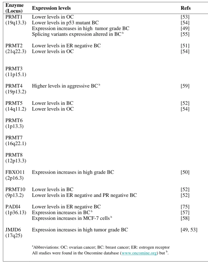
Prmt knockout mice have been developed, providing interesting findings on the relevance of
244
arginine methylation in vivo [1]. For instance, Prmt1 and Carm1 gene disruptions result in an 245
embryonically lethal phenotype and neonatal death respectively, confirming a fundamental 246
role for these enzymes in cellular metabolism [47, 48]. However, until now, these genetically 247
modified animals have not provided information regarding roles for PRMT in estrogen-248
dependent physiological processes. By contrast, recent studies using microarray and 249
quantitative PCR-basedapproaches described the aberrant expression of arginine methylation 250
enzymes in estrogen-dependent cancers (Table 2). 251
252
PRMT expression in estrogen-related cancers
253
Several recent papers analyzed PRMT expression in estrogen-dependent cancers. Prmt1 and 254
Fbxo11 expression is up-regulated in high tumor grade breast carcinomas [49, 50] and Prmt2, 255
5 and 10 expression is down-regulated in breast carcinomas [51, 52]. In ovarian 256
adenocarcinomas, Prmt1, 2 and 5 expression is down-regulated compared to normal tissues 257
[53, 54]. 258
Prmt1 isoforms exist as a result of alternative mRNA splicing, and amino acid sequence 259
comparison indicates that they are all enzymatically active, but with different N-terminal 260
hydrophobic regions. Goulet et al. foundthat the expression profile of Prmt1 splicingvariants 261
is altered in breast cancer [55]. Furthermore, this study showed that increased Prmt1 262
expression was detected in human breast tumor samples compared with adjacent normal 263
breast tissue, confirming the studies mentioned previously. Strikingly, increased arginine-264
methylated protein levels were also observed in breastcancer cell lines. Therefore, an altered 265
Prmt1 isoform expression profilecorrelates with a differential pattern of arginine methylation 266
in breast cancer cell lines, suggesting that misregulation of arginine methylation could 267
contribute to the propagation of breastcancer. 268
269
Increased PADI4 expression in breast tumors
270
Immunohistochemistry detected significant PADI4 expression in various malignancies 271
including breast carcinomas, endometrial carcinomas and uterine adenocarcinomas, with no 272
detectable PADI4 expression in benign and healthy tissues [56]. Quantitative PCR and 273
western blot analyses also showed higher PADI4 mRNA and protein levels in malignant 274
tissues compared to benign and non-tumor tissues [57]. Interestingly, in MCF7 breast cancer 275
cells, PADI4 mRNA expression gradually increased with time after estradiol stimulation 276
through both classical and non-classical ER-mediated pathways [58]. Altogether, these results 277
suggest increased PADI4 expression in breast cancer tissues, probably in response to 278
estradiol. 279
280
CARM1/E2F1 breast cancer growth induced by estrogens
281
ER controls the expression of cell cycle genes which in turn mediate breast cancer 282
proliferation. Frietze et al. showed that CARM1 is essential for estrogen-induced cell cycle 283
progression in MCF-7 breast cancer cells. Upon silencing of CARM1 by siRNA, the E2-284
mediated stimulationof MCF-7 cell cycle progression was strongly reduced. This silencing 285
resulted in decreased expression of E2F1 and E2F1-target genes (cyclin E1, cyclin A, 286
cdc25A), providing a direct link between CARM1 and cell cycle regulation and identifying 287
CARM1 as a potential new target in the treatment of estrogen-dependent breast cancer [46]. 288
Aberrant expression of CARM1 has also been linked to human breast cancer [59], with 289
elevated CARM1 levels found in aggressive breast tumors that also express high levels of the 290
oncogenic coactivator AIB1 (amplified in breast cancer 1). Compiled high levels of CARM1 291
and AIB1 could work in synergy to enhance target gene expression and thereby cell 292
proliferation. 293
294
ER methylation in breast cancer
295
In MCF-7 cells, E2-induced ER methylation is transitory suggesting the involvement of a 296
not yet identified arginine demethylase whose expression or activity could be deregulated in 297
breast cancer [20]. The evaluation of methylated ER with a specific antibody showed that 298
ER is hypermethylated in 50% of human breast tumors [20]. Because ER methylation is 299
necessary for estrogen-induced cellular kinase pathways, this deregulation in breast cancer 300
could lead to sustained activation of those kinases which in turn would activate estrogen 301
signaling. This bidirectional crosstalk appears to play a critical role in breast cancer 302
development by maintaining the activation of signaling pathways and survival of breast 303
cancer cells even in the presence of tamoxifen [60]. It is then tempting to speculate that the 304
deregulation of ER methylation may be involved in breast tumorigenesis and resistance to 305
hormonal therapy. Further analyses are needed to consider methylated ER a new prognostic 306 marker. 307 308 Concluding remarks 309
Arginine methylation impacts various levels of regulation in estrogen signaling, and thereby 310
appears to be a key regulatory event in genomic and non-genomic estrogen actions. In 311
genomic events, arginine methylation of transcriptional coregulators influences coregulators 312
complex formation, activity, subcellular localization, and stability. Methylation of histones 313
participates in chromatin remodeling in concert with other PTMs according to the histone 314
code. In non-genomic events, estrogen receptor methylation is necessary for the formation of 315
a transduction signaling complex and may participate in downstream events. Although, recent 316
findings strongly enhanced our knowledge of the role of arginine methylation in estrogen 317
signaling, a complete understanding will only be realized through answering fundamental 318
questions (Box 3). For instance, since expression of other PRMTs beside PRMT1 and 319
CARM1 is deregulated in estrogen-dependent cancers (Table 2), could other PRMTs be also 320
involved in genomic and non-genomic estrogen regulatory mechanisms? Are other substrates 321
implicated in estrogen signaling? 322
Deregulation of arginine methylation, methyltransferases and demethylases are described in 323
estrogen-related cancers, strengthening the notion of a connection between arginine 324
methylation patterns and cancer progression. Moreover, hypermethylation of ER in human 325
breast cancers may indicate that methylarginine proteins represent novel interesting 326
prognostic biomarkers. Moreover, inhibiting methyltransferase activity, in particular that of 327
PRMT1 and CARM1, by selective molecules appears as a potential therapeutic tool. 328
Recently, using an approach based on a protein virtual screen, Spannhoff et al. identified 329
specific PRMT1 inhibitors. These compounds operate as a brake onsteroid hormone actions, 330
suggesting their potential for future drug development in cancer therapy [61]. There is little 331
doubt that detailed insights into the function and regulation of arginine methylation will 332
unravel the pathogenesis of various diseases, in particular hormone-dependent cancers, and 333
eventually contribute to the discovery of novel biological markers or therapeutic targets. 334
Box 1. Arginine methylation enzymes
336
The enzymes responsible for arginine methylation are called protein arginine 337
methyltransferases (PRMTs). So far, eleven members have been identified in the PRMT 338
family [62, 63]. PRMT-encoding genes are well-conserved through evolution [63], sharing 339
common structural and functional domains (Figure I). Although circumstantial evidence over 340
the past 40 years depicted arginine methylation as an irreversible PTM, some data suggest 341
that this modification could be reversed. For instance, histone H4 is transiently and cyclically 342
methylated on arginine 3 [64] and estrogen receptor α methylation also appears transient [20]. 343
While enzymes capable of removing or preventing such methylation have been identified, 344
their roles are still controversial. Peptidylarginine deiminase 4 (PADI4) has been described to 345
convert monomethylated arginine to citrulline by deimination [27, 65, 66]. However, a full 346
reversion would then need an enzyme to convert citrullines to arginine residues. More 347
importantly, PADI enzymes do not deiminate methylated arginine residues in vitro [67, 68], 348
and it seems rather that histone citrunillation simply interferes with methylation of arginine 349
residues. The first histone demethylase removing asymmetrical dimethylation at arginine 2 of 350
histone H3 and symmetrical dimethylation at arginine 3 of histone H4 is the Jumonji domain-351
containing 6 protein (JMJD6) [69]. However, no publication has confirmed these results and, 352
very recently, the Bottger’s group demonstrated that JMJD6 is a lysine hydroxylase involved 353
in RNA splicing indicating that demethylation of methylarginine residues is not its major 354
activity [70]. Altogether, this suggests that additional arginine demethylases remain to be 355
identified. 356
357 358
Box 2. Estrogen receptor and coregulator complexes
359
Estrogens or anti-estrogens induce ER ligand binding domain (LBD) conformational change, 360
DNA binding to specific response elements in promoter regions of target genes and 361
recruitment of coregulator complexes [4]. Agonist-induced LBD conformational change 362
allows recruitment of coactivator complexes composed of p160 coactivators or PGC-1 and 363
of histone-modifying enzymes (p300, CARM1 and PRMT1), whereas antagonists permit 364
recruitment of corepressors complexes containing SMRT, NCoR and HDAC enzymes. 365
Recruitment of coactivator complexes helps to pull down chromatin remodeling ATPase 366
complexes (e.g. BRG1, SWI/SNF1), which participate in chromatin remodeling. This event 367
facilitates the recruitment of the Mediator complex and thereby of the transcription machinery 368
(which contains among others the RNA Polymerase II and the TATA Binding Protein, TBP) 369
to the initiation start point [6]. Enzymes associated with coactivator complexes are 370
acetyltransferases (p300, SRC-1, SRC-3) and methyltransferases (CARM1, PRMT1). They 371
modify histone tails inducing the open state of chromatin and gene expression. On the 372
contrary, HDACs deacetylate histones leading to the closed conformation of chromatin and 373
gene repression. In the presence of estradiol, ER can also bind corepressors, such as RIP140 374
which interacts with HDACs, leading to gene repression (Figure I). 375
376 377 378
Box 3. Outstanding questions
379
- Arginine methylome and methylarginine target proteins
380
A fundamental issue is to define the entire arginine methylome in estrogen signaling, i.e. to 381
set up proteomic approaches with high-performance mass spectrometry methods in order to 382
describe all the methylated arginine residues in proteins involved in the estrogen pathway. In 383
addition, although several methyl lysine-binding proteins have been identified [71], effectors 384
for arginine-methylated proteins remain to be found. Actually, only three mammalian proteins 385
have been demonstrated to bind methylarginine motifs through their Tudor domains [72, 73]. 386
Defining the proteins which recognize methylated arginines of estrogen receptors and 387
coregulators will certainly enhance our understanding of the downstream cascades dependent 388
on arginine methylation in estrogen signaling. 389
- Regulation of arginine methylation
390
Studies addressing the integration of signal transduction and arginine methylation pathways 391
are also needed. Specifically, how are arginine methylation and PRMT activities regulated by 392
PTMs such as phosphorylation, acetylation, ubiquitination, sumoylation and trans- or 393
automethylation? How are PRMT activity and expression regulated by estrogens or other 394
stimuli? Finally, we also clearly need a better characterization of the enzymes that 395
demethylate arginine residues. 396
- In vivo function of arginine methylation
397
Most of the published studies discuss the importance of arginine methylation in a cellular 398
context. It will be of great interest to elucidate further the in vivo function of the modifications 399
in estrogen target tissues. To achieve this goal, conditional knock-out approaches targeting 400
specifically breast, ovarian or uterus tissues together with knock-in strategies in mice with 401
unmethylatable mutants will be of a great help. 402
Acknowledgments
404 405
This work was supported by a grant from the “Association pour la Recherche sur le Cancer” 406
(#3169) to L.C. and V.C. and the Ligue Contre le Cancer (L.C.). C.T. is funded by the “Ligue 407 Contre le Cancer” 408 409 410 References 411 412
1. Bedford, M.T., and Clarke, S.G. (2009) Protein arginine methylation in mammals: who, 413
what, and why. Mol Cell 33, 1-13 414
2. Bedford, M.T., and Richard, S. (2005) Arginine methylation an emerging regulator of 415
protein function. Mol Cell 18, 263-272 416
3. Aletta, J.M., and Hu, J.C. (2008) Protein arginine methylation in health and disease. 417
Biotechnol Annu Rev 14, 203-224
418
4. O'Malley, B. (2008) The Year in Basic Science: nuclear receptors and coregulators. Mol 419
Endocrinol 22, 2751-2758
420
5. Lonard, D.M., and O'Malley, B.W. (2006) The expanding cosmos of nuclear receptor 421
coactivators. Cell 125, 411-414 422
6. Rosenfeld, M.G. et al. (2006) Sensors and signals: a coactivator/corepressor/epigenetic 423
code for integrating signal-dependent programs of transcriptional response. Genes Dev 20, 424
1405-1428 425
7. Lee, D.Y. et al. (2005) Role of protein methylation in regulation of transcription. 426
Endocr Rev 26, 147-170
427
8. Augereau, P. et al. (2006) The nuclear receptor transcriptional coregulator RIP140. Nucl 428
Recept Signal 4, e024
429
9. Jones, P.L., and Shi, Y.B. (2003) N-CoR-HDAC corepressor complexes: roles in 430
transcriptional regulation by nuclear hormone receptors. Curr Top Microbiol Immunol 274, 431
237-268 432
10. Bjornstrom, L., and Sjoberg, M. (2005) Mechanisms of estrogen receptor signaling: 433
convergence of genomic and nongenomic actions on target genes. Mol Endocrinol 19, 833-434
842 435
11. Migliaccio, A. et al. (2006) Crosstalk between EGFR and extranuclear steroid receptors. 436
Ann N Y Acad Sci 1089, 194-200
437
12. Acconcia, F. et al. (2005) Palmitoylation-dependent estrogen receptor alpha membrane 438
localization: regulation by 17beta-estradiol. Mol Biol Cell 16, 231-237 439
13. Greger, J.G. et al. (2007) Phosphorylation of MNAR promotes estrogen activation of 440
phosphatidylinositol 3-kinase. Mol Cell Biol 27, 1904-1913 441
14. Azuma, K. et al. (2009) Association of estrogen receptor alpha and histone deacetylase 442
6 causes rapid deacetylation of tubulin in breast cancer cells. Cancer Res 69, 2935-2940 443
15. Litt, M. et al. (2009) Histone arginine methylations: their roles in chromatin dynamics 444
and transcriptional regulation. Biosci Rep 29, 131-141 445
16. Wysocka, J. et al. (2006) Histone arginine methylation and its dynamic regulation. 446
Front Biosci 11, 344-355
447
17. Pal, S., and Sif, S. (2007) Interplay between chromatin remodelers and protein arginine 448
methyltransferases. J Cell Physiol 213, 306-315 449
18. Koh, S.S. et al. (2001) Synergistic enhancement of nuclear receptor function by p160 450
coactivators and two coactivators with protein methyltransferase activities. J Biol Chem 276, 451
1089-1098 452
19. Chen, D. et al. (1999) Regulation of transcription by a protein methyltransferase. 453
Science 284, 2174-2177
454
20. Le Romancer, M. et al. (2008) Regulation of estrogen rapid signaling through arginine 455
methylation by PRMT1. Mol Cell 31, 212-221 456
21. Lahusen, T. et al. (2009) The role and regulation of the nuclear receptor co-activator 457
AIB1 in breast cancer. Breast Cancer Res Treat 116, 225-237 458
22. Harigopal, M. et al. (2009) Estrogen receptor co-activator (AIB1) protein expression by 459
automated quantitative analysis (AQUA) in a breast cancer tissue microarray and association 460
with patient outcome. Breast Cancer Res Treat 115, 77-85 461
23. Torres-Arzayus, M.I. et al. (2004) High tumor incidence and activation of the 462
PI3K/AKT pathway in transgenic mice define AIB1 as an oncogene. Cancer Cell 6, 263-274 463
24. Kuang, S.Q. et al. (2004) AIB1/SRC-3 deficiency affects insulin-like growth factor I 464
signaling pathway and suppresses v-Ha-ras-induced breast cancer initiation and progression in 465
mice. Cancer Res 64, 1875-1885 466
25. Feng, Q. et al. (2006) Signaling within a coactivator complex: methylation of SRC-467
3/AIB1 is a molecular switch for complex disassembly. Mol Cell Biol 26, 7846-7857 468
26. Naeem, H. et al. (2007) The activity and stability of the transcriptional coactivator 469
p/CIP/SRC-3 are regulated by CARM1-dependent methylation. Mol Cell Biol 27, 120-134 470
27. Lee, Y.H. et al. (2005) Regulation of coactivator complex assembly and function by 471
protein arginine methylation and demethylimination. Proc Natl Acad Sci U S A 102, 3611-472
3616 473
28. Teyssier, C. et al. (2005) Activation of nuclear receptor coactivator PGC-1alpha by 474
arginine methylation. Genes Dev 19, 1466-1473 475
29. Mostaqul Huq, M.D. et al. (2006) Suppression of receptor interacting protein 140 476
repressive activity by protein arginine methylation. Embo J 25, 5094-5104 477
30. Kouzarides, T. (2007) Chromatin modifications and their function. Cell 128, 693-705 478
31. Taverna, S.D. et al. (2007) How chromatin-binding modules interpret histone 479
modifications: lessons from professional pocket pickers. Nat Struct Mol Biol 14, 1025-1040 480
32. O'Malley, B.W. et al. (2008) Cracking the coregulator codes. Curr Opin Cell Biol 20, 481
310-315 482
33. Mostaqul Huq, M.D. et al. (2008) Post-translational modifications of nuclear co-483
repressor RIP140: a therapeutic target for metabolic diseases. Curr Med Chem 15, 386-392 484
34. Huq, M.D. et al. (2009) Lysine methylation of nuclear co-repressor receptor interacting 485
protein 140. J Proteome Res 8, 1156-1167 486
35. Gupta, P. et al. (2008) PKCepsilon stimulated arginine methylation of RIP140 for its 487
nuclear-cytoplasmic export in adipocyte differentiation. PLoS ONE 3, e2658 488
36. Berthet, C. et al. (2002) Interaction of PRMT1 with BTG/TOB proteins in cell 489
signalling: molecular analysis and functional aspects. Genes Cells 7, 29-39 490
37. Lin, W.J. et al. (1996) The mammalian immediate-early TIS21 protein and the 491
leukemia-associated BTG1 protein interact with a protein-arginine N-methyltransferase. J 492
Biol Chem 271, 15034-15044
38. Rouault, J.P. et al. (1998) Interaction of BTG1 and p53-regulated BTG2 gene products 494
with mCaf1, the murine homolog of a component of the yeast CCR4 transcriptional 495
regulatory complex. J Biol Chem 273, 22563-22569 496
39. Robin-Lespinasse, Y. et al. (2007) hCAF1, a new regulator of PRMT1-dependent 497
arginine methylation. J Cell Sci 120, 638-647 498
40. Wang, H. et al. (2001) Methylation of histone H4 at arginine 3 facilitating 499
transcriptional activation by nuclear hormone receptor. Science 293, 853-857 500
41. Huang, S. et al. (2005) Methylation of histone H4 by arginine methyltransferase 501
PRMT1 is essential in vivo for many subsequent histone modifications. Genes Dev 19, 1885-502
1893 503
42. Prevot, D. et al. (2001) Relationships of the antiproliferative proteins BTG1 and BTG2 504
with CAF1, the human homolog of a component of the yeast CCR4 transcriptional complex: 505
involvement in estrogen receptor alpha signaling pathway. J Biol Chem 276, 9640-9648 506
43. Xu, W. et al. (2004) A methylation-mediator complex in hormone signaling. Genes Dev 507
18, 144-156 508
44. Higashimoto, K. et al. (2007) Phosphorylation-mediated inactivation of coactivator-509
associated arginine methyltransferase 1. Proc Natl Acad Sci U S A 104, 12318-12323 510
45. Feng, Q. et al. (2009) Biochemical control of CARM1 enzymatic activity by 511
phosphorylation. J Biol Chem 512
46. Frietze, S. et al. (2008) CARM1 regulates estrogen-stimulated breast cancer growth 513
through up-regulation of E2F1. Cancer Res 68, 301-306 514
47. Pawlak, M.R. et al. (2000) Arginine N-methyltransferase 1 is required for early 515
postimplantation mouse development, but cells deficient in the enzyme are viable. Mol Cell 516
Biol 20, 4859-4869
517
48. Yadav, N. et al. (2003) Specific protein methylation defects and gene expression 518
perturbations in coactivator-associated arginine methyltransferase 1-deficient mice. Proc Natl 519
Acad Sci U S A 100, 6464-6468
520
49. Miller, L.D. et al. (2005) An expression signature for p53 status in human breast cancer 521
predicts mutation status, transcriptional effects, and patient survival. Proc Natl Acad Sci U S 522
A 102, 13550-13555
523
50. van 't Veer, L.J. et al. (2002) Gene expression profiling predicts clinical outcome of 524
breast cancer. Nature 415, 530-536 525
51. Richardson, A.L. et al. (2006) X chromosomal abnormalities in basal-like human breast 526
cancer. Cancer Cell 9, 121-132 527
52. Finak, G. et al. (2008) Stromal gene expression predicts clinical outcome in breast 528
cancer. Nat Med 14, 518-527 529
53. Ivshina, A.V. et al. (2006) Genetic reclassification of histologic grade delineates new 530
clinical subtypes of breast cancer. Cancer Res 66, 10292-10301 531
54. Hendrix, N.D. et al. (2006) Fibroblast growth factor 9 has oncogenic activity and is a 532
downstream target of Wnt signaling in ovarian endometrioid adenocarcinomas. Cancer Res 533
66, 1354-1362 534
55. Goulet, I. et al. (2007) Alternative splicing yields protein arginine methyltransferase 1 535
isoforms with distinct activity, substrate specificity, and subcellular localization. J Biol Chem 536
282, 33009-33021 537
56. Chang, X., and Han, J. (2006) Expression of peptidylarginine deiminase type 4 (PAD4) 538
in various tumors. Mol Carcinog 45, 183-196 539
57. Chang, X. et al. (2009) Increased PADI4 expression in blood and tissues of patients 540
with malignant tumors. BMC Cancer 9, 40 541
58. Dong, S. et al. (2007) Estrogen-enhanced peptidylarginine deiminase type IV gene 542
(PADI4) expression in MCF-7 cells is mediated by estrogen receptor-alpha-promoted 543
transfactors activator protein-1, nuclear factor-Y, and Sp1. Mol Endocrinol 21, 1617-1629 544
59. El Messaoudi, S. et al. (2006) Coactivator-associated arginine methyltransferase 1 545
(CARM1) is a positive regulator of the Cyclin E1 gene. Proc Natl Acad Sci U S A 103, 546
13351-13356 547
60. Tokunaga, E. et al. (2006) The association between Akt activation and resistance to 548
hormone therapy in metastatic breast cancer. Eur J Cancer 42, 629-635 549
61. Spannhoff, A. et al. (2009) Cancer treatment of the future: inhibitors of histone 550
methyltransferases. Int J Biochem Cell Biol 41, 4-11 551
62. Lee, Y.H., and Stallcup, M.R. (2009) Minireview: protein arginine methylation of 552
nonhistone proteins in transcriptional regulation. Mol Endocrinol 23, 425-433 553
63. Krause, C.D. et al. (2007) Protein arginine methyltransferases: evolution and 554
assessment of their pharmacological and therapeutic potential. Pharmacol Ther 113, 50-87 555
64. Metivier, R. et al. (2003) Estrogen receptor-alpha directs ordered, cyclical, and 556
combinatorial recruitment of cofactors on a natural target promoter. Cell 115, 751-763 557
65. Cuthbert, G.L. et al. (2004) Histone deimination antagonizes arginine methylation. Cell 558
118, 545-553 559
66. Wang, Y. et al. (2004) Human PAD4 regulates histone arginine methylation levels via 560
demethylimination. Science 306, 279-283 561
67. Hidaka, Y. et al. (2005) Methylation of the guanidino group of arginine residues 562
prevents citrullination by peptidylarginine deiminase IV. FEBS Lett 579, 4088-4092 563
68. Raijmakers, R. et al. (2007) Methylation of arginine residues interferes with 564
citrullination by peptidylarginine deiminases in vitro. J Mol Biol 367, 1118-1129 565
69. Chang, B. et al. (2007) JMJD6 is a histone arginine demethylase. Science 318, 444-447 566
70. Webby, C.J. et al. (2009) Jmjd6 catalyses lysyl-hydroxylation of U2AF65, a protein 567
associated with RNA splicing. Science 325, 90-93 568
71. Kim, J. et al. (2006) Tudor, MBT and chromo domains gauge the degree of lysine 569
methylation. EMBO Rep 7, 397-403 570
72. Cote, J., and Richard, S. (2005) Tudor domains bind symmetrical dimethylated 571
arginines. J Biol Chem 280, 28476-28483 572
73. Cheng, D. et al. (2007) The arginine methyltransferase CARM1 regulates the coupling 573
of transcription and mRNA processing. Mol Cell 25, 71-83 574
74. Bauer, U.M. et al. (2002) Methylation at arginine 17 of histone H3 is linked to gene 575
activation. EMBO Rep 3, 39-44 576
75. Saal, L.H. et al. (2007) Poor prognosis in carcinoma is associated with a gene 577
expression signature of aberrant PTEN tumor suppressor pathway activity. Proc Natl Acad 578
Sci U S A 104, 7564-7569
579
76. Cook, J.R. et al. (2006) FBXO11/PRMT9, a new protein arginine methyltransferase, 580
symmetrically dimethylates arginine residues. Biochem Biophys Res Commun 342, 472-481 581
77. Troffer-Charlier, N. et al. (2007) Functional insights from structures of coactivator-582
associated arginine methyltransferase 1 domains. Embo J 26, 4391-4401 583
78. Teyssier, C. et al. (2002) Requirement for multiple domains of the protein arginine 584
methyltransferase CARM1 in its transcriptional coactivator function. J Biol Chem 277, 585 46066-46072 586 587 588 589 590
Figure legends
592
Figures inside Boxes:
593
Box 1. Figure I– The PRMT family
594
The nine members of the mammalian PRMT family are shown. Recently, two related 595
members, FBXO11 (also called PRMT9) and FBXO10, have been added to the family [63, 596
76]. The common methyltransferase domain consists of a series of short conserved motifs 597
(blue bars) that are important for binding the methyl donor and for catalysis. This catalytic 598
core region is formed by a Rossman fold and two helices (black boxes). The less conserved 599
-barrel structure (gray boxes) folds against the catalytic region to form the protein substrate 600
binding cleft [77]. Additional specific motifs, such as the SH3 domain (SH3), the zinc finger 601
(ZnF), the myristylation motif (Myr), the FBox motif and the tetratricopeptide repeat motif 602
(TPR) are represented in green boxes. CARM1 uniquely contains a substantial C-terminal 603
region which contains an autonomous transcriptional activation domain [78]. 604
605
Box 2. Figure I- Gene regulation mechanism by estrogen receptor and its coregulators
606
Estrogen receptor (ER) binds to specific regions called estrogen response element (ERE) 607
represented by a pink box on the DNA target gene drawn as a thick black line wrapped 608
around histones symbolized by yellow plots. The initiation start point is represented by a 609
black arrow. The green and red arrows represent the impacts of coactivator and corepressor 610
complexes on chromatin state and gene transcription. 611
612
Figure 1 –Arginine methylation and estrogen signaling
613
Methylarginine proteins are involved in genomic and non-genomic estrogen actions. 614
(a) In estrogen genomic action, histones and numerous estrogen receptor (ER) coregulators
615
are substrates for PRMTs. Arginine methylation regulates their transcriptional activity (PGC-616
1 ), their subcellular localization (RIP140), their stability (SRC-3) and their complex 617
assembly (p300-GRIP1). The removing methyl mark enzymatic function of PADI4 regulates 618
the assembly of the complex containing GRIP1, CARM1 and p300. This action is represented 619
by a yellow dotted line. 620
(b) In non-genomic estrogen action, induction of arginine methylation of cytoplasmic ER by
621
estradiol leads to activation of downstream kinase cascades and corresponding target gene 622
activation. The role of methylated RIP140 in this non-genomic pathway remains to be 623
demonstrated. In both figures, the methyl mark is represented by a pink circle. 624
625 626 627
361 PRMT1 SH3 433 PRMT2 637 PRMT5 375 PRMT6 692 PRMT7 608 CARM1 394 PRMT8 Myr 845 PRMT10 TPRTPR PRMT9/ FBXO11 843 FBox ZnF FBXO10 FBox 956 531 PRMT3 ZnF Figure
p300 ER + Partial antiestrogens (tamoxifene, raloxifene ...) HDACs Corepressor complexes + Estrogens RNA Pol II Gene repression Coactivator complexes + Estrogens -Ac Ac-ERE p160 PRMT1 PGC-1a CARM1 RIP140 Met Met Mediator complex SMRT N-CoR Gene activation Chromatin remodeling ATPases BRG1 SWI/SNF1 TRAP/DRIP TBP CtBPs HDACs Figure
PI3K FAK
ER ER
p300 SRC-3 RIP140 Cytoplasm Nucleus GRIP1 RIP140Target gene regulation
H3
(a) Genomic action
(b) Non-genomic action
PRMT1ER
E2
H4
Activated
MAPK and Akt
pathways
SrcER
Src PI3K Cytoplasm RIP140 FAK PADI4 PGC-1 ERE FigureTable 1. Methylarginine proteins involved in estrogen pathway
Enzyme Substrates Arginine (R) Impact Refs
PRMT1 Histone H4 R3 Gene activation [40]
ER R260 Interaction with Src and PI3K [20] PGC-1 R665, 667, 669 Stimulation of coactivator activity [28] RIP140 R240, 650, 948 Inhibition of repressive activity [29]
CARM1 Histone H3 R2, 17, 26 Gene activation [19, 74]
p300/CBP R2142 Complex assembly regulation [27]
SRC-3
R1171
R1178, 1184, 1195
Complex assembly regulation Activity and stability regulation
[25, 26] Table
Table 2. Arginine methylation enzymes in estrogen-dependent cancers Enzyme
(Locus) Expression levels Refs
PRMT1 (19q13.3) PRMT2 (21q22.3) PRMT3 (11p15.1) PRMT4 (19p13.2) PRMT5 (14q11.2) PRMT6 (1p13.3) PRMT7 (16q22.1) PRMT8 (12p13.3) FBXO11 (2p16.3) PRMT10 (9p13.2) PADI4 (1p36.13) JMJD6 (17q25) Lower levels in OC
Lower levels in p53 mutant BC
Expression increases in high tumor grade BC Splicing variants expression altered in BC b Lower levels in ER negative BC
Lower levels in OC
Higher levels in aggressive BC b
Lower levels in BC Lower levels in OC
Expression increases in high grade BC
Lower levels in BC
Lower levels in ER negative and PR negative BC Lower levels in ER negative BC
Expression increases in BC b
Expression increases in MCF-7 cells b
Expression increases in high tumor grade BC
aAbbreviations: OC: ovarian cancer; BC: breast cancer; ER: estrogen receptor
All studies were found in the Oncomine database (www.oncomine.org) but b.
[53] [54] [49] [55] [51] [54] [59] [52] [54] [50] [52] [52] [75] [57] [58] [49, 53] Table