Direction des bibliothèques
AVIS
Ce document a été numérisé par la Division de la gestion des documents et des archives de l’Université de Montréal.
L’auteur a autorisé l’Université de Montréal à reproduire et diffuser, en totalité ou en partie, par quelque moyen que ce soit et sur quelque support que ce soit, et exclusivement à des fins non lucratives d’enseignement et de recherche, des copies de ce mémoire ou de cette thèse.
L’auteur et les coauteurs le cas échéant conservent la propriété du droit d’auteur et des droits moraux qui protègent ce document. Ni la thèse ou le mémoire, ni des extraits substantiels de ce document, ne doivent être imprimés ou autrement reproduits sans l’autorisation de l’auteur.
Afin de se conformer à la Loi canadienne sur la protection des renseignements personnels, quelques formulaires secondaires, coordonnées ou signatures intégrées au texte ont pu être enlevés de ce document. Bien que cela ait pu affecter la pagination, il n’y a aucun contenu manquant.
NOTICE
This document was digitized by the Records Management & Archives Division of Université de Montréal.
The author of this thesis or dissertation has granted a nonexclusive license allowing Université de Montréal to reproduce and publish the document, in part or in whole, and in any format, solely for noncommercial educational and research purposes.
The author and co-authors if applicable retain copyright ownership and moral rights in this document. Neither the whole thesis or dissertation, nor substantial extracts from it, may be printed or otherwise reproduced without the author’s permission.
In compliance with the Canadian Privacy Act some supporting forms, contact information or signatures may have been removed from the document. While this may affect the document page count, it does not represent any loss of content from the document.
University of Montreal
The role of the TGN in the transport of
Herpes simplex virus type 1 capsids
By Constantina Mihai
Department of Pathology and Cell Biology
Faculty of Medicine
University of Montreal
Faculty of Graduate Studies
This thesis called:
The role of the TGN in the transport
of·
Herpes simplex virus type 1 capsids
Presented by Constantina Mihai
Is evaluated by the jury comity:
Dr. Ghitescu Dorin-Lucian
President reporter
Dr. Roger Lippe
Research director
Dr. Fernandes Karl J.L.
Member of jury
IIThis thesis is dedicated to my 4 years old
nephew,
Alexandru,
currently
under
the
acyclovir
treatment
for
his
recently
discovered Herpes
simplex virus
type
1
Résumé
Le virus Herpès Simplex de type 1 (HSV -1) est un membre de la famille des Herpesviridae et
cause une variété de maladies chez les humains et les animaux (Roizman and Knipe, 2001). HSV-l peut demeurer latent dans les neurones sensoriels et occasionnellement se réactiver et causer une maladie récurrente. Lorsqu'il est réactivé, HSV-l cause des feux sauvages ainsi que de sérieuses maladies telles que la kératoconjonctivite et l'encéphalite. Les traitements antiviraux tels que les vaccins n'ont toutefois pas réussi à éradiquer HSV -1.
La meilleure méthode cepedant pour empêcher HSV-l de causer des maladies est d'utiliser des
v~ccins qui bloquent l'infection initiale. Au niveau cellulaire, une méthode pour bloquer la propagation virale aux cellules voisines serait la plus utile. En vue de trouver une méthode préventive, les détails du cycle viral doivent être explorés, y compris la manière dont le virus entre et infecte les cellules. Nous espérons qu'une meilleure compréhension du transport de HSV-l dans les cellules infectées nous aidera dans le traitement des maladies causées par HSV -1. Une fois dans la cellule, HSV -1 produit de nouvelles capsides dans le noyau des cellules infectées.
En 2007, parmi de nombreuses autres études,. Rémillard-Labrosse et
aI.
suggèrent que les capsides nouvellement assemblées, trop grosses pour sortir par les pores nucléaires, bourgeonnent dans l'espace périnucléaire et fusionnent ensuite avec la membrane nucléaire externe. Par la suite, les capsides cytoplasmiques nues migrent au site de ré-enveloppement, présumé être le TGN ou les endosomes (Turcotte at al., 2005). Plusieurs laboratoires, dont Turcotte et al. en 2005, ont démontré le rôle du TGN dans le cycle viral de HSV -1. Ils ont constaté que, dans les membranes du TGN, les capsides cytoplasmiques acquièrent leur enveloppe finale pour devenir des particules infectieuses dans le milieu extracellulaire. Le TGN est le lieu de triage des protéines avant d'être délivrées à la surface de la celluleet
dans diverses organelles; toutefois, le processus par lequel les capsides de HSV -1 quittent ce compartiment n'est· pas encore clair. JDans cette étude, nous suggérons l'implication de la protéine kinase D (PKD) dans le transport du virus du TGN à la membrane plasmique. Dans l'étude du transport intracellulaire des protéines, PKD est présenté comme un important médiateur pour le transport de cargos du TGN à la surface des cellules. Son activité est dépendante du DAG et la réduction de la synthèse de DAG inhibe le transport de molécules du TGN à la membrane plasmique. De plus, une mutation dans le domaine kinase de PKD entraîne la formation de tubules au TGN et la rétention de cargos dans ces tubules. Nos résultats montrent que les virions de HSV-l sont également pris au piège dans les tubules du TGN formés lors de l'expression de
PKD sous sa forme mutante. Ces résultats proposent l'utilisation par HSV-l de cette même voie de sécrétion dans son transport à la surface des cellules.
Su
ITl ITlary
Herpes Simplex Virus Type 1 (HSV -1) is a member of the Herpesviridae, which causes a variety of diseases in humans and animaIs (Roizman and Knipe, 2001). HSV-l can remain latent in sensory neurons and occasionally reactivates to cause recurrent disease. When it is reactivated, HSV cause cold sores as weIl as other serious diseases such as, kerato-conjunctivitis and encephalitis. Anti-viral drugs as well as vaccines have been unsuccessful in eradicating HSV-l.
The best way to prevent HSV -1 from causing diseases,however, is still to utilize vaccine~ which prevent of the initial infection. At the cellular level a method to stop viral spread to neighboring cells would be most useful. In order to search for a viral prevention method, the details of the virus lifecycle must be explored, including how it enters and infects cells. We hope that a better understanding of the HSV egress from the infected cells will help in the treatment ofHSV-l diseases. Once HSV-l is in a cell, it produces new capsids within the infected cell nucleus.
Rémillard - Labrosse et al., 2007, among many other studies, suggest that newly HSV-l assembled capsids which are to big to escape via nuclear pores bud into the lumen of the nuclear envelope and then fuse with the outer nuclear membrane. The subsequent cytoplastp.ic naked .capsids travel to the re-envelopment site, presumed to be the TGN or endosomes (Turcotte at al., 2005). Many laboratories among with Turcotte at
al.,
2005, demonstrated the TGN role in the HSV-l life cycle. They found that in TGN membranes, cytoplasmic capsids acquire their mature envelope to become infectious particles within the extracellular medium. TON represents the station from where the proteins are sorted and delivered to the cell surface and other various organelles; but it is not clear by which pathway HSV-l capsids leave this compartment.In this study, we suggest the implication ofProtein Kinase D (PKD) in the viral egress from the TGN to the plasma membrane. In intracellular protein transport studies, PKD is presented as an important mediator of cargo transport from the TGN to the cell surface. Its activity is DAO dependent and reduction in DAO synthesis inhibits the transport of molecules from the TGN to the plasma membrane. Also, a mutation in the PKD kinase domain produces TGN tubule formation and cargo retention in these tubules.
Our
results show that the HSV -1 virions are also trapped in the TGN tubules formed by the expression of PKD mutant. These results propose that HSV -1 utilizes the same pathway as secretory molecules in their transport to the cell surface.Keywords: HSV -1, capsids, TGN, PKD, plasma membrane.
Table of contents
Résumé ... IV Summary ... VI Table of contents ... VII LIST OF FIGURES ... VIII Included in Chapter 1 ... VIII Included in Chapter 3 ... VIII Included in Chapter 4 ... IX LIST OF ABBREVIA TION ... X ACKNOWLEDGEMENTS ... XII
CHAPTER 1: Literature Review ... 1
1. Introduction ... 1
1.1 Brief history of herpesviruses ... 1
1.2 Proprieties and classification of herpesviruses ... 2
1.3 The HSV -1 virion structure and genome organization ... 3
1.4 Pathogenesis ... 5
1.5 Treatment ... 6
1.6 HSV-l LIFE CyCLE ... 7
1.6.1 HSV-l entry ... 7
1.6.2 Uncoating and genome release ... 8
1.6.3 Gene expression ... 8
1.6.4 Viral DNA replication ... 9
1.6.5 Capsid assembly and maturation ... Il 1.6.6 Nuclear egress ... 13
1.6.7 Cytoplasmic capsid assembly and secondary envelopment.. ... 16
1.6.8 TGN to plasma membrane viral egress ... 17
1.6.9 Host transport from the TGN to the plasma membrane ... 17
CHAPTER II: Objectives of research ... 22
3.3 Results ... 26
3.4 Discussion ... 32
3.5 Materials and Methods ... 35
Chapter IV Discussion ... 60
Conclusions ... 66
Bibliography ... 67
LIST OF FIGURES
Included in Chapter 1
Figure 1 1: Herpesvirus virion structure ... 3Figure 1 2: The HSV-1 virion genome organization ... 4
Figure 1 3: Transport of the capsid to the nuc1ear pores with release of the virion DNA into the nucleus ... 8
Figure 1 4: Mechanism of HSV DNA replication ... 10
Figure 1 5: HSV -1 latent infection ... Il Figure 1 6: Maturation of herpes simplex capsids ... 12
Figure 17: Models of herpes simpex virus egress ... 14
Figure: 1 8. Kinase dead PKD block HSV -1 transport from the TGN to the plasma membrane ... 19
Included in Chapter 3
Fig. 1: Synchronization of HSV -1 egress to the TGN ... .43Fig. 2: Inhibition of TGN to cell surface transport by PKD inhibitors ... .46
Fig. 3: Viral egress is hampered by PKD inhibitors ... .47
Fig. 4: Viral egress is hampered by PKD inhibitors ... .48
Fig. 5: PKD inhibitors trap HSV -1 in the TGN ... 50
Fig. 6: HSV-1 co-Iocalizes with PKD K618N induced tubules at the TGN ... 52
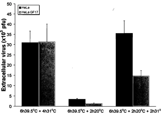
Fig. 7: Cellline expressing kinase dead PKD mutant has reduced viral output ... 54
Fig. 8: 143B cells solely express PKD3 ... 55
Fig. 9: Inhibition of PKD3 prote in expression by siRNA ... 57
Fig. 10: Trapping HSV-1 at the TGN by siRNA targeted against PKD3 ... 59
Included in Chapter 4
Figure: 1 9. PKD inhibitors block TGN to plasma membrane transport ... 62 Figure 1 10: siRNA PKD3 block the virus containing vesicles from the TGN to the ceU surface ... 64 Figure 1 Il: Model for HSV -1 egress ... 65
LIST OF ABBREVIATION
AP ARF bp BFA CMV OMEM dsDNA E EM ER FBS gB gC gO gE GFP GGA FAPPs gH gI gK gL adaptor protein AOP-Ribosylation Factor base pair brefeldin A CytoMegalo VirusOulbecco's modified Eagle's medium double-stranded deoxyribonucleic acid early
electron microscopy endoplasmic reticulum fetal bovine serum
glycoprotein B glycoprotein C glycoprotein 0
glycoprotein E
green fluorescent protein
Golgi-iocalizing, y-adapter ear homology domain ARF- binding protein four-phosphate adaptor proteins
glycoprotein H glycoprotein 1
glycoprotein K glycoprotein L
gM gN GST ICP HSV-l lE L LAT MOI PA PBS
PKD
PI4P P14,5P2 SDS-PAGE SM TGN UL US VP VZV glycoprotein M glycoprotein N glutathione-S-transferase Infected Cell Prote inHerpes simplex virus type 1 immediate early
late
latency associated transcript multiplicity of infection phosphatidic acid
phosphate- buffered saline prote in kinase D
phosphatidylinositol-4-phosphate phosphatidylinositol 4, 5-biphosphate
sodium dodecyl sulfate-polyacrylamide gel electrophoresis sphingomyelin trans-Golgi network unique long unique short Virion Protein varicella-zoster virus
ACKNOWLEDGEMENTS
There is not way that 1 can thank enough everyone who was part of creating this diseration; however, 1 would like to highlight sorne persons whom 1 am particularly indebted to.
First, and foremost, 1 would like to express my gratitude to Dr. Roger Lippe, who provided invaluable guidance, assistance, encouragement, suggestions, and support during the time that 1 have spent as a graduate student in his laboratory. He showed me different ways to approach a research problem and the need to be persistent to accomplish any goal. Thank you for giving me the opportunity to work with you.
ln addition, 1 owe many thanks to ail members of the Lippe's laboratory, who gave me helpful advices whenever 1 asked for. A special thanks goes to Sandra Loret, who is most responsible for helping me shape many ideas the support in pursuit of our goals. 1 would like to thank Gaudeline Remillard-Labrose, lohanne Duron, Ginette Guy, lie Zhang, and Pascal Raymond. Each of you was for me a great source of encouragement and inspiration.
1 would like to thank the faculty members that served on my graduate committee, Dr. Ghitescu Dorin-Lucian, Dr. Karl Frenandes, and Dr.Guy Doucet, for their advices and recommendations in performing my Master research and my leaming experience.
Words cannot explain the role of my lovely husband Marian in my pursuits; 1 would like to thank him who was the most responsible for helping me complete the writing of this dissertation, his encouragement, constant guidance, and steadfast support in achiving my goals. 1 could never have done this without you.
Last, but not at least, 1 wou Id like to thank my family for providing me with unconditionally love and support and a special thanks to my oldest brother and his wife for making my dream true.
CHAPTER 1: Literature Review
1. Introduction
Viruses are the smallest infectious particles, with diameters ranging between 18 and 300 nm. Like all viruses, these particles cannot be seen with a light microscope and are unable to reproduce by themselves because they lack certain functions. The Latin word virus means poison. Viruses establish an obligate intracellular parasitism into many different biological organisms in order to produce new virions (infecti ve viral particles).
1.1 Brief history of herpesviruses
Evidences for herpesviruses have been signaled in the 5th century RC. when the ancient Greek physician and father of medicine Hippocrates mentioned for the first time about skin les ions (Roizman and Whitley 200 1, Smith and Cyr 1988). The family name is derived from the Greek word herpein "to creep" which refers to the latent, re-occurring infections typical of this group of viruses. Herpesviridae can cause latent or lytic infections (Wikipedia definition). Herodotus, in the Roman civilization period, described the presence of the fever associated with these lesions, and he named it "herpes febrilis" (Thomas Bateman, 1814). Other references for herpes virus infection are dated from the Shakespeare' sera in England, and much 1ater in the king's court of France (Astruc, 1736). The advanced methods of herpesvirus studies began in the late 19th century when researchers started to test scientific hypothesises about how the viruses interact with the host. In the early 20th century, Lowenstein and Gruter demonstrated that human HSV could also produce lesions on the rabbit's cornea (Gruter, 1924). In 1920 and 1930, it was found that many lab animaIs were susceptible to HSV infections. In 1939, Bumett and Williams published an article describing the nature of latency, noting that HSV seemed to persist for life and could be reactivated under stressful conditions to produce visible lesions (Bumet and Williams, 1939). The work with the cell culture allowed for the discovery of the other human herpesviruses, including cytomegalovirus (CMV) and Varicela zoster virus (VZV) (Craig et al., 1957). Until now, there were about 100 Herpesviruses isolated from many animal species and 9 from humans.
After the 1960s the new technologies such as electron microscopy (EM), DNA sequencing, and DNA cloning made it possible to determine the structure of herpesvirus particles, the sequence of their genomes, the viral gene expression pattern, and the identification of many individual gene products.
Moreover, advanced studies over the last 40 years have resulted in new treatments and vaccines for herpesvirus infections (e.g. VZV) (Epstein, Achong, and Barr, 1964). Recently, herpesviruses have been re-examined for use as viral vectors for certain treatments of human diseases. The modem experimental period has facilitated a better understanding of herpesvirus diseases and made it possible to utilize herpesviruses to potential human health benefit.
1.2 Proprieties and classification of herpesviruses
Herpesviridae find hosts in amphibians, reptiles, fish, birds, and marnmals and consist of a wide variety of viruses. The International Committee on the Taxonomy of Viruses defined Herpesviridae for the first time as being capable of establishing a latent infection in their natural hosts in a specific set of cells, which varies from one virus to another. There are also other biological properties, such as the length of the reproductive cycle. These were used as the basis of classification, before DNA sequences of the viruses were known. Members of the family Herpesviridae were classified by the Herpesvirus Study
Group into three subfamilies: the Alphaherpesvirinae, the Betaherpesvirinae, and the
Gammaherpesvirinae. This classification is based on host range, length of replication cycle and cell tropism (Roizman, Bartha, and Biggs, 1973; Roizman et al., 1992; Van Regenmortel et al., 2000).
Alphaherpesviruses (a) is represented by Herpes simplex virus type 1 (HSV-l), Herpes simplex
virus type 2 (HSV -2), Pseudorabies virus (PRV), VZV and Marek's diseases virus (MDV). They are characterized by a short replication cycle, a rapid multiplication in cell culture, and a large host variety in
vitro and in vivo, and could be latent within neuronal cells (Van Regenmortel et al., 2000).
In contrast, betaherpesviridae (8) replicate slower than a; they have a limited host range, and establish latency in numerous tissues, such as secretory glands and lymphoreticular cells. During infection with herpesviruses of this subfamily, host cells frequently become enlarged (cytomegalia) leading to the name of the cytomegaloviruses, the representative members of the ~-herpesvirus subfamily (Van Regenmortel et al., 2000).
Although y-herpesviruses have a limited host range similar to the j3--herpesviruses, the length of the replication cycle of these viruses varies between species. The y-herpesviruses infect cells of the lymphatic system, like B or T lymphocytes, and the latent virus is frequently demonstrated in lymphoid
tissue. The Epstein-Barr virus (EBV) is the principal member of this subfamily (Van Regenmortel et al., 2000; Roizman, 1996; Roizman and Sears 1996; Mettenleiter 1994).
There are eight species of human herpesviruses. (HSV -1) and (HSV -2) are etiological agents for oral and genitallesions, keratoconjuctivitis and encephalitis. Varicela zoster virus is the primary cause for chickenpox and shingles, while HCMV causes cytomegalic inclusion disease. EBV causes mononucleosis and tonsillitis. African Burkitt Lymphoma causes B- and T - cell carcinomas. Human Herpes virus 6 (HHV6) and Human Herpes virus 7 (HHV 7) are known to cause T-cell lymphomas. The newly discovered human Herpes virus 8 (HHV 8) is a causative agent of Kaposi's sarcoma and has been renamed as Kaposi Sarcoma Associated Herpes Virus (KSHV).
1.3 The HSV-1 virion structure and genome organization
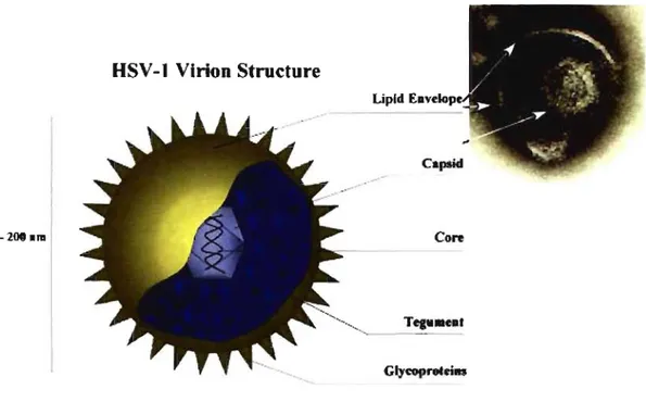
HSV -1 produces spherical particles that range in size from 120-200 nm (Wildy P. et al., 1963). They contain more than 35 different virally-encoded gene products that assemble into three major structures: the nuc\eocapsid, tegument and envelope (Mettenleiter, T.C. 2004).
HSV -1 Virion Structure Upld t;av,elOov
-
--2iMhra Core GlycoproteiM --..Figure Il: Herpesvirus virion structure (Roizman and Furlong, 1974).
HSV -1 virion consists of: an electrondense core containing the viral genome, an icosadeltahedral caps id around the core, an amorphous tegument around the caps id, and an enveJope derived from cellular membranes containing glycoprotein spikes (Roizman and Furlong, 1974; Travis J. Taylor et al.,2002).
The linear, 152 kbp DNA genome consists of two unique segments (unique long (UL) and unique short (Us) flanked by inverted repeats (fig.2) (McGeoch, D.J. et al., 1988, McGeoch, D.J. et al., 1986, McGeoch, D.J., et al., 1985).
Ra. Ra Ra Ra.
[ C J r
-a b
UL
,---1=II
]:1:::::It-
Ua-c:Db' a' c' c a
Figure 1 2: The HSV-l virion genome organization.
The HSV genome consists of a long (L) and short (S) compone nt. Each component includes a unique sequence (UL and US) tlanked by inverted repeats (RL and RS). The repeat sequence of the long compone nt is designated.
Within infected cells, the "a" repeats that flank both unique segments help to promote the inversion of UL and Us resulting in the production of four genomic isomers (Hayward G.S. et al., 1975). At present, the viral genome is thought to contain over 80 genes that occasionally overlap with one another and have very few introns (Hardy W.R. et al., 1994, Roizman Band AE. Sears. 2001).
The icosahedral capsid is composed of four predominant virion proteins (VP5, VPI9c, VP23, and VP26) and severalless abundant species (Gibson W. and B. Roizman, 1972). The VP5 protein is part of 162 capsomers (150 hexons and 12 pentons) which are linked together by triplex complexes composed of the VP19c and VP23 pro teins (Newcomb W.W., et al., 1993, Trus B.L. et al., 1992, Zhou Z.H., et al.
1994). The VP26 protein is bound to the distal tips of each hexon-associated VP5 protein (Booy F. P. et al., 1994, Trus B.L. et al., 1995). The tegument contains more than 20 different virally-encoded proteins, which lie between the nuc1eocapsid and envelope. This makes the tegument very difficult for study, being the least defined virion substructure (Roizman Band AE. Sears. 1996). However, recent data suggestthat it is a flexible network structure containing extensive protein-protein interactions. The tegument pro teins perform several essential functions for the virus, such as host gene expression shut-off, viral gene transactivation and assembly. Surrounding the tegument is the lipid envelope that is derived from host TGN/endosome membranes (Epstein M.A and S.J. Holt. 1963, Watson D.H. and P. Wildy, 1963, Wildy P. and Watson 1963). The envelope has embedded at least 14 different virally-encoded integral membrane proteins, 12 glycosylated and 2 non-glycosylated with a role in the processes of entry and immune evasion (Campadelli- Fiume G. et al., 2000, Spear P.A. et al., 1970). Cellular components are also present in the
HSV -1 capsids. For example, HSV -1 packages large quantities of polyamines, spermine and spermidine (Gibson W and Roizman 1971). The role of these molecules is to neutralize the negative charges present on the viral DNA, and allow the large genome to fold into preasembeld capsids (Pohjanpelto P. et al., 1988, Raina A. et al. 1988). In addition to cationic molecules, growing evidence indicates that HSV-l packages both viral and cellular mRNA molecules into virions (Sciortino M.T., et al., 2001). Sorne hypotheses assume this RNA plays a role in "priming" newly infected cells by delivering transcripts which encode proteins that work early in the replication cycle (Bresnahan W.A. and T. Shenk. 2000). AIso, it is possible that packaged RNA serves no function and is only a result of becoming trapped during assembly.
1.4 Pathogenesis
Infections caused by HSV occur worldwide in both developed couritries and underdeveloped countries (Black, 1975). Virus transmission, from an infected to a susceptible individual, occurs during close personal contact (Whitley, 2001). Due to HSV infection, more than half of the world's population probably has recurrent HSV infections, enabling the transmission of HSV. The mouth area is the most common location of infection (Whitley, 2001). After primary infection, usually in oral or genital mucosal tissue, the viral replication results in the infection of sensory nerve endings; and the virus is then transported to the dorsal root ganglia (Baringer and Swoveland, 1973; Bastian et al., 1972). In HSV-l infection, the trigeminal ganglia become the site of the latent virus; whereas in HSV-2 infection, the sacral ganglia is the site of latency (Whitley, 2001). After the establishment of latency, certain stimuli can cause reactivation to occur, and the virus becomes evident at mucocutaneous sites as vesicles or u1cers. Cellular changes, induced by viral infection, include enlargement of infected cells and the appearance of condensed chromatin within the nuclei, followed by degradation of the nuclei. Cells lose intact plasma membranes and form multinucleated giant cells. In infected dermal regions, there is an intense inflammatory response whose intensity decreases substantially with recurrent disease (Whitley, 2001). Primary HSV-l infection can be either totally asymptomatic or can result in symptoms including fever, sore throat, vesicular or ulcerative lesions. However, asymptomatic infection is generally the rule rather than the exception (Whitley, 2001). Neonatal HSV infections occur at a rate of about 1 in 3000 per year (Nahmias, Keyserling, and Kerrick, 1983; Nahmias, Keyserling, and Lee, 1989), and the highest mortality
rate occurs in babies with disseminated infection (Whitley et al., 1991). Keratoconjunctivitis can also occur in either a single eye or both eyes, and if not treated, causes comeal blindness (Binder, 1977). HSV is one of the most common causes of sporadic, fatal encephalitis (OIson et al., 1967). Sorne studies estimate a rate as high as 1250 cases per year in the United States (Whitley, 2001). Encephalitis is caused when the virus spreads past the dorsal root ganglia, in which latency is usually established, to the CNS. The mechanisms responsible for this aberrant event in the virus life cycle are unclear. The manifestations of HSV encephalitis include primarily focal encephalitis along with fever, altered behavior, and localized neurological findings. There is usually evidence of localized temporal lobe disease (Whitley et al., 1977; Whitley et al., 1981). In untreated patients, mortality exceeds 70% and only 2.5% of patients retum to normal neurological function (Whitley, 2001).
1.5 Treatment
The two methods for control ofHSV infections are antiviral therapy and prevention. In theory, any step in the viral cycle, such as attachment, entry, DNA replication, gene expression, virion assembly and egress could be a potential target for antiviral therapy (Coen and Schaffer, 2003). Practically all antiherpesvirus drugs used are nucleoside analogs that target the viral DNA polymerase. Acyclovir, pencyclovir, valacyclovir and famciclovir are members of this class (Wagst ff, Faulds, Goa, 1994). Vaccination would be the ideal method of HSV prevention; however to date no HSV vaccine has been completely successful. AIso, patient education can prevent many potential fetus exposures. To date, only one vaccine with 70-90% effectiveness against VZV disease has been accepted for use in humans. The serum was originally isolated from a culture taken in 1970, from a three year old boy named K.Oka in Japan (Asano Y et al., 1977; Takahashi M. 1986; Takahashi M. et al., 1974; Takahashi M. et al., 1985). Moreover, LUPIDON H is an anti-HSV-l heat inactivated vaccine. Its subcutaneous administration produces cell-mediated immunity in patients (De Maria A., et al., 1995). In order to induce both the cellular and humoral immunity, a disabled infectious single cycle HSV -1 virus (DISC) vaccine was developed. This virus lacks glycoprotein H in the progeny virus and is therefore not infectious (Farrell J.E et al., 1994; McLean C.S, M.Erturk and R. Jennings, 1994).
1.6 HSV-1 LlFE CYCLE 1.6.1 HSV-1 entry
Knowledge about the molecular details of the HSV -1 life cycle has come mostly from the tissue culture systems. The HSV-l enve10pe glycoproteins play a central role in virus entry. The initial interaction between the virus and its host begins with the attachment of the viral enve10pe glycoproteins C and B, to heparin sulphate proteoglycan on the cell surface (Andreas Jacobs et al., 1999; Herold BC et al., 1994; Laquerre S et al., 1998). This binding is followed by the fuzion of the viral enve10pe glycoproteins gB, gD and the heterodimer H (gH) and L (gL) with their cell surface receptors on the cell membrane, name1y Herpes Entry Mediator (HVEM), a member of the tumor necrosis factor receptor family, or nectin-l, a member of the immunoglobulin superfamily, (Montgomery et al., 1996; Nicola et al., 1998 ; Forrester et al., 1992; Sarmiento et al., 1979, Fuller et al., 1989, and Johnson & Ligas, 1988; Manservigi et al., 1977; Roop et al., 1993). De1etion of any one of these glycoproteins results in viruses that are able to bind to cells, but can not penetrate them (Cai W.H. et al., 1988). AIso, neutralizing antibodies that target each of these four essential glycoproteins has been isolated, which prove the requirements for each of them in the entry process (Gompe1s U. and A. Minson 1986). gD binding to its receptor induces a conformational change in gD, that allows gB and the gH:gL heterodimer to complete the fusion process. The gD receptor interaction is extreme1y important for HSV -1 entry (Montgomery et al., 1996, Whitbeck et al., 1997). It takes between 15 and 30 minutes for HSV-l to enter into Vero cells (Adi Reske et al., 2007). Originally, HSV was be1ieved to enter into cells by fusion at the cell surface. However, several studies that have been pub li shed showed that HSV entry can occur by endocytosis. Nicola et al. demonstrated that HSV entry into CHO and HeLa cells can be inhibited by energy depletion or hypertonic medium, which inhibits endocytosis. Aiso using lysosomotropic drugs (e.g. bafilomycin Al) the endosome acidification is prevented; thereby the fusion of the virus with endocytic compartment is blocked. These studies suggest that HSV infection of CHO cells occurs through a pH-dependent endocytic pathway by showing that lysosomotropic drugs inhibit productive infection, while entry into Vero cells, in which the original HSV entry pathway studies were conducted, was not affected O~iCola A. et al., 2003, Nicola A. et al., 2004).
1.6.2 Uncoating and genome release
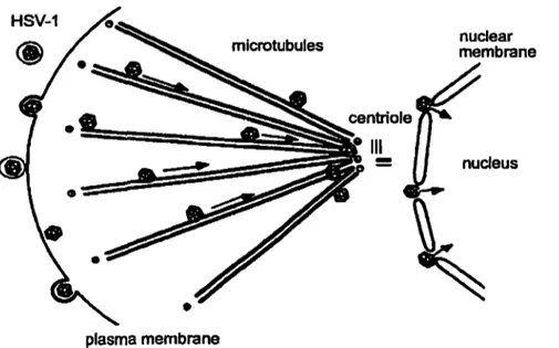
HSV-1
plasma membrane
microtubules nuclear membrane
nucleus
Figure 1 3: Transport of the capsid to the nuclear pores with release of the virion DNA into the nucleus (Sodeik et al., 1997).
Once the viral and cellular membranes fuse, the capsids still surrounded by tegument enter into the cell. Unenveloped capsids travel inside the cytoplasm to the nucleus along microtubules and dock to the nuclear pore. At this point, the viral DNA is ejected from the capsid and is released into the nucleoplasm (Sodeik et al., 1997). At the same time, the tegumentprotein vhs (ho st shutoff) escapes from the naked capsids and degrades the cellular mRNA, which allows ribosomes to preferentially synthesize viral proteins (Read and Frenkel. 1983). In addition, viral tegument proteins down-regulate cellular proteins that interfere with virus detection by the host's immune system (Triezenberg et aL, 1988; Baarr &
Skulstad, 1994).
1.6.3 Gene expression
Inside the nucleus of infected cells, HSV -1 synthesis involves a synchronized cascade of three phases of gene expression: immediate early (lE or a), early (E or ~), and late (L or y) (Honess and Roizman, 1975).
The cellular RNA polymerase II is responsible for aIl viral gene transcription. The lE genes are transcribed in the absence of any previous prote in synthesis and their products act to mediate the expression of the early and late genes. a genes expression occurs when VPl6 (aTIF) is released from the tegument and forms a complex with the two host cell proteins: the POU domain protein, Oct-l and a host cell factor HCF (Thomas S et al., 1998). This complex activates the TATA GARAT elements and initiates the transcription of viral lE genes into the ceIl nucleus (Goding and O'Hare, 1988, Hagmann et al., 1995; Preston et al., 1988). Expression of early gene products occurs at 4-5 hours postinfection and they are mostly enzymes necessary for the replication of the viral genome. The L genes expression starts at 6-7 hours postinfection and encodes mostly structural elements, such as capsid, tegument and glycoproteins that will be assembled into the progeny virions (Godowski P.J., and Knipe D. M.1986).
1.6.4 Viral DNA replication
Studies in vivo have demonstrated that once ~ genes have been expressed and translated, there are several proteins that are localized into the nucleus, where they assemble on the parental viral DNA in punctuate "prereplicative sites" near nuclear ND 1 0 structures (Ishov and Maul, 1996; Uprichard and Knipe, 1996). The viral DNA replication initiates on the circular viral DNA, which creates a "theta" structure, and then changes to a rolling circle mechanism producing head-to-tail concatemers of viral DNA (Jacob, Morse, and Roizman, 1979). At this point, replication takes place in "replication compartments" that consist of accumulating DNA molecules and replication complexes (Quinlan, Chen, and Knipe, 1984).
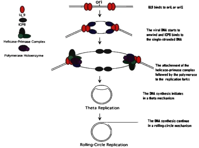
There are seven viral proteins absolutely required for viral DNA replication into cells. These are the viral DNA polymerase (UL30) (Purifoy, Lewis, and Powell, 1977), its accessory protein (UL42) (Conley et al., 1981), an origin-binding protein (UL9), the single stranded DNA binding protein (ICP8), and the helicase-primase complex that consists ofthree proteins: UL5, UL8, and UL52 (Challberg, 1986). Host cell factors may also be involved in DNA synthesis, and host enzymes that include the DNA polymerase a- primase, DNA ligase, and topoisomerase II are also required. The viral genome contains also the origins of replication, named oriS, and oriL (Mocarski and Roizman, 1982; Vlazny, K wong, and Frenkel, 1982; Weller et al., 1985). The basic model for the replication of HSV viral DNA proceeds as follows. First, the parental viral DNA is circularized in the nucleus of the infected cell. After the expression of a and ~ gene, UL9 binds to either oriL or oriS and begins to unwind the viral DNA. Then,
UL9 recruits the ssDNA binding prote in ICP8 to the unwound portion of the viral DNA. At this point, UL9 and ICP8 recruit the remaining five proteins to the replication forks. The helicase-primase and viral DNA polymerase complexes assemble at each replication fork and initiate the the ta replication. Through an unknown mechanism, replication switches from the theta form to the rolling circle form. The rolling circle replication results in long head-to-tail concatamers of viral DNA, which become cleaved into individual units during packaging of viral DNA into empty capsids (Roizman and Knipe, 2001)
ori
=====t
ll
l===::!=' ===4
11
F===
+
Hellcase-Primase Complex•
Polymerase Holoenzyme Theta Replicationo
Roiling-Circie Replication 11.1 blnds 10 oriL Dr oriSThe V'irallIII star1s 18 Inwlnd and ICPI binds ID thl! sinill-strandl!d IlIA
The allachmllnt Df thl! helicase..primase complu fallo.ed 1Iy!he polyrnerase la III! replication fortes
ThllllA synlllesis initi_s
ln il
.I!ta
mechanlsmThl l1li synlhlsis call1lllli in Il ralllng..circii lIechanism
Figure 1 4: Mechanism of "SV DNA replication (adapted from Travis J. Taylor et al., 2002)
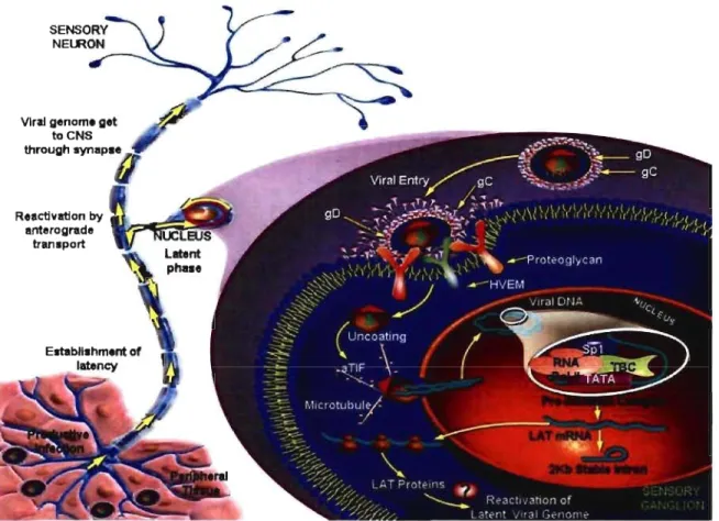
AIso, viral genome is able to stay latently in neuronal cells. Unlike a persistent infection where the virus is constantly replicating, the viral genome is not replicated during the latent infection, with the exception of a subset of HSV genes termed latency associated transcript (LAT) which is abundantly expressed. The role of LAT is still unclear; however recent studies have shown the importance of LATin limiting viral gene expression, and for the maintenance of the latent state. The major site of HSV latent infection is sensory neurons in ganglion tissue such as trigeminal ganglia for HSV -1 or sacral ganglia for HSV-2. During the latent state, the viral genome remains in the nucleus of the neuron as circular,
chromosomal DNA. After its reactivation by various stimuli, latently infected cells enter into a Iytic phase with the production of infectious virus particles (Whitley, 2001). The reactivation from latency occurs upon UV irradiation as a result of excessive sun exposure, stress, fever, damage or perturbation of the ganglia, or menstruation.
Reactivation by
anterograde w.,"::JIIII~~1III! transport
Figure 1 5: HSV-llatent infection (adapted from Protein Lounge Presentation).
1.6.5 Capsid assembly and maturation
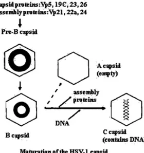
As structural proteins accumulate, capsid assembly begins. The viral capsid assembly requires the expression of late genes, the synthesis of the viral structural proteins such as: VP5, VP26, VP23, VP19c and two other polypeptides, UL26 and UL26.5 (lCP35) (Liu and Roizman, 1993). The UL26.5 (ICP35) product is the major scaffold prote in for capsid formation, and UL26 encodes a protease (Pra) that cleaves the scaffold. Both products form complexes with VP5 and triplex proteins, consisting of VP23 and VP 19. This immature capsid, called procapsid, is characterized as being fragile, thin waled, porous, and easily disassembled at 4°C (Newcomb et al., 1996; Rixon et al., 1996; Trus et al., 1996).
li
~
'II
So as to mature the procapsid into the stable rigid angularized capsid, scaffolds are cleaved by Pra and ejected from procapsid. Subsequent capsid angularization is coupled to packaging of the viral genome. The mature nucleocapsid is known as a C capsid, and is supposed to be capable of becoming an infectious particle. Beside the formation of the C capsid, maturing procapsids may also result in formation of A capsids and B capsids (Gibson and Roizman, 1972; Gibson and Roizman, 1974). The lightest A capsids are composed only of an empty shell, whereas B capsids (intermediate) have sorne additional internaI scaffold proteins Pra and ICP35. Capsid A does not package DNA and is thought to result from an abortive process ofDNA packaging (Gibson & Roizman, 1972, Perdue et aL, 1975).
capsidproteins:Vp5,19C;23,26 assellihlyprotebls:Vp21, 22a, 24
,
Pre-B capsido
~
O·
Acapsid (empty)l
'
@
.,,'
-...,j---::o--..
/r~~~y
c v ·
~
.
X/ '
. ~ Q DNA B capsid C capsid (contains DNA)Maturation of the HSV-~ capsid
Figure 1 6: Maturation of herpes simplex capsids (adapted from D. R. Harper)
Procapsids are observed in cells infected with HSV mutants such as the ts1201 (Preston et al. 1983), tsProt
A (Gao et aL, 1994), or V701 (Register et aL, 1996). In these strains, the UL26-encoded Pra has point mutations that inhibit its protease activity in a temperature dependent manner.
The phenotype of these mutant strains has been well used in the study of herpes virus life cycle. Cells infected with these mutants accumulate procapsids in the nucleus at the non-permissive temperature of 39°C (Preston et aL, 1983). A significant feature of this mutation is the fact that the mutation is reversible. As the protease function is restored upon retum to the permissive temperature of 31°C, subsequent procapsid maturation, angularization and DNA packaging occur in a single synchronized wave (Church and Wilson, 1997). This is a very well used tool in the study of complex biological phenomena as
diverse as protein trafficking through the secretory pathway, endocytosis, control of the cell cycle because of the ability to monitor the accumulation of a protein at a certain stage in its biogenesis, and then to release it as a synchronous wave.
1.6.6 Nuclear egress
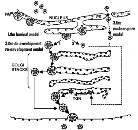
After their assembly, the nucleocapsids have to leave the nucleus going into the extracelular space and initiate a new round of infection. The first step is to bud through the inner nuclear membrane. Row this is achieved is not well understood, and the literature proposes three concurrent models of egress (Enquist et al., 1998; Mettenleiter 2002, Johnson and Juber, 2002).
The luminal model, also known as the single envelopment model, was described by Johnson and Spear in 1982. This model involves virus envelopment at the inner nuclear membrane, which already contains viral glycoproteins. The enveloped capsids leave the perinuclear space and enter into the ER and Golgi vesicles. Following the exocytic partway, the viral envelope interacts with the membrane of the surrounding vesicle where the final maturation of viral glycoproteins can take place, and the viral particle is released from the cell.
The second model of egress is named de-envelopment re-envelopment (Skepper et al., 2001). The model proposes that the primary envelopment of RSV capsids at the inner nuclear membrane is followed by a fusion with the outer nuclear membrane, resulting in its de-envelopment, and release of naked capsids into the cytoplasm. The nucleocapsid surrounded by sorne tegument proteins, further accumulates other tegument proteins, buds into the TGN/endosome membranes where it undergoes a secondary envelopment (re-envelopment), and is released into the extracellular environment (Cheung et al., 1991). This model is supported by studies in which the cellular retention signaIs of viral glycoproteins were mutated. For example, the construction of a mutant gR protein with an ER retention signal failed to package gR into the virus (Browne et al., 1996), and a gD mutant constructed with an ER localization signal produced significantly less of the virus, than a mutant with a Golgi retention signal (Whiteley et al., 1999). In addition, the extracellular viral envelope is more similar to plasma membrane composition than to that of the nuclear membrane (vanGenderen et al., 1994).
The third model of egress, proposed by Wild et al. 2005, and Leuzinger et al, 2005 implies a disassembly and dilatation of the nucleare pores, resulting in a direct passage of the capsid from the nucleus to the cytoplasm.
n _.,..---...-_ , 3.the
:...cs.---- l nuclear-(JOre l.the luminal model
""-..4..~~~;r--';;:QP
~
mDdel 2.the de.envelopment@..-1
~~® __________
.1.. __ _
re.envelopmentmodel~~
1
Gocœ
_~ 1 STACKS ~ 1 • 1 1L--~_J
--. • • •. •• ifi;... ·
..---Figure 1 7: Models of herpes simpex virus egress.
1-the luminal model ; 2-the de-envelopment re-envelopment model; 3- the nuclear pore exit model , NM- the nuclear membrane; TGN - trans-Golgi network.; PS, perinuclear space (Campadelli-Fiume G. and Gianni T. 2006).
The correct pathway of HSV -1 nuclear egress is still controversial and much examination is still needed to elucidate it. There are molecular studies that involve a crucial role of two virally encoded proteins, UL31 and UL34 in primary envelopment (Chang et al., 1997; RoBer et al., 2000). UL31 and UL34
interact with each other, co-Iocalizing at the nuclear membrane in infected ceUs (Bjerke et al., 2003; Fuchs et al., 2002; Reynolds et al., 2001). In the absence of either protein, primary envelopment is inhibited and capsids accumulate in the nucleus (Chang et al., 1997; Fuchs et al., 2002; RoBer et al., 2000; Klupp et aIl., 2000). Both the UL 31 and UL 34 proteins have been found associated with newly enveloped virions in the
perinuclear space, but not with cytoplasmic or extracellular virions (Reynolds et al., 2001; Reynolds et al., 2002). UL34 is a substrate for the Us3 who is itselfphosphorylated by ULI3. The interaction among these
proteins has been demonstrated to depolymerize the nuclear lamina through the cellular protein kinase C (PKC) pathway (Bjerke, S. L., and R. J. RoUer. 2006). This locallarpina depolymerization (Reynolds et
al., 2004; Simpson Holley et al., 2004) presumably allows the capsids to reach the mner nuclear membrane (Muranyi et al., 2002, Reynolds et al., 2004).
Sorne evidence which supports the de-envelopment re-envelopment model considers that naked capsids are necessarily present in the infected cell cytoplasm. Others evidence considers them a result of the enveloped virus's disassembly found in a non-productive state (Roizman and Sears, 1993). In addition, glycoproteins with an ER retention motif are not incorporated into extracellular virions (Browne et al., 1996; Whiteley et al., 1999). For example, Browne and coworkers have constructed a recombinant HSV in which the glycoprotein gH has been modified to contain a KKXX endoplasmic reticulum (ER) retenti on motif. When cells are infected with this recombinant, the amount of viruses released into the medium is the same as that in cells infected with the wild-type virus. However, these viruses are completely devoid of gH and have a 100-fold-lower infectivity than cells infected with the wild-type virus. This result suggests that the ER nuclear membrane is not a donor of viral envelope, and virions acquire their final envelope in a post-ER compartment, from which the modified gH is absent because of the ER retention motif. The work of van Gederen and coworkers have shown that the lipid composition of isolated HSV envelopes is very different from that of nuclear membranes. This means the viral envelope formed at the nuclear membrane is lost and the naked capsids in the cytoplasm will acquire a different lipid bilayer from the other intracellular compartment, which is post ER (van Genderen et al., 1994).
The validity of the two models of envelopment was, also studied using a fungal metabolite known to block the anterograde transport of cargo through the secretory pathway, named brefeldin A (BF A) (Klausner et al., 1992; Lippincott-Schwartz et al., 1989). In the case of single envelopment model, the virus in cells treated with BF A accumulates inside the perinuclear space and does not pass through the secretory pathway because of the block of export from ER by BF A (Anindya Dasgupta and Duncan W. Wilson, 2001). In contrast, in the de-envelopment re-envelopment model, the enveloped virus in the perinuclear space fuses with the external nuclear membrane and leads to an accumulation~of naked capsids in the cytoplasm. BF A blocked its traffic through Golgi and naked capsids accumulated in the cytoplasm. These experiments support the second model where the Golgi complex, the most sensitive BF A-organelle, is the major envelopment site of HSV -1 nucleocapsids leading to the formation of the infectious progeny virus (Chatterjee and Sarkar, 1992; Cheung et al. 1991; Anindya Dasgupta and Duncan W. Wilson, 2001; Koyama and Uchida, 1994).
'1
1
_ _ 1
1.6.7 Cytoplasmic capsid assembly and secondary envelopment
The mechanism of secondary envelopment is not well understood. The final envelopment requires a combination of capsid, tegument, and the final envelope at the site of re-envelopment, i.e. the TON or endosomes. After nuclear egress, intracytoplasmic capsids were found to contain pUL36 and pUL37 proteins which mediate transport of nucleocapsids to the envelopment site. That indicates that UL36 is necesary for the attachment of tegument components to the capsids (Desai, 2000). Its gene deletion results in accumulation of unenveloped capsids in the cytoplasm of infected cells. The same effect was observed by deletion of the UL37 gene, but in this case nucleocapsids were shown to accumulate also in the nucleus, which suggests UL37 prote in may be involved in both stages of viral egress, at the nucleus and in the cytoplasm (Desai et al., 2001). In principle, all components of the mature viral envelope need to be present in the correct compartment for the virion incorporation. From Il glycoproteins encoded by HSV -1, most ofthem have been reported to be present at the TON including gB, gD, gE/gI, gK, gM, and gH. gB and gL do not completely colocalise with the TON as the other cited glycoproteins that implies they play another role in other compartiments (Turcotte et al., 2005). However, other envelope glycoproteins such as the gH/L complex and gD do not contain any TON signal and it is thought they localize to the plasma membrane (Hutchinson et al., 1992; McMillan & Johnson, 2001) even that Turcotte et al., 2005 found a partial presence of gL in the TON at 20°C. The mechanisms by which the envelope glycoproteins could be targeted to the final envelopment compartment are unclear. Many studies have shown that gM forms a complex with gN and together have important roles in viral assembly and egress. gMIN colocalize with the TON marker TON46, and cause a relocalization of several membrane proteins from the plasma membrane such as gD and gH/L to the TON (Crump et al., 2004). The ability of gMIN to cause localization of the herpesvirus envelope pro teins gD and gH/L to the TON, could be part of the mechanism by which herpes viruses maintain sufficient concentrations of envelope proteins in the secondary envelopment compartment, thus allowing efficient assembly and viral egress. Even if the entire process still remains poorly understood, these results provide clues about how HSV -1 virion components are driven to the site of envelopment.
1.6.8 TGN to plasma membrane viral egress
Another step in HSV -1 envelopment that is po orly understood is the process by which virion initiate budding at TGN membranes. The budding process comprises an aggregation of virion proteins on the inner surface of a membrane followed by induction of membrane curvature and subsequent "pinching off' of the particle. Unlike for the cellular proteins, the HSV -1 machinery to initiate the membrane curvature and the pinching-off has not been identified. The discovery of this machinery is important because it provides an indication about how the virion components are selectively packaged during assembly. For example, which proteins are packaged by forming interactions with components of this machinery and which are packaged in the correct compartment during envelopment?
l'
li
1.6.9 Host transport from the TGN to the plasma membrane
1Various intracellular transport studies have revealed the complex formation of vesicles at the TGN membranes and they consider the TGN as a major prote in exit station towards various destinations inside the cell. After their maturation in the Golgi, proteins are sorted in the TGN, and transported to different locations inside the cell, with respect to biochemical sorting signaIs that are found on the individual proteins (Balch W. E., and B. A. Bernard. 1999). In this sense, the TGN is considered a critical gateway
for protein transit. In the lumen of TGN, proteins interact with specific receptor molecules. It is considered that after proteins find their specific receptors, they accumulate within TGN subdomains, then bud off in order to form diverse secretory vesicles (Mark A. McNiven and Heather M. Thompson. 2006).
In addition, sorne proteins are retained within the TGN due to the presence of a phosphosorting acidic
cluster motif adaptor (McNiven M. A. and Thompson H. M. 2006). The decision of sorting and transporting for a given protein from the TGN, is taken in association with resident adaptor molecules, such as ARF and Golgi-iocalized gamma-ear which contains ADP ribosylation factor (ARF)-binding proteins (GGAs) (Boman A. L. 2001).
Sorne proteins exit inside COPI-coated vesicles in the retro grade direction back to the ER (Fernandez-Ulibarri D.V. 1. et al., 2007). Finally, TGN vesicles that travel to endosomes, or plasma membrane, depend of the presence of GGAs and/or PKD (Kirchhausen, 2000; Brodsky et al., 2001;
Coat proteins induce the membrane curvature or regulate the association of motor proteins with membranes. This prevents the vesicles detachment from the organelle before the protein sorting has been completed (Kirchhausen 2000; Boehm and Bonifacino 2001; Bonifacino and Lippincott-Schwartz 2003). Clathrin coats found on the cytosolic face of the membranes mediate the releasing of transmembrane proteins at the plasma membrane, TGN or endosomes (Kirchhausen, 2000; Brodsky et al., 2001). At each location, clathrin coats contain adaptor protein (AP) complexes that mediate both the attachment of the clathrin to membranes and the concentration of specific transmembrane proteins. While AP-2 complex functions at the plasma membrane, AP-l complex is present at the TGN and/or endosomes (Kirchhausen, 2000; Brodsky et al., 2001; Robinson and Bonifacino, 2001). A protein family, named, GGAs (GGA1, GGA2, and GGA3 in humans) was presented to promote the recruitment of clathrin and the releasing of transmembrane proteins at the TGN (Robinson and Bonifacino, 2001; Boman, 2001). The ARP family (for ADP-Ribosylation Factor), of small GTPases plays an important role in vesicle formation. They recruit the Golgi-associated adaptor AP-l and GGAs to the Golgi membranes, and interfere in the releasing of proteins in endocytic pathways (Taylor et al., 1994). De Matteis and collegues further reported a new family of coat proteins, called F APPs (the four-phosphate adaptor proteins), that function at the TGN. Like clathrin, F APPs are coat proteins that bind to the vesicles or tubules which extend from the TGN and contain cargo. FAPPI and 2 are recruited in TGN by phosphatidylinositol-4-phosphate (PI, IPI4P), and they are considered as mediator of the apical protein transport (Go di et al. 2004, Vieira et al. 2005). Overexpression ofa dominant-negative ofFAPPs or depleting FAPPIIeveis with small-interfering RNAs inhibits protein transport from the TGN to the plasma membrane (Godi et al. 2004). Although FAPPs control the formation of transport carriers at the TGN, they are not present on transport intermediates that detach from the TGN, which means they disassemble from transport vesicles after budding.
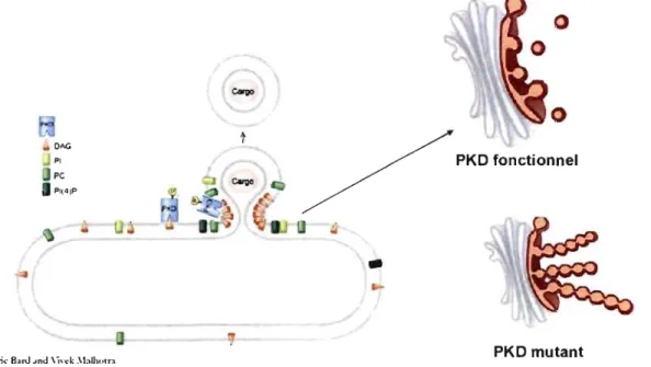
While GGA proteins play a role in regulating the TGN vesicular egress to endosomes, PKD has been identified as a molecule which regulates TGN vesicular transport to the plasma membrane (Liljedahl et al., 2001). PKD is a family of serine/threonine protein kinases that belongs to the Ca2+/calmodulin-dependent kinase superfamily. Three members of the PKD family have been identified so far in humans: PKD1, PKD2, and PKD3. They share homology in their catalytic do main and distinct sequences located . between the conserved motifs in the regulatory region confer isoform specific functions (Rykxa A., et al. 2003).
The three PKD isofonns are implicated in basolateral protein transport from TGN to the plasma membrane (Yeaman et al. 2004). They bind to DAG from the TGN membranes via their cysteine rich (Cl) domain (Maeda et al. 2001). DAG depletion inhibits their binding to the TGN and consequently, their activation (Baron and Malhotra 2002). The kinase-inactive fonn of PKD, the lysine to asparagine K618N mutant, has been shown to accumulate at the TGN and cause tubulation of the TGN. PKD-K618N tubules contain cargo, but do not detach from the TGN. The mutant also produces an inhibition of cargo transport between the TGN and plasma membrane. In contrast, PKD overactivation by ilimaquinone induces fragmentation of the Golgi apparatus (Keller et al. 200 1, Liljedahl et al. 200 1, and Polishchuk et al. 2003.
PKD-K618N
···1
"f"
•
:
.
:
.
carQo
Golgi
Figure: 1 8. Kinase dead PKD block HSV-l transport from the TGN to the plasma membrane.
production in the Golgi depends on the conversion of phosphatidic acid (PA) mediated by phosphatidic acid phosphohydrolases (PAPs). Finally, PA could be a result of phosphatidylcholine (PC)-specific phospholipase D enzymes (PLD) activity (Siddhanta and Shields, 1998). Therefore, PLD and PAP are important in DAG production in Golgi membranes. Another source of DAG is represented by sphingomyelin synthase (SMS) activity, which generates sphingomyelin (SM) and DAG (Ichikawa and Hirabayashi, 1998). Moreover, phosphoinositides such as (PI, PI4P) or phosphatidylinositol 4, 5-biphosphate (PI4, 5P2) are converted to DAG and inositol bis- or tris-phosphate through phosphoinositide-specific phospholipase C (PI-PLC) (Claro et al., 1993; Rhee, 2001).
Intracellular VSV -G post-Golgi transport has been studied in the presence of sorne drugs, such as fumonisin BI (FB-l), l-cycloserin (L-CS) and propranolol (Liebisch G. Schmitz G. Hoekstra D. 2004). Those drugs all cause an inhibition in the TGN-derived transport carriers as a result of Golgi-associated DAG level reduction (Baron and Malhotra, 2002; Brindley & Waggoner 1998, Pyne et al. 2004). These results show the DAG is a lipid involved in neck formation of the Golgi-derived vesicles or tubules. Consequently, DAG creates membrane insertion sites that allow peripheral membrane proteins to gain access to the hydrophobic portion of the bilayer, where they induce the generation of membrane curvature (Nie and Randazzo, 2006). Moreover, a reduction in DAG levels of Golgi membranes cause the inactivation of the molecular machinery necessary to induce membrane fission (Bard and Malhotra, 2006).
o
PKD fonctionnel
..
Fri'dêric B;1ro .Jnd \i\'(:k .\blhotn PKD mutant
Figure 1 9: Recruitment and activation of protein kinase D (PKD) at the TGN (adopted from V. Malhotra and modified by CM).
CHAPTER II: Objectives of research
Unlike the cellular proteins, nothing is known about the machinery that HSV -1 virions utilize to initiate the TGN to plasma membrane egress. The present work tries to identify whether HSV-l virions utilise the same machinery as the cellular proteins in their exit from TGN to the plasma membrane. In the cellular biosynthetic pathway it is known that the serine-threonine protein kinase D (PKD) plays a central role in vesicle formation at the TGN. As discussed above, PKD function itself depends on the pool of DAG in the TGN membranes. We have investigated the role of PKD in virus cargo exit from TGN. To verify this hypothesis we made use of the synchronized infection with HSV -1 termosensitive mutant virus V701 previously used (Turcotte et al. 2005). Since pharmacological inhibitors could acts at multiple stages of the viral life cycle, a mutant form of PKD to and RNA interference were also used to determine the real implication of this machinery in the HSV -1 virion egress.
. 1
CHAPTER III: Article
Summited to the Cel! Biology Journal
Protein kinase D dependent trafficking of the large HSV -1 capsids from the TGN to plasma membrane
by
Constantina Mihai, Gaudeline Rémillard-Labrosse, Johanne Duron and Roger Lippé* Department ofPathology and Cell Biology, University of Montreal,
Montreal, Quebec, Canada H3C 317
. Running tille: PKD regulates large cargo transport from the TGN
Character counts: 32087
*Corresponding author: Dr. Roger Lippé
Department of Pathology and Cel! Biology University of Montreal
PO Box 6128, Succursale Centre- Ville
Montreal, Quebec Canada H3C 3J7 Tel: (514) 343-5616
Fax: (514) 343-5755
3.1 Abstract
The conventional biosynthetic pathway has been extensively studied for small cargo. Interestingly, large partic1es such as procollagen, chylomicron and various virions reach the TGN by alternative routes. Given this dichotomy, we probed which machinery large cargo uses downstream of the TGN. Using Herpes simplex virus type 1 virions as a model, a collection of specific inhibitors and a synchronized infection protocol, the data surprisingly revealed a role in HSV -1 egress for the cellular serine-threonine protein kinase D. This established mediator of TGN to cell surface transport for small cargo unexpectedly highlights the trafficking of entities as large as virions by the common machinery used by small cargo. This substantially alters the range of cargo that the conventional biosynthetic pathway can accommodate. Given the apical release of HSV -1 in neurons, it also raises the possibility that PKD might regulate basolateral sorting. Lastly, it addresses for the time the molecular basis of egress of any viral partic1e transiting at the TGN.
3.2 Introduction
The biosynthetic pathway is a well studied route by which membrane bound and secreted proteins sequentially travel in the ER, the Golgi apparatus and the TGN before being sorted to their final destination. Along this route, the serine-threonine protein kinase D (PKD) is a key player at the TGN, since it regulates the fission of cargo filled carriers destined for the cell surface (Liljedahl et al., 2001). Three PKD isoforms exist in humans and differentially modulate cargo transport to the basolateral surface (Sanchez- Ruiloba et al., 2006; Yeaman et al., 2004). Interestingly, recruitment of PKD from cytosol requires its interaction with diacyl glycerol (DAG), a lipid critical to PKD function and whose presence at the TGN is determining (Baron and Malhotra, 2002). Procollagen, chylomicron and virions are large particles reaching several hundred nanometers (Canty and Kadler, 2005; Fromme and Schekman, 2005). Their substantial size poses major challenges for their intracellular transport (Fromme and Schekman, 2005; Mettenleiter, 2004). For instance, it has been reported that both procollagen and chylomicron bypass the classical COPII coated vesicles to escape the ER (Siddiqi et al., 2003; Starkuviene and Pepperkok, 2007; Stephens and Pepperkok, 2002). Similarly, Herpes simplex virus type 1 (HSV-l) is a large particle assembled in the nucleus that also reaches the TGN by an alternative route. Too large to leave the nucleus via the pores, the 125 nm capsids sequentially bud across the inner nuclear membrane and fuse with the second nuclear envelope to be released in the cytoplasm (Mettenleiter, 2004; Remillard- Labrosse et al., 2006). They then bypass the Golgi apparatus and acquire an envelope from the TGN to form mature 200-300 nm virions (Harley et al., 2001; Turcotte et al., 2005). The virions finally leave this last compartment by a completely unknown mechanism.
Given the unconventional transport pathways employed by large cargo to arrive at the TGN, we sought to examine ifthey use the conventional transport machinery further downstream. Using HSV-I as a model and a collection of inhibitors, the egress of virions from the TGN to the plasma membrane was 1
monitored using a recently developed protocol (Turcotte et al., 2005). The data surprisingly revealed a 1
critical contribution of PKD in HSV -1 virion egress, a unique finding for any of the known viruses transiting at the TGN. These results clearly indicate that large particles can share the same route as small cargo to escape the TGN, in sharp contrast to earlier steps of transport. Given the exceptional size of HSV -1 virions, this also substantially broadens the range of cargo the classical transport machinery can accommodate. Finally, given the apical release of HSV - 1 in neurons, it raises the possibility that PKD
3.3 Results
Synchronization
0/
HSV-l intracellular transport/rom the TGN to the cell surface.There is little information regarding the mechanism by which HSV -1 leaves the TGN. Unfortunately, the rapid life cycle of HSV-l makes it difficult to characterize tbis viral transport step. Nonetheless, others and we have showed that viral egress can be synchronized using mutants of the viral i
protease UL26, a protein required for encapsidation of the herpes DNA and capsid maturation (Church , and Wilson, 1997).
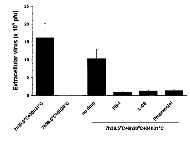
Hence, thermosensitive UL26 viral mutants such as ts1201 (Preston et al., 1983), tsProt.A (Gao et al., 1994) and V701 (Register and Shafer, 1996) accumulate immature viral capsids in the nucleus at the non permissive temperature of 39.5°C but release mature extracellular virus at 31°C. To ensure a tight wave ofviral egress, cycloheximide - an inhibitor of prote in synthesis - is typically added after the 39.5°C incubation to prevent the assembly of new capsids (Church and Wilson, 1997; Turcotte et al., 2005). Viral egress can further be dissected as HSV -1 capsid transport is reversibly arrested at the TGN at 20°C, much like host proteins along the biosynthetic pathway (Turcotte et al., 2005). This reversible 20°C block represents an ideal mean to define the molecular requirements of HSV -1 transport from the TGN to the plasma membrane. It is also optimal to probe the potential role of PKD in the transport of cargo as massive as fully assembled virus. To first confirm the efficacy ofthis block, 143B cells were infected with the thermosensitive HSV -1 stain V701 and the virus was monitored at 39.s°C, 20°C and 31°C (fig. 1). To detect the virus, the samples were stained with an ICP5 antiserum, which recognizes the major viral capsid protein. As previously reported, the virus was retained in the nucleus at 39.5°C, was efficiently retained in the TGN at 20°C and could reach the cell surface at 31°C (fig. 1; Turcotte et al., 2005). Most important, the 20°C block was reversible as the virus could escape the TGN when followed by a chase at 31°C. It was therefore possible to synchronize the transport of the large HSV -1 virus from the TGN to the cell surface.
PKD inhibitors arrest TGN to plasma membrane transport.
Recruitment of cytosolic PKD to the TGN is essential for cargo release from that compartment. This requires an interaction between PKD and the TGN bound pool of DAG (Baron and Malhotra, 2002). Fumonisin BI (FB-l) and L-cycloserine (L-CS) block DAG production by preventing at two distinct steps the synthesis of ceramide, which is ultimately converted into DAG and sphingomyelin (Baron and Malhotra, 2002; van Ooij et al., 2000). In contrast, propranonol blocks DAG synthesis by inhibiting the