Deletion of the ORF9p Acidic Cluster Impairs the Nuclear Egress of
Varicella-Zoster Virus Capsids
Laura Riva,a* Marc Thiry,bMarielle Lebrun,aLaurent L’homme,cJacques Piette,a,cCatherine Sadzot-Delvauxa
University of Liege, GIGA Infection Immunity and Inflammation, Laboratory of Virology and Immunology, Liege, Belgiuma; University of Liege, GIGA Neurosciences, Laboratory of Cellular and Tissular Biology, Liege, Belgiumb; University of Liege, GIGA Signal Transduction, Laboratory of Virology and Immunology, Liege, Belgiumc
The protein encoded by ORF9 is essential for varicella-zoster virus (VZV) replication. Previous studies documented its presence
in the trans-Golgi network and its involvement in secondary envelopment. In this work, we deleted the ORF9p acidic cluster,
destroying its interaction with ORF47p, and this resulted in a nuclear accumulation of both proteins. This phenotype results in
an accumulation of primary enveloped capsids in the perinuclear space, reflecting a capsid de-envelopment defect.
O
ne of the crucial steps in herpesviruses infection is capsid exit
from the nucleus. This process mainly follows the
envelop-ment/de-envelopment model, strongly documented by
transmis-sion electron microscopy (TEM) observations (
1
). According to
this model, nuclear capsids bud at the inner nuclear membrane
(INM), thereby acquiring a primary envelope, which is then lost
after fusion with the outer nuclear membrane (ONM), resulting
in the release of naked capsids into the cytoplasm (
1
).
Viral glycoproteins seem to play a role during this
de-envelop-ment fusion process (
2
). In the case of herpes simplex virus
(HSV), it is known that gB and gH/gL, components of the viral
entry machinery, are present at nuclear membranes and are likely
responsible for this fusion (
2
,
3
). Moreover, the fusogenic role of
gB seems to be mediated by its pUS3-dependent phosphorylation
(
4
). These hypotheses are supported by observations that HSV
mutants lacking pUS3 or both gB and gH accumulate primary
enveloped virions in the perinuclear space (
3
,
4
). The components
of the nuclear export complex (NEC), pUL31 and pUL34, have
been described to mediate the primary envelopment (
2
).
Phos-phorylation of pUL31, another substrate of pUS3, also promotes
the de-envelopment process (
5
).
Unfortunately, the mechanisms leading to varicella-zoster
vi-rus (VZV) nuclear egress are still poorly understood, and it is not
clear whether the role of these proteins is conserved in VZV egress.
In this work, we destroyed the interaction of the essential VZV
tegument protein ORF9p (
6
) with the viral kinase ORF47p,
ho-mologous to HSV-1 VP22 and UL13, respectively, and this
af-fected their localization and impaired VZV de-envelopment.
ORF9 is the most transcribed VZV gene during infection (
7
),
and ORF9p has been observed to be present in the trans-Golgi
network (TGN) (
8
), playing a role in secondary envelopment (
9
).
Within its sequence is an acidic motif corresponding to residues
85 to 93: EDDFEDIDE (
Fig. 1B
). Acidic clusters are described as
targeting signals to the TGN (
10
), and the HSV VP22 acidic cluster
has been shown to have a role in both correct protein subcellular
localization and virion incorporation (
11
). We thus deleted this
acidic region to generate BAC-VZV-ORF9-⌬AC-V5 C-ter (
Fig.
1A
and
B
). Transfection of this bacterial artificial chromosome
(BAC) into MeWo cells led to VZV-ORF9-⌬AC-V5 virus.
Infec-tion analysis revealed a significant defect in infectivity for this
mutant, compared to that of the previously described
VZV-ORF9-V5 (
Fig. 1C
) (
9
). Generation of the revertant
VZV-ORF9-rev-V5, achieved by replacing the ORF9-
⌬AC sequence with the
wild-type one, rescued this defect (
Fig. 1C
).
In order to determine if this acidic region influences ORF9p
localization, immunofluorescence analysis was performed on
MeWo cells infected with VZV-ORF9-V5 (
Fig. 2A
to
C
),
VZV-ORF9-
⌬AC-V5 (
Fig. 2D
to
F
), or VZV-ORF9-rev-V5 (
Fig. 2G
to
I
). This experiment revealed the nuclear accumulation of
ORF9p-⌬AC-V5 (
Fig. 2D
), compared to ORF9p-V5 or ORF9p-rev-V5,
which appeared to be mainly cytoplasmic (
Fig. 2A
and
G
). ORF9p
has been previously observed in the nucleus of infected cells by
TEM analysis after immunogold labeling (
8
) and is characterized
by the presence of two in silico-predicted nuclear localization
sig-nals (NLS) with the PSortII software (
12
); the first one has been
confirmed to be active (
13
). Moreover, it contains an active
nu-clear export signal (NES) (
13
). This suggests that ORF9p can
shut-tle between the nucleus and cytoplasm, where its acidic region is
thought to mediate its targeting to the TGN. Deletion of the
ORF9p acidic region does not affect its isoelectric point or its in
silico-predicted localization (PSortII prediction [
12
]), but it
strongly modifies its subcellular localization.
This acidic cluster overlaps the consensus sequence SEDD (
Fig.
1B
), which we have previously described as being responsible for
ORF47p-dependent phosphorylation (
9
). We thus decided to
check the ORF9p-⌬AC Western blotting pattern in the context of
infection. As shown in
Fig. 2J
to
K
(lanes 2 to 4), ORF9p-
⌬AC
migrated more quickly than ORF9p or ORF9p-rev in the gel, likely
due to a reduced level of phosphorylation. In addition,
coimmu-noprecipitation experiments confirmed that the ORF9p-ORF47p
Received 5 November 2014 Accepted 26 November 2014 Accepted manuscript posted online 3 December 2014
Citation Riva L, Thiry M, Lebrun M, L’homme L, Piette J, Sadzot-Delvaux C. 2015. Deletion of the ORF9p acidic cluster impairs the nuclear egress of varicella-zoster virus capsids. J Virol 89:2436 –2441.doi:10.1128/JVI.03215-14.
Editor: L. M. Hutt-Fletcher
Address correspondence to Catherine Sadzot-Delvaux, csadzot@ulg.ac.be. * Permanent address: Laura Riva, Center for Infection and Immunity of Lille, Lille Pasteur Institute, University Lille North of France, Lille, France.
Copyright © 2015, American Society for Microbiology. All Rights Reserved. doi:10.1128/JVI.03215-14
on May 22, 2015 by UNIVERSITE DE LIEGE
http://jvi.asm.org/
interaction observed for the wild type and the revertant was lost in
cells infected with this
⌬AC mutant (
Fig. 2J
and
K
, lanes 8).
Moreover, immunofluorescence analysis indicated a stronger
nuclear accumulation of ORF47p in VZV-ORF9-
⌬AC-V5-in-fected cells (
Fig. 2E
) than in VZV-ORF9-V5- or
VZV-ORF9-rev-V5-infected cells (
Fig. 2B
and
H
). This observation suggests an
involvement of ORF9p in exporting ORF47p from the nucleus to
the cytoplasm or in its cytoplasmic retention. All experiments
were performed as previously described (
9
).
In order to better understand the phenotype of this
⌬AC
mu-FIG 1 Deletion of the ORF9p acidic region impairs VZV infectivity. (A) Primer sequences used for the construction of recombinant viruses. (B) Schematicrepresen-tation of the ORF9p mutant inserted in BAC-VZV, via a two-step BAC recombineering technique described by Warming et al. (21), generating the BAC-VZV-ORF9-⌬AC-V5 construct. ⌬AC, deletion of the acidic cluster corresponding to amino acids (AA) 85 to 93 of ORF9p. (C) Analysis of VZV replication in MeWo cells. Uninfected cells were inoculated at day 0 with 200 infected cells for each infection (VZV-ORF9-V5, VZV-ORF9-⌬AC-V5, or VZV-ORF9-rev-V5), based on analysis by flow cytometry. Inoculated flasks were harvested daily for 4 days to perform serial dilutions. Infection quantification was performed after 2 days; we counted the fluorescent foci on melanoma cell monolayers. Each point represents the mean number of foci from three independent experiments. Error bars represent the standard errors of the means. RH, alphaherpesvirus conserved region of homology. Statistical significance is indicated by the following symbols (calculated by a two-way analysis of variance): **, P⬍ 0.01, and ***, P ⬍ 0.001 (compared with VZV-ORF9-V5); §, P ⬍ 0.05, and §§§, P ⬍ 0.001 (compared with VZV-ORF9-rev-V5).
ORF9 Acidic Cluster Removal Impairs VZV Nuclear Egress
on May 22, 2015 by UNIVERSITE DE LIEGE
http://jvi.asm.org/
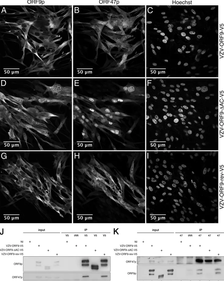
FIG 2 Deletion of the ORF9p acidic region leads to ORF9p and ORF47p nuclear accumulation. MeWo cells infected with V5 (A to C),
VZV-ORF9-⌬AC-V5 (D to F), or VZV-ORF9-rev-V5 (G to I) for 48 h were fixed using 4% paraformaldehyde and immunostained using the anti-V5 antibody (A, D, and G) or ORF47p antiserum (B, E, and H). Alexa 568 anti-rabbit and Alexa 633 anti-mouse were used as secondary antibodies. (C, F, and I) Nuclear labeling with Hoechst stain. Imaging was performed using a Zeiss 780 confocal microscope with a 63⫻ oil objective. (J and K) Uninfected MeWo cells or MeWo cells infected for 24 h with VZV-ORF9-V5, VZV-ORF9-⌬AC-V5, or VZV-ORF9-rev-V5 were harvested in lysis buffer, and cell extracts were incubated with beads coated with anti-V5 antibody (J) or ORF47 antiserum (K). The immunoprecipitated proteins were resolved by SDS-PAGE and immunoblotted using antibody against the V5 tag and ORF47p antiserum. NI, noninfected cells; IRR, irrelevant antibody.
on May 22, 2015 by UNIVERSITE DE LIEGE
http://jvi.asm.org/
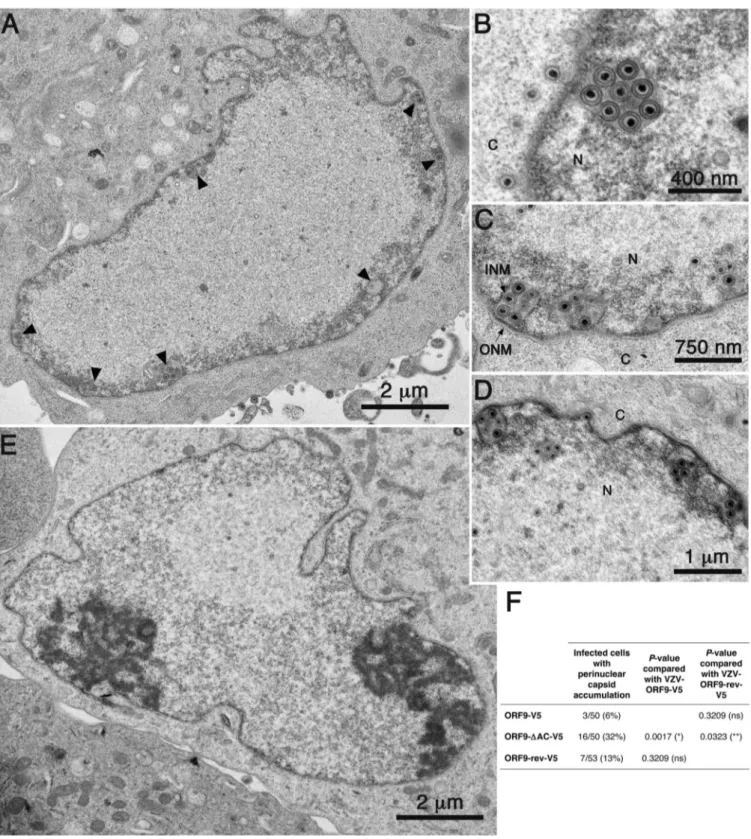
FIG 3 Deletion of the ORF9p acidic region causes an accumulation of primary enveloped capsids in the perinuclear space. MeWo cells were infected with
VZV-ORF9-V5, VZV-ORF9-⌬AC-V5, or VZV-ORF9-rev-V5 for 48 h and then fixed and analyzed by TEM. (A to D) Nucleus of a VZV-ORF9-⌬AC-infected cell, representative of nuclear capsid accumulation (A), and higher magnifications of perinuclear spaces showing accumulations of primary enveloped VZV-ORF9-⌬AC capsids (B to D). (E) A nucleus representative of a VZV-ORF9-V5-infected cell. (F) Quantification of the observed phenotype. Cells infected with VZV-ORF9-V5, VZV-ORF9-⌬AC-V5, or VZV-ORF9-rev-V5 were analyzed for the presence or absence of perinuclear capsid accumulation, and the results are displayed in the table. Statistical significance was calculated by using Fisher’s exact test and is indicated by one of the following symbols: *, P⬍ 0.05; **, P ⬍ 0.01; ns, nonsignificant. N, nucleus; C, cytoplasm; INM, inner nuclear membrane; ONM, outer nuclear membrane. Arrowheads indicate perinuclear capsid accu-mulation.
ORF9 Acidic Cluster Removal Impairs VZV Nuclear Egress
on May 22, 2015 by UNIVERSITE DE LIEGE
http://jvi.asm.org/
tant, VZV-ORF9-V5-, VZV-ORF9-
⌬AC-V5-, and
VZV-ORF9-rev-V5-infected MeWo cells were analyzed by TEM 48 h
postin-fection (
Fig. 3
). Surprisingly, the VZV-ORF9-
⌬AC-V5-infected
cells showed primary enveloped capsids that had accumulated in
the perinuclear space (
Fig. 3A
to
D
versus E). Such perinuclear
accumulation was observed in 32% of infected cells (up to 11
capsids/perinuclear structure), while it was observed in only 6%
(maximum, 4 capsids) and 13% (maximum, 6 capsids) of
wild-type- or revertant-infected cells, respectively (
Fig. 3F
). Altogether,
these observations could explain the infection defect of
VZV-ORF9-
⌬AC-V5. ORF9p could be directly involved in capsid
egress or have an indirect role, regulating ORF47p localization,
which could itself regulate VZV de-envelopment. Another
possi-bility is that neither ORF9p nor ORF47p are involved in capsid
egress, but nuclear misaccumulation of one or both of these
pro-teins could have an adverse effect on de-envelopment.
Nuclear and cytoplasmic extracts (
14
) from VZV-ORF9-V5-,
VZV-ORF9-⌬AC-V5-, or VZV-ORF9-rev-V5-infected MeWo
cells were further analyzed by Western blotting 24 h postinfection,
in order to determine if the de-envelopment defect could be due to
an altered expression level, stability, or localization pattern of gB,
gH, ORF66p, ORF24p, or ORF27p, which are homologous to the
HSV proteins involved in de-envelopment. The glycoprotein gE,
described as an additional fusion protein during VZV entry (
15
),
was also analyzed, while IE63 was chosen as an infection control.
No apparent difference was observed for any of these proteins
(
Fig. 4
), suggesting instead an independent role for ORF9p, which
may act as a negative regulator of the fusion process. Observations
of a pseudorabies mutant virus deleted for both gB and gH showed
no defect in nuclear egress (
16
); indeed, this suggests that
glyco-protemediated fusion is probably not the only mechanism
in-volved in the de-envelopment process.
In summary, we showed that ORF9p is somehow involved in
the ORF47p nuclear/cytoplasmic balance, and its acidic cluster
was identified as an important determinant for ORF9p subcellular
localization, ORF47p interaction, and VZV infectivity. We also
found evidence that VZV nucleocapsid egress is impaired when
the ORF9p acidic cluster is deleted.
ACKNOWLEDGMENTS
This work was supported by the Fonds pour la Recherche dans l’Industrie et l’Agriculture (Belgium) and by the Fonds National pour la Recherche Scientifique (Belgium). The study was cofunded by the University of Liege and the European Union via Marie Curie BeIPD DG Research-FP7-PEOPLE PCOFUND-GA-2012-600405 Fellowship.
We thank H. Zhu for BAC-VZV-pOka-WT (17), the Biology Research Branch at the NCI for the pGalK plasmid and SW102 bacterial strain, M. Sommer for gB and ORF66 antisera (18,19), and S. Jonjic for anti-gH, ORF24, and ORF27 antibodies (20). We also thank P. Piscicelli for TEM preparations and the GIGA Imaging and GIGA Genotranscriptomics platforms for their technical support.
REFERENCES
1. Mettenleiter TC, Muller F, Granzow H, Klupp BG. 2013. The way out: what we know and do not know about herpesvirus nuclear egress. Cell Microbiol 15:170 –178.http://dx.doi.org/10.1111/cmi.12044.
2. Johnson DC, Baines JD. 2011. Herpesviruses remodel host membranes for virus egress. Nat Rev Microbiol 9:382–394.http://dx.doi.org/10.1038 /nrmicro2559.
3. Farnsworth A, Wisner TW, Webb M, Roller R, Cohen G, Eisenberg R,
Johnson DC. 2007. Herpes simplex virus glycoproteins gB and gH
func-tion in fusion between the virion envelope and the outer nuclear
mem-FIG 4 Proteins described as important players in the de-envelopment process are apparently not influenced by ORF9p mutation. (A) Noninfected MeWo cells
or MeWo cells infected with VZV-ORF9-V5, VZV-ORF9-⌬AC-V5, or VZV-ORF9-rev-V5 for 24 h were harvested in cytoplasmic lysis buffer. After centrifuga-tion, the pellet was washed and resuspended in nuclear lysis buffer. Cytoplasmic and nuclear proteins were resolved by SDS-PAGE and immunoblotted using antibodies against VZV IE63, gE, gH, ORF24p, and ORF27p and gB and ORF66p antisera. Cellular p100 and nucleolin C23 were used as the cytoplasmic and nuclear controls, respectively. NI, noninfected cells.
Riva et al.
on May 22, 2015 by UNIVERSITE DE LIEGE
http://jvi.asm.org/
brane. Proc Natl Acad Sci U S A 104:10187–10192.http://dx.doi.org/10 .1073/pnas.0703790104.
4. Wisner TW, Wright CC, Kato A, Kawaguchi Y, Mou F, Baines JD,
Roller RJ, Johnson DC. 2009. Herpesvirus gB-induced fusion between
the virion envelope and outer nuclear membrane during virus egress is regulated by the viral US3 kinase. J Virol 83:3115–3126.http://dx.doi.org /10.1128/JVI.01462-08.
5. Mou F, Wills E, Baines JD. 2009. Phosphorylation of the U(L)31 protein of herpes simplex virus 1 by the U(S)3-encoded kinase regulates localiza-tion of the nuclear envelopment complex and egress of nucleocapsids. J Virol 83:5181–5191.http://dx.doi.org/10.1128/JVI.00090-09.
6. Tischer BK, Kaufer BB, Sommer M, Wussow F, Arvin AM, Osterrieder
N. 2007. A self-excisable infectious bacterial artificial chromosome clone
of varicella-zoster virus allows analysis of the essential tegument protein encoded by ORF9. J Virol 81:13200 –13208.http://dx.doi.org/10.1128/JVI .01148-07.
7. Cohrs RJ, Hurley MP, Gilden DH. 2003. Array analysis of viral gene transcription during lytic infection of cells in tissue culture with varicella-zoster virus. J Virol 77:11718 –11732.http://dx.doi.org/10.1128/JVI.77.21 .11718-11732.2003.
8. Che X, Reichelt M, Sommer MH, Rajamani J, Zerboni L, Arvin AM. 2008. Functions of the ORF9-to-ORF12 gene cluster in varicella-zoster virus replication and in the pathogenesis of skin infection. J Virol 82:5825– 5834.http://dx.doi.org/10.1128/JVI.00303-08.
9. Riva L, Thiry M, Bontems S, Joris A, Piette J, Lebrun M,
Sadzot-Delvaux C. 2013. ORF9p phosphorylation by ORF47p is crucial for the
formation and egress of varicella-zoster virus viral particles. J Virol 87: 2868 –2881.http://dx.doi.org/10.1128/JVI.02757-12.
10. Bonifacino JS, Traub LM. 2003. Signals for sorting of transmembrane proteins to endosomes and lysosomes. Annu Rev Biochem 72:395– 447.
http://dx.doi.org/10.1146/annurev.biochem.72.121801.161800. 11. O’Regan KJ, Brignati MJ, Murphy MA, Bucks MA, Courtney RJ. 2010.
Virion incorporation of the herpes simplex virus type 1 tegument protein VP22 is facilitated by trans-Golgi network localization and is independent of interaction with glycoprotein E. Virology 405:176 –192.http://dx.doi .org/10.1016/j.virol.2010.06.007.
12. Nakai K, Horton P. 1999. PSORT: a program for detecting sorting signals in proteins and predicting their subcellular localization. Trends Biochem Sci 24:34 –36.http://dx.doi.org/10.1016/S0968-0004(98)01336-X.
13. Cai M, Wang S, Xing J, Zheng C. 2011. Characterization of the nuclear import and export signals, and subcellular transport mechanism of vari-cella-zoster virus ORF9. J Gen Virol 92:621– 626.http://dx.doi.org/10 .1099/vir.0.027029-0.
14. Bureau F, Delhalle S, Bonizzi G, Fievez L, Dogne S, Kirschvink N,
Vander-plasschen A, Merville MP, Bours V, Lekeux P. 2000. Mechanisms of
per-sistent NF-kappa B activity in the bronchi of an animal model of asthma. J Immunol 165:5822–5830.http://dx.doi.org/10.4049/jimmunol.165.10.5822. 15. Maresova L, Pasieka TJ, Grose C. 2001. Varicella-zoster virus gB and gE coexpression, but not gB or gE alone, leads to abundant fusion and syncytium formation equivalent to those from gH and gL coexpression. J Virol 75:9483– 9492.http://dx.doi.org/10.1128/JVI.75.19.9483-9492.2001.
16. Klupp B, Altenschmidt J, Granzow H, Fuchs W, Mettenleiter TC. 2008. Glycoproteins required for entry are not necessary for egress of pseudora-bies virus. J Virol 82:6299 – 6309.http://dx.doi.org/10.1128/JVI.00386-08. 17. Zhang Z, Rowe J, Wang W, Sommer M, Arvin A, Moffat J, Zhu H. 2007. Genetic analysis of varicella-zoster virus ORF0 to ORF4 by use of a novel luciferase bacterial artificial chromosome system. J Virol 81:9024 –9033.
http://dx.doi.org/10.1128/JVI.02666-06.
18. Oliver SL, Sommer M, Zerboni L, Rajamani J, Grose C, Arvin AM. 2009. Mutagenesis of varicella-zoster virus glycoprotein B: putative fusion loop residues are essential for viral replication, and the furin cleavage motif contributes to pathogenesis in skin tissue in vivo. J Virol 83:7495– 7506.http://dx.doi.org/10.1128/JVI.00400-09.
19. Schaap A, Fortin JF, Sommer M, Zerboni L, Stamatis S, Ku CC, Nolan
GP, Arvin AM. 2005. T-cell tropism and the role of ORF66 protein in
pathogenesis of varicella-zoster virus infection. J Virol 79:12921–12933.
http://dx.doi.org/10.1128/JVI.79.20.12921-12933.2005.
20. Lenac Rovis T, Bailer SM, Pothineni VR, Ouwendijk WJ, Simic H,
Babic M, Miklic K, Malic S, Verweij MC, Baiker A, Gonzalez O, von Brunn A, Zimmer R, Fruh K, Verjans GM, Jonjic S, Haas J. 2013.
Comprehensive analysis of varicella-zoster virus proteins using a new monoclonal antibody collection. J Virol 87:6943– 6954.http://dx.doi.org /10.1128/JVI.00407-13.
21. Warming S, Costantino N, Court DL, Jenkins NA, Copeland NG. 2005. Simple and highly efficient BAC recombineering using galK selection. Nu-cleic Acids Res 33:e36.http://dx.doi.org/10.1093/nar/gni035.
ORF9 Acidic Cluster Removal Impairs VZV Nuclear Egress