ORIGINAL ARTICLE
Taxonomic revision of Endoraecium digitatum (rust fungi,
Uredinales) with description of four new species
from Australia and Hawaii
Reinhard BerndtReceived: 21 April 2010 / Revised: 31 May 2010 / Accepted: 16 June 2010 / Published online: 19 November 2010 # German Mycological Society and Springer 2010
Abstract Endoraecium digitatum was described on Acacia notabilis from South Australia but has been reported on more than 20 Acacia spp. throughout the Australian and the Pacific regions. Results are presented on the micro-morphology and taxonomy of E. digitatum from different Acacia species and geographic provenience. E. digitatum revealed a highly non-uniform morphology and is considered a species complex comprising at least six morphologically distinct rust fungi. The name E. digitatum could not be applied to any of the distinguished species by a comparison with available type specimens or the original description as types were depleted and the diagnosis not detailed enough. An epitype is therefore proposed to supplement the lectotype of E. digitatum selected here and to allow delimitation of the new species E. parvum, E. violae-faustae and E. walkerianum from Australia, and E. kauaianum from Hawaii. Endoraecium phyllodiorum is shown to be different from E. digitatum and is proposed as a new combination for Uromyces phyllodiorum. Speci-mens from Hawaii formerly considered to be E. digitatum represent two species, E. kauaianum and another one preliminarily assigned to E. phyllodiorum. The investigated species show differences between and plasticity within their life cycles. E. parvum, E. violae-faustae and E. kauaianum are macrocyclic. In E. walkerianum, only demicyclic speci-mens were found, while demi- and macrocyclic specispeci-mens occurred in E. digitatum. E. phyllodiorum revealed variable combinations of spore states as well and comprised macro-, demi- and microcyclic life cycle variants. Life cycle variants were considered to express specific variability and were not
used to create new taxa. The present results indicate that members of the E. digitatum complex in Australia are not narrowly host specific. Keys are presented for the Australian and Hawaiian species. Taxonomical novelties: Endoraecium kauaianum R. Berndt, Endoraecium parvum R. Berndt, Endoraecium phyllodiorum (McAlp.) R. Berndt, Endorae-cium violae-faustae R. Berndt and EndoraeEndorae-cium walkerianum R. Berndt.
Keywords Acacia . Racospermyces . Life cycle . Epitype . Species complex
Introduction
The rust fungus Uromyces digitatus G. Winter was described on Acacia notabilis F. Müll. from South Australia (Winter 1886). It was later regarded as a member of the genus Pileolaria Castagne (Dietel1921; sub P. phyllodiorum) or Atelocauda Arth. & Cummins (Cummins and Hiratsuka 1983). Walker (2001) erected the new genus Racospermyces J. Walker to accommodate U. digitatus and five other species, four of which had been assigned to Atelocauda.
In 1984, Hodges and Gardner (1984) reported two endocyclic rust fungi with Uredo-like teliospores on Acacia spp. from Hawaii, Endoraecium acaciae Hodges & D. Gardner and E. hawaiiense Hodges & D. Gardner. They hypothesized them to be derivatives of the morphologically similar U. digitatus but placed them in a new genus, Endoraecium Hodges & D. Gardner. Scholler and Aime (2006) presented further evidence for a close relationship between U. digitatus and Endoraecium based on combined sequence data of the LSU and SSU region of nuclear DNA. Regarding Racospermyces and Endoraecium as congeners, Scholler and Aime (2006) stated that the name Endorae-cium had priority over Racospermyces and recombined all
R. Berndt (*)
ETH Zurich, Institute of Integrative Biology (IBZ), Universitätstr. 16,
8092 Zurich, Switzerland
e-mail: reinhard.berndt@env.ethz.ch DOI 10.1007/s11557-010-0719-9
known Racospermyces species into Endoraecium: E. bicinctum (McAlp.) M. Scholler & Aime and E. tierneyi (J. Walker & R.G. Shivas) M. Scholler & Aime, only known from Australia, E. angustiphyllodium (D. Gardner) M. Scholler & Aime and E. koae (Arth.) M. Scholler & Aime, only known from Hawaii, E. digitatum (G. Winter) M. Scholler & Aime reported from Australia, New Caledonia, Hawaii and East Asia, and E. hyalosporum (Sawada) M. Scholler & Aime that is distributed in China, Japan and Taiwan. All known Endoraecium species are autoecious and restricted to Acacia subgen. Phyllodineae (DC.) Seringe (= Racosperma C. Martius; comp. Pedley 1986) of Leguminosae (Walker 2001).
Endoraecium digitatum is a variable rust fungus. Hodges and Gardner (1984) described some morphological differ-ences between aeciospores of E. digitatum from Kauai and other Hawaiian islands and noticed that the fungus induced either witches’ brooms or hypertrophy of shoots, flowers and pods or large bullate swellings on phyllodes, according to geographic location and host. Similarly, Walker (2001) pointed out to a considerable morphological variability of E. digitatum in Australia and hypothesized that this rust fungus could represent a complex of closely related taxa that may not only differ morphologically but also in life cycle, host range and geographical distribution. Specimens of E. digitatum recently collected in Queensland by the author revealed characteristic differences as well, especially in the morphology of the aecio- and urediniospores, and were considered to belong to several distinct species.
The finding that rust fungi assigned to E. digitatum belong to different albeit very similar species, the reportedly broad host range of E. digitatum and its wide distribution including a disjunct area in the Hawaiian Islands, raises a number of questions. (1) What does authentic E. digitatum look like in detail? (2) Which of the different species discovered in Australia complies with E. digitatum? (3) Is E. digitatum indeed the same as U. phyllodiorum and U. phyllodii as McAlpine (1906) affirmed? And (4) do speci-mens from Hawaii assigned to E. digitatum really belong to this species?
It is the aim of the present paper to answer these questions, to clarify the identity of E. digitatum and to delimit and describe similar species. The work is based on numerous vouchers from different host plants and geo-graphic provenience and resulted in the recognition of E. digitatum as a species complex comprising at least six species, among them four new ones.
Materials and methods
Spores and hand sections of herbarium material were mounted in lactophenol on a slide glass and gently heated to accelerate
soaking of the fungal structures. Preparations were examined with an Olympus BX51 light microscope and micrographs taken with a ColorView IIIu camera. The“Cell^B” software package (Software Imaging System GmbH) was used to capture micrographs and to measure details of spore walls and their ornamentation. Such measurements are given to 0.1μm while measurements made by the use of an ocular micrometer scale are given to the nearest 0.5μm. Forty to fifty spores, but at least 30, were measured routinely for each specimen and spore state; occasional lower numbers are mentioned in the descriptions. Arithmetic means are presented in brackets after the measurement ranges.
Teliospores were very variable. Their size and shape, the apical thickening and the shape, number and length of the apical processes were considered to describe them. Measure-ments of the teliospore length include the apical thickening but not the processes. In aecio- and urediniospores, spore size and shape, number and location of germ pores, features of the surface reticulum, shape and thickness of the spore apex, the morphology of the spore base and the thickness of the spore wall were analyzed. Measurements of spore size and wall thickness include the reticulate ornamentation.
Infected host leaves were scanned with a HP Scanjet 4890 flatbed scanner.
Investigated specimens are listed under the rust species. For each specimen the occurring spore states are noted. The sori are designated according to their position in the life cycle. Aecia looking like the anamorph genus Uredo are called“Uredo-like aecia”, indicated by the Roman numeral I. Uredinia are indicated by II, telia by III and spermogonia by 0. Roman numerals were used for brevity only and one should keep in mind that they had been established originally to designate morphological types of sori (comp. Hennen and Hennen 2000). The names of herbaria are abbreviated by their acronyms according to‘Index Herbariorum’ (Holmgren et al.1990);‘HeRB’ stands for the author’s herbarium. The Australian States or Territories are abbreviated ACT (Australian Capital Territory), NSW (New South Wales), NT (Northern Territory), QLD (Queensland), SA (South Australia), WA (Western Australia), and VIC (Victoria).
Taxonomy
Endoraecium digitatum (G. Winter) M. Scholler & Aime. Mycoscience 47:163 (2006) (Figs.1,2,3and 4).
≡ Uromyces digitatus G. Winter. Rev. Mycol. (Toulouse) 8:209 (1886). Basionym.
≡ Atelocauda digitata (G. Winter) Cummins & Hirats. f. Illustrated Genera of Rust Fungi, 2 ed.:81 (1983).
≡ Racospermyces digitatus (G. Winter) J. Walker. Aus-tralas. Mycol. 20:13 (2001).
= Melampsora phyllodiorum auct. McAlpine (1906). The rusts of Australia: p. 95, not Berk. & Broome. Trans. Linnean Soc. London, 2. ser., 2:67 (1882).
≡ Uromyces phyllodiorum McAlp. The rusts of Aus-tralia: p. 95 (1906) [as U. phyllodiorum (Berk. & Broome) McAlp.].
≡ Pileolaria phyllodiorum (McAlp.) Dietel. Ann. Mycol. 19:302 (1921) [as P. phyllodiorum (Berk. & Broome) Dietel].
= Uromyces phyllodii Cooke & Massee in Cooke. Grevillea 17:70 (1889) (as U.“phyllodiae”).
= Uredo notabilis Ludwig. Bot. Centralbl. 43:6 (1890). = ?Uredo gemmata Pat. & Hariot. Bull. Soc. Mycol. France 20:84 (1905). Fide Sydow and Sydow1915.
Endoraecium digitatum has been reported from Australia (McAlpine 1906), New Zealand (introduced; McKenzie 1998), Indonesia, Papua New Guinea, China (Walker 2001), New Caledonia (Huguenin 1966), and Hawaii (Gardner 1994) on at least 20 species of Acacia (e.g. McAlpine 1906, Old et al.2000, Walker 2001). The type specimen was collected by JGO Tepper on A. notabilis near Gawler in South Australia.
The diagnosis given by Winter (1886) characterized the urediniospores as“ovatae vel ellipticae, aureo-fulvae, dense verrucosae, membrana apice interdum parum incrassata, 32–35 μm longae, 20–25 μm crassae” and the teliospores as“oblongo-cuneatae, in stipitem ... attenuatae, apice valde incrassatae et processus plures (3–6) digitiformes erectos vel divaricatos, saepe recurvatos, obtusos gerentes, primo aureae, demum expallentes, hyalinae, 50–56 μm longae, 14–18 μm crassae”. The sori were described as “Acervuli in centro maculae atrae, rotundatae vel orbicularium ...”. This description is not detailed enough to distinguish E. digitatum from similar species with certainty. The alleged wartiness of the urediniospores is a misinterpretation of the reticulate spore surface, and the figures given for the urediniospore length may not aptly characterize the real interval of spore length.
To assess the morphological characters in more detail available types of E. digitatum, U. phyllodiorum, and of U. phyllodii were investigated. The original material of E. digitatum kept in B and S either did not bear any rust (B 700014001) or was almost entirely depleted (SF35352) so that spore characters could not be assessed. Another specimen, B 700014002, may represent a fragment of the original collection but this could not be verified due to incomplete data on the label. Micrographs and a line drawing prepared from this specimen were kindly provided by J. Walker. They showed only old and compressed urediniospores and three teliospores (teliospores illustrated in Walker2001, Fig. 22).
Because existing original material of E. digitatum is not useful any more for identification purposes, the question, what authentic E. digitatum looks like, cannot be answered satisfactorily. This means that none of the species distin-guished in the E. digitatum complex can be identified as E. digitatum s. str. and that it is impossible to name the species which are different from it.
In this situation, the creation of an epitype is justified to settle the outlined problems. According to Article 9.7. of the “Vienna Code” (McNeill et al. 2006)“an epitype is a specimen or illustration selected to serve as an interpreta-tive type when the holotype, lectotype, or previously designated neotype, or all original material associated with a validly published name, is demonstrably ambiguous and cannot be critically identified for purposes of the precise application of the name of a taxon ...”. In order to find an appropriate epitype of E. digitatum the type locality was visited and rust-infected A. notabilis collected. Further-more, all specimens of A. notabilis kept in AD were scrutinized for the presence of Endoraecium. A specimen collected at the type locality is selected as epitype to supplement the lectotype of E. digitatum that is designated here as well:
Lectotypus hic designatus: Australia, SA: near Gawler, on A. notabilis, leg. JGO Tepper, 1 July 1885 (SF35352).
Epitypus hic designatus ad E. digitatum SF35352 (lecto-typus): Australia, SA: W of Gawler, along road to Mallala, 34°33′59.5″ S / 138°43′06.3″ E, on A. notabilis, 16 Oct. 2009, leg. V. Faust-Berndt & R. Berndt (AD s. n., isoepitype ZT Myc 2100; II, III).
The following description is based on the epitype (for uredinia and telia) and other available specimens on A. notabilis listed below (for the remaining spore states).
Spermogonia on bullate, blister-like swellings or at least on hypertrophied areas of phyllodes together with aecia. Aecia Uredo-like, subepidermal, large and early naked, ochraceous, pulverulent; aeciospores pedicellate, ellipsoid to broadly ellipsoid, with rounded to conical apex and a short wedge-shaped rugose socket-like base or without socket-like base, 31–47.5×20.5–29 μm (39.1×24.3 μm; in type of U. notabilis 40.7×24.2 μm), spore wall golden-yellow, often slightly more deeply pigmented towards apex, 3–4 μm thick, at apex to 5 μm, asymmetrically thickened in equatorial region to 5–6 μm (asymmetric thickening is variable and not always easily visible depending on orienta-tion of spores), germ pores 7–8 more or less equatorial, without caps, spore surface reticulate, meshes 1.6–2.9 μm (2.1μm) diam. with rugulose bottom, separated by delicate, rather high irregular ridges which give spores an almost spinulose aspect in optical section. Uredinia on more or less unaltered parts of phyllodes, subepidermal, small, often partly overarched by the rigid epidermis, ferrugineous and pulverulent; urediniospores very similar to aeciospores,
ellipsoid, obovoid to broadly ellipsoid, often slightly com-pressed laterally, apex rounded or broadly conical, without or with a short rugose to almost smooth socket-like base, 31– 49×19–26(29) μm (37.2×21.3 μm for epitype; range of averages: 37.2–42.8×21.2–23.6 μm), spore wall 2.5–3 μm thick, asymmetrically thickened at equator to 4–5 μm, at apex to 3–5 μm, germ pores 5–8 (often 6) more or less equatorial, spore surface reticulate, meshes ca. 1.5–2.5 μm wide, bounded by delicate ridges, with rugulose bottom. Telia scattered on more or less unaltered parts of phyllodes, pronouncedly pulvinate, semicompact with fibrous texture,
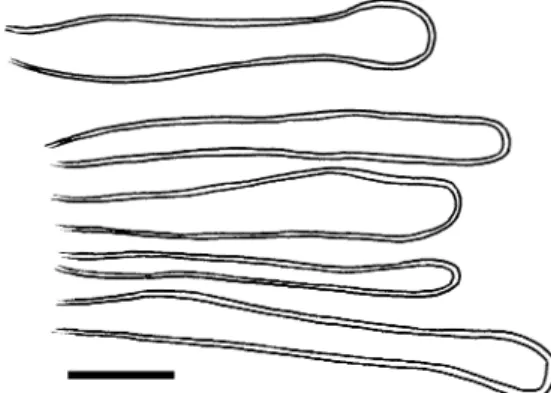
first ferrugineous later assuming a sooty or dirty brownish tinge; teliospores fusiform, narrowly ellipsoid or obconical, sometimes slightly bent, apex generally much thickened, narrowly conical and simple or rather variably digitate, ranging between 2–3 simple and stout branches and rather long slender, sometimes distorted or weakly branching appendages, 35–80×11–21 μm (55.9×15.5 μm for epitype), spore wall smooth, pallid yellowish to golden yellow, apex generally more deeply pigmented (greenish-)golden, germ pore proximal to apical thickening, spores germinating upon maturity.
Fig. 1 Endoraecium digitatum. a. Teliospores from Acacia notabilis (isoepitype). b, c. Teliospores from A. irrorata (BRIP 14165). In (b), teliospores with profusely digitate apices are shown, in (c), spores with more simple apical digitations. Bar=20μm
Additional material examined and used to complete the description of E. digitatum:
On phyllodinous Acacia spp. On A. notabilis. Australia, SA: Northern Lofty Region, South Hummocks, 34°02′ S / 138°08′ E, leg. JS Womersley no. 41, 24 Feb. 1963 (AD
966070527; 0, I). Head of Gulf of St. Vincent, Hummock Mt., ca. 20 km NNW of Port Wakefield, leg. HM Cooper, 11 Sept. 1963 (AD 96414049; [I, II], III). Eyre Peninsula, main highway between Kimba and Kyancutta, leg. F. Kutsche, 12 July 2001 (AD 123547; II, III). Eyre Peninsula, ca. 10 km E of Uno Station (“Mt. Helen”), 32°28′ S / 136°
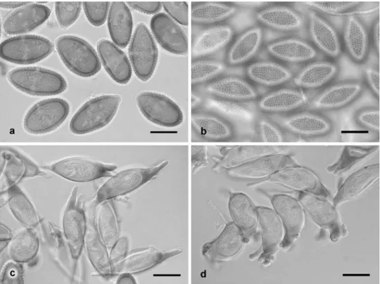
Fig. 2 Endoraecium digitatum from Acacia notabilis. a, b. Urediniospores, optical section and spore surface (AD 96045074). c, d. Aeciospores, optical section and spore surface (AD 966070527). Bars =20μm
Fig. 3 Endoraecium digitatum. a, b. Urediniospores from Acacia podalyriifolia (ZT Myc 3015) in optical section and with focus on spore surface. c, d. Aeciospores from A. irrorata (BRIP 14165) in optical section and with focus on spore surface. Bars=20μm
44′ E, leg. DJE Whibley no. 7979, 25 Sept. 1981 (AD 98150264; II). Eyre Peninsula, on roadside ca. 1 km S of Cummins (ca. 55 km NNW of Port Lincoln), leg. DJE Whibley no. 1931, 26 Aug. 1967 (AD 96741090; [0, I], II, [III]). Eyre Peninsula, Refuge Rocks area, track entrance to microwave tower, 33°11.5′ S / 136°49.5′ E, leg. JD Briggs no. 1053, 25 Aug. 1983 (AD 98527220; 0, I, III). Eyre Peninsula, County Buxton, Hundred Pinkawillinie, south corner of section 99–94, ca. 25 km W of Kimba, leg. KD Rohrlach no. 793, 27 Aug. 1960 (AD 96045074; II, III). Eyre Peninsula, Whyalla, Hummock Hill, 33°02′08″ S / 137°35′15″ E, leg. JD Sandham, D. Bosworth & D. New, 9 Dec. 1997 (AD 99845440; II). Roadside to Roseworthy, leg. FM Hilton, 3 Aug. 1954 (AD 98583644; II). Eyre Peninsula, Gawler Ranges, Paney Station (quadrate 3), woodland, 32°40′24″ S / 135°30′20″ E, collector unknown, 10 Oct. 1985 (AD 98715184; [I], II, III). Southern Lofty Ranges, Roseworthy Scrub, leg. JGO Tepper, 11 Sept. 1889 (MEL 1054135, sub Uromycladium notabile McAlp.; 0, I. Type of Uredo notabilis?). The following specimens of A. notabilis from AD bear E. digitatum as well but very scarcely or old: AD 123506 (0, I), AD 95921146 (I), AD 95708078 (II, III), AD 97803461 (II), AD 98583618 (II), AD 98548141 (0, I), AD 97940386 (II, III), AD 97135213 (II), AD 95928050 (I), AD 97806122 (I), AD 99616850 (I). On A. podalyriifolia A. Cunn. ex Don. Australia, QLD: Carnarvon Gorge, at main trail through gorge, leg. V. Faust-Berndt & R. Faust-Berndt no. Austr 9, 2 Aug. 2006 (ZT Myc 3015; II, III). Brisbane, leg. RF Langdon no. 32, 19 June 1930 (K(M) 146702, sub At. digitata; II, III). Redlands District, leg. K. Bodman, without date (BRIP 22895, sub R. digitatus; II, III). Brisbane, leg. RJ McAllister, 19 June 1930 (BRIP 6056 and 7546 ex BRIU 32, sub R. digitatus; II, III). Brisbane, leg. RFN Langdon, 23 June 1951 (BRIP 6057; probably II, 0 not seen). On A. falciformis DC. Australia, NSW: Brown Mountain 40 km W of Bega, leg. J. Walker, 11 Dec. 1975 (BRIP 14088 ex DAR 30202, sub R. digitatus; III).
On compound-leaved Acacia spp. On A. dealbata Link. Australia, VIC: Midlands, Murramurrangbong Ranges, leg. GH Robinson, 5 Jan. 1905 (MEL 1054763, sub U. notabile; I). Eastern Highlands, Cumberland Forest area, leg. GA Crichton, 20 Feb. 1966 (MEL 1054549, sub U. notabile; I). Midlands, Simpsons Place, Alexandra, leg. GA Crichton, 15 Nov. 1965 (MEL 2060932, sub U. notabile; 0, I). On A. deanei M.B. Welch, Coombs & McGlynn. Australia, QLD: Brisbane, Indooroopilly, DPI complex, leg. J. Barrett, 30 Aug. 1983 (BRIP 14106, sub R. digitatus; 0, I, III). Mt. Cotton, leg. L. Forsberg, 31 May 1990 (BRIP 17076, sub R. digitatus; I, II, III). On A. irrorata Sieber ex Spreng. Australia, QLD: Passchendaele, leg. J. Tierney, 7 March 1983 (BRIP 14165, sub R. digitatus; II [I?], III). The Summit, leg. KC Goulter, 29 Dec. 1983 (BRIP 14256, sub R. digitatus; 0, I, III. With telia of Uromycladium aff. maritimum McAlp.). On A. oshanesii F. Müll. & JH Maiden. Australia, QLD: Mt. Mee, leg. J. Tierney, 23 Nov. 1983 (BRIP 14170, sub R. digitatus; 0, I, III). On Acacia sp. (“Black Wattle”). Australia, VIC: Eastern Highlands, Mt. Margaret—Marysville Road, leg. GA Crichton, 11 April 1965 (MEL 2060933, sub U. notabile; 0, I).
Endoraecium digitatum differs from similar species by aecio- and urediniospores whose apex is not or hardly thickened and which have a delicate, wide-meshed, reticulate ornamentation with irregular narrow and rather high ridges and a conspicuously rugulose bottom (Figs. 2 and 3). The spore base is not or only inconspicuously socket-like. The teliospores have generally a thickened apex with one to several long, sometimes tortuous and ramifying processes (Fig. 1). As in other Endoraecium species, aecio-, uredinio- and teliospores are formed in conspicuous fascicles comprising slightly thick-walled and elongated basal cells bearing several pedicellate spores and many collapsed remnants of pedicels whose spores have already become detached. In the uredinia of ZT Myc 3015 and the aecia of AD 98527220, slenderly cylindrical and rather inconspicuous paraphyses were found (Fig. 4). Their walls were slightly thickened and pigmented yellowish.
Besides A. notabilis a number of other Acacia spp. bore rust infection assignable to E. digitatum. These are the phyllodinous A. podalyriifolia and A. falciformis and four species with bipinnate adult leaves, A. dealbata, A. deanei, A. irrorata, and A. oshanesii. The specimen on A. falciformis bore only telia and was assigned to the present species tentatively based on the strongly digitate teliospores. All investigated specimens on A. dealbata were only aecial (with or without spermogonia). Aeciospore morphology showed them to belong to E. digitatum. At present E. digitatum is the only known species distinguished in the E. digitatum complex occurring on both phyllodinous and compound-leaved members of Phyllodineae.
Fig. 4 Endoraecium digitatum. Paraphyses from urediniospore fascicles (ZT Myc 3015). Bar=20μm
The specimens from A. notabilis tended to have longer teliospores and slightly larger aecio- and urediniospores than collections from the other Acacia spp. It was observed in addition that teliospores from specimens on compound-leaved acacias generally had longer and more profusely branching apical processes (Fig. 1b). As the digitate teliospores of Endoraecium species are often variable no taxonomic consequence was drawn upon this difference.
E. digitatum was originally described with uredinio- and teliospores (Winter 1886). McAlpine (1906) stated that it comprised spermogonia, uredinia and telia, while Hodges and Gardner (1984) established a macrocyclic life cycle by inoculation experiments using specimens from Hawaii. In the present study, all but one specimens on A. notabilis bore uredinia and telia separately from spermogonia and aecia that were produced together on blister-like swellings or galls. In one specimen, uredinia were lacking and telia were produced together with the aecia on galls. Demicyclic specimens were also encountered on A. irrorata and A. oshanesii. In BRIP 14256 on A. irrorata, the sori occurred in variable asso-ciation, spermogonia with telia, spermogonia with aecia and telia, or telia unaccompanied by other sori. Because of the morphological similarity of the spores of the specimens, I consider all these variants to belong to E. digitatum. One can conclude that E. digitatum is a species with unfixed life cycle. Uredo notabilis, hitherto considered the uredinial state of Uromycladium notabile (McAlpine 1906), is assigned to the present species as anamorph name of the aecial state. I compared a specimen collected by Tepper near Roseworthy on 11 Sept. 1889 (MEL 1054135), most probably a part of the type of U. notabilis, with the aecial state of E. digitatum and could not find any differences. Ludwig (1890) stated that the type was collected on 11 Feb. 1889 at Roseworthy and presented a detailed description and excellent drawings which strongly suggest as well that U. notabilis represents the aecial state of E. digitatum. In addition, the similarity between U. notabilis and the uredinial state of E. digitatum is striking. Uredo gemmata described from Melbourne on Acacia sp. is listed as a synonym of U. notabilis by Sydow and Sydow (1915) but was not investigated in this study.
Is E. digitatum the same as Uromyces phyllodiorum and U. phyllodii?—McAlpine (1906) gave a detailed descrip-tion of U. phyllodiorum based on type material and of U. phyllodii which he considered synonymous. He reported the presence of spermogonia, uredinia (in fact Uredo-like aecia) and telia on “tubercles” of the phyllodes and described the aeciospores as 35–54 μm long. His micro-graphs show that the aeciospores are apically thickened. McAlpine’s observations were confirmed by a study of the syntypes of U. phyllodiorum (K(M) 146703, K(M) 146704) and an isotype of U. phyllodii (K(M) 146705). E. digitatum has shorter aecio- and urediniospores with more numerous germ pores and a different surface ornamentation. The
aeciospores are not or hardly thickened at the apex. In addition, most specimens of E. digitatum were found to be macrocyclic though spermogonia, aecia and telia associated together on swellings of the phyllodes were also encoun-tered occasionally. It is concluded that U. phyllodiorum and U. phyllodii are names for the same fungus, as already stated by McAlpine (1906), but that both are different from E. digitatum. The new combination Endoraecium phyllo-diorum, based on Uromyces phyllophyllo-diorum, is proposed for the separate species.
Endoraecium phyllodiorum (McAlp.) R. Berndt, comb. nov. (Figs. 5,6,7b,9)
≡ Melampsora phyllodiorum Berk. & Broome. Trans. Linnean Soc. London, 2. ser., 2:67 (1882). On Acacia sp. A telial name applied to Uredo-like aecia (cf. Berkeley and Broome1883, Plate 15, Figs.6,7and8). The telial stage is present, however, according to McAlpine (1906).
≡ Uromyces phyllodiorum McAlp. The rusts of Australia: p. 95 (1906) [as U. phyllodiorum (Berk. & Broome) McAlp.]. This is not a recombination but a new name for the holomorph based on teliospores encountered in the type specimen of M. phyllodiorum and described in McAlpine (1906). Basionym.
≡ Pileolaria phyllodiorum (McAlp.) Dietel. Ann. Mycol. 19:302 (1921) [as P. phyllodiorum (Berk. & Broome) Dietel]. A recombination of U. phyllodiorum McAlp., not of M. phyllodiorum Berk. & Broome.
= Uromyces phyllodii Cooke & Massee in Cooke. Grevillea 17:70 (1889) (as U. “phyllodiae”). On Acacia sp. A telial name applied to Uredo-like aecia (comp. protologue by Cooke (1889): “Maculis ... bullatis ... Teleutosporis ... episporio minute verruculoso.”). The telial stage is present, however.
Sori in combination 0/I, 0/I/III or 0/III on blister-like swellings of phyllodes which are up to 6×3 mm large, sometimes together with uredinia and/or telia on ± unaltered parts of phyllodes, or uredinia and/or telia on ± unaltered parts of phyllodes the only spore states present. Aecia small, Uredo-like, subepidermal, ferrugineous, pulverulent, associ-ated with spermogonia; aeciospores ellipsoid, subpyriform or more rarely subclavate, provided with a short socket-like base, (33)36–52(58)×17–26 μm (K(M) 146703: 44.3×20.2 μm, means ranging between 37.9–44.5×20.2–22.6 μm), rather coarsely reticulate, mesh width 1.5–2.6 μm, spore wall laterally about 2–3 μm thick, apically (5)6–9(10) μm, germ pores 4–5, more or less equatorial, without caps. Uredinia tiny, subepidermal, scattered or in loose groups on both sides of phyllodes, pulverulent, ferrugineous, no paraphyses observed; urediniospores more or less like the aeciospores, (31)35–48(52)×18–23(25) μm (means ranging between 39.0–43.3×19.9–23.4 μm), with 3–5, mostly four germ pores, apex not or slightly to conspicuously thickened (4–
9μm), with conspicuous smooth socket-like contraction of spore base. Gall-borne telia small, semi-immersed, compact, greyish, no paraphyses observed; non gall-borne telia tiny, scattered on both sides of phyllodes, pulvinate, yellowish brown to ferrugineous, semi-compact and with fibrous
texture, no paraphyses observed; teliospores (gall-borne) broadly ellipsoid to ellipsoid or cuneate, 36–52×(18)20– 27μm (means ranging between 43.4–48.3×20.4–22.8 μm), smooth and thin-walled, at apex slightly to moderately thickened (≤10 μm), greenish-yellow to golden-yellow, normally with 1–3 narrowly conical to cylindrical rather short outgrowths, more rarely more profusely digitate, pedicels subhyaline, stout and persistent, spores germinating upon maturity, non gall-borne teliospores ellipsoid, broadly ellipsoid or obconical, tapering towards the pedicel, apex rounded, conical or truncate, (33)35–50(52)×16–25.5 μm (means ranging between 40.5–54.6×18.1–21.2 μm), spore wall smooth, subhyaline, light yellowish or golden, ca. 1(2) μm thick, ca. (3)6–11 μm at apex, with 2–7, most often shortly cylindrical or knob-like or sometimes rather stout horn-like processes which are more rarely bifurcate or shifted to the subapical region, pedicels subhyaline to pale yellowish, delicate and thin-walled, up to 60μm long.
Material examined:
Specimens with sori (0/I/III) on bullate swellings or galls of phyllodes (only the presence of additional spore stages or of swellings with other combinations of stages are mentioned with the particular specimens). Australia, QLD: On Acacia sp. Brisbane, leg. FM Bailey no. 643 [ex herb. Massee?] (K(M) 146705, isotype of Uromyces phyllodii; 0, I, [III]). Locality not given, from Bailey’s herbarium, in Dept. of Agriculture Herbarium, VIC (PUR F 19689, type of Uromyces phyllodii; labelled 0, II, III. I saw only 0, I macroscopically on rather depleted specimen). Brisbane, leg. FM Bailey no. 269 [ex herb. Broome] (K(M) 146703 and 146704, syntypes of Melampsora phyllodiorum; 0, I). Paluma Range, hiking trails near Paluma village, at‘Witt’s lookout’, leg. V. Faust-Berndt & R. Berndt no. Austr 35, 9 Aug. 2006 (ZT Myc 3016; 0/I, 0/I/III or 0/III on galls, III not on galls). Cairns, Kuranda, Barron Gorge Natl. Park, ‘Mc Donald’s trail’, open Acacia woodland, leg. V. Faust-Berndt & R. Faust-Berndt no. Austr 44 and 46, 18 Aug. 2006 (ZT Myc 3020 and ZT Myc 3021; 0/I, 0/III or 0/I/III on galls, some III not on galls). On A. aulacocarpa Cunn. ex Benth. Mossman, Mt. Molloy, leg. I. Hood, 8 April 1995 (BRIP 23281, sub R. digitatus; 0/III, 0/I and 0/I/III). Bellenden Ker Natl. Park, Behana Gorge, leg. RG Shivas, 29 July 1992 (BRIP 20500, sub R. digitatus). Cape Tribulation, Donovan Range, leg. RG Shivas, 7 Aug. 1992 (BRIP 20514, sub R. digitatus; II not on galls). Mission Beach, leg. I. Hood, 5 April 1995 (BRIP 23284, sub R. digitatus; 0/I and 0/III on galls, III not on galls). On A. crassicarpa Cunn. ex Benth. Cape York Peninsula, Coen, Peninsula Road, leg. RG Shivas & M. Gunther, 16 July 1999 (BRIP 26562, sub R. digitatus). On Acacia cf. mangium Willd. Cairns, Kuranda, Barron Gorge Natl. Park, ‘Mc Donald’s
Fig. 5 Endoraecium phyllodiorum, teliospores. a. Spores from specimen bearing II and III on unaltered parts of phyllode and galls with spore states 0/I/III. Delineated spores are from telia not on galls (ZT Myc 3023). b. Teliospores from galls bearing spore states 0/I/III (BRIP 23281). c. Teliospores from microcyclic specimen with 0/III on galls (BRIP 39796). Bars=20μm
trail’, leg. V. Faust-Berndt & R. Berndt no. Austr 48, 18 Aug. 2006 (ZT Myc 3023; 0/I/III on galls, some II, III not on galls).
Specimens with sori not on bullate swellings or galls of phyllodes. Australia, QLD: On Acacia sp. 10 km S of
Townsville, leg. RG Shivas, 7 May 2005 (BRIP 44604, sub R. digitatus; II, III). Cairns, Mt. Whitfield conservation area, first (main) lookout, leg. V. Faust-Berndt & R. Berndt no. Austr 38, 24 Aug. 2006 (ZT Myc 3017; II). On A. aulacocarpa. Brisbane, leg. E. Jarvis, April 1911 (BRIP 6041 sub R. digitatus; II, III). Marcus Beach, leg. JL
Fig. 6 Endoraecium phyllodio-rum (ZT Myc 3023). a. Aecio-spores in optical section with thickened spore apex and equa-torial germ pores. Note presence of more or less smooth stalk-like spore base (arrows). b. Aecio-spores, focus on reticulate spore surface. c, d. Urediniospores in optical section and with focus on spore surface. Bars=20μm
Fig. 7 a, b. Endoraecium cf. phyllodiorum from Hawaii (K(M) 146700). Urediniospores in optical section and with focus on spore surface. Note the presence of a more or less smooth stalk-like spore base (a, arrow). c, d. Endoraecium kauaianum (holotype). Urediniospores in optical section and with focus on spore surface. Bars =20μm
Alcorn, 17 Aug. 1972 (BRIP 8769, sub R. digitatus; II, III). On A. holosericea. Eliott, leg. JH Simmonds, Aug. 1974 (BRIP 8934, sub R. digitatus; II). Gillies Range Natl. Park, leg. RG Shivas, 31 July 1992 (BRIP 20477, sub R. digitatus; II). Peach no. 1, Coen, leg. RG Shivas & M. Gunther, 18 July 1999 (BRIP 25819, sub R. digitatus; II). Coen Airport, leg. RG Shivas & M. Gunther, 16 July 1999 (BRIP 26550, sub R. digitatus; II). Australia, NT: Renner Springs, on A. holosericea, leg. RN Pitkethley no. 638, 25 Sept. 1975 (K(M) 146701, sub A. digitata; II). Australia, WA: Broome, Golf Course, on A. holosericea, leg. J. Ray no. JR 137, Oct. 2006 (BRIP 49108, sub R. digitatus; II, III).
Microcyclic specimens with sori (0/III) on swellings or galls of phyllodes. Australia, QLD: On Acacia sp. Thursday Island, leg. FM Bailey, without date (BRIP 6064, sub R. digitatus). On A. mangium. Glenbora, leg. MH Ivory no. P9289, 23 Oct. 1997 (BRIP 39796, sub R. digitatus). Nursery Ingham, leg. B. Brown, 28 June 1990 (BRIP 30420a, sub R. digitatus). Tully, Mission Beach Rd., collector unknown, 27 June 1990 (BRIP 30422, sub R. digitatus). Garner’s Beach, collector unknown, 27 June 1990 (BRIP 30423, sub R. digitatus). Wooloogin Creek near Japoonvale, collector unknown, 27 June 1990 (BRIP 30425, sub R. digitatus). Mena Creek Expt. 730, collector unknown, 27 June 1990 (BRIP 30426, sub R. digitatus). South Johnstone, collector unknown, 27 June 1990 (BRIP 30429, sub R. digitatus). Meunga, leg. MH Ivory, 23 Oct. 1997 (BRIP 39807, sub R. digitatus). Utchee Creek, leg. MH Ivory, 7 Dec. 1997, (BRIP 39804, sub R. digitatus). Ingham, Forestry Nursery, leg. M. Ivory, 3 July 2000 (BRIP 29567, sub R. digitatus). Mission Beach, leg. ex Forestry Dept., 10 Nov. 1983 (BRIP 14167, sub R. digitatus). Near Mission Beach, leg. I. Hood, 5 April 1995 (BRIP 23268, sub R. digitatus). Asherton, leg. M. Ivory, 3 March 1999 (BRIP 29732, sub R. digitatus). Innisfail, leg. A. Bragg, 7 Nov. 1988 (BRIP 31167, sub R. digitatus). Lannercost Expt. 734, leg. B. Brown, 28 June 1990 (BRIP 30418, sub R. digitatus). Lannercost, leg. M. Ivory, 9 Aug. 1999 (BRIP 29677, sub R. digitatus). Vanuatu, Prov. Tafea: Tanna, Middlebush, on Acacia sp., leg. JG Wright, 17 June 2003 (BRIP 44904, sub R. digitatus).
Fig. 8 Endoraecium kauaianum (holotype). a, b. Aeciospores in optical section and with focus on spore surface. c, d. Teliospores. Bars=20μm
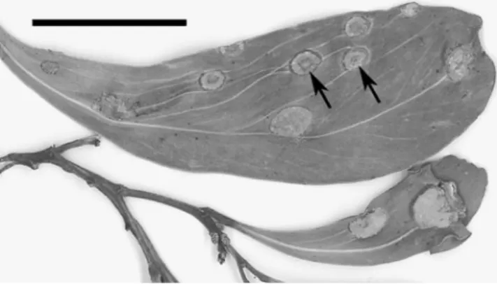
Fig. 9 Endoraecium phyllodiorum (ZT Myc 3023). Infected phyllodes of Acacia sp. with blister-like swellings bearing spermogonia and Uredo-like aecia. Bar=20 mm
Specimens preliminarily assigned to R. phyllodiorum Australia, QLD: Mossman, Mossman Gorge, on A. aulaco-carpa, leg. RG Shivas, 27 July 1993 (BRIP 21806, sub R. digitatus; 0, I, III on galls). Rifle Creek Bridge at Mt. Molloy, on A. auriculiformis Cunn. ex Benth., leg. I. Hood, 7 April 1995 (BRIP 23263, sub R. digitatus; 0, I on galls, III not on galls).
Endoraecium phyllodiorum revealed a remarkable plas-ticity with regard to the presence of spore states. In most specimens, sori were only produced on galls of which some bore the spore states 0/I, others 0/I/III, or 0/III. A considerable number of specimens produced exclusively galls with spermogonia and telia (microcyclic forms). In some specimens, galls occurred together with telia or with uredinia plus telia that were scattered over non-hypertrophied phyllodes or parts of the phyllodes. Sori on galls and scattered sori most likely belong to the same species as the aeciospores (from galls) and the urediniospores (from scattered sori) are closely similar (Fig. 6). In addition, both the galls and scattered uredinia and telia could be present on a single phyllode. It is concluded that E. phyllodiorum, like E. digitatum, has a variable life cycle with the potential to show itself macro-, demi- or microcyclic or even to produce different combinations of sori at the same time.
Teliospores were variable but similar regardless of their origin from gall-borne or scattered telia (Fig. 5). A considerable variability regarding spore length and thick-ness of the spore apex was also noted among uredinio-spores from different specimens.
Specimens of E. phyllodiorum were investigated from A. aulacocarpa A. Cunn. ex Benth., A. holosericea A. Cunn. ex G. Don., A. mangium Willd. and probably A. auriculi-formis Benth. (determination of rust uncertain). McAlpine (1906) reported it on A. dallachiana F. Müll., A. dealbata Link, A. microbotrya Benth., A. neriifolia A. Cunn., A. notabilis, A. penninervis Sieber, and A. pruinosa A. Cunn. Most probably his reports do not only represent E. phyllodiorum but similar species as well. Outside of Australia the rust occurs in Vanuatu, and photographs presented in Old et al. (2000) indicate its presence in Kalimantan. Huguenin (1966) reported it from New Caledonia on A. spirorbis Labill.
What is‘Endoraecium digitatum’ from Hawaii? The native phyllodinous acacias of Hawaii are A. kauaiensis Hbd., A. koa A. Gray with vars. koa, latifolia (Benth.) St. John and waianaeensis St. John and A. koaia Hbd. (St. John 1979). Pedley (1975) accepted only A. kauaiensis and A. koa, the latter comprising the varieties described at that time and A. koaia. Hodges and Gardner (1984) and Gardner (1994) reported the presence of E. digitatum on A. koa vars. koa and latifolia. Gardner (1991) supposed that the rust had been introduced to Hawaii, while in Gardner (1994) it is
regarded as indigenous. If, as hypothesized by Gardner (1994), the Hawaiian endemic E. angustiphyllodium is a derivative of E. digitatum, the latter assumption would be more probable.
A study of Hawaiian specimens determined as E. digitatum showed that aecio- and urediniospores were clearly distinct from E. digitatum s. str. but similar to E. phyllodiorum. A closer examination revealed that two different species were involved, one preliminarily left with E. phyllodiorum, the other one new for science.
Endoraecium kauaianum R. Berndt, sp. nov. (Figs.7c, d and 8).
Etymology: After Kauai, island of the Hawaiian Archi-pelago, location of the type.
Spermogonia sparsim adsunt inter aecia. Aecia generi Uredo similia, cinnamomea ad ferruginea, pulverulenta, inflorescentias et fructua (scoparum hospiti?) omnino denseque tegentia; aeciosporae urediniosporis generaliter similes sed formae plus variabilis, ellipsoideae ad late ellipsoideae, subfusiformes, apice rotundato vel conico, contractionem basalem pedicelliformem generatim carentes vel cum contractione brevi rugoso-subreticulato, 28–44× 19–23(25.5) μm (37.4×21.3 μm), pariete aureo ca. 3– 4.5 μm crasso in parte aequatoriali, apicaliter non vel paulum incrassato usque ad 3.5–6 μm, reticulato, poris germinationis 7–10 aequatorialibus sine papillis. Uredinia in phyllodia vix mutata sparsa, teliis consociata, cinnamo-mea, pulverulenta; urediniosporae ellipsoideae ad late ellipsoideae, subfusiformes, apice rotundato vel conico, contractionem basalem pedicelliformem generatim carentes vel cum contractione brevi rugoso-subreticulato, (31)35–44 (46)×(17)19–22.5 μm (38.1×20.4 μm), pariete pallide aureo vel aureo, ca. 3–4.5 μm crasso in parte aequatoriali, paulum tenuiore basim versus, apicaliter non vel paulum incrassato usque ad 4–5(7) μm, reticulato maculis ca. 1.5– 2.6 μm (2.0 μm) diam. et ca. 1 μm altis, poris germi-nationis 6–8 aequatorialibus sine papillis. Telia minuta, singularia, urediniis consociata in phyllodia vix mutata; teliosporae formae et processuum apicalium variabilissimae, plerumque ellipsoideae ad subfusiformes, rarius fusiformes vel late ellipsoideae, 29–48(50)×14.5–19 μm (38.9× 16.6 μm), pariete 1–1.5 μm crasso, subhyalino ad pallide aureo, apice valde incrassato, pallide aureo vel viridi-flavo, ornato processu singulo ad paucis brevibus vel longis, rectis vel incurvatis, plerumque integris; per maturitatem ex poro germinationis subapicali germinantes.
In phyllodiis, inflorescentiis fructibusque Acaciae koae (vel A. kauaiensis?).
Spermogonia scarcely present among aecia. Aecia Uredo-like, cinnamon to ferrugineous, pulverulent, densely covering what may have been an inflorescence with young pods (on witches’ brooms?); aeciospores essentially like urediniospores though more variable in shape, ellipsoid to
broadly ellipsoid, subfusiform, apex rounded to conical, generally without basal stalk-like contraction or with a short and rugose to subreticulate one, 28–44×19–23(25.5) μm (37.4×21.3 μm), spore wall golden brown, ca. 3–4.5 μm thick around equator, at apex not or slightly thickened to 3.5– 6μm, reticulate, germ pores 7–10, equatorial, without caps. Uredinia scattered on unaltered(?) phyllodes and associated with telia, cinnamon, pulverulent; urediniospores ellipsoid to broadly ellipsoid, subfusiform, apex rounded to conical, generally without basal stalk-like contraction or with a short and rugose to subreticulate one, (31)35–44(46)×(17)19– 22.5μm (38.1×20.4 μm), spore wall pale golden or golden brown, about 3–4.5 μm thick around equator, slightly thinner proximal, apex not or slightly thickened to 4–5(7) μm, reticulate, meshes ca. 1.5–2.6 μm (2.0 μm) diam., with delicate up to 1μm high ridges, germ pores 6–8, equatorial, without caps. Telia very small, singly and associated with uredinia on unaltered(?) phyllodes; teliospores very variable with regard to shape and apical processes, most often ellipsoid to subfusiform, more rarely fusiform or broadly ellipsoid, 29– 48(50)×14.5–19 μm (38.9×16.6 μm), spore wall 1–1.5 μm thick, subhyaline to pale golden brown, apex strongly thickened, pale golden with greenish tinge, bearing one to several, short to long, straight to bent, most often entire horn-like processes, germinating at maturity from a germ pore located proximal to the apical thickening.
Holotype (PUR 89860): USA, Hawaiian Islands: Kauai, Puu Ki-Waialae trail, on A. koa A. Gray, leg. A. Kyono, Feb. 1991 (sub At. digitata, det. DE Gardner; I, II, III).
Additional material examined:
E. kauaianum. USA, Hawaiian Islands, Kauai: Collection site not indicated, on A. koa var. koa, leg. CS Hodges, May 1982 (PUR 66863, sub A. digitata, det. DE Gardner; 0, I). Collection site not indicated, on A. koa, leg. Swezey, without date (PUR F2890, sub At. digitata, det. DE Gardner, det. as Uromyces koae by JC Arthur; probably I, spermogonia not observed).
E. cf. phyllodiorum. USA, Hawaiian Islands, Oahu: Pacific Palisades, on A. koa var. koa, leg. CS Hodges, 9 Sept. 1982 (K(M) 146700, sub At. digitata; II). Waimea Falls Park, on A. koa var. koa, leg. CS Hodges, 23 Oct. 1981 (PUR 66873, sub At. digitata, det. DE Gardner; II). Wahiawa, Ahrens Ditch trail, on A. koa cf. var. koa, leg. FL Stevens no. 291, 8 June 1921 (PUR F2887, sub At. digitata; II). Hawaiian Islands, Kauai: Makaha Ridge, Acacia host not specified, from infection experiment with aeciospores, collector not indicated, 29 July 1982 (PUR 66861, sub At. digitata; II).
Endoraecium koae+E. angustiphyllodium USA, Hawaiian Islands, Hawaii: Hawaii Volcanoes Natl. Park,
on A. koa var. latifolia, leg. DE Gardner, Dec. 1986 (BISH 510514, sub At. digitata; III, III).
Endoraecium kauaianum differs from E. phyllodiorum mainly in aecio- and urediniospore characters. Spores of both states lack a smooth stalk-like base or have only a short, inconspicuous one that is subreticulate or rugose (Figs. 7c, d and 8a, b). The aeciospores have 7–10, the urediniospores 6–8 germ pores vs. 4–5 and 3–5 in E. phyllodiorum. E. digitatum differs in aecio- and uredinio-spores not thickened at apex and a differently reticulate spore surface.
PUR 66863 and PUR F2890 were only aecial (0, I and I, respectively) and had slightly larger aeciospores than the type of E. kauaianum. In PUR F2890, assigned to E. digitatum by Gardner and reported to have uredinia and telia (Gardner1994), aeciospores measured (33)37–50×20– 25 μm (42.5×22.6 μm), in PUR 66863, 35–45(51)×19– 25 μm (40.9×21.7 μm). I did not see in any investigated specimen the slender paraphyses observed by Gardner (1994) in the aecia and uredinia of ‘E. digitatum’. Hosts of E. kauaianum were given as A. koa or A. koa var. koa, but a comparison of the phyllodes present in the type specimen with illustrations provided by St. John (1979) suggests that the type host may be A. kauaiensis. Up to know, E. kauaianum is only known from Kauai Island.
Specimens K(M) 146700, PUR 66873 and PUR F2887, all from Oahu and most probably on A. koa var. koa, and PUR 66861 from Kauai on an undetermined Acacia showed only uredinia and were different from E. kauaianum. The urediniospores of these specimens (Fig. 7a, b) had a prominent, smooth stalk-like spore base. In this they resembled urediniospores of Australian E. phyllodiorum from which they slightly differed, however, by the presence of 5–7, most often six germ pores. Urediniospores and aeciospores of Australian E. phyllodiorum have generally 3–5, most often four pores. As there is overlap in this character the Hawaiian specimens are preliminarily assigned to E. phyllodiorum.
Hodges and Gardner (1984) described that spermogonia and Uredo-like aecia of‘E. digitatum’ were mostly formed on“compact witches’ brooms” on A. koa var. latifolia, less commonly on “large, discrete, bullate swellings”. In contrast, no witches’ brooms were observed on A. koa var. koa where spermogonia and aecia were associated with a variety of other symptoms. It is difficult to interpret these observations given the possibility that E. kauaianum and E. cf. phyllodiorum were involved or even E. koae. The connection of uredinia and telia of ‘E. digitatum’ with spermogonia and aecia formed on witches’ brooms and other malformations of the hosts was proven by Hodges and Gardner (1984) with inoculation experiments. The authors reported that the aeciospores showed minor morphological differences (shape of the apex) between the
islands of Kauai and those of Hawaii and Maui but that inoculation experiments carried out with both forms led to “typical uredinia and telia”.
BISH 510514 on A. koa var. latifolia is listed in Gardner (1994) under ‘E. digitatum’ (spore stages 0, I). Its reinvestigation revealed spermogonia, associated Uredo-like aecia and very few telia. The fungus is different from E. phyllodiorum and E. kauaianum in aeciospores with 7– 8 germ pores, a not thickened spore apex and the lack of a socket-like spore base. The aeciospores measured 31–39× 18–23.5 μm (34.9×21.4 μm). In these characters they closely resemble aeciospores of E. koae. It is concluded that BISH 510514 bears both the spermogonial/aecial state of E. koae and teliospores that belong to another rust species, probably E. angustiphyllodium.
SEM micrographs of aecio- and urediniospores of E. digitatum-like rusts from Hawaii can be found in Gardner and Hodges (1985). A key to the rusts on endemic acacias in Hawaii was provided by Hodges and Gardner (1984). The following key is partly based on the latter.
Spore wall not reticulate, either with apical and lateral tubercles or laterally smooth and with one to several apical processes. Teliospores.
Spores apically and laterally with numerous short, blunt tubercles, generally associated with spermogonia and Uredo-like aecia . . . E. koae Spores laterally smooth, apically with one to several ± long processes (‘digitate’)
Telia densely covering witches’ brooms, associated with spermogonia . . . E. angustiphyllodium Telia scattered on ‘normal’ phyllodes, not on witches’ brooms, not associated with spermogonia but often with uredinia . . . . . . . E. cf. phyllodiorum / E. kauaianum1 Spore wall reticulate. Aecio- or urediniospores, or Uredo-like teliospores
Spores with conspicuous, smooth, stalk-like basal con-traction, apically thickened to 4–8 μm, on average longer than 40μm (urediniospores) . . . E. cf. phyllodiorum Spores without smooth basal contraction; if stalk-like process present than short and usually subreticulate or rugose
Spores (aeciospores) not thickened apically, on average shorter than 40 μm, associated with spermogonia, often with teliospores . . . E. koae
Spores slightly to strongly thickened apically, associated with spermogonia or not
Sori covering witches’ brooms, spermogonia often present
Spore apex thickened to 6–15 μm (Uredo-like teliospores) . . . E. acaciae Spore apex thickened to 4–7 μm (Uredo-like teliospores or aeciospores) . . . . . . . E. hawaiiense / E. kauaianum2 Sori not on witches’ brooms, not with spermogonia, spore apex thickened to 4–7 μm (urediniospores) . . . . . E. kauaianum A number of Australian specimens determined as E. digitatum tallied with the latter in essential morphological characters but were found to be sufficiently distinct to be described as separate species. For them the new species E. walkerianum, E. parvum and E. violae-faustae are proposed. Endoraecium walkerianum R. Berndt, sp. nov. (Figs.10, 11and12).
Etymology: Dedicated to John Walker, excellent mycolo-gist and authority on rust fungi.
Spermogonia aeciis consociata in tumoribus phyllodiorum et fructuum usque ad 1 cm diam. Aecia generi Uredo similia primum spermogonia annuliforme circumdantia deinde tumores plus minusve omnino tegentia subepidermalia pulverulenta ochracea vel sordide luteo-aurantia; aecio-sporae ellipsoideae rariter late ellipsoideae nonnunquam contractione basali breviter stipitiformi delicate sulcato-rugosa praeditae, 35–48(50)×19–25 μm (42.5×21.6 μm), pariete aureo ad aureo-brunneo magis saturate pigmentato apicem versus, inaequaliter crasso in parte aequatoriali, partibus crassioribus ca. 3–4.5 μm, partibus tenuioribus ca. 1.5–2 μm crasso, in apice (3)4–7(8) μm incrassato, reticulato maculis ca. 1.5–2.2 μm diam. margine ca. 1 μm alto limitatis, poris germinationis 5–7 praecipue 6 plus minusve aequatorialibus sine papillis. Uredinia et uredinio-sporae ignota. Telia in phyllodiis sparsa minuta rotunda pulvinata aurea semicompacta et fibrosa; teliosporae fusi-formes ellipsoideae vel cuneatae hilum versus attenuatae, 44–60×15–21 μm (pro 20 sporis 51.2×17.4 μm), pariete levi ca. 1.5 μm crasso in apice 7–16 μm subhyalino ad aureo in apice, processus apicales absunt vel 1–4 conici.
In phyllodiis leguminibusque Acaciae penninervis Sieber ex DC. et A. obliquinerviae Tindale probabiliter.
Spermogonia together with aecia on large blister-like hypertrophies (up to 1 cm diam.) scattered on the phyllodes
2Intermediate forms occur with regard to the thickness of the apex
that are difficult to assign to either E. acaciae or E. hawaiiense. Aeciospores of E. kauaianum unaccompanied by uredinio- or teliospores are hardly distinguishable from aeciospore-like teliospores of E. hawaiiense.
1
For a safe distinction of both species aecio- or urediniospores are necessary. Teliospores not reported hitherto from Hawaiian E. cf. phyllodiorum.
and pods. Aecia Uredo-like, first surrounding the spermo-gonia like a ring, later more or less covering the entire hypertrophies, subepidermal, pulverulent, ochraceous or dull yellow-orange; aeciospores ellipsoid, rarely broadly ellipsoid, sometimes with a short, rugose or grooved stalk-like basal process, 35–48(50)×19–25 μm (42.5×21.6 μm), spore wall golden-yellow to golden-brown, more deeply pigmented towards spore apex, unevenly thickened around equator, thinner parts ca. 1.5–2 μm, thicker parts ca. 3– 4.5 μm thick, (3)4–7(8) μm at the apex, reticulate with meshes ca. 1.5–2.2 μm wide and ca. 1 μm high, germ pores 5–7, most often six, more or less equatorial, without caps. Uredinia and urediniospores unknown. Telia scattered on phyllodes, tiny, round, pulvinate, golden-yellow, semi-compact and with fibrous texture; teliospores fusiform, ellipsoid or wedge-shaped, tapering towards base, 44–60× 15–21 μm (mean for 20 measured spores 51.2×17.4 μm), spore wall smooth, ca. 1.5 μm thick, apically 7–16 μm thick, subhyaline to golden-yellow at thickened apex, apex without or with mostly 1–4 stout, conical appendages.
On phyllodes and pods of A. penninervis and A. cf. obliquinervia.
Holotype (SF35354, Reliq. Petrak. 1333, sub U. phyllo-diorum): Australia, NSW: Cavan Gap, near Yass, on A. penninervis, leg. E. Gauba, 20 Nov. 1951. Isotypes distributed as Reliq. Petrak. 1333 (sub U. phyllodiorum; I [spermogonia not seen], III).
Additional material examined:
Australia, ACT: Mt. Corree near Canberra, alt. 4600 ft., on A. penninervis, leg. E. Gauba, 22 Dec. 1954 (SF35353, Reliq. Petrak. 1332, sub U. phyllodiorum; I [spermogonia not seen]). Australia, NSW/ACT: Brindabella Range, on A. penninervis, leg. E. Gauba, April 1959 (PUR F16457, sub At. digitata; 0, I). Australia, VIC: Ca. 35 km ENE of Melbourne, Mt. Donna Buang, alt. ca. 1220 m, on A. cf. obliquinervia, leg. C. & K. Vánky, 2 March 1996 (HeRB 5962; 0, I). Australia, NSW: Bald Rock, on A. penninervis, leg. J. Tierney, 26 Feb. 1984 (BRIP 14205, sub R. digitatus; 0, I). Endoraecium walkerianum is very similar to E. digitatum which is basically different in the wider and higher meshes of the aeciospores provided with a more conspicuously rugulose bottom, a not or hardly thickened apex of the aeciospores and – generally – the presence of a uredinial state. Both species are probably closely related. E. walkerianum differs from E. phyllodiorum in several characters. The aeciospore wall is asymmetrically thickened in the equator region, the aeciospores lack a conspicuous smooth socket-like basal contraction and have a less thickened apex (Fig.12). The aecial acervuli (Fig. 10) are large, conspicuous and have a saturated ochre colour. The teliospores of E. walkerianum appear to be larger than in E. phyllodiorum. Whether this is a constant character is uncertain given the generally high variability of the teliospores in E. phyllodiorum and the small number of teliospores measured in the present species. PUR F16457 was slightly different from the type in aeciospores with generally 7–8 germ pores and a finer meshed reticulum (mesh diam. ca. 1.2–1.8 μm). As far as known, E. walkerianum lacks a uredinial state and has a demicyclic life cycle. The Hawaiian endemic E. koae is demicyclic, too (Gardner1994). It differs from the present species by smaller aeciospores with equally thickened walls and by different teliospores. The known host plants of E. walkerianum, A. penninervis and A. cf. obliquinervis, are morphologically rather similar and possibly closely related.
Endoraecium parvum R. Berndt, sp. nov. (Figs.13and14). Etymology: Denominating the comparatively small aecio- and urediniospores.
Spermogonia aeciis vel teliis consociata, amphigena, in maculis obscuris phyllodiorum non vel lenissime hyper-trophicis vel in pedicellis. Aecia parva generi Uredo similia
Fig. 11 Endoraecium walkerianum (holotype). Teliospores. Bar= 20μm
Fig. 10 Endoraecium walkerianum (HeRB 5962). Infected phyllodes of Acacia cf. obliquinervia with blister-like hypertrophies bearing spermogonia and Uredo-like aecia. The sori are first ring-like (arrows) but cover the entire hypertrophies later. Bar=20 mm
crebre aggregata subepidermalia ferruginea; aeciosporae ellipsoideae vel late ellipsoideae apicaliter rotundatae, plerumque contractione basali breviter stipitiformi levi, 27– 38(42)×16–22 μm (33.0×18.5 μm), pariete pallide aurantio-brunneo, (2.5)3–4 μm crasso circa aequatorem, non vel leniter incrassato in apice, reticulato maculis ca. 1.2–2.3 μm diam. margine delicato limitatis, poris germinationis 3–5 praecipue 4 plus minusve aequatorialibus ad subaequatorialibus sine papillis. Uredinia sparsa vel laxe aggregata amphigena in maculis obscuris vel senescentibus phyllodiorum; uredinio-sporae ellipsoideae vel late ellipsoideae late obovoideae magis rariter oblongae vel subclavatae, apicaliter rotundatae ad late conicae, plerumque contractione basali breviter stipitiformi subhyalina levi, 27–38(44)×16–22.5 (31.9×19.3 μm), pariete pallide aurantio-brunneo, ca. 3 μm crasso circa aequatorem, non vel leniter incrassato (ad 4–5 μm) in apice, reticulato maculis ca. 1.2–2.1 μm diam., poris germinationis 3–5 praecipue 4 plus minusve aequatorialibus sine papillis. Telia in phyllodiis spermogoniis consociata et aggregata vel sparsa et secum viventia nonnunquam ex aeciis nascentia, minuta pulvinata semi-compacta fibrosa, ferruginea albescentes post germinationem basidiorum; teliosporae formae varabili ellipsoideae vel late ellipsoideae magis rariter subfusifomes subrhomboideae vel subtriangulares, 32–52(56)×15–22 μm (42.0×18.7 μm), pariete levi ca. 1–2 μm crasso subhyalino ad pallide aureo, apice magis saturate pigmentato non-nunquam flavo-virescenti, 5–15 μm crasso formae variabili simpliciconico vel digitato processibus apicalibus 1–paucis conicis verruciformibusve, per maturitatem ex poro germi-nationis subapicali germinantes.
Fig. 13 Endoraecium parvum, teliospores. a. BRIP 7543 (holotype). b. BRIP 23071a. Bars=20μm
Fig. 12 Endoraecium walkeria-num. a, b. Aeciospores in optical section (holotype) and with focus on spore surface (SF35353). c. Aeciospores showing apical thickening and unevenly thickened lateral wall of the spores (arrows) depending on their orientation (SF35353). d. Teliospores among aecio-spores (holotype). Bars =20μm
In phyllodiis Acaciae cunninghamii Hook. et A. specierum Spermogonia associated with aecia or telia, amphigenous on dark, not or hardly swollen spots of phyllodes or pedicels. Aecia small, Uredo-like, in dense groups, subepi-dermal, ferrugineous; aeciospores ellipsoid to broadly ellipsoid, apically rounded, generally with a smooth, contracted stalk-like base, 27–38(42)×16–22 μm (33.0× 18.5μm), spore wall pale orange-brown, (2.5)3–4 μm thick in equator region, not or hardly thickened at the apex, reticulate, meshes 1.2–2.3 μm wide, confined by delicate, smooth ridges, germ pores 3–5, often 4, equatorial to sub-equatorial, without caps. Uredinia single or in small groups, amphigenous on dark or senescent spots of phyllodes, subepidermal, pulverulent, ferrugineous; urediniospores ellipsoid to broadly ellipsoid, broadly obovoid, more rarely oblong or subclavate, apically rounded or sometimes broadly conical, generally with a subhyaline, smooth, contracted stalk-like base, 27–38(44)×16–22.5 (31.9× 19.3μm), spore wall pale orange-brown, ca. 3 μm thick in equator region, not or hardly thickened at the apex (to 4– 5 μm), reticulate, meshes 1.2–2.1 μm wide, confined by smooth ridges, germ pores 3–5, often 4, more or less equatorial, without caps. Telia tiny, pulvinate, semi-compact and with fibrous texture, orange-brown or with whitish bloom after germination of basidia, either in groups and associated with spermogonia, some probably deriving from old aecia, or scattered and not closely associated with other sori; teliospores variable in shape, ellipsoid to broadly ellipsoid, more rarely subfusiform, subrhomboid or sub-triangular, 32–52(56)×15–22 μm (42.0×18.7 μm), spore
wall smooth, subhyaline to pale golden, sometimes with greenish tinge, at the apex slightly more deeply pigmented, ca. 1–2 μm thick, at the apex thickened to 5–15 μm, apex variable in shape, simple and conical or with one to few knob-like outgrowths or digitate with one to few, generally short and simple processes, germination upon maturity from a germ pore located proximal to the thickened spore apex.
On phyllodes of A. cunninghamii and Acacia spp. Holotype (BRIP 7543): Australia, QLD: Caloundra, on A. cunninghamii (= A. leiocalyx (Domin) Pedley var. leioca-lyx?), leg. ST Blake, 25 Aug. 1932 (sub R. digitatus; 0, I, III). The same collection is represented in BRIP 6045 (isotype).
Additional material examined:
Australia, QLD: Brisbane, Mt. Coot-tha, on Acacia sp. (narrow-‘leaved’), leg. RE Harrison, 5 May 1955 (BRIP 6063 and 7548, sub R. digitatus; 0, I, III). Cairns, Mt. Whitfield, on Acacia sp. (possibly A. mangium), leg. V. Faust-Berndt & R. Berndt no. Austr 41, 20 Aug. 2006 (ZT Myc 3019; II). On A. concurrens Pedley. Brisbane, Indooroopilly, Meiers Road, leg. DE Shaw, 30 Sept. 1996 (BRIP 23071a, sub R. digitatus; II, III). On A. mangium. Near Mission Beach, leg. I. Hood, 5 April 1995 (BRIP 23266, sub R. digitatus; II). Australia, NT: On A. mangium. Sylvatech Pine Forest, leg. PM Stephens, 16 June 2003 (BRIP 42379, sub R. digitatus; 0, I, III).
The short aecio- and urediniospores (Fig.14) distinguish E. parvum from other species of the E. digitatum complex. In addition, the teliospores are relatively short and their
Fig. 14 Endoraecium parvum. a, b. Aeciospores in optical section, together with some teliospores, and with focus on surface reticulum (BRIP 6063). c, d. Urediniospores in optical section and with focus on sur-face reticulum (BRIP 23071a). Bars=20μm
apices quite often not or only slightly digitate (Fig.13). The life cycle appears to be variable. In BRIP 7543/6045 and BRIP 6063/7548, spermogonia were associated with aecia or with telia or with aecia and telia, of which some possibly developed from old aecia. The sori occurred on darkly decoloured but otherwise more or less unaltered phyllode spots, not on galls or blister-like swellings. In BRIP 23071a and BRIP 23266, no spermogonia and aecia were present but telia and/or uredinia on non-hypertrophied parts of phyllodes as well. These specimens could be linked to the forms with aecia by the virtually identical teliospores and the urediniospores which closely resembled the aecio-spores. The known hosts, A. concurrens, A. cunninghamii and A. mangium belong to Phyllodineae section Juliflorae. Endoraecium violae-faustae R. Berndt, sp. nov. (Figs.15 and16).
Etymology: Dedicated to Viola Faust-Berndt, indefatigable collector and companion on field stays, discoverer of the rust fungus.
Spermogonia aeciis consociata in maculis tumidis phyllodiorum usque ad 8×6 mm largis obscure olivaceis. Aecia generi Uredo similia, parva pulverulenta ferruginea; aeciosporae plus minusve ellipsoideae ad subfusiformes,
saepe contractione basali pedicelliformi brevi levique praeditae, apice conico vel subapiculato, 45–60×18– 25 μm (52.1×21.6 μm), pariete aurantiaco-brunneo foveolato-reticulato maculis ca. 1.1–1.8 μm diam. basim et apicem versus angustioribus, ca. 3 μm crasso in parte aequatoriali, incrassato usque ad (5)6–9(10) μm in apice, poris germinationis 4–5 plus minusve aequatorialibus sine papillis. Uredinia minuta subepidermalia, in phyllodiis utrinque sparsa vel laxe aggregata pulverulenta ferruginea, saepe paraphysibus flavescentibus vel stramineis, ut in teliis, circumdata; urediniosporae generaliter aeciosporis similes, ellipsoideae ad subfusiformes, plerumque contrac-tione basali pedicelliformi brevi levique praeditae, apice conico vel subapiculato, 40–57(64)×20–25.5 μm (48.5× 22.5μm pro typo; magnitudo variationis 44.6–52.1×22.4– 27.1μm), pariete aurantiaco-brunneo interdum inconspicue laminato foveolato-reticulato maculis ca. 1.1–1.8(2.0) μm diam. basim et apicem versus angustioribus et tenuioribus, ca. 3–3.5(4) μm crasso in parte aequatoriali incrassato usque ad 5–12 μm in apice, poris germinationis 3–5, plerumque 4 plus minusve aequatorialibus sine papillis. Telia subepider-malia, in phyllodiis inter uredinios sparsa, minuta vix confluentia flavo-brunnea ad aurantiaco-brunnea deinde obscure ferruginea semicompacta et textura fibrosa; telio-sporae ellipsoideae ad late ellipsoideae, interdum cuneatae ad obconicae, 32–66(76)×16–25 μm (57.4×19.5 μm), pariete dilute citrino ad aureo levi ca. 1–1.5 μm crasso, in apice incrassato 5–10(12) μm, processu singulo ad paucis conicis cylindricis vel gongylodibus integris praeditae, interdum cum processibus singulis inferioribus, per maturitatem ex poro germinationis subapicali germinantes; pedicellis hyalinis pariete leniter incrassato persistentibus; paraphysibus numero-sis interpositis, filiformibus ad leniter clavatis, 80–136(150)× 8–16 μm, levibus, parte apicali incrassato ca. 10–35 μm longo processibus uno ad paucis brevibus variabilissimis saepe cylindricis vel conicis praeditis.
In phyllodiis Acaciae spp.
Spermogonia together with aecia on blister-like, up to 8× 6 mm large, dark or brownish green hypertrophied swellings of phyllodes. Aecia Uredo-like, small, subepidermal, pul-verulent, ferrugineous; aeciospores ellipsoid to subfusiform, often with a short smooth stalk-like base, apically conical to subapiculate, 45–60×18–25 μm (52.1×21.6 μm), spore wall dull orange-brown, reticulate-foveolate with ca. 1.1–1.8 μm wide meshes which become finer and shallower towards the spore apex and base, laterally ca. 3 μm thick, apically thickened to (5)6–9(10) μm, germ pores 4–5, more or less equatorial, without caps. Uredinia tiny, subepidermal, scat-tered over entire surface of phyllodes or in loose groups, pulverulent, ferrugineous, often bordered by a fringe of yellowish or straw-coloured paraphyses which are similar to telial paraphyses; urediniospores essentially like the aecio-spores, ellipsoid to fusiform, often with a subhyaline, short,
Fig. 15 Endoraecium violae-faustae (ZT Myc 3018). a. Teliospores. ‘pTS’ marks a teliospore resembling a paraphysis. b. Paraphyses from telia.‘TS’ indicates a single teliospore. Bars=20 μm
smooth stalk-like base, apically conical to subapiculate, 40– 57(64)×20–25.5 μm (48.5×22.5 μm for type; range of means 44.6–52.1×22.4–27.1 μm), spore wall dull orange-brown, sometimes indistinctly layered, reticulate-foveolate with ca. 1.1–1.8(2.0) μm wide meshes which become finer and shallower towards the spore apex and base, laterally about 3–3.5(4) μm thick, apically thickened to 5–12 μm, germ pores 3–5, mostly 4, more or less equatorial, without caps. Telia subepidermal, scattered on phyllodes among the uredinia, tiny and hardly confluent, yellowish-brown to orange-brown, later dull orange-brown, semi-compact with fibrous texture; teliospores ellipsoid to broadly ellipsoid, sometimes wedge-shaped to obconical, 32–66(76)×16– 25 μm (57.4×19.5 μm), spore wall faint lemon-yellow to golden-yellow, smooth, ca. 1–1.5 μm thick, apically thick-ened to 5–10(12) μm, at apex with one to several conical, cylindrical or knob-like entire projections, more rarely with single projections more proximal, germinating at maturity from below of the apical thickening, pedicels hyaline, slightly thick-walled and persistent though collapsing; paraphyses numerous, interspersed among teliospores, fili-form or slenderly clavate, most often partially compressed, 80–136(150)×8–16 μm, smooth, with a 10–35 μm long apical thickening which bears one to few very irregular, short, often cylindrical or conical outgrowths.
On phyllodes of Acacia spp.
Holotype (BRIP 53388). Australia, QLD: Cairns, Kuranda, Barron Gorge Natl. Park,‘Mc Donald’s trail’, on Acacia sp., leg. V. Faust-Berndt & R. Berndt no. Austr 47, 18 Aug. 2006 (ZT Myc 3022, isotype; 0, I, II, III).
Additional material examined:
Australia, QLD: On Acacia sp. Cairns, Mt. Whitfield conservation area, first (main) lookout, leg. V. Faust-Berndt & R. Berndt no. Austr 39, 24 Aug. 2006 (ZT Myc 3018; II, III). Cape York, leg. GS Pegg, Nov. 2001 (BRIP 28985 and 28987, sub R. digitatus; II). On A. aulacocarpa. Rockingham Bay, without collector and date (SF35351, sub U. phyllodio-rum, ex herb. Sydow; II, III). Cape Tribulation, Emmagen Creek, leg. RG Shivas, 4 Aug. 1993 (BRIP 21805, sub R. digitatus; II). On A. crassicarpa A. Cunn. ex Benth. Cape York Peninsula, 9 km N of Bamaga, leg. JL Alcorn, 30 May 1981 (BRIP 13711, sub R. digitatus; II, III). Injinoo, leg. RI Davis, 15 Jan. 1998 (BRIP 26599, sub R. digitatus; II). On A. flavescens A. Cunn. ex Benth. Peach no. 7, Coen, leg. RG Shivas & M. Gunther, 18 July 1999 (BRIP 25816, sub R. digitatus; II). Australia, NT: Darwin, Fog Dam, on Acacia sp., leg. R. Berndt & V. Faust-Berndt, 23 Aug. 2003 (BRIP 44497; II, III).
Endoraecium violae-faustae differs from E. phyllodio-rum by larger aecio- and urediniospores which are foveolate rather than reticulate, which have a more pronounced apical thickening and are apically more acute (Fig. 16). The most obvious difference is the presence of characteristic paraphyses in the uredinia and telia (Fig. 15b). Their abundance is variable. McAlpine (1906) described uredinial and telial paraphyses for E. phyllodio-rum. One may assume that his observation was based on specimens of E. violae-faustae as I could not find paraphyses in E. phyllodiorum. His description suggests a
Fig. 16 Endoraecium violae-faustae. a, b. Aeciospores in optical section and with focus on spore surface (holotype). c, d. Urediniospores in optical section and with focus on spore surface (ZT Myc 3018). Bars=20μm
different morphology of the paraphyses, however, as they are said to be“variously shaped, but generally somewhat cylindrical, thickened and rounded at the apex”. The teliospores of E. violae-faustae (Fig. 15a) were very variable especially with regard to spore size. In the type collection, they measured 32–50×18–24 μm (40.5× 21.4 μm), while they were much longer, (47)50–66(76)× 16–23(25.5) μm (57.4×19.5 μm), in ZT Myc 3018. As all other characters tallied well I have no doubt that this specimen belongs to E. violae-faustae as well. BRIP 44497 was preliminarily assigned to the present species. Its urediniospores were considerably smaller than in the other specimens (mean 39.0×20.8 μm) and the meshes of the spore reticulum wider. The typical uredinial paraphyses were present, however.
E. violae-faustae is known up to now on A. aulacocarpa, A. crassicarpa (both Phyllodineae section Juliflorae), and A. flavescens (Phyllodineae section Plurinerves).
The following key covers the known Australian species of the E. digitatum complex and is based on aecial and/or uredinial characters. Telial characters were included only in as far as they aid identification.
Aecio- and urediniospores with foveolate spore surface, large (generally longer than 44μm on average), often with subacute apex. Uredinia and telia with ± cylindrical or club-shaped, apically thick-walled and often shortly digitate paraphyses (Figs.15and16) . . . E. violae-faustae Aecio- and/or urediniospores with reticulate surface, gener-ally shorter than 44μm on average and apically rounded or conical, sori without paraphyses, if paraphyses are present with other characters than in E. violae-faustae
Aecio- and urediniospores generally with a smooth, stalk- or socket-like basal contraction
Majority of aecio- and urediniospores between 35 and 48 μm long, apically slightly to conspicuously (5– 10μm) thickened (Figs.6and7a, b) . . . . . . . E. phyllodiorum Aecio- and urediniospores mostly between 27 and 38 μm long, apex not or only slightly thickened (Fig.14) . . . E. parvum Aecio- and urediniospores without a stalk-like spore base or with a short, subreticulate to rugose, not a smooth one
Aecio- and urediniospores apically not or hardly thickened, reticulate with wide meshes (to 3 μm) confined by delicate, somewhat uneven and high ridges, bottom of meshes conspicuously rugulose (Figs. 2 and 3); teliospores (Fig. 1) often profusely digitate with long, tortuous and branching processes . . . . . E. digitatum
Uredinial state unknown. Aeciospores with thick-ened apex, more narrowly meshed reticulum and coarser, even ridges between meshes (Fig.12) . . . . . . . . E. walkerianum
Conclusions and general discussion
Endoraecium digitatum, a species complex– For more than a century E. digitatum was regarded as a single species with a wide distribution in Australasia and a disjunct area in Hawaii. Only recently Walker (2001) pointed out to the high variability of this rust fungus and suggested that it may comprise several taxa. In the present paper, four new species and the known E. phyllodiorum are distinguished from E. digitatum based on characters observable with light microscope. Another species apparently related to this complex is the Hawaiian E. angustiphyllodium.
E. digitatum, as understood hitherto, has been reported on numerous Acacia species and from an extensive area of distribution. It could be shown in the present study that reports of E. digitatum from the Hawaiian Islands do not belong to this species whose proven records are restricted to Australia (SA, VIC, NSW, QLD). The Hawaiian records of ‘E. digitatum’ were demonstrated to represent two species, the new E. kauaianum and E. cf. phyllodiorum. The latter would be the only species shared between Australia and Hawaii. Only uredinia were present on specimens from Hawaii and their identity with Australian representatives of E. phyllodiorum is therefore only indicated by the overall similarity of the urediniospores. Yet, slight differences noticed between the urediniospores allow for the possibility that two species are involved. Another clue for this hypothesis would be that, according to Gardner (1994), ‘E. digitatum’ has never been observed in Hawaii on any of the introduced Australian acacias. A more definite decision will only be possible after the remaining spore states of the supposed E. phyllodiorum have been discovered in Hawaii and by DNA sequence comparisons.
The host range of E. digitatum s. str. was shown to be narrower than assumed but is still rather broad comprising presently three phyllodinous and four compound-leaved Acacia spp. from subgen. Phyllodineae. Specimens from compound-leaved acacias revealed minor morphological differences to specimens from phyllodinous acacias but did not seem to represent a distinct species. It should be remarked in this connection that the morphological difference between phyllodinous and compound-leaved adult foliage needs not reflect a wide phylogenetic distance of the respective acacias. This was demonstrated by Murphy et al. (2010) who found out that some phyllodinous and pinnately-leaved acacias, among them the hosts A. falciformis and A. irrorata