BRAIN
A JOURNAL OF NEUROLOGYLETTER TO THE EDITOR
Microcystic macular degeneration from optic neuropathy
Mathias Abegg, Martin Zinkernagel and Sebastian Wolf
Universita¨tsklinik fu¨r Augenheilkunde, Universitaet Bern, Inselspital, CH-3010 Bern, Switzerland Correspondence to: Mathias Abegg MD, PhD,
Universita¨tsklinik fu¨r Augenheilkunde, Universita¨t Bern, Inselspital,
CH-3010 Bern, Switzerland E-mail: [email protected]
Sir, We read with great interest the article by Gelfand et al. (2012) in the June issue of Brain. The authors described in detail vacuolar macular changes in the inner nuclear layer of patients with mul-tiple sclerosis. This ‘microcystic macular oedema’ was associated with decreased visual acuity and a higher disability score. We found similar microcysts in the macular inner nuclear layer of a 13-year-old male suffering from neurofibromatosis type 1 and chronic compressive optic neuropathy in both eyes due to optic glioma (Fig. 1). Since the incidental finding of the glioma 15 months ago, the bilateral microcystic macular inclusions re-mained unchanged and vision was stable at one line below normal in both eyes. Our observation raises the possibility that microcystic macular changes may be caused by optic nerve damage rather than by multiple sclerosis-specific processes. Not surprisingly Gelfand et al. (2012) found significantly reduced ret-inal nerve fibre layer thickness in their patients, supporting the view that optic neuropathy is a prerequisite for this phenomenon. The authors speculated that the macular cysts originate from breakdown of the blood–retinal barrier or from focal inflammation. The fact that our patient had chronic compressive optic neur-opathy suggest that retrograde trans-synaptic degeneration (Syc et al., 2012) leads to cystic degeneration of the inner nuclear layer. In fact the phenotype of cystic cavitations in the inner nuclear layer has been described in enucleated human eyes with lesions of the optic nerve (Gills and Wadsworth, 1967) and in optic nerve crush experiments in non-human primates (VanBuren, 1963). These reports showed histological evidence that retrograde trans-synaptic degeneration from optic neuropathy results in degeneration of the inner nuclear layer with formation of
cystic spaces. On this basis the term microcystic macular oedema might be misleading and the term microcystic degeneration more appropriate. However, it remains to be clarified why only a frac-tion of patients with optic neuropathy display this phenotype while others do not.
If microcystic macular degeneration is a direct consequence of optic neuropathy, the suggestion of Gelfand et al. (2012) that microcystic macular changes might be of use as a marker in mul-tiple sclerosis, should be reconsidered. The direct measurement of the causal reduction of retinal nerve fibre layer might represent a better option for this purpose. Nevertheless, this anatomically dis-tinct and well defined macular alteration in some patients with optic neuropathy is intriguing and may lead to new insights into the pathophysiology of optic neuropathies.
References
Gelfand JM, Nolan R, Schwartz DM, Graves J, Green AJ. Microcystic macular oedema in multiple sclerosis is associated with disease sever-ity. Brain 2012 Jun; 135 (Pt 6): 1786–93.
Syc SB, Saidha S, Newsome SD, Ratchford JN, Levy M, Ford E, et al. Optical coherence tomography segmentation reveals ganglion cell layer pathology after optic neuritis. Brain 2012 Feb; 135 (Pt 2): 521–33.
Gills JP, Wadsworth JAC. Retrograde Transsynaptic Degeneration of the Inner Nuclear Layer of the Retina. Invest Ophthalmol Vis Sci 1967 Aug; 6 (4): 437–48.
VanBuren JM. Trans-Synaptic Retrograde Degeneration in the Visual System of Primates. J Neurol Neurosurg Psychiatry 1963 Oct; 26: 402–9.
doi:10.1093/brain/aws215 Brain 2012: 135; 1–2
|
e225Advance Access publication September 7, 2012
ßThe Author (2012). Published by Oxford University Press on behalf of the Guarantors of Brain. All rights reserved. For Permissions, please email: [email protected]
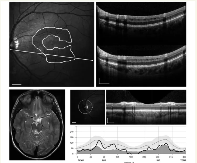
Figure 1 Bilateral microcystic macular degeneration in a 13-year-old male with neurofibromatosis type 1 and optic atrophy from a chiasmal glioma. Only the left eye is shown, the right eye showed similar changes. Asterisks show chiasmal and dorsal midbrain glioma in an axial MRI section. Top left panel: A fundus photograph with the extent of microcystic degeneration outlined in white; the straight line indicates localization of optical coherence tomography line scans. Top right: Macular line scans show microcysts exclusively in the inner nuclear layer in the macular region. This pattern remained stable during 15 months (upper panel at beginning, lower panel at end of observation period) and was restricted to the macular region. Bottom right: Peripapillary line shows significant thinning of retinal nerve fibre layer thickness, indicating severe optic nerve atrophy. Horizontal scale bars correspond to 1 mm, vertical bars to 200 mm.