HAL Id: inserm-02933692
https://www.hal.inserm.fr/inserm-02933692
Submitted on 8 Sep 2020HAL is a multi-disciplinary open access archive for the deposit and dissemination of sci-entific research documents, whether they are pub-lished or not. The documents may come from teaching and research institutions in France or abroad, or from public or private research centers.
L’archive ouverte pluridisciplinaire HAL, est destinée au dépôt et à la diffusion de documents scientifiques de niveau recherche, publiés ou non, émanant des établissements d’enseignement et de recherche français ou étrangers, des laboratoires publics ou privés.
On phagocytes and macular degeneration
Xavier Guillonneau, Chiara Eandi, Michel Paques, José-Alain Sahel,
Przemyslaw Sapieha, Florian Sennlaub
To cite this version:
Xavier Guillonneau, Chiara Eandi, Michel Paques, José-Alain Sahel, Przemyslaw Sapieha, et al.. On phagocytes and macular degeneration. Progress in Retinal and Eye Research, Elsevier, 2017, 61, pp.98-128. �10.1016/j.preteyeres.2017.06.002�. �inserm-02933692�
On Phagocytes and Macular Degeneration
Xavier Guillonneau1, Chiara M Eandi2, Michel Paques1,3, José-Alain Sahel1,3, Przemyslaw Sapieha4, Florian Sennlaub1
1Institut de la Vision, 17 rue Moreau, Sorbonne Universités, UPMC Univ Paris 06, INSERM, CNRS, 75012 Paris, France.
2 University of Torino, Department of Surgical Science, Eye Clinic, Torino, Italy
3Centre Hospitalier National d'Ophtalmologie des Quinze-Vingts, INSERM-DHOS CIC 503, Paris, France.
4Department of Ophthalmology, Maisonneuve-Rosemont Hospital Research Centre, University of Montreal, Quebec, Canada.
†Correspondence should be addressed to: Dr Florian Sennlaub, Inserm, UMR_S 968, Institut de la Vision, Paris, F-75012, France. Tel: (33) 1 53 46 26 93,
Abstract:
Age related macular degeneration (AMD) is a complex multifactorial disease caused by the interplay of age and genetic and environmental risk factors. A common feature observed in early and both forms of late AMD is the breakdown of the physiologically immunosuppressive subretinal environment and the protracted accumulation of mononuclear phagocytes (MP). We here discuss the origin and nature of subretinal MPs, the mechanisms that lead to their accumulation, the inflammatory mediators they produce as well as the consequences of their chronic presence on photoreceptors, retinal pigment epithelium and choroid. Recent advances highlight how both genetic and environmental risk factors directly promote subretinal inflammation and tip the balance from a beneficial inflammation that helps control debris accumulation to detrimental chronic inflammation and destructive late AMD. Finally, we discuss how changes in life style or pharmacological intervention can help to break the vicious cycle of inflammation and degeneration, restore the immunosuppressive properties of the subretinal space, and reestablish homeostasis.
ON PHAGOCYTES AND MACULAR DEGENERATION 1. Age related macular degeneration 4 2. Mononuclear phagocytes 10 2.1. Mononuclear phagocytes in AMD 11 2.2. Mononuclear phagocytes and inflammation 15 2.2.1. Inflammation, a crucial process for survival 15 2.2.2. Nonresolving inflammation 17 3. The immuno-suppressive environment of the retina 17 3.1. Immune privilege and immune suppression 17 3.2. Tonic inhibitory signals in the retina 19 3.3. The immunosuppressive retinal pigment epithelium 22 4. The origin of infiltrating mononuclear phagocytes 25 5. Function and consequences of chronic subretinal mononuclear phagocyte accumulation 31 5.1. A “homeostatic” role 31 5.2. Collateral damage 34 5.2.1. M1 and M2 polarization 34 5.2.2. Phagocyte-derived cytokines and collateral damage 35 5.2.2.1. Interleukin-1β 35 5.2.2.2.2. Tumor Necrosis Factor α (TNFα) 38 5.2.2.3. Interleukin 6 (IL-6) 39 5.2.2.4. Chemokine (C-C motif) ligand 2 40 6. AMD-risk factors and subretinal mononuclear phagocytes 41 6.1. Genetic risk factors 41 6.2. Aging and environmental risk factors 57 7. Patho-mechanistic conclusions 63 8. Therapeutic considerations 65 Life style adaptations 65 “Anti-inflammatory” pharmaceutical therapies 66 Acknowledgments: 68 References 69
Abbreviations:
ABCA1 ATP-binding cassette transporter A1 AD Alzheimer’s disease
aHUS atypical hemolytic uremic syndrome AMD Age related macular degeneration APOA-1 Apolipoprotein A-1 APOE apolipoprotein E
ARM age-related maculopathy ATP adenosine triphosphate AZ atrophic zone
Blam basal laminar deposits BlinD basal linear deposits BM Bruch’s membrane
CCL2 chemokine (C-C motif) ligand 2 CCR2 C-C chemokine receptor type 2
CD11b/CD18 Complement 3 Receptor, Mac-1 CEPT cholesteryl transfer protein
CFH complement factor H CNS central nervous system CNV choroidal neovascularization CS cone segments
CX3CL1 chemokine (C-X3-C motif) ligand 1 CX3CR1 CX3C chemokine receptor 1
DAMP damage-associated molecular pattern DC dendritic cells
FASL Fas ligand
GA geographic atrophy GAG glycoaminoglycans HDL high-density lipoprotein
IBA1 ionized calcium-binding adapter molecule 1 IIRC innate immunity receptor cluster
IL-1β Interleukin 1 β
iMf inflammatory macrophages KPSG keratan sulfate proteoglycans LAP latency-associated peptide
LCAT lecithin:cholesterol acyltransferase LDL low-density lipoprotein
LPS lipopolysaccharide
LTBP latent TGF-β binding protein MAC membrane attack complex MC microglial cells
Mo monocytes
MP mononuclear phagocytes
MPGN II Membranoproliferative glomerulonephritis, type II, dense-deposit disease NFκB nuclear factor κB
NLR Nod-like receptors
NLRP3 NOD-like receptor family, pyrin domain containing 3 OTX2 orthodenticle homeobox 2
P2RX7 P2X purinoceptor 7
PAMP pathogen-associated molecular pattern RAP retinal angiomatous proliferations RCT reverse cholesterol transport RDH5 retinol dehydrogenase 5 rMf tissue resident macrophages RP retinitis pigmentosa
RPE retinal pigment epithelium SCR short Consensus Repeat domains SIRP1α Signal regulatory protein α
TGF-β tumor growth factor β TLR toll like receptor
TNF-α Tumor necrosis factor α
TRE2/3/4 targeted replacement mice expressing the human APOE-isoforms TSP-1 Thrombospondin 1
TTR transthyretin TZ transitional zone
1. Age related macular degeneration
The macula consists of a small cone-dominated fovea, responsible for high acuity vision, surrounded by a rod-dominated parafovea and peripheral retina. The degeneration of the macula and central retina is a common medical problem called age-related macular degeneration (AMD), and a leading cause of visual impairment in the world (Wong et al., 2014). Early AMD, also called age-related maculopathy (ARM) (Bird et al., 1995), is characterized by pigmentary abnormalities and accumulation of membranous, lipoproteinaceous debris located between the basal lamina of the retinal pigment epithelium (RPE) and the inner collagenous layer of Bruch’s membrane (BM) of the central retina. They can either take the form of basal linear deposits (BlinD) or clinically visible sizeable (>125 μm), ill-defined protrusions called large (soft) drusen (Curcio and Millican, 1999; Sarks, 1976). The protrusions deform the over-lying retina leading to metamorphopsia, and are associated with a local thinning of the photoreceptor cell layer (Schuman et al., 2009) and local loss of sensitivity (Midena et al., 2007). Eyes with large-sized soft Drusen can progress and develop late AMD (~ 15% in the Beaver Dam study over 10 years; ~30% in the Blue mountain study over 6 years), regress (~ 25%, Beaver Dam study) or stay stable for years (Klein et al., 2004; Wang et al., 2003). It is not clear whether the constituents of drusen are derived from the RPE, choroidal vasculature or both. Additionally, reticular pseudodrusen are strongly associated with AMD (Klein et al., 2008b; Mimoun et al., 1990). They are ~30-150 μm-sized pale fundus lesions that are believed to be caused by subretinal drusenoid deposits located between the RPE and photoreceptors (Alten and Eter, 2014; Rudolf et al., 2008; Zweifel et al., 2009). Small, globular, well-defined, hard drusen (<63 μm) are not associated with AMD when few are present, but large areas of macular hard drusen are a risk factor for ARM (Klein et al., 2004).
from late AMD (Wong et al., 2014). The reasons that patients progress to late AMD are incompletely understood, but the interactions of certain protective and predisposing genetic variants, alongside with environmental factors, seem to play a role (Yu et al., 2012). There are two clinical forms of late AMD: exudative AMD and geographic atrophy (GA) (Sarks, 1976): In exudative AMD, subretinal neovascularization develops below (occult) or above (classic) the RPE. They originate for the most part from the choroid (choroidal neovascularization; CNV), but in about 10-15% of exudative AMD, subretinal neovascularization arises intra-retinally from the retinal vasculature (retinal angiomatous proliferations; RAP) (Ghazi, 2002; Yannuzzi et al., 2001). Interestingly, eyes with CNV also present a loss of choriocapillaries in the surrounding areas (McLeod et al., 2009). CNV is commonly associated with retinal edema, subretinal exudation as well as blood and lipid deposits. Untreated, exudative AMD leads to the formation of a neovascular fibrous membrane and the formation of a disciforme subretinal scar (Sarks, 1976). In recent years, treatments for neovascular AMD have dramatically improved with the development of anti-angiogenic therapy (anti-vascular endothelial growth factor (VEGF)). The anti-angiogenic therapy decreases the permeability and inhibits the formation of neovessels and the associated degenerative changes. It does however not halt vessel-independent degenerative processes and the decline in visual functions in 30% of patients that occurs in the long term (Rofagha et al., 2013).
In GA, an extending atrophic zone (AZ) forms, characterized by the loss of the RPE and degeneration of the photoreceptor cell layer (Sarks, 1976). The initial lesion in GA often develops parafoveally (Sarks et al., 1988) and slowly expands through the central retina and eventually the fovea. In the AZ, despite the absence of RPE, residual cones (and to a lesser extent rods) survive, but they lack their inner and outer cone segments (CS) necessary for light perception (Bird et al., 2014; Eandi et al., 2016). Similar findings are observed in disciform subretinal scars, the end-stage of exudative AMD (Curcio, 2001). In a perilesional
transitional zone (TZ), directly peripheral to the area of RPE-loss of the scar and AZ, the number of rods drops dramatically compared to regions more distant from the lesion, despite the presence of the RPE (Bird et al., 2014; Curcio, 2001; Eandi et al., 2016). These anatomical changes translate clinically to decreased perilesional retinal sensitivity (Meleth et al., 2011). The number of cones changes little in the TZ, but they lack their cone segments (CS) (Bird et al., 2014; Curcio, 2001; Eandi et al., 2016). One could assume that CS loss in the TZ of AMD patients is due to a primary RPE dysfunction, however CS loss is also observed in patients with retinitis pigmentosa (RP) with rod-gene mutations and unremarkable RPE (Mitamura et al., 2013). Clinically GA lesions provoke central scotomas, which severely affect visual acuity when the lesion involves the fovea itself. Patients with late AMD also suffer from impaired dark adaptation, which can be measured as an increase in recovery time after bleach, (Flamendorf et al., 2015; Owsley et al., 2001). During dark adaptation, the bleached retinal (all-trans retinal) of the rod outer segments is transported into the RPE where it is re-isomerized into 11-cis-retinal by the isomerohydrolase RPE65 and retinol dehydrogenase 5 (RDH5). Together, this complex process is called the visual cycle. Any slowing of the visual cycle, will increase both dark adaptation and recovery times after bleach, as observed in RDH5-deficient patients (Cideciyan et al., 2000). Interestingly, a significant increase in recovery time after bleach is already observed with reticular pseudodrusen in ARM (Flamendorf et al., 2015; Owsley et al., 2001) and is associated with an increased incidence of AMD (Owsley et al., 2016) and this quite some time before visible RPE or photoreceptor lesions occur. There is currently no therapy that has been approved for atrophic AMD.
AMD is a common, complex disease that results from interplay of aging, and genetic and environmental risk factors. The importance of genetic factors is illustrated by the fact that the AMD-risk increases between 5- and 10-fold when a parent or sibling is affected
(Chakravarthy et al., 2010; Shahid et al., 2012), making AMD one of the most heritable complex diseases. Polymorphisms in the complement factor H (CFH), the Apolipoprotein E isoforms and on chromosome 10q26 (that harbors the gene for the high-temperature requirement A serine peptidase 1 [HTRA1] and age-related maculopathy susceptibility 2 [ARMS2]) are the major genetic factors that predispose to AMD (Swaroop et al., 2007). Age, smoking, obesity, and possibly exposure to light are the main environmental factors, but other factors such as hypertension might also play a role (Adams et al., 2011; Chakravarthy et al., 2010; Schick et al., 2016). How the coaction of these mainly non-ocular risk factors fuels the pathomechanisms of AMD remains unclear but will be discussed in this article.
Current hypotheses on the pathomechanisms of AMD can be grouped into three main axis: (1) choriodal vascular insufficiency results in hypoxia, CNV and dysfunction and death of RPE and photoreceptors; (2) dysfunction of RPE (induced by oxidative stress or complement activation) impairs photoreceptor renewal and leads to progressive accumulation of deposits at the basal membrane of the RPE, visible as drusen, that ultimately cause hypoxia and CNV, or progressive loss of RPE and photoreceptor cells; (3) chronic non-resolving inflammation (mainly the accumulation of MPs and over-activation of the innate immune system) leads to neovascularization and degeneration of RPE and photoreceptor cells. None of theses hypothesis is exclusive and they likely all play into the disease development to different degrees in individual patients. This review will concentrate on the possible implication of phagocytes and their mediators in the pathogenesis of AMD.
2. Mononuclear phagocytes
Mononuclear phagocytes (MPs) comprise a family of cells that include monocyte (Mo), monocyte-derived inflammatory macrophages (iMf), dendritic cells (DC), and tissue resident macrophages (rMf) such as microglial cells (MCs) (Chow et al., 2011; Gautier et al., 2012; Ransohoff and Cardona, 2010; Wynn et al., 2013). rMfs, which serve trophic as well as
sentinel roles, are present in virtually all organs and develop early during embryogenesis or pre-natal life from either embryonic Mfs found in the yolk-sac or from embryonic monocyte found in the liver, respectively (Gautier and Yvan-Charvet, 2014). In the retina, microglia are involved in the clearance of apoptotic neurons and pruning of synapsis in retinal development, and constitutively required for the maintenance of synaptic structure in the adult retina and for synaptic transmission underlying normal visual function (Wang et al., 2016). The vast majority of long-lived rMφs are not replaced by Mos in the adult, but rather self-renew by proliferation. Few exceptions might remain as for a sub-population of rMφs in the gut (Gautier and Yvan-Charvet, 2014). During inflammation, short-lived circulating Mos that strongly express the chemokine receptor CCR2 are recruited to the inflammatory site where they differentiate into iMfs (Geissmann et al., 2003). Thus, under inflammatory conditions, Mfs from different origins cohabite but little is known about their specificity of function and action. At the end of the inflammatory response, iMfs disappear from the site, contrary to rMφs that are dedicated to remain in tissues after resolution and return to homeostasis.
In summary, distinct types of resident and infiltrating MPs, exert distinct functions during tissue development, inflammation and homeostasis. Insight on individual roles of each type of MP has been complicated given the limitation of currently available tools and the immunohistochemical similarities of these cell populations in diseased tissues, as they constitutively express or induce similar markers (Gautier et al., 2012; Ransohoff and Cardona, 2010).
2.1. Mononuclear phagocytes in AMD
Physiologically, the posterior segment of the eye contains several types of MPs. Numerous rMfs reside in the choroid, a network of MCs is located in the inner layers of the retina, perivascular rMfs are found along the major retinal and choroidal vessels, while DCs are rare in the retina (Forrester et al., 2010; Kumar et al., 2014; Streilein, 2003). The adult
central photoreceptor cell layer and subretinal space (located between the RPE and the photoreceptor outer segments) are physiologically devoid of resident and infiltrating MPs in healthy subjects (Combadiere et al., 2007; Eandi et al., 2016; Gupta et al., 2003; Lad et al., 2015; Levy et al., 2015a; Sennlaub et al., 2013). Subretinal MPs, can however be found in the extreme periphery and during fetal development of the photoreceptor cell layer (McMenamin and Loeffler, 1990).
Histological evidence of the association of subretinal leucocytes and exudative AMD has first been described in 1916 (Hegner, 1916) and in numerous publications since (Penfold et al., 2001). A number of studies have observed inflammatory cells, particularly MPs, but also lymphocytes in excised neovascular membranes from patients with AMD (Gehrs et al., 1992; Lopez et al., 1991; Oh et al., 1999; Seregard et al., 1994 ).
In GA, MPs have been observed within the atrophic area (Combadiere et al., 2007; Eandi et al., 2016; Gupta et al., 2003; Lad et al., 2015; Levy et al., 2015a; Penfold et al., 2001), on RPE cells of the TZ and on large drusen (Eandi et al., 2016; Gupta et al., 2003; Lad et al., 2015; Levy et al., 2015a; Sennlaub et al., 2013), within large drusen and BlinD (Lad et al., 2015; Sennlaub et al., 2013), and their numbers in the choroid increase compared to controls (McLeod et al., 2016). The MPs were identified using the MP markers Ricinus communis agglutinin-I (Gupta et al., 2003), CX3CR1 (Combadiere et al., 2007), CD18 (Combadiere et al., 2007; Levy et al., 2015a; Sennlaub et al., 2013), IBA1 (Sennlaub et al., 2013), CCR2 (Sennlaub et al., 2013), CD163 (Lad et al., 2015), and CD14 (Eandi et al., 2016). Interestingly, the classically used marker CD68 only reveals negligible numbers of MPs in sections of patients with AMD, which has been suggested to be the reason why changes in the MP population went unnoticed in some earlier studies (Lad et al., 2015). IBA-1-staining (to visualize subretinal MPs) of central RPE flatmounts, reveal that IBA-1+MPs are seldom present in healthy age-matched central donor RPE (Eandi et al., 2016; Levy et al., 2015a) and
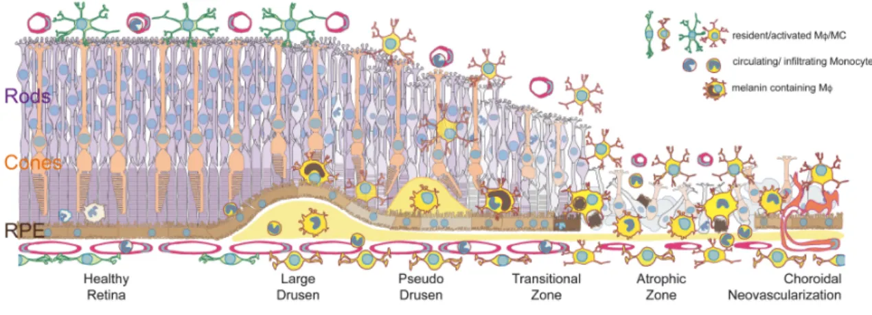
in donors with small drusen that are not associated with AMD (Fig. 1 A and B). However, they are invariably observed on large drusen, within the AZ of patients with GA, and on the apical side of the RPE in the TZ adjacent to GA lesions (Levy et al., 2015a) (Fig. 1 C and D).
Interestingly, a subset of MPs might be clinically visible in AMD patients. A significant number of MPs (identified using immunohistological markers) in AMD donor eyes contain melanosomes, presumably from ingested RPE debris (Lad et al., 2015; Sennlaub et al., 2013). We have recently shown, using serial in vivo flood-illumination adaptive optics imaging (FIAO), that highly mobile melanin-containing cells (MCCs), of a similar size to the observed MPs, precede and accompany the emergence and progression of atrophy in GA (Gocho et al., 2013). Examination by Optical Coherence Tomography (OCT), identifies similarly sized hyper-reflective dots, proposed to be MCCs, that are associated with photoreceptor loss in ARM (Schuman et al., 2009), predictive progression from ARM to GA (Christenbury et al., 2013; Leuschen et al., 2013) and associated with AMD-genetic risk variants (Altay et al., 2016). It is thus tempting to speculate that MCCs are melanin-containing MPs, possibly additionally to migrating RPE cells (Zanzottera et al., 2015). Surprisingly this view was recently contested (Curcio and Ach, 2016), even though evidence for MCCs being MPs is supported by immunohistochemical data using three MP markers (CD163, IBA-1 and CCR2), while the existence of migrating RPE cells in AMD is solely based on the histological observation of melanin containing cells without the use of an additional RPE cell marker.
Contrary to fast evolving autoimmune lesions, characterized by cytotoxic T lymphocytes, neutrophils and MPs (Caspi, 2002; Kerr et al., 2008), infiltrating leukocytes in slowly-evolving GA are predominantly MPs which is similar to other protracted age-related diseases including atherosclerosis, neurodegenerative diseases and cancer (Grivennikov et al., 2010; Hotamisligil, 2010), However, the choroid of patients with AMD and surgically removed neovascular membranes are infiltrated by lymphocytes, (Ezzat et al., 2008; Lopez et
al., 1991; Penfold et al., 1984; Penfold et al., 1985), including IL17+T lymphocytes (Camelo et al., 2016). Patients with GA are also characterized by an increased number of degranulated (activated) choroidal mast-cells (Bhutto et al., 2016). While it has been suggested that the absolute number of infiltrating MPs during progression of AMD is too low to play a meaningful pathogenic role in either CNV or retinal degeneration, a growing number of animal studies have demonstrated that a comparable infiltrate in mice plays an important role in both, CNV and photoreceptor degeneration (see 4. and 5.2).
Additionally, increased intra-ocular concentrations of CCL2, a potent chemokine that attracts Mo to diseased tissue, are observed in humans with neovascular- (Fauser et al., 2015; Jonas et al., 2010; Kramer et al., 2012; Rezar-Dreindl et al., 2016) and atrophic-AMD (Newman et al., 2012; Sennlaub et al., 2013). Moreover, from a systemic perspective, patients whose Mos express the greatest amount of TNFα, have a higher prevalence of choroidal neovascularization (Cousins et al., 2004), and circulating blood Mo of patients with AMD express higher levels of CCL2, IL8, and VEGF (Lechner et al., 2017). The percentage of IL-6-expressing circulating Mos is higher in patients with neovascular AMD (Lechner et al., 2017) and these Mos more generally display an altered immune-related transcription signature (Grunin et al., 2016). Increased serum levels of IL-6 correlate with the incidence of ARM (Klein et al., 2014) and with late AMD (Klein et al., 2008a; Seddon et al., 2005).
In summary, AMD is associated with systemic pre-activation of circulating Mos, and a local infiltration of the photoreceptor cell layer by MPs (iMφs and rMφs) around large drusen, in and around GA lesions and adjacent to choroidal neovascularization (Fig. 2). Their presence on AMD-associated large drusen, in the advancing TZ of GA lesions as well as in CNVs puts them at the right time and place to possibly partake in early pathogenic changes of AMD, the growth of the GA lesion, and ultimately, in the formation CNV.
2.2. Mononuclear phagocytes and inflammation 2.2.1. Inflammation, a crucial process for survival
MPs survey the local tissue environment, and are involved in innate and adaptive immune responses that occur during inflammation induced by tissue injury (sterile inflammation), or infection (the invasion of potentially disease-causing agents). Most often tissue injury, such as injury of the skin and mucous membranes, is associated with the invasion of microbes from the commensal flora, or worse, with pathogens. This invasion will quickly become life threatening if their proliferation and spread is not controlled. The inflammatory process is typically triggered by activation of tissue mast cells and rMfs, followed by early neutrophil recruitment, and an infiltration of blood Mos that differentiate into Mfs. These recruited neutrophils and Mfs first display an anti-microbial and pro-inflammatory phenotype (iMfs), characterized by bactericidal mechanisms (release of reactive oxygen species and complement, and phagocytosis of opsonized pathogens) and secretion of inflammatory cytokines (such as IL-1β, TNF-α, IL-6, and CCL2) that alter the tissue to facilitate the immune response (vaso-dilation, increased permeability, increased sensitivity (Wynn et al., 2013)). The rapid differentiation of Mos into iMfs requires considerable amounts of energy and building materials, as the small precursor Mo with little cytoplasm, transforms into an iMf that is several times its initial size and secretes a number of bactericidal mediators and cytokines. iMfs meet this demand in energy and substrates via increased glucose consumption, and a predominantly glycolytic metabolism with minimal reliance on oxidative phosphorylation in the Krebs cycle (similar to the Warburg effect in tumors) (Kelly and O'Neill, 2015). This switch allows iMfs to use oxygen to produce bactericidal reactive oxygen species and citrate to (i) produce antimicrobial itaconic acid, (ii) generate large amounts of fatty acids, required for the synthesis of new membrane lipids, arachidonic acid, and prostaglandins (iii) synthesize oxaloacetate, necessary to generate
reactive oxygen species and nitric oxide (El Kasmi and Stenmark, 2015). Due to this metabolic switch, iMfs are very reliant on surrounding glucose concentrations for survival and function, which they insure by inducing insulin resistance via IL-1β and TNF-α in adjacent stromal cells, thereby decreasing their competing glucose consumption (Medzhitov, 2008; Shoelson et al., 2006). The release of inflammatory cytokines in microscopic injuries of our skin and mucosae that characterize our daily life does not cause symptoms, but they are at the origin of the cardinal features of inflammation in bigger localized- (calor, dolor, rubor, tumor (Nathan, 2002)) and systemic- (sleepiness (Roerink et al., 2017), fever, hyperglycemia (McGuinness, 2005)) infections and can participate in shock and organ failure if the infection gets out of hand.
At the site of injury, once the wound is disinfected, neutrophils undergo death within hours and are cleared together with tissue debris by Mfs. Phagocytosis of dead neutrophils and other stimuli trigger the production of mediators, which generate Mfs that facilitate tissue repair, scar formation, and inflaation resolution (Fadok et al., 2001; Huynh et al., 2002). Finally, iMfs disappear from the site and the tissue is left with the tissue-specific rMf, as before the injury. Similar to neutrophils, iMfs are mostly eliminated by local death rather than clearance through emigration to lymph nodes (Gautier et al., 2013). It is not clear what triggers the death of iMfs in inflammation resolution. It might be due to the decrease of growth factors and inflammatory cytokines necessary for their survival, an intrinsic maximal life span and a death rate that surpasses the recruitment of new iMfs, or an active induction of apoptosis by stromal cells or other leukocytes.
In summary, injury and infection potentially coincide. The innate immune system reacts very quickly, to terminate the spread of a possible infection that would otherwise cause sepsis and death but there may be a cost of further tissue damage. In Carl Nathan’s words:
“Evolution did not anticipate surgery with aseptic technique. Thus, the body reacts to trauma as if the emergency is infection, until proven otherwise.” (Nathan, 2002).
2.2.2. Nonresolving inflammation
In order to re-establish tissue homeostasis after the elimination of persistent tissue stress, invading pathogens and tissue repair, the inflammatory reaction needs to rapidly and efficiently resolve. If the inflammatory response is not quickly controlled, it can become pathogenic and contribute to disease progression, as seen in many chronic inflammatory diseases. Nonresolving and low-grade chronic inflammation (also called dysregulated parainflammation, see 5.1. (Medzhitov, 2008)) signifies that tissue homeostasis was not reestablished. It is observed in contexts such as metabolic diseases (obesity, atherosclerosis), neurodegenerative diseases and cancers (Glass et al., 2010; Grivennikov et al., 2010; Hotamisligil, 2010). Nonresolving inflammation is not a primary cause or trigger of disease, but it contributes significantly to pathogenesis as pro-inflammatory mediators produced by neutrophils and iMfs (reactive oxygen species, proteases and inflammatory cytokines, etc.) can also cause considerable collateral damages to host cells, which itself fuels inflammation. It is often not clear if chronic inflammation persists because of a continuous primary problem or the incapacity to exit a vicious circle that leads to collateral damage and fosters inflammation. Perhaps no single phenomenon contributes more to the medical burden in industrialized societies than nonresolving inflammation, as it is involved in many of the most prevalent diseases (Nathan, 2002).
3. The immuno-suppressive environment of the retina 3.1. Immune privilege and immune suppression
Certain sites of the human body (eye, brain, testis, ovaries) are considered as “immune privileged”, meaning that innate and adaptive immune responses are dampened or suppressed in these tissues, possibly because they are particularly vulnerable to inflammation induced collateral damage (Streilein, 2003), and the damage to these tissues puts the fitness of the
individual at risk (survival or reproductive capacity). All tissues are potentially threatened by pathogen invasion. The immune response consists of a compromise between fast effective elimination and neutralization of pathogens, and avoidance of collateral tissue damage that interferes with vital functions. In particular, the skin and mucous membranes are most exposed to tissue injury and microbial invasion, but also have one of the greatest regenerative capacities. On the other extreme, the retina and brain are especially vulnerable to immunopathogenic damage as they have very limited regenerative capacities, but they are endowed with structures that protect them particularly well from direct infection (cranium, sclera, eye lids), and also from blood-born microbial invasion (blood-tissue barriers). Additionally, these tissues are sites of “immune privilege”, which further protects them from inflammation-mediated injury.
Strictly speaking the term “immune privilege” describes the observation first reported by Medawar in 1948 (Medawar, 1948) that skin allografts introduced into an “immune privileged” site or organ such as the brain and the eye anterior chamber do not elicit an immune response and are not rejected, unless the recipient animal was previously immunized against the graft. Nowadays, the term immune privilege is often more widely used to describe organs or sites where the innate and adaptive immune reaction is diminished and inflammation reduced. Factors that determine immune privilege include the lack of DCs and a lymphatic drainage system (e.g. eye and brain) through which antigen-presenting cells migrate to the lymph nodes, the lack of blood vessels through which effector cells infiltrate the tissue (cornea, subretinal space), and locally produced factors that induce immune tolerance. Importantly, this privilege is also mediated by tonic inhibitory signals in the retina that set the threshold for activation high and the particularly efficient clearance of infiltrating inflammatory cells (immunosuppressive microenvironment) compared to non-immune privileged tissues (Streilein et al., 2002) (Fig. 3). In that way, potential antigen-presenting
cells and effector cells (lymphocytes, Mfs) can be neutralized before they develop cytotoxicity.
3.2. Tonic inhibitory signals in the retina
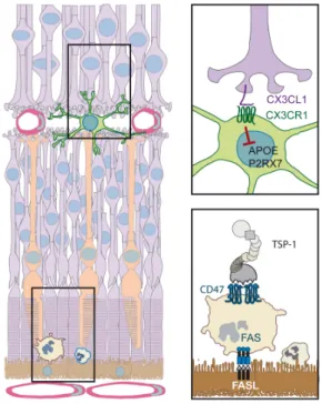
Physiologically, neurons express a number of factors that continually repress MC activation, such as CX3CL1, CD200L, SIRP1α, CD22 (Galea et al., 2007), and translocator protein (Karlstetter et al., 2014; Wang et al., 2014b) among others. CX3CL1 is an atypical chemokine. It is expressed as a transmembrane protein that mediates integrin-like intracellular adhesion and can be cleaved by proteases into a soluble form that has chemotactic properties (Bazan et al., 1997). CX3CL1 has only one receptor, CX3CR1. In blood, CX3CR1 is expressed on “non inflammatory” or “patrolling” Mos (~10% of circulating Mos in humans) (Geissmann et al., 2003) and its genetic deletion in mice reduces MP accumulation in diseases of peripheral tissues, where “patrolling” Mos partake in the MP infiltrate (Combadiere et al., 2003). Among MPs, rMφs express high levels of CX3CR1, which is by far the highest in MCs (Gautier et al., 2012). CX3CR1/CX3CL1 signaling between neurons and MCs plays important roles in synaptic pruning, transmission and plasticity and additionally mediates a tonic inhibitory signal on MCs (Limatola and Ransohoff, 2014). In mouse experimental models of brain pathologies, Cx3cr1-deficiency most often leads to increased neuroinflammation and degeneration (Parkinson’s disease, amyotrophic lateral sclerosis, multiple sclerosis, epilepsy (Cardona et al., 2006; Limatola and Ransohoff, 2014 ; Wolf et al., 2013 )) except in models of Alzheimer’s disease, where uninhibited Apolipoprotein E secretion from Cx3cr1-/-MPs might explain its protective effect (Lee et al., 2010) (see 6.1.1.).
In the eye, transmembrane CX3CL1 is constitutively expressed in inner retinal neurons (Zieger et al., 2014). We and others showed that genetic deletion of Cx3cr1 in mice leads to an accelerated age-dependent increase of subretinal MPs in pigmented animals in both
2011; Combadiere et al., 2007; Levy et al., 2015a; Sennlaub et al., 2013) compared to wildtype animals kept in the same conventional light conditions (~250lux). Cx3cr1-deletion also increases MP accumulation in young albino mice (Chinnery et al., 2011; Combadiere et al., 2007), and in pigmented mice exposed to a non-toxic light-challenge that does not induce degeneration in control mice (Levy et al., 2015a; Sennlaub et al., 2013). Raising albino
Cx3cr1-/-mice in darkness (Combadiere et al., 2007) or letting pigmented C57BL/6
Cx3cr1GFP/GFP mice age in very dim light conditions prevents the accumulation (Combadiere et al., 2013; Luhmann et al., 2013), which confirms the importance of light in subretinal MP accumulation (Ng and Streilein, 2001). Furthermore, the age- and light-dependent accumulation of subretinal Cx3cr1-deficient MPs is associated with a significant loss of rods (Calippe et al., 2017; Chinnery et al., 2011; Combadiere et al., 2007; Hu et al., 2015; Levy et al., 2015a; Sennlaub et al., 2013), cone segments (in acute models such as light-challenge) (Eandi et al., 2016), and cone numbers (in chronic age-related inflammation) (Calippe et al., 2017). The subretinal accumulation of MPs in Cx3cr1-deficient mice is not associated with drusen development or RPE atrophy, but the MP accumulation in combination with rod and cone degeneration observed in these mice is strikingly similar to the TZ of GA patients (Eandi et al., 2016). Taken together, the deletion of Cx3cr1, a gene that is exclusively expressed on MPs (and not on RPE or retinal neurons) in combination with aging under normal light conditions is sufficient to trigger a pathogenic non-resolving subretinal inflammation with age. The Cx3cr1 knockout mouse therefore represents a model of “primary” subretinal inflammation, where inflammation is not the consequence of a tissue injury but solely due to “over reacting” MPs, triggered by a non-toxic initiating stimulus (aging under normal light conditions). In the inflammatory reaction that appears secondary to tissue injury, such as a laser-injury, Cx3cr1-/--mice develop exaggerated MP accumulation and associated amplified CNV (Combadiere et al., 2007). A strikingly similar phenotype is observed in CD200R-/-mice
(Horie et al., 2013) that lack the tonic inhibitory signal of neuronal CD200 (Broderick et al., 2002).
Mechanistically, we demonstrated that Cx3cr1-deficient MPs are characterize by an over-expression of surface P2RX7, which stimulates IL-1β maturation and secretion (Hu et al., 2015) (see 5.2.2.1.) and by increased apolipoprotein E secretion (Levy et al., 2015a) (also observed in muscle repair (Arnold et al., 2015)) that activates the expression of inflammatory cytokines (see 6.1.1.). The increased expression of inflammatory cytokines leads to the recruitment of neurotoxic blood-derived iMϕs (CCL2), the resistance to subretinal MP clearance (IL-6), and photoreceptor toxicity (IL-1β) in Cx3cr1-deficient mice (see 4., 5.2.2.3, and 5.2.2.1. respectively) (Eandi et al., 2016; Hu et al., 2015; Sennlaub et al., 2013). Cx3cr1 deletion also increases MP accumulation and associated degenerative changes in other retinal disease models, such as diabetes (Beli et al., 2016; Cardona et al., 2015; Kezic et al., 2013; Mendiola et al., 2016), retinitis pigmentosa (Peng et al., 2014; Zabel and Kirsch, 2013), Glaucoma (Wang et al., 2014a) and paraquat-induced retinopathy (Chen et al., 2013). Importantly, increased CCL2, APOE and IL6 expression are also observed in AMD (Anderson et al., 2001; Jonas et al., 2010; Klaver et al., 1998; Klein et al., 2008a; Levy et al., 2015a; Seddon et al., 2005; Sennlaub et al., 2013). Cx3cr1-deficient mice might therefore share important pathogenic similarities with human disease. Although they do not mimic all aspects of AMD such as drusen formation and RPE atrophy, they do model subretinal MP accumulation and associated photoreceptor degeneration, which are important features of AMD (Combadiere et al., 2007; Eandi et al., 2016; Gupta et al., 2003; Levy et al., 2015a; Sennlaub et al., 2013). Furthermore, CX3CL1 expression in the brain decreases with age (Fenn et al., 2013) and Cx3cr1 polymorphisms have been associated with AMD in several studies (Anastasopoulos et al., 2012; Combadiere et al., 2007; Schaumberg et al., 2014; Tuo et al., 2004; Zhang et al., 2015), which suggest a possible direct role of CX3CL1/CX3CR1
signaling in AMD. In addition, in animal models, the injection of soluble CX3CL1 has been shown to restore tonic MP inhibition and curb pathogenic inflammation (Mendiola et al., 2016; Zabel and Kirsch, 2013) and might be useful to treat inflammation in patients.
In summary, constitutive repression of MP activation by neuronal inhibitory signals is essential for retinal homeostasis. The lack of only one of these signals, as observed in Cx3cr1-deficient mice, can be sufficient to trigger a vicious cycle of chronic inflammation and collateral damage under normal “healthy” aging conditions that do not cause any significant degeneration in age-matched Cx3cr1-competent mice.
3.3. The immunosuppressive retinal pigment epithelium 3.3.1. Fas/FasL signaling
Additionally to the tonic inhibitory signals of the inner retina that restrict the activation of retinal MCs, the subretinal space is an actively immunosuppressive environment. The subretinal space harbors the photoreceptor outer segments and is shielded from the surrounding tissue by tight junctions of the RPE and zonula adherens between the photoreceptors and Müller glial cells that form the outer limiting membrane (van de Pavert et al., 2004). It is devoid of blood and lymphatic vessels that facilitate leukocyte infiltration and egress in other tissues and the RPE expresses immunosuppressive mediators that induce the death of infiltrating immune cells. In fact RPE allografts survive in non-immunosuppressed hosts for prolonged periods of time even when grafted to non-immune privileged sites, such as the kidney capsule (Wenkel and Streilein, 2000), where conjunctival control cells are quickly rejected. Interestingly, this immunosuppressive capacity is dependent on RPE FasL expression, as FasL-deficient RPE (obtained from FasLgld/gld mice with inactive FasL) is
quickly destroyed under the same circumstances (Wenkel and Streilein, 2000). However, FasL over-expression alone in an allograft, such as a Langerhans cell graft, is not sufficient to convey immune privilege (Kang et al., 1997). Therefore, FasL expression by RPE allografts
seems a necessary but not sufficient factor to escape immune rejection and other factors expressed by the RPE or MPs must be involved. In agreement with the transplant experiments described above, we reported that FasL-defective (FasLgdl/gld-mice) and Fas-defective
(Faslpr/lpr-mice) mice develop significant subretinal MP accumulation after a light-challenge
(designed to cause little inflammation and no damage in wild-type mice) (Levy et al., 2015a). This increase in subretinal MPs is likely the result of their deficient elimination, as subretinally injected MPs survived better when FasL/Fas signaling was defective (WT MPs injected in FasLgld/gld mice and Faslpr/lpr MPs injected in WT mice compared to WT MPs
injected in WT mice) (Levy et al., 2015a). Interestingly, in the resolution phase of lung injury
in vivo, FasL preferentially eliminates iMfs and not rMfs (Janssen et al., 2011) and a Fas
agonist induced monocyte but not Mfs apoptosis in vitro in our hands (Levy et al., 2015a). In contrast, our experiments showed that Mo, Mf and MCs all undergo apoptosis when adoptively transferred to the subretinal space, suggesting that a synergism of different factors acts to physiologically eliminate all types of MPs in contact with the RPE (Levy et al., 2015a).
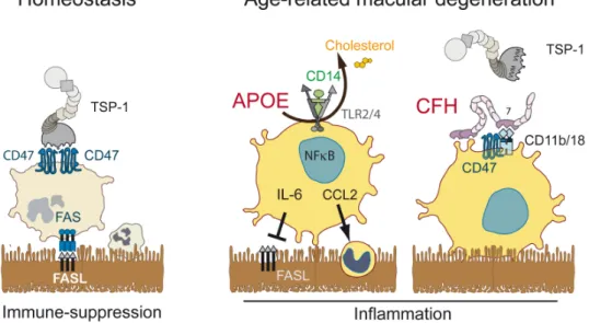
3.3.2. TSP-1/CD47 signaling
Similarly to FasLgld/gld and Faslpr/lpr mice, Thrombospondin 1 (TSP-1)- deficientmice
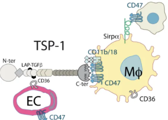
display increased and prolonged subretinal inflammation with age, and after light- and laser-induced injury (Ng et al., 2009; Wang et al., 2012), and develop more severe uveitis (Chen et al., 2012a). They also develop increased inflammation in a variety of peripheral diseases (extensive acute pneumonia, leukocytosis, pancreatitis, and inflammatory infiltrates in the lacrimal glands) (Lopez-Dee et al., 2011). TSP-1 is synthesized by a wide variety of cell types, notably the RPE (Miyajima-Uchida et al., 2000), iMφs (Fordham et al., 2012) and rMφs (Gautier et al., 2013) and plays a role in multiple biological processes including phagocytosis, angiogenesis, and immune regulation (Bornstein, 2009; Housset and Sennlaub, 2015; Lopez-Dee et al., 2011). It contains a Willebrand type C domain, necessary for its trimerization,
followed by three properdin-like type 1 repeats, which include the regions that bind the CD36 receptor (anti-angiogenic signals (Jimenez et al., 2000)) and the latency-associated peptide (LAP) of the latent TGF-β binding protein (LTBP). TSP-1 thereby binds CD36 and LTBP and liberates active TGF-β (Bornstein, 2001; Lawler and Hynes, 1986). The COOH-terminal cell-binding domain (CBD) of TSP-1 contains two valine-valine-methionine (VVM) sequences that can each interact with a CD47 receptor (Bornstein, 2001; Lawler and Hynes, 1986) (Fig. 4). Independently of its role as a TSP-1 receptor, the N-terminal domain of CD47, also acts as a ligand for the signal regulatory protein alpha (SIRPα) of MPs and inhibits phagoptosis (“don’t eat me” signal) (Navarro-Alvarez and Yang, 2011). The efficient activation of CD47 by TSP-1 is dependent on the presence of both VVM sites of TSP-1 (McDonald et al., 2003), but can be mimicked by the TSP-1-derived 4N1K peptide (KRFYVVMWKK) (Martinez-Torres et al., 2015), at 100 fold higher molar concentrations (McDonald et al., 2003). CD47 activation has been shown to sensitize endothelial cells and lymphocytes to FasL-induced death in a CD47-dependent manner (Manna et al., 2005; Quesada et al., 2005). We recently confirmed that Tsp1-/--mice develop age-, and exaggerated light- and laser-induced subretinal MP accumulation. Interestingly, this phenotype was shared by CD47-/-, but not CD36-/-mice (Calippe et al., 2017). Tsp1-/-- and Cd47-/--MCs, adoptively transferred to the subretinal space of wildtype recipients significantly resisted the elimination observed in wildtype MCs and recombinant TSP1 very significantly accelerated the elimination of wiltype-MCs, reversed the phenotype of Tsp1-/--MCs but had no effect on Cd47-/--MCs. This suggests that the interaction of TSP1 and CD47 mediates subretinal macrophage elimination (Calippe et al., 2017). Taken together, these results demonstrate that the activation of CD47 on infiltrating subretinal MPs participates in their homeostatic elimination and during the resolution of acute inflammation. CD47 signaling likely sensitizes MPs to FasL-induced death as described for other cell types (Manna et al., 2005; Quesada et al., 2005) (Fig. 3). In diseases where subretinal MPs
accumulate on the RPE, in areas such as the TZ and drusen of AMD, subretinal RPE-mediated immune-suppression has clearly become ineffective. It is currently not clear if the deficient immune-suppression arises due to insufficient RPE signals, increased resistance of MPs to elimination, or both.
Taken together, the capacity of the RPE to induce leukocyte death is a crucial part in repressing the accumulation of potentially pathogenic MPs and maintaining homeostasis in the photoreceptor cell layer and the subretinal space. TSP-1 mediated activation of CD47 and FasL signaling through Fas are essential, but likely not the only molecular pathways involved in this mechanism.
4. The origin of infiltrating mononuclear phagocytes
The photoreceptor cell layer and the subretinal space are physiologically devoid of MPs, and any subretinal MPs found in AMD must therefore have actively infiltrated the space through the OLM, or the RPE. Infiltrating MPs could originate from retinal MCs, circulating Mos or rMφs from the choroid or ciliary body. Most studies concerning MP accumulation in AMD patients and animal models do not allow for the differentiation of distinct types and origins of MPs, as they express or can induce similar markers (Gautier et al., 2012; Ransohoff and Cardona, 2010). Mo-derived MPs are also not morphologically distinguishable from MCs, best evidenced by the blood borne GFP+microglia-like cells that replace MCs in whole-body irradiated, GFP+bone marrow transplanted mice (Chen et al., 2012b). The distinction between rMφ or Mo-derived iMφs might be important, as the sustained presence of iMfs, evolved to neutralize pathogens, has been shown to be detrimental in neurodegenerative conditions such as multiple sclerosis and stroke (Conductier et al., 2010; Ransohoff, 2009).
A molecular marker that differentiates inflammatory Mos and iMfs from MCs is CCR2, the receptor of the major chemokine CCL2 (Geissmann et al., 2010). Contrary to MCs, inflammatory Mos express high levels of CCR2 (Geissmann et al., 2010; Mizutani et al.,
2011; Sennlaub et al., 2013) and CCR2 cannot be induced in MCs (Saederup et al., 2010; Sennlaub et al., 2013). CCR2 is also not constitutively expressed or induced (light-challenge) in RPE cells, clearly demonstrated by the absence of red fluorescence protein (RFP) in the RPE of Ccr2RFP/RFP-mice (Sennlaub et al., 2013). CCR2 is therefore a good marker to identify subretinal Mo-derived Mφs, but likely underestimates this population, as Ccr2 transcription is quickly downregulated when Mo differentiate to Mφ (Sennlaub et al., 2013; Wong et al., 1997).
CCL2 (the main ligand of CCR2) expression in the retina is physiologically low but it increases with age and is induced in situations of stress such as laser- and light-injury or retinal detachment in animal models (Chen et al., 2012a; Nakazawa et al., 2007; Sennlaub et al., 2013; Yamada et al., 2007). Initially CCL2 is likely secreted by resident MCs that sense local danger signals, but it is also strongly produced by infiltrating iMφs (Sennlaub et al., 2013), and other resident cells, such as Müller glial cells (Rutar et al., 2012) and the RPE (Chen et al., 2008; Elner et al., 1991). Importantly, increased intra-ocular levels of CCL2 are observed in human neovascular- (Fauser et al., 2015; Guymer et al., 2011; Jonas et al., 2010; Kramer et al., 2012; Newman et al., 2012; Rezar-Dreindl et al., 2016) and atrophic-AMD (Newman et al., 2012; Sennlaub et al., 2013). Using CCR2 immunohistochemistry, we showed that subretinal MPs observed in human sections of GA patients are in part blood-derived inflammatory CCR2+ Mos (Sennlaub et al., 2013). Indeed, all studied diseased eyes, but no healthy control eye, contained CCR2+ Mos in the atrophic lesions and CCR2+ Mos were observed on the RPE of the TZ, in BlinD and in and around large drusen (Sennlaub et al., 2013). In inflammation-prone Cx3cr1-deficient mice (in which CCL2 is induced with age and during a light-challenge) we demonstrated using genetic Ccl2 or Ccr2 deletion, monocyte depletion, and inhibitors of CCR2 that approximately 50% of the MPs infiltrating the photoreceptor cell layer are recruited from circulation. This recruitment from blood occurred,
despite the absence of a visible breech in the blood retinal barrier (Sennlaub et al., 2013), indicating that the blood retinal barrier is not an obstacle for Mo infiltration. Importantly, the inhibition of CCR2+ monocyte recruitment nearly completely prevents the
inflammation-associated photoreceptor degeneration in these mice. These results suggest that (i) the CCR2+
monocyte derived MPs are the main mediators of inflammation-associated neurotoxicity in this model and (ii) that the subretinal accumulation of MPs is in part due to CCR2neg cells,
possibly retinal microglial cells that display no or little toxicity. Our in vitro experiments comparing wildtype and Cx3cr1-/- Mo and MC toxicity on photoreceptors of retinal explants in a co-culture system confirmed increased toxicity of Mos compared to MCs and of Cx3cr1-/- MPs compared to wildtype MPs (Sennlaub et al., 2013).
Similarly, infiltrating MPs are in part derived from circulating Mos in RD models (Guo et al., 2012; Kohno et al., 2015) and photooxydative stress (O'Koren et al., 2016; Suzuki et al., 2012). Mo-derived iMφs contribute significantly to photoreceptor degeneration in light-induced models (Hu et al., 2016; Rutar et al., 2012; Suzuki et al., 2012), in the Abca4-/-Rdh8
-/-mouse Stargardt/AMD model (Kohno et al., 2013), the rd10 -/-mouse (Guo et al., 2012), and in the carboxyethylpyrrole immunization-induced AMD model (Cruz-Guilloty et al., 2013). Although we observed little MC neurotoxicity in light-challenged Cx3cr1-deficient mice, MCs have been shown to contribute to photoreceptor degeneration in rd10 mice (Zhao et al., 2015). The rd10 mice lack the exon 13 of the phopshodiesterase 6b that is physiologically expressed in rods. MCs participate in photoreceptor degeneration in rd10 mice as they phagocytose diseased cells, but living, rods in a process called phagoptosis (Zhao et al., 2015). It will be interesting to see to what degree photoreceptor phagoptosis by MCs occurs in other degeneration models, where the instigator event does not lie within the photoreceptors.
In experimental, laser-induced CNV approximately 50-70% of MPs are Mo-derived and 30-50% are derived from resident MPs (MCs, and choroidal rMφs) (Caicedo et al., 2005).
Depletion of circulating Mo (Sakurai et al., 2003a) and inhibition of Mo-recruitment by genetic deletion of Ccr2 and Ccl2 (Liu et al., 2013b; Luhmann et al., 2009; Robbie et al., 2016; Tsutsumi et al., 2003), or CD18 / ICAM-1 signaling (important for Mo diapedesis (Sakurai et al., 2003b)) very significantly inhibits CNV formation in numerous studies. These results might seem somewhat surprising as CCR2+Mo derived iMφs are generally supposed to be anti-angiogenic (M1 polarized, see 5.) (Sica and Mantovani, 2012; Wynn et al., 2013), contrary to MCs (M2 polarized) that participate in physiological retinal angiogenesis (Checchin et al., 2006) and are pro-angiogenic in the subretinal space (Ma et al., 2009). However, the phagocytosis of damaged RPE by the Mo-derived Mφs (Liu et al., 2013b), which characterize the microenvironment of CNV, might lead to this “quick switch” to a pro-angiogenic profile of the infiltrating subretinal iMφs.
Taken together, MP populations that infiltrate the photoreceptor cell layer and subretinal space are likely constituted of resident Mφs such as MCs and possibly choroidal rMφs but also of blood Mo-derived iMφs as seen in animal models and in the human disease (Sennlaub et al., 2013). As outlined above, iMφs evolved to react as quickly as possible to terminate the spread of potential deadly infections, even at the cost of tissue damage (Nathan, 2002). The threshold to induce the secretion of potentially damaging inflammatory cytokines in iMφs is therefore much lower compared to rMφs (Nathan, 2002). Experimental data from a number of “secondary” subretinal inflammation models, where inflammation occurs secondary to RPE or photoreceptor disease or injury, demonstrate that the chronic presence of subretinal Mo-derived iMφs aggravate photoreceptor degeneration additionally to promoting CNV (Fig. 5). Importantly, this was also the case in “primary” models of subretinal inflammation, in which the inflammation is not a secondary event to photoreceptor or RPE dysfunction but due to (i) the lack a tonic inhibitory signal in the inner retina (such as Cx3cr1-deficient mice (Calippe et al., 2017; Combadiere et al., 2007; Eandi et al., 2016; Hu et al., 2015; Sennlaub et al., 2013))
or (ii) an induced autoimmune reponse (carboxyethylpyrrole immunization-induced AMD model (Cruz-Guilloty et al., 2013)). These primary models strongly suggest that the chronic infiltration of iMφs can be sufficient to induce degenerative changes in the outer retina.
Crb1rd8 mutation induced confusion in the field
In 2003 Ambati et al. reported that aged Ccl2-/- and Ccr2-/- mice develop drusen, spontaneous neovascularization and photoreceptor degeneration (Ambati et al., 2003). The authors suggested that deficiency in CCL2-/CCR2-dependent Mφ recruitment from the choroidal circulation may prevent the clearance of accumulating debris in BM (Ambati et al., 2003), which, over time, would lead to drusen formation. In an attempt to accelerate the development of AMD-like features, Tuo et al. generated Ccl2-/-Cx3cr1-/--mice that develop “drusen,” pigment alterations, and retinal degeneration by the age of 6 weeks in 100% of mice and CNV in 15% of mice (Tuo et al., 2007). This mouse strain was subsequently shared with a number of laboratories and used in numerous publications as a model of AMD, because of its phenotypical similarities. How deficiency in CCL2/CCR2- and CX3CL1/CX3CR1-signaling led to the phenotype was not investigated. In these studies, it remained unclear if CCL2-deficiency protected against the more pronounced phenotype observed in Cx3cr1-deficient mice (or vice versa), as data from single knockout controls were never presented.
Later reports first failed to reproduce spontaneous CNV in aged Ccl2-/- and Ccr2-/- mice and the appearance of drusen was recognized due to subretinal lipid bloated MPs (Chen et al., 2011; Luhmann et al., 2009) similar to those observed in Cx3cr1-deficient mice (Combadiere et al., 2007). In our laboratory, aged Ccl2-/- mice only developed discreet subretinal
accumulation of MP, which represented only a fraction of the accumulation observed in age-matched Cx3cr1-deficient mice kept under age-matched conditions in the same animal facilities (see supplementary data of Sennlaub et al. (Sennlaub et al., 2013)). Furthermore, none of our independently bred Ccl2-/-Cx3cr1-/--, Ccl2-/-Cx3cr1GFP/GFP-, and Ccr2RFP/RFPCx3cr1GFP/GFP
-mice developed the early onset phenotype. In fact CCL2 or CCR2 deficiency, pharmacological inhibition of CCR2, or circulating Mo depletion, protected against the pathogenic accumulation of subretinal MPs observed in Cx3cr1-/- and Cx3cr1GFP/GFP-mice.
These observations are in line with the tonic inhibitory role of CX3CL1/CX3CR1 signaling and the neurotoxicity of CCR2+-Mo-derived Mφs observed not only in the retina, but also in the central nervous system in general (see above). It is still however not clear how deletion of CCL2 or CCR2 induces subretinal MP accumulation and what the significance this observation has for AMD research as intraocular CCL2 concentrations are increased in both late forms and CCR2+Mos accumulate in GA (see above).
Ultimately, in 2012, the early onset phenotype of Ccl2-/-Cx3cr1-/- mice was explained
by a contamination with a mutation in the Crumbs homologue-1 gene (Crb1), known as the
retinal degeneration 8 (Crb1rd8) mutation (Luhmann et al., 2013; Mattapallil et al., 2012). The Crb1rd8 mutation produces a secreted truncated CRB1 protein (Mehalow et al., 2003). CRB1 localizes specifically to the adherens junction complex at the outer limiting membrane in the retina. Homozygous Crb1rd8 mice can develop prominent focal inferior retinal degeneration, photoreceptor loss, and retinal thinning (Mehalow et al., 2003) as observed in Crb1-/- mice (van de Pavert et al., 2004). At later stages the mice might present vascular lesions that resemble CNV, but are in fact derived from the retinal vasculature, and subretinal MP accumulation similar to AMD (Luhmann et al., 2014). Crb1 mutations in humans can lead to a comparable range of symptoms (Bujakowska et al., 2012) but there are currently no studies that would link variants of Crb1 to AMD pathology. Akin to human mutations the phenotype in homozygous Crb1rd8 mice is dependent on additional yet unknown genetic modifiers (located on chromosome 15 for the mouse) and the absence of macroscopic lesions does not exclude the presence or even homozygosity for the mutation (Luhmann et al., 2014). The
not seem to have been contaminated with the Crb1rd8 mutation as their pro-inflammatory phenotype was since consistently reproduced in mice that tested negative for the Crb1rd8 mutation (Calippe et al., 2017; Eandi et al., 2016; Hu et al., 2015; Levy et al., 2015a; Sennlaub et al., 2013).
It is likely that numerous mouse studies using knockout- and transgenic-mice contaminated Crb1rd8 wrongly implicated genes in subretinal inflammation and the pathomechanism of AMD because of the similarity of the features caused by the Crb1rd8 mutation. Taken together, it is crucial that any transgenic mouse strain used in retinal and in particular AMD research must be genotyped and the Crb1rd8 mutation eliminated. In our hands, the targeted replacement mice that express human APOE isoforms (TRE2-, 3-, and 4 - C57BL/6N mice; a generous gift from Dr. Patrick Sullivan (Sullivan et al., 1997)) and TSP-1 -/- C56BL/6J-mice (purchased from Jackson laboratories) were all homozygote for the Crb1rd8 contamination and were backcrossed to eliminate the mutation before experimentation. The absence of the typical Crb1rd8 phenotype or the background strain (C57BL/6N versus C56BL/6J) does not exclude its presence and interference with ocular phenotypes.
5. Function and consequences of chronic subretinal mononuclear phagocyte accumulation
5.1. A “homeostatic” role
In tissues with high regenerative potential such as the skin, a homeostatic role of MPs is clearly observed in the resolution of acute inflammation, where they enhance the proliferation of epithelial progenitor cells and promote wound healing (Wynn and Vannella, 2016). Similarly, in the central nervous system of fish that have retained the capacity to regenerate neurons after injury, MPs trigger the regeneration of neurons and photoreceptors (Kyritsis et al., 2012; White et al., 2017). However, this reparative aspect of the resolution of acute inflammation is not observed in mammals, as we lost the regenerative capacity of neurons and photoreceptors.
In chronic low-grade tissue inflammation triggered by tissue stress and malfunction MPs might help preserve tissue function for some time, albeit the persistence of the noxious conditions. This might be in particular the case in situations where tissue stress is accentuated or caused by accumulation of toxic extracellular material that MPs can remove by phagocytosis. For example, we showed that infiltration of iMφs can be partially neuroprotective in the (admittedly extreme) case of experimental subretinal injection of toxic doses of β-amyloid, as iMφs neutralize this protein (Bruban et al., 2011). Theoretically, in this context, MP infiltration is beneficial as long as the toxicity associated with the MP infiltration is outweighed by the toxicity of the material it removes. It depends therefore on the presence, amount, and nature of the accumulating toxins and the composition and activation state of the
infiltrate (iMφs, rMφs and others). Medzhitov proposed to call the beneficial, “homeostatic”
accumulation of rMφs, “which helps a tissue to adapt to the noxious conditions and restore tissue functionality” that is observed with milder tissue malfunction, “para-inflammation”. Pathogenic chronic low grade inflammation that includes Mo-derived Mφs on the other hand would be called “dysregulated para-inflammation” (Medzhitov, 2008). “Para-inflammation” might take place in patients with early AMD, as MPs that are observed in and around large drusen might participate in the removal of debris, preventing drusen growth or participate in drusen absorption. It has become increasingly clear that large drusen are dynamic structures that grow, but are also absorbed (Querques et al., 2016; Smith et al., 2010; Toy et al., 2013; Yehoshua et al., 2011) and MPs are likely candidates to play an important part in this mechanism. Furthermore, patients with large sized soft drusen do not necessarily progress to late AMD. Over 10 years, only 15% (Beaver Dam study)-30% (Blue mountain study) progressed to late AMD in these population based epidemiological studies (Klein et al., 2004; Wang et al., 2003) and MP-mediated “para-inflammation” might have prevented patients from progression to late AMD. As mentioned above such a mechanism has been proposed,
based on the observation for spontaneously appearing drusen-like fundus lesions in Ccl2-/- and Ccr2-/--mice (Ambati et al., 2003). However, these lesions were later identified to be caused by subretinal lipid-bloated MPs (Chen et al., 2011; Luhmann et al., 2009) and not drusen. It is generally difficult to study the implication of MPs in drusen regression with current rodent animal models, as none develop lesions similar to human drusen. In humans, AMD-associated drusen are thick (10-50μm) lipoproteinaceous debris located between the basal lamina of the RPE and the inner collagenous layer of BM (in the same anatomical location as BlinD). In Mice, sub RPE debris accumulation can be observed in APOE-/- mice (Ong et al., 2001), TRE4-mice (Malek et al., 2005) and Cfh-/-mice (Toomey et al., 2015) but these accumulations are thin (<3μm) and located between the RPE and their basal infoldings. They are therefore similar to basal laminar deposits (BlamD), that are associated with age but not with AMD (Curcio and Millican, 1999).
Aged C57BL/6 wildtype mice also present subretinal MP accumulation (at a significantly older age compared to Cx3cr1-/- mice) that does not seem to be associated with photoreceptor degeneration. This accumulation has been suggested to be a form of para-inflammation (Chen and Xu, 2015), but experimental evidence that the infiltrate protects these mice from photoreceptor or RPE dysfunction is lacking so far. Contrary to mice, donor eyes from healthy humans (even of advanced age) do not present significant subretinal MP accumulation that could be akin of a para-inflammatory infiltrate and subretinal MPs are only observed in diseased eyes (Eandi et al., 2016; Gupta et al., 2003; Levy et al., 2015a; Sennlaub et al., 2013).
In summary, during the resolution phase of acute inflammation MPs fulfill an important role in the regeneration and reestablishment of homeostasis in tissues with high regenerative potential. In vertebrates such as fish this mechanism is preserved in the central nervous system (CNS) including the retina that has retained the ability to regenerate, but is restricted
to tissues such as the skin in mammals. In chronic inflammation MPs can help prevent the
accumulation of toxic extracellular material and help preserve tissue function. Even though
experimental data is difficult to produce due to the lack of appropriate models, this might well be the case for hard drusen during “healthy” aging and for large drusen in non-progressing ARM patients, where moderate MP accumulation might help control drusen growth.
5.2. Collateral damage
In some patients the compounded combination of several AMD risk factors likely leads to particularly inflammatory MPs (see 6.), which pushes the balance towards a pathogenic chronic inflammation and late AMD. The degree to which chronic inflammation constitutes a major or minor pathogenic factor in these patients likely depends on the extent of the infiltrate and on its production of pathogenic mediators.
5.2.1. M1 and M2 polarization
Polarization states of MPs are often divided into two activation states in analogy to the adaptive immune system, where immunologists observed that two distinct subpopulations of CD4+ T helper cells could be subdivided on the basis of the pattern of their cytokine production, which predicted their role in the immune response (Th1/Th2 polarization) (Liew, 2002). In vitro, Mϕs activated by bacterial molecular patterns (such as LPS) are defined as classically activated Mϕs (M1). They are pro-inflammatory, anti-microbial, anti-angiogenic, potentially neurotoxic and defined by the expression of mediators such as IL-1β, TNF-α, IL-6, CCL2 and iNOS. Alternative activation of Mϕs (M2) is traditionally induced by 4 and IL-13 which polarizes them towards a phenotype that is anti-inflammatory, promotes phagocytosis, neovascularization, wound healing and ultimately fibrosis (scarring). They are characterized by the expression of VEGF, Arginase, IL-10, and IL-1RA among others (Sica and Mantovani, 2012; Wynn et al., 2013). However these two types of polarization are only two of a multitude of activation states and the M1/M2 defining proteins are not regulated by a general transcriptional switch but individually. In many studies the identification of M1 or M2
markers is used to infer if the MPs are “good” or “bad”, without investigating the effect of the individual mediators that are used as markers. However, in vivo subretinal MPs have been shown to express M1 and M2 markers simultaneously (Camelo et al., 2012; Horie et al., 2013; Liu et al., 2013b) and they are often both, neurotoxic and angiogenic (see 4.). Hence, in subretinal inflammation, but also in peripheral and central inflammation in other settings, the dated differentiation of M1 and M2 polarized MPs is not helpful to characterize Mo-derived Mφ polarization (Wynn et al., 2013) or MC polarization (Ransohoff, 2016) and should no longer be used. Therapeutically, it would also seem ill advised to direct subretinal MP polarization from M1 to M2, as their pro-angiogenic/fibrotic phenotype would promote CNV and fibrosis.
5.2.2. Phagocyte-derived cytokines and collateral damage
As outlined above, Mo-derived iMφs play a pathogenic role in many models of subretinal inflammation and AMD (Cruz-Guilloty et al., 2013; Guo et al., 2012; Hu et al., 2016; Kohno et al., 2013; Rutar et al., 2012; Sennlaub et al., 2013; Suzuki et al., 2012), in addition to MCs (Zhao et al., 2015). As Mo-derived iMφs lack the receptors for tonic inhibitory signals from the retina (see above) at the beginning of their differentiation into Mφs in the retinal microenvironment, they are prone to produce high levels of inflammatory cytokines, including the classical inflammatory cytokines IL-1β, TNF-α, IL-6, CCL2 when they infiltrate the tissue. Over the last years, the understanding of the multiple modes of action of these cytokines and how they affect chorioretinal homeostasis has evolved significantly (Fig. 6):
5.2.2.1. Interleukin-1β
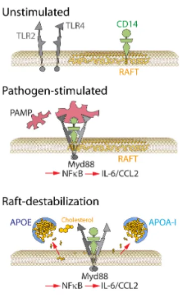
The Il-1β gene is transcribed and pro-IL-1β protein produced after pro-inflammatory stimuli, such as those ensuing from the activation of toll-like receptors (TLR) that recognize pathogen- or damage-associated molecular patterns (PAMPs, DAMPs) (Allan et al., 2005; Simi et al., 2007a). Activation by a second stimulus, can trigger the assembly of the NLRP3