High-resolution and high-sensitivity blood flow estimation using deconvolution and optimization approaches with application to thyroid vascularization imaging
Texte intégral
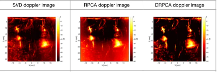
Figure

Documents relatifs
Cependant, bien que ces infirmières aient une moyenne de 11 ans d'expérience avec des personnes démentes, qu’elles avaient reçu un vaste enseignement dans la
We evaluated the effects of the two main kiwifruit cultivars (gold kiwifruit (GOK) and green kiwifruit (GRK)) and their active phenolic compound, quercetin, on H 2 O 2
Given a profile of public opinions and an influence net- work, we model the process of opinion diffusion by means of an aggregation function, which shapes the private opinion of
In contrast to the optical birefringence, the dielec- tric permittivity of Py4CEH in the quasistatic limit is char- acterized by a positive dielectric anisotropy in the
Si vous en voyez une autre pour rendre heureux les enfants mariés qui vivent avec leurs parents, n'ayez crainte de nous faire part de votre point de vue.. On est là
While some neuroimaging studies have found evidence of greater activation of the MTL during the retrieval of recent relative to remote episodic memories ( Haist
The real challenge in membrane based gaseous separation is developing the right polymeric materials having all the desired properties, such as high selectivity, high permeability,
Four hypothetical AML patients aged 60 years or older were selected as representative of clinical practice and were summa- rized by 3 local specialists (PB, SB and
