Strahlenther Onkol 2014 · 190:386–393 DOI 10.1007/s00066-013-0508-x Received: 20 August 2013 Accepted: 8 November 2013 Published online: 7 February 2014 © Springer-Verlag Berlin Heidelberg 2014
S. Janssen1 · C. Glanzmann1 · G. Huber2 · G. Studer1 1 Department of Radiation Oncology, University Hospital Zurich
2 Department of Otorhinolaryngology, Head and Neck Surgery, University Hospital Zurich
Individualized IMRT treatment
approach for cervical lymph node
metastases of unknown primary
In approximately 3–5% of patientspre-senting with squamous cell cancer (SCC) metastases in cervical lymph nodes, the primary is not found despite extensive clinical evaluation [1, 2]. Treatment of those patients remains controversial lack-ing evidence from prospective random-ized trials. Recommendations include neck dissection and/or radio(chemo)ther-apy. For more advanced stages, multimod-al treatment is recommended. Induction chemotherapy before radio(chemo)ther-apy followed by surgery depending on re-sponse may be an additional option [2, 3, 4]; however, its value is not proven. The main controversial aspect is the radio-therapy (RT) treatment volume. Planning treatment volumes (PTVs) often include the affected lymph node region as well as contralateral cervical lymph nodes and the mucosal sites of the pharynx in order to cover a putative primary (comprehen-sive RT) [5].
Several retrospective single institution studies compared comprehensive con-ventional radiation to involved field radi-ation. While some groups demonstrated better local outcome using extended fields [5, 6, 7], others did not show an advantage for more extensive RT [4, 8, 9, 10]. Fur-thermore, omitting hypopharyngeal and laryngeal mucosa seems to be feasible as well [1, 11].
Introduction of intensity-modulat-ed radiotherapy (IMRT) in patients with head and neck cancer showed an im-proved therapeutic ratio compared to historic conventional three-dimension-al techniques [12]. Recently, in the era of IMRT, several study groups used IMRT in patients with cervical lymph node
metas-tases of unknown primary cancer (UPC) and confirmed those good results [13, 14, 15, 16, 17, 18, 19].
The purpose of this article is to evalu-ate the outcome of risk-adapted PTVs in patients with cervical lymph node metas-tases of UPC treated with IMRT.
Material and methods
Patients
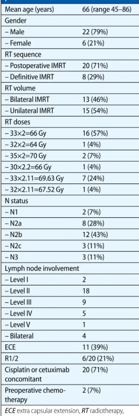
From January 2006 to November 2012, 28 consecutive patients presenting with cer-vical lymph node metastases of UPC were treated in our department with IMRT ei-ther postoperatively (n=20) or as defini-tive treatment (n=8). Diagnosis was prov-en histologically in all patiprov-ents show-ing metastases of SCC. PET-CT was per-formed routinely before treatment start and was considered for delineation. Pa-tient- and treatment-related parameters are summarized in . Tab. 1. Mean/me-dian follow-up time was 31.6/30.5 months (range 3–78 months). Details regarding surgical neck dissection prior to radiation (n=20) or as postradiation elective neck dissection (END) are listed in . Tab. 2.
Regular follow-up visits were carried out in our joint clinic at the Department of Otorhinolaryngology, Head and Neck Surgery. Institutional standards for patient assessment included physical examination and flexible fiber optic endoscopy approx-imately every 2 months in the first year of follow-up, every 3 months in the second to third year and every 6 months in the fourth to fifth years.
Treatment
IMRT
Unilateral irradiation was preferred. Based on individual risk factors includ-ing clinical, surgical, histopathological, and imaging information, we enlarged treatment fields to the putative mucosal site or the contralateral neck. One crucial risk factor was suspicious enhancement of contralateral lymph nodes or oropharyn-geal structures in PET-CT (n=4). In those cases, a biopsy of the questionable region was carried out revealing no malignancy. Nevertheless a certain risk of involvement was supposed in that case—which was the reason to extend PTVs. Other risk factors suggestive for bilateral nodal irradiation were status after pT1 floor of the mouth carcinoma in the past (n=1), recurrent lymph node metastases (n=1), and level I involvement (n=2). “Classical” extensive bilateral treatment was reserved for N2c and bilateral N3 patients (n=5; postoper-ative: n=1; definitive: n=4). One patient with extensive N3 disease (116 ccm) was only treated unilaterally due to advanced age and poor general condition. Doses and PTVs based on the affected cervical node levels are shown in . Tab. 3.
. Fig. 1, 2, and. Fig. 3 demonstrate uni- and bilateral RT volumes. We used si-multaneously integrated boost (SIB) tech-nique in all patients [20, 21]. SIB-IMRT was performed using the schedules below.
In definitive IMRT patients (n=8):
F SIB2.00: 35 fractions with daily SIB doses of 2.00 Gy (PTV1)/1.70 Gy (PTV2) and 1.54 Gy (PTV3) to a total boost dose of 70.00 Gy (five fractions a week).
F SIB2.11: 33 fractions with daily SIB doses of 2.11 (PTV1)/1.80 Gy (PTV 2) and 1.64 Gy (PTV3) to a total boost dose of 69.60 Gy (five fractions a week).
In postoperative patients (n=20):
F SIB2.00: 32–33 fractions with daily SIB doses of 2.00 Gy (PTV 1)/1.80 Gy (PTV2)/1.64 Gy (PTV3) to a total boost dose of 64–66.00 Gy (five frac-tions a week).
F SIB2.11: 33 fractions of daily SIB dos-es of 2.11 (PTV1)/1.80 Gy (PTV 2)
and 1.64 Gy (PTV3) to a total boost dose of 69.60 Gy (five fractions a week).
F SIB2.2: 30 fractions with daily SIB doses of 2.2 Gy (PTV1)/2.0 Gy (PTV2) and 1.64 Gy (PTV3) to a to-tal dose of 66.00 Gy (five fractions a week) (carried out in 1 patient) In 1 patient two IMRT series were carried out instead of SIB (50 Gy, 16 Gy boost, sin-gle dose: 2.0 Gy, 6 fractions/week).
The dose was normalized to the mean dose in PTV1. For intensity optimization,
the prescribed dose encompassed at least 95% of the PTV. Additionally, no more than 2% of any PTV received >110% of its prescribed dose, whereas no more than 1% of any PTV received <93% of the pre-scribed dose.
Target volumes were delineated as fol-lows: the involved lymph nodes included the gross extent of primary disease, tak-ing clinical and radiological findtak-ings in-to account; clinical target volume (CTV) was defined by adding 10–15 mm margin to the gross tumor volume (GTV), anoth-er 2–3 mm margin was added from CTV to PTV 1 dependent on proximity to crit-ical structures (e.g., spinal cord or brachi-al plexus); PTV2 covered areas consid-ered at high risk for potential microscop-ic disease; and PTV3 included the clinmicroscop-ical- clinical-ly negative mucosa or clinical-lymphatic pathways (elective PTV coverage).
In patients with substantial parts of the pharynx and/or larynx involved in the PTV, 2.0 Gy per session was given. To ensure sufficient dose delivery to the skin close to GTVs, bolus material (0.5–1 cm thickness) was used in patients with skin involvement and/or <5 mm between GTV and the overlying skin.
Irradiation was delivered with five or seven coplanar beam angles by a 6-MV dynamic MLC system (Varian Medical Systems, Palo Alto, CA, USA) using the sliding window technique, or using the volumetric modulated rapid arc technique (VMAT, since April 2010). Patients were immobilized from head to shoulders us-ing a commercially available thermoplas-tic mask in supine position.
Systemic therapy
Systemic therapy preferably consisted of cisplatin (40 mg/m2 weekly) and was
switched to cetuximab in case of cisplatin-related adverse effects (cetuximab load-ing dose: 400 mg/m2 followed by weekly
applications of 250 mg/m2 [22]). For
pa-tients with contraindications against cis-platin, cetuximab was favored primarily. The indication for systemic therapy was made based on extent of nodal involve-ment, resection status, extra-nodal ex-tension, age, and Karnofsky performance score. In 2 patients with extended disease (cN3 and cN2c), three cycles of neoadju-vant chemotherapy with TPF (docetaxel Tab. 1 Patient- and treatment-related
parameters
Mean age (years) 66 (range 45–86) Gender – Male 22 (79%) – Female 6 (21%) RT sequence – Postoperative IMRT 20 (71%) – Definitive IMRT 8 (29%) RT volume – Bilateral IMRT 13 (46%) – Unilateral IMRT 15 (54%) RT doses – 33×2=66 Gy 16 (57%) – 32×2=64 Gy 1 (4%) – 35×2=70 Gy 2 (7%) – 30×2.2=66 Gy 1 (4%) – 33×2.11=69.63 Gy 7 (24%) – 32×2.11=67.52 Gy 1 (4%) N status – N1 2 (7%) – N2a 8 (28%) – N2b 12 (43%) – N2c 3 (11%) – N3 3 (11%)
Lymph node involvement
– Level I 2 – Level lI 18 – Level III 9 – Level IV 5 – Level V 1 – Bilateral 4 ECE 11 (39%) R1/2 6/20 (21%) Cisplatin or cetuximab concomitant 20 (71%) Preoperative chemo-therapy 2 (7%) ECE extra capsular extension, RT radiotherapy, IMRT intensity-modulated radiotherapy.
Tab. 2 Extent of neck dissection and number of positive lymph nodes for all 20 patient receiving surgery prior to radia-tion. In 4 of 8 patients with definitive ir-radiation, an elective neck dissection (END) was performed Extent of neck dissection (level) Positive lymph nodes 1 II–IV left 4/31 2 II–IV left 1/12 3 II–IV left 9/24 4 I–IV right 2/6 5 II–III left 2/18 6 I–V right 18/30 7 I–IV right 3/14 8 II–IV right 1/25 9 II–IV right 1/27 10 II–V right 1/14 11 II–V left 3/14 12 I–V right 1/39 13 I–IV right 1/21 14 I 1/1 15 I–II left 2/4
16 I–IV left 0/29 (after lymph node excision) 17 I–IV right 0/12 (after lymph
node excision) 18 II–IV left 1/9
19 I–IV both sides 8/51 right, 4/37 left 20 II–V left 1/13 END 1 II–III right 0/17 END 2 performed exter-nally (no informa-tion)
END 3
II–IV right and II–III left 0/15 right, 0/6 left END 4 II–V right 0/2
Abstract · Zusammenfassung
75 mg/m2 day 1, cisplatin 75 mg/m2 day 1,
and 5-fluorouracil 750 mg/m2 days 1–5)
were administered.
Surgery
The extent of neck dissection and the number of positive lymph nodes in oper-ated patients is shown in . Tab. 2.
Statistics
Statistical calculation was performed us-ing the statistic program implemented in StatView (Version 4.5; SAS Institute, Cary, NC, USA).
Results
Outcome
Mean/median follow-up was 31.6/30.5 months (range 3–78 months). The 3-year overall survival rate was 76%. The 3-year mucosal control rate, nod-al control rate, and distant metastasis-free survival were 100, 93, and 88%, re-spectively (. Fig. 4). Two patients with a nodal mass of 63 and 116 ccm, respec-Strahlenther Onkol 2014 · 190:386–393 DOI 10.1007/s00066-013-0508-x
© Springer-Verlag Berlin Heidelberg 2014
S. Janssen · C. Glanzmann · G. Huber · G. Studer
Individualized IMRT treatment approach for cervical lymph node metastases of unknown primary
Abstract
Purpose. The goal of the present study
was to evaluate the outcome of risk-adapt-ed planning treatment volumes (PTVs) in pa-tients with cervical lymph node metasta-ses of unknown primary cancer (UPC) treat-ed with intensity-modulattreat-ed radiotherapy (IMRT).
Patients and material. Between January
2006 and November 2012, 28 patients with cervical lymph node metastases of UPC were treated in our institution with IMRT either postoperatively (n=20) or as definitive treat-ment (n=8). Nodal involvetreat-ment distributed as follows: N1 (n=2), N2a (8), N2b (10), N2c (4), and N3 (4). Systemic therapy with cispl-atin or cetuximab was added concomitant-ly in 20 of 28 patients (71%). Radiotherapy
using simultaneously integrated boost (SIB-IMRT) was carried out with 2.0 or 2.11 Gy sin-gle doses up to 66/70 Gy.
Results. Mean/median follow-up was
31.6/30.5 months (range 3–78 months). In all, 15 of 28 patients were treated with unilater-al SIB-IMRT (54%). An elective PTV to the con-tralateral oropharynx and concon-tralateral lev-el II–III lymph nodes was carried out in 8 pa-tients with PET-CT suspected but not histo-logically proven involvement, recurrences or former tumor of the oropharynx. More ex-tended treatment fields were reserved for pa-tients with N2c or bilaterally N3 status (n=5). The 3-year overall survival, mucosal control, neck control and distant metastasis-free sur-vival rates were 76, 100, 93, and 88%,
respec-tively. No patient suffered from a locoregional recurrence. Two patients treated with radio-therapy alone had persistent nodal disease. No grade II or higher late sequel has been ob-served.
Conclusion. Our single center approach to
treat patients with cervical lymph node me-tastases of UPC with individualized, risk-adapted SIB-IMRT resulted in high locore-gional tumor control and was well tolerated.
Keywords
Radiotherapy, intensity-modulated · Treatment outcome · Neoplasm metastasis, unknown primary · Cisplatin · Cetuximab
Individualisierter IMRT-Bestrahlungsansatz bei zervikalen
Lymph-knotenmetastasen mit unbekanntem Primärtumor
Zusammenfassung
Ziel. Evaluation von
intensitätsmoduliert-er Radiothintensitätsmoduliert-erapie (IMRT) mit risikoadaptiintensitätsmoduliert-erten Planungszielvolumina („planning treatment volumes“, PTVs) bei Patienten mit zervikalen Lymphknotenmetastasen bei unbekanntem Primarius („unknown primary cancer“, UPC).
Patienten und Methoden. Zwischen
Janu-ar 2006 und November 2012 wurden 28 Pa-tienten mit zervikalen Lymphknotenmetas-tasen eines UPC in unserer Abteilung mit IMRT entweder postoperativ (n=20) oder definitiv (n=8) behandelt. Das Ausmaß des Lymphknotenbefalls stellte sich folgender-maßen dar: N1 (n=2), N2a (n=8), N2b (n=10), N2c (n=4) und N3 (n=4). Bei 20/28 Patient-en (71%) wurde eine simultane Systemthe-rapie mit Cisplatin oder Cetuximab appliziert. Die Bestrahlung mit integriertem Boost (SIB-IMRT) erfolgte in Einzeldosen von 2,0 oder
2,11 Gy bis zu einer Gesamtdosis von 66 bis 70 Gy.
Ergebnisse. Die
durchschnittliche/me-diane Nachbeobachtungszeit betrug 31,6/30,5 Monate (Spanne 3–78 Monate). Von 28 Patienten wurden 15 mit einer uni-lateralen SIB-IMRT behandelt (54%). Bei 8 Pa-tienten mit histologisch nicht bestätigten, suspekten Befunden im PET-CT, Rezidiven oder Zustand nach Oropharynxtumoren wurde eine elektive Bestrahlung des kontra-lateralen Oropharynx und der kontrakontra-lateralen Level-II- bis Level-III-Lymphknoten durchge-führt. Darüberhinausgehende Erweiterungen des PTV wurden bei Patienten mit N2c- oder bilateralem N3-Status durchgeführt (n=5). Das Gesamtüberleben, die Mukosa-kontrolle, die lokale Tumorkontrolle und das fernmetastasenfreie Überleben nach 3 Jahren
betrugen 76, 100, 93 und 88%. Kein Patient erlitt ein lokoregionäres Rezidiv. Bei 2 Patient-en persistierte der LymphknotPatient-enbefund nach definitiver Radiotherapie. Grad II oder höher-gradige Spätnebenwirkungen wurden nicht beobachtet.
Schlussfolgerung. Die Behandlung von
Pa-tienten mit zervikalen Lymphknotenmetas-tasen eines UPC mit einer individualisierten, risikoadaptierten SIB-IMRT führt zu einer ho-hen lokoregionären Tumorkontrolle und ist gut tolerabel.
Schlüsselwörter
Intensitätsmodulierte Strahlentherapie · Behandlungsergebnis ·
Neoplasiemetastasierung, unbekannter Primarius · Cisplatin · Cetuximab
tively, suffered from nodal persistence following definitive and radio-chemotherapy. No patient developed a nodal recurrence. By the time of analy-sis 9 patients were dead: 6/9 for other rea-sons than UPC [sepsis, severe pneumo-nia (not aspiration induced) and crapneumo-nial bleeding], 3 patients suffered from a pro-gressive second malignancy (1 sigma-car-cinoma, 2 lung cancers). Five of 28 pa-tients (18%) developed distant metasta-ses [brain (n=2), bones (n=4), liver (n=2), lung (n=1)], 4, 5, 6, 19, and 36 months after completion of RT. The patients with nod-al persistence were inoperable and due to their poor general condition, comorbidity (gastrointestinal malignancy) and/or age, no systemic therapy was carried out.
Lo-cally no complications were observed dur-ing follow-up.
Planning treatment volumes
Unilateral SIB-IMRT was performed in 15/28 patients (54%), either postopera-tively (n=11) or as definitive treatment (n=4). In 2 of 15 patients unilateral lymph nodes with contralateral mucosa was ir-radiated (10 to <20 Gy to the contralateral pathways, . Fig. 3). In case of risk factors (see methods) we enlarged treatment vol-umes to the contralateral lymph node sites [n=13 (46%), postoperatively: 9, defini-tive: 4]. Contralateral elective PTVs usu-ally included level II and III and the con-tralateral oropharyngeal mucosa (54 Gy).Dose prescriptions for different treatment volumes are summarized in . Tab. 3. An overview of the lymphatic pathways and the mucosal areas included in the PTVs is shown in . Tab. 4.
Systemic therapy
A total of 20 patients received systemic therapy. In 14 of 17 patients receiving che-motherapy, cisplatin had to be stopped af-ter 1 (n=3), 2 (n=2), 3 (n=4), and 4 (n=5) cycles due to rising levels of creatinine (n=6), cytopenia (n=3), reduced gener-al condition (n=4), or tinnitus (n=2). In 6 of the latter a switch to cetuximab was performed. Three patients received ce-tuximab as first choice due to previously diagnosed chronic renal failure or hear-ing impairment. Side effects of cetux-imab therapy were acneiform skin reac-tion (grade III, n=3). One patient devel-oped a grade IV allergic reaction.
Treatment tolerance
Early side effects
Grade III acute radiation induced der-matitis was observed in 11 patients (39%). Three patients suffered from cetuximab-related acneiform skin reaction grade III (11%). One patient reacted with a grade IV anaphylactic shock on loading dose cetux-imab and had to be treated on intensive care unit before he fully recovered. One patient developed a grade III glottis ede-ma in the last week of definitive radiother-apy which resolved a few weeks after treat-ment completion without any invasive therapy (glucosteroids only). This patient had a N3 disease and was only treated uni-laterally due to reduced general condition. He died from cranial bleeding 3 months after completion of radiotherapy.
During radiochemotherapy, 3 patients had to be hospitalized due to reduced general condition. One patient having re-ceived bilateral IMRT rere-ceived a gastric tube to ensure nutrition. All acute adverse effects were reversible.
Late term effects
No grade II or higher late sequel was seen.
Tab. 3 Simplified description of doses and planning treatment volumes applied, based on lymph node involvement in patients with cervical lymph node metastases of unknown pri-mary cancer treated in our institution with SIB-IMRT
Affected lymph node levelsa 70/66 Gy 60 Gy 54 Gy Level I (n=2) Bilateral level I Bilateral I, III
Bilateral OP
Ipsilateral SCR Ipsilateral level IV, V Ipsilateral RPN Level II (n=18) Ipsilateral level
II–III
Ipsilateral OP Ipsilateral SCR Ipsilateral level IV, V Ipsilateral level IB Ipsilateral RPN Level III (n=9) Ipsilateral level
II–III
Ipsilateral IV Ipsilateral SCR Ipsilateral level IV, V Ipsilateral RPN Ipsilateral HP Level IVb (n=5) Ipsilateral level
(II),I, IV
Ipsilateral SCR Ipsilateral level V Ipsilateral RPN Ipsilateral HP Higher risk for bilateral
involve-ment (additionally to the vol-umes described above) (n=9)
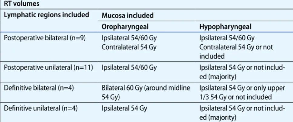
Contralateral level II–III Contralateral OP OP oropharynx, HP hypopharynx, RPN retropharyngeal nodes, SCR supraclavicular lymph node region. aIn case of N2c and bilateral N3 status bilaterally radiation was performed. bLevel IV and V were not involved solitary. Tab. 4 Elective mucosal irradiation. Simplified description (individual adaption due to in-volved lymph node levels). Examples shown in . Fig. 1 and . Fig. 2
RT volumes
Lymphatic regions included Mucosa included
Oropharyngeal Hypopharyngeal
Postoperative bilateral (n=9) Ipsilateral 54/60 Gy Contralateral 54 Gy
Ipsilateral 54/60 Gy Contralateral 54 Gy or not included
Postoperative unilateral (n=11) Ipsilateral 54/60 Gy Ipsilateral 54 Gy or not includ-ed (majority)
Definitive bilateral (n=4) Bilateral 60 Gy (around midline 54 Gy)
Ipsilateral 54 Gy or only upper 1/3 54 Gy or not included Definitive unilateral (n=4) Ipsilateral 54 Gy Ipsilateral 54 Gy or not
Discussion
The goal was to evaluate the effectiveness of individualized IMRT target volumes in patients with cervical lymph nodes of UPC.
Planning treatment volumes
Several study groups compared extended treatment fields as described above with volumes confined to the unilateral lymph node side. As some mostly older studies show extensive radiation of bilateral neckand entire mucosal axis to be superior in locoregional control [5, 6, 7], other stud-ies did not reveal any differences between bilateral and unilateral neck irradiation [8, 9, 10]. In a different approach Barker et al. [11] showed larynx-sparing radio-therapy to result in a high likelihood of locoregional control and survival. Wal-lace et al. [1] also practicing elimination of larynx and hypopharynx from RT por-tals showed comparable outcomes in a larger patient collective (n=179). Patel et al. [23] reserved extended RT for patients at higher risk of locoregional failure such
as N2–3 status. Studies dealing with dif-ferent PTVs in UPC patients are summa-rized in . Tab. 5. Except for sporadic pa-tients being treated with IMRT in those series most of the patients received RT in the pre-IMRT area.
Intensity-modulated radiotherapy
IMRT offers the ability to discriminate be-tween different target volumes, to deliver multiple doses to different targets simulta-neously and to reduce the rate and severity of toxicity [15, 19]. Taking those potentialFig. 1 9 Example of a post-operative bilateral SIB-IMRT. Planning treatment volumes (PTV) with affect-ed lymph nodes in left lev-el II, contralateral levlev-el II– III were treated electively up to 54 Gy, red line: PTV1 (TD 66 Gy/2 Gy), blue line: PTV2 (TD 60 Gy/1.8 Gy), green line: PTV 3 (TD 54 Gy/1.64 Gy)
Fig. 2 9 Example of a de-finitive unilateral SIB-IMRT. Planning treatment vol-umes (PTV) with affect-ed lymph nodes in left lev-el III.red line: PTV1 (TD 69.63 Gy/2.11 Gy), blue line: PTV2 (TD 59.4 Gy/1.8 Gy), green line: PTV 3 (TD 54.12 Gy/1.64 Gy)
Original article
advantages of IMRT into account several recent studies have been published show-ing the feasibility of this technique in pa-tients with UPC. The study with the larg-est patient group (n=52) presented a lo-cal control rate and a locoregional control rate after 5 years of 94 and 88%, respec-tively ([15], Tab. 6). Nevertheless, in all IMRT studies standardized bilateral radi-ation fields were used for all patients.
Individualized IMRT
treatment volumes
For every patient in our cohort the RT vol-ume was defined depending on the poten-tial risk of lymph node involvement ac-cording to Eisbruch et al. [24, 25]. This
ap-proach was adapted to patients with UPC. A summary of our dose and PTV sched-ule is shown in . Tab. 3 and. Tab. 4. The primary goal was to avoid extensive vol-umes while treating patients without on-cological compromise. Boost dose was de-livered to the involved lymph node areas which were usually level II–III/IV. Adja-cent unilateral lymph node levels were al-so included and treated up to 54 Gy or 60– 66 Gy, respectively. The rates of mucosal and nodal control and overall survival are comparable with findings in other stud-ies (see . Tab. 5 and. Tab. 6). The 2 pa-tients in our study with persistent disease had large tumor masses of 63 and 116 ccm, respectively. No patient developed a newly diagnosed locoregional failure.
The advantages of IMRT in sparing the parotid glands, pharyngeal tissues, oral mucosa and mandible bone are well documented. In our study, bilateral SIB-IMRT was carried out in 13/28 (46%) pa-tients. In those cases, the mean dose of the contralateral parotid was kept under 20 Gy. Chen et al. [13] observed a median dose of 23.3 Gy to the contralateral parot-id gland (compared to 50.5 Gy in conven-tional 3D treatment). In case of unilateral treatment the contralateral parotid gland only received a median dose of 6.9 Gy in our study which translated to no grade II or higher xerostomia. Another advan-tage of IMRT in unilateral treatment is the ability to reduce dysphagia while sparing the pharyngeal constrictors. In our series only one patient was in need of a tempo-rary gastric tube.
Chen et al. [14] showed concurrent chemoradiation to be associated with sig-nificant more toxicity without a clear ad-vantage to overall survival and locore-gional control in UPC patients. Lacking larger randomized trials in patients with UPC, concomitant systemic therapy was generously given as it was shown to be su-perior in head in neck cancer patients in general especially in presence of certain risk factors like positive resection mar-gins and extra capsular spread [22, 26, 27, 28]. This is supported by Shoushtari et al. [17] who recommend the addition of
che-Fig. 3 9 Example of a post-operative unilateral SIB-IMRT including the contra-lateral mucosa. Planning treatment volumes (PTV) with affected lymph nodes in left level II. red line: PTV1 (TD 66 Gy/2 Gy), blue line: PTV2 (TD 60 Gy/1.8 Gy), green line: PTV 3 (TD 54 Gy/1.64 Gy) 0 .2 .4 .6 .8 1 Cum. Sur vival 0 10 20 30 40 50 60 70 80 months NC OAS DMFS
Fig. 4 9 Neck con-trol (NC), distant me-tastasis-free surviv-al (DMFS), and over-all survival (OAS) for patients with cervi-cal lymph nodes of un-known primary can-cer (UPC) treated with SIB-IMRT
Tab. 5 Studies using different radiation–volume concepts in patients with unknown primary cancer Study, year Patients (n) Surgery only (%) RT (%) RT + ND (%) CTx (%) RT volume 5-year OS (%) 5-year NC (%) 5-year DFS (%) Unilateral Bilateral Reddy and Marks 1997 [5]
52 0 40 60 0 Neck: 31% Neck incl. NP, OP,
HP: 69% 40 73 ipsilat 51 Grau et al. 2000 [7] 277 8 77 9 0 Neck only: 9%
Neck incl. NP OP, HP,LA: 91%
36 51 48
Christiansen 2005 [29]
28 0 39 61 18 Neck: 11% Neck (incl. possible
primary site): 89% 40 73 Barker et al. 2005 [11] 17 0 29 71 6 Neck + NP, OP 82 88 Beldi et al. 2007 [6] 113 0 48 52 19 Neck: 29% Neck:12% Neck + NP, OP, HP: 59% 41 27 Patel et al. 2007 [23]
70 11 65 14 0 Neck: 82% Neck + NP, OP, HP,
LA 18% 56 84 ipsilat 93 contralat. 62 Ligey et al. 2009 [8] 95 0 17 83 45 Neck: 57% Neck + mu-cosa: 5% Neck: 1% Neck + mucosa: 37% 24 Wallace et al. 2011 [1]
179 0 39 61 7 Neck: 3% Neck plus: OC, OP:
4% OP, NP: 15% OP, HP: 1% NP, OP, HP: 77% 52 81 73 Fakhrian et al. 2012 [9]
65 0 6 94 56 Neck: 26% Neck, NP, OP, HP,
LA: 74% 48 48 Total 844 0 6–77 9–94 0–56 47 range: 24–82 76 range: 51–93 57 range: 27–88 RPN retropharyngeal nodes, NP nasopharynx, OP oropharynx, HP hypopharynx, LA larynx, OC oral cavity, CTx chemotherapy, OS overall survival, NC neck control, DFS disease-free survival.
Tab. 6 Selective studies using IMRT in patients with cervical lymph node metastases of unknown primary cancer
Study (ref) year Pat RT (%) RT + ND (%) CTx (%) RT volume OS (%) NC (%) DFS (%) Unilateral Bilateral
Lu 2008 [16] 18 50 50 33 Bilateral neck + putative mucosal site incl. NP, OP, RPN
74.2 (2 years) 88.3 (2 years)
88.2 (2 years) Madani et al.
2008 [19]
23 17 83 13 Bilateral neck + extended putative
mucosal 74.8 (2 years) 87 (2 years) 76.3 (2 years) Frank et al. 2010 [15]
52 75 25 27 Bilateral neck (contralateral without Level I/V) entire pharyngeal axis 33%: without HP, LA 89 (5 years) 94 (5 years) 88 (5 years) Shoushtari et al.2011 [17]
27 15 75 30 bilateral neck + RPN, Waldeyer’s
ring 70.9 (5 years) 88.5 (5 years) 85.2 (5 years) Sher 2011 [30]
24 54 46 92 Bilateral neck + musosa incl. NP, OP, HP, LA 92 (2 years) 100 (2 years) 96 (2 years) Chen et al. 2011 [13, 14]
27 30 70 63 Bilateral neck + mucosal axis (incl, NP, OP, LA, HP) 86 (2 years) 89 (2 years) 84 (2 years) Villeneuve et al. 2012 [18]
25 93 7 72 Bilateral neck + ipsilateral putative pharyngeal mucosa (NP, OP, HP, LA)
100 (3 years) 100 (3 years) 100 (3 years) Own cohort 2014 28 29 71 71 Risk adapted
Unilateral: 54% bilateral: 46% 76 (3 years) 93 (3 years) 81 (3 years) Total 224 15–93 7–83 13–92 n=15 (own) n=209 (93%) 83 (2–5 years) range: 71–100 93 (2–5 years) range: 87–100 86 (2–5 years) range:76–100
RPN retropharyngeal nodes, NP nasopharynx, OP oropharynx, HP ypopharynx, LA larynx, ND neck dissection, RT radiotherapy, CTx chemotherapy, OS overall survival, NC neck control, DFS disease-free survival.
motherapy in UPC cancer patients with extracapsular extension and bulky N2 or N3 disease.
To summarize, our results are com-parable to modern IMRT studies as well as studies evaluating reduced treatment fields (. Tab. 5, 6). We observed no grade II or higher late squeals so far. We tried to establish some factors standing for a high-er risk for potential contralathigh-eral disease and/or mucosa involvement. Those risk factors are of course not evidenced based and to some extend subjectively motivat-ed by the treating radio-oncologist. Nev-ertheless, in the lack of randomized tri-als, this approach seems to be effective in terms of locoregional control so far and should be confirmed in a larger patient cohort with longer follow-up.
Conclusion
Risk-adapted individualized reduction of PTVs is feasible in IMRT treatment of cervical lymph node metastases of UPC showing high mucosal and nodal con- trol rates and a very good treatment tol-erance.Corresponding address
Prof. Dr. G. StuderDepartment of Radiation Oncology, University Hospital Zurich
Rämistr. 1000, 8091 Zurich Switzerland
gabriela.studer@usz.ch
Compliance with ethical
guidelines
Conflict of interest. S. Janssen, C. Glanzmann, G. Hu-ber, and G. Studer state that there are no conflicts of interest.
The accompanying manuscript does not include stud-ies on humans or animals.
References
1. Wallace A, Richards GM, Harari PM et al (2011) Head and neck squamous cell carcinoma from an unknown primary site. Am J Otolaryngol 32:286– 290
2. Jereczek-Fossa BA, Jassem J, Orecchia R (2004) Cer-vical lymph node metastases of squamous cell car-cinoma from an unknown primary. Cancer Treat Rev 30:153–164
3. Balaker AE, Abemayor E, Elashoff D, St John MA (2012) Cancer of unknown primary: does treat-ment modality make a difference? Laryngoscope 122:1279–1282
4. Strojan P, Ferlito A, Medina JE et al (2013) Contem-porary management of lymph node metastases from an unknown primary to the neck: I. A review of diagnostic approaches. Head Neck 35:123–132 5. Reddy SP, Marks JE (1997) Metastatic carcinoma in
the cervical lymph nodes from an unknown pri-mary site: results of bilateral neck plus mucosal ir-radiation vs. ipsilateral neck irir-radiation. Int J Radiat Oncol Biol Phys 37:797–802
6. Beldi D, Jereczek-Fossa BA, D’Onofrio A et al (2007) Role of radiotherapy in the treatment of cervical lymph node metastases from an unknown prima-ry site: retrospective analysis of 113 patients. Int J Radiat Oncol Biol Phys 69:1051–1058
7. Grau C, Johansen LV, Jakobsen J et al (2000) Cervi-cal lymph node metastases from unknown prima-ry tumours—results from a national survey by the Danish Society for Head and Neck Oncology. Ra-diother Oncol 55:121–129
8. Ligey A, Gentil J, Crehange G et al (2009) Impact of target volumes and radiation technique on lo-co-regional control and survival for patients with unilateral cervical lymph node metastases from an unknown primary. Radiother Oncol 93:483–487 9. Fakhrian K, Thamm R, Knapp S et al (2012)
Radio(chemo)therapy in the management of squamous cell carcinoma of cervical lymph nodes from an unknown primary site. A retrospective analysis. Strahlenther Onkol 188:56–61
10. Nieder C, Gregoire V, Ang KK (2001) Cervical lymph node metastases from occult squamous cell carci-noma: cut down a tree to get an apple? Int J Radiat Oncol Biol Phys 50:727–733
11. Barker CA, Morris CG, Mendenhall WM (2005) Lar-ynx-sparing radiotherapy for squamous cell carci-noma from an unknown head and neck primary site. Am J Clin Oncol 28:445–448
12. Lee N, Puri DR, Blanco AI, Chao KS (2007) Intensi-ty-modulated radiation therapy in head and neck cancers: an update. Head Neck 29:387–400 13. Chen AM, Li BQ, Farwell DG et al (2011) Improved
dosimetric and clinical outcomes with intensity-modulated radiotherapy for head-and-neck cancer of unknown primary origin. Int J Radiat Oncol Biol Phys 79:756–762
14. Chen AM, Farwell DG, Lau DH et al (2011) Radi-ation therapy in the management of head-and-neck cancer of unknown primary origin: how does the addition of concurrent chemotherapy affect the therapeutic ratio? Int J Radiat Oncol Biol Phys 81:346–352
15. Frank SJ, Rosenthal DI, Petsuksiri J et al (2010) In-tensity-modulated radiotherapy for cervical node squamous cell carcinoma metastases from un-known head-and-neck primary site: M. D. Ander-son Cancer Center outcomes and patterns of fail-ure. Int J Radiat Oncol Biol Phys 78:1005–1010 16. Lu H, Yao M, Tan H (2009) Unknown primary head
and neck cancer treated with intensity-modulat-ed radiation therapy: to what extent the volume should be irradiated. Oral Oncol 45:474–479 17. Shoushtari A, Saylor D, Kerr KL et al (2011)
Out-comes of patients with head-and-neck cancer of unknown primary origin treated with intensity-modulated radiotherapy. Int J Radiat Oncol Biol Phys 81:e83–e91
18. Villeneuve H, Despres P, Fortin B et al (2012) Cervi-cal lymph node metastases from unknown prima-ry cancer: a single-institution experience with in-tensity-modulated radiotherapy. Int J Radiat Oncol Biol Phys 82:1866–1871
19. Madani I, Vakaet L, Bonte K et al (2008) Intensity-modulated radiotherapy for cervical lymph node metastases from unknown primary cancer. Int J Radiat Oncol Biol Phys 71:1158–1166
20. Studer G, Huguenin PU, Davis JB et al (2006) IMRT using simultaneously integrated boost (SIB) in head and neck cancer patients. Radiat Oncol 1:7 21. Studer G, Furrer K, Davis BJ et al (2006)
Postoper-ative IMRT in head and neck cancer. Radiat Oncol 1:40
22. Bonner JA, Harari PM, Giralt J et al (2006) Radio-therapy plus cetuximab for squamous-cell carcino-ma of the head and neck. N Engl J Med 354:567– 578
23. Patel RS, Clark J, Wyten R et al (2007) Squamous cell carcinoma from an unknown head and neck primary site: a “selective treatment” approach. Arch Otolaryngol Head Neck Surg 133:1282–1287 24. Eisbruch A, Marsh LH, Dawson LA et al (2004)
Re-currences near base of skull after IMRT for head-and-neck cancer: implications for target delinea-tion in high neck and for parotid gland sparing. Int J Radiat Oncol Biol Phys 59:28–42
25. Eisbruch A, Foote RL, O’Sullivan B et al (2002) In-tensity-modulated radiation therapy for head and neck cancer: emphasis on the selection and delin-eation of the targets. Semin Radiat Oncol 12:238– 249
26. Bernier J, Domenge C, Ozsahin M et al (2004) Post-operative irradiation with or without concomitant chemotherapy for locally advanced head and neck cancer. N Engl J Med 350:1945–1952
27. Forastiere AA, Goepfert H, Maor M et al (2003) Concurrent chemotherapy and radiotherapy for organ preservation in advanced laryngeal cancer. N Engl J Med 349:2091–2098
28. Cooper JS, Pajak TF, Forastiere AA et al (2004) Post-operative concurrent radiotherapy and chemo-therapy for high-risk squamous-cell carcinoma of the head and neck. N Engl J Med 350:1937–1944 29. Christiansen H, Hermann RM, Martin A et al. Neck
lymph node metastases from an unknown prima-ry tumor retrospective study and review of litera-ture. Strahlenther Onkol. 2005; 181: 355–356 30. Sher DJ, Balboni TA, Haddad RI et al. Efficacy and
toxicity of chemoradiotherapy using intensity-modulated radiotherapy for unknown primary of head and neck. Int J Radiat Oncol Biol Phys. 2011; 80:1405–1411