Open Peer Review F1000 Faculty Reviews are commissioned from members of the prestigious F1000 . In order to make these reviews as Faculty comprehensive and accessible as possible, peer review takes place before publication; the referees are listed below, but their reports are not formally published.
Discuss this article (0) Comments
REVIEW
Endoplasmic reticulum turnover: ER-phagy and other flavors in
selective and non-selective ER clearance [version 1; referees: 2
approved]
Ilaria Fregno
, Maurizio Molinari
1,2,4
Università della Svizzera italiana, Via G. Buffi, CH-6900 Lugano, Switzerland Institute for Research in Biomedicine, Via V. Vela 6, CH-6500 Bellinzona, Switzerland Department of Biology, Swiss Federal Institute of Technology, Wolfgang-Pauli-Strasse 27, CH-8093 Zurich, Switzerland École Polytechnique Fédérale de Lausanne, School of Life Sciences, EPFL Station 19, CH-1015 Lausanne, Switzerland Abstract The endoplasmic reticulum (ER) is a highly dynamic organelle in eukaryotic cells. It is deputed to lipid and protein biosynthesis, calcium storage, and the detoxification of various exogenous and endogenous harmful compounds. ER activity and size must be adapted rapidly to environmental and developmental conditions or biosynthetic demand. This is achieved on induction of thoroughly studied transcriptional/translational programs defined as “unfolded protein responses” that increase the ER volume and the expression of ER-resident proteins regulating the numerous ER functions. Less understood are the lysosomal catabolic processes that maintain ER size at steady state, that prevent excessive ER expansion during ER stresses, or that ensure return to physiologic ER size during recovery from ER stresses. These catabolic processes may also be activated to remove ER subdomains where proteasome-resistant misfolded proteins or damaged lipids have been segregated. Insights into these catabolic mechanisms have only recently emerged with the identification of so-called ER-phagy receptors, which label specific ER subdomains for selective lysosomal delivery for clearance. Here, in eight chapters and one addendum, we comment on recent advances in ER turnover pathways induced by ER stress, nutrient deprivation, misfolded proteins, and live bacteria. We highlight the role of yeast (Atg39 and Atg40) and mammalian (FAM134B, SEC62, RTN3, and CCPG1) ER-phagy receptors and of autophagy genes in selective and non-selective catabolic processes that regulate cellular proteostasis by controlling ER size, turnover, and function. Keywords Autophagy and macroautophagy, ER turnover, ER-phagy and recovER-phagy receptors, ER stress, LC3-interacting region (LIR), live bacteria, nutrient deprivation, proteostasis1-3
1,2,4
1 2 3 4 Referee Status: Invited Referees version 1 published 13 Apr 2018 1 2 , Telethon Institute of Carmine Settembre Genetics and Medicine (TIGEM), Italy Federico II University, Italy 1 , George Wise Gerardo Lederkremer Faculty of Life Sciences, Tel Aviv University, Israel 2 13 Apr 2018, (F1000 Faculty Rev):454 (doi: First published: 7 ) 10.12688/f1000research.13968.1 13 Apr 2018, (F1000 Faculty Rev):454 (doi: Latest published: 7 ) 10.12688/f1000research.13968.1v1
Maurizio Molinari ( )
Corresponding author: maurizio.molinari@irb.usi.ch
: Writing – Original Draft Preparation; : Conceptualization, Funding Acquisition, Writing – Original Draft
Author roles: Fregno I Molinari M
Preparation No competing interests were disclosed. Competing interests: MM is the recipient of a Sinergia grant of the Swiss National Science Foundation (SNF) and is supported by Signora Grant information: Alessandra, the AlphaONE Foundation, the Foundation for Research on Neurodegenerative Diseases, the Novartis Foundation, SNF, and the Comel and Gelu Foundations.
The funders had no role in study design, data collection and analysis, decision to publish, or preparation of the manuscript.
© 2018 Fregno I and Molinari M. This is an open access article distributed under the terms of the
Copyright: Creative Commons Attribution
, which permits unrestricted use, distribution, and reproduction in any medium, provided the original work is properly cited. Licence
Fregno I and Molinari M.
How to cite this article: Endoplasmic reticulum turnover: ER-phagy and other flavors in selective and
2018, (F1000 Faculty Rev):454 (doi:
non-selective ER clearance [version 1; referees: 2 approved] F1000Research 7
) 10.12688/f1000research.13968.1
13 Apr 2018, (F1000 Faculty Rev):454 (doi: )
The history behind regulated endoplasmic reticulum turnover
The volume (and the activity) of the endoplasmic reticulum (ER) can be expanded on demand. Activation of quiescent B cells into antibody-producing plasma cells is a textbook example of the daunting expansion of this biosynthetic organelle to accom-modate and assist the maturation of a flood of cargo entering the secretory pathway (in this specific case, thousands of immu-noglobulin chains produced per second)1. In general terms, ER
expansion is one of the many cellular responses to ER stresses, which collectively are defined as unfolded protein responses (UPR). Logically, recovery of pre-stress homeostasis upon ER stress resolution or cessation of drug (e.g. phenobarbital) treatments requires dismantling of the excess ER and degrada-tion of the ER chaperones produced during the stress phase2. It
is in these contexts that first indications of catabolic control of ER size emerged3–5. However, hints of lysosomal-regulated
ER turnover can be found in earlier literature (for example, in 6). Selective ER turnover is a constitutive process that maintains ER size in Eukarya. It can be enhanced or induced on micro-tubule de-polymerization7 and, as described below, during ER
stress, during recovery from acute ER stress, on ER overload with membrane or proteasome-resistant misfolded proteins, under nutrient deprivation, or on pathogen attack. It relies on intricate mechanisms that we are just starting to understand.
Yeast flavors of ER-phagy
Chapter one: the origin of a name. ER-phagy as ER stress-induced microautophagy of the ER
The term ER-phagy is a contraction of the words “ER” and “macroautophagy”. It was coined by the group of Peter Walter to define the process of selective ER delivery (not of selec-tive ER clearance) in the vacuole of yeast cells exposed to per-turbation of the redox homeostasis to induce a UPR8. Initially,
ER-phagy was described as a conventional macroautophagic pathway with autophagy genes alleviating cell growth defects under ER stress conditions and regulating the engulfment of ER fragments within double-membrane autophagosomes that eventually fuse with vacuoles to clear their content8,9. None of
these original findings survived the test of time. In fact, further analyses by the same group revealed that neither autophagy genes nor autophagosomes are actually involved in the vacuolar delivery of excess ER upon perturbation of redox homeostasis or play a role in the survival of yeast cells challenged with the reducing agent dithiothreitol (DTT). Moreover, it turns out that ER delivery to the vacuole under these experimental conditions
does not result in ER clearance10 (Table 1). Rather, on chemical
UPR induction, excess ER membranes are delivered within the vacuole by poorly understood micropinocytosis events. Importantly, because DTT inactivates vacuolar activity, the ER delivered into the vacuole under these experimental conditions accumulates in the vacuolar lumen as ER whorls10. The
path-ways and the yeast genes regulating ER fragmentation, selec-tive delivery of ER whorls within vacuoles (is it controlled by ER-phagy receptors?), and capture of ER by microau-tophagic invagination of the vacuolar membrane remain to be identified.
Chapter two: membrane protein-induced macroautophagy of the ER
Overexpression of integral membrane proteins is yet another manner to activate/enhance delivery of ER to the yeast vacuole11,12. This process is highly selective because
endog-enous markers of the ER membrane (Sec61 and Hmg1) but not intrinsic or peripheral proteins of ER exit sites (Sec12 and Sec13) or the luminal ER protein Kar2p are delivered to the vacuole with the exogenous integral protein(s) to be cleared from cells11. Membrane protein-induced delivery of ER material
to the yeast vacuole is under the control of Ypt1 (the orthologue of mammalian RAB1) to generate ER-to-autophagy membrane structures, of Ypt51 (Vps2) to promote fusion of ER membrane proteins-containing structures with the vacuole, and of select macroautophagy gene products (Atg1, Atg2, Atg8, Atg9, and Atg11 but not Atg17, Atg18, Atg19, Atg32, and Atg3611; for
a description of the function of the various autophagy gene products mentioned in this commentary, please refer to 13,14) (Table 1). The role of Atg39 and Atg40 as ER-phagy receptors in nutrient deprivation-induced yeast ER-phagy was published subsequently (chapter three in this commentary and 15). To our knowledge, a possible involvement of Atg39 or Atg40 (or both) in membrane protein-induced selective macroautophagy remains to be investigated.
Chapter three: nutrient deprivation-induced, receptor (Atg39 and Atg40)-mediated macroautophagy of the ER (and of the nuclear envelope)
Nutrient deprivation and rapamycin are conventional activators of macroautophagy16. Like cell exposure to DTT10, these
experimental conditions enhance the delivery of ER frag-ments within the yeast vacuole. However, as crucial differences (compare with chapter one), vacuolar delivery eventually results in ER clearance15,17 and relies on macroautophagy-like
Table 1. Yeast ER-phagy types and their requirements.
ER-phagy type (yeast) Receptor Gene products required Gene products dispensable
Dithiothreitol-induced microER-phagy8–10 Unknown Unknown Atg1, Atg7, Atg8, Atg16, Ego1, Ego3, Vtc4, Nvj1, Pep4, Vps4, Vps23 Membrane protein-induced
macroER-phagy11 Unknown Atg1, Atg2, Atg8, Atg9, Atg11, Ypt1, Ypt51 Atg17, Atg18, Atg19, Atg32, Atg36 Nutrient
programs, where receptor-mediated engulfment of ER frag-ments into double-membrane autophagosomes that eventually fuse with lysosomes does indeed occur15,17,18. Atg39 spans the
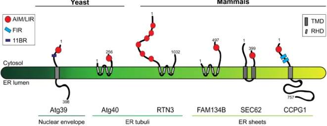
perinuclear ER membrane and decorates it for capture by autophagosomes and destruction. Instead, Atg40 is in the peripheral ER membrane, where it co-localizes with Rtn1, a marker of ER tubules and sheet edges, but not with the ER sheet marker Sec63 and regulates the turnover of highly curved ER subdomains (Figure 1). Atg39 and Atg40 contain an Atg8- interacting motif (AIM) that bridges the ER membrane with the phagophore membrane via Atg8 association. Moreover, Atg40 (and possibly Atg39) contains an Atg11-binding region (11BR,
Figure 1) and harbors two transmembrane domains that have some similarities with reticulon domains displayed by some mammalian ER-phagy receptors (15, see below). These domains supposedly curve the ER membrane to facilitate the ER fragmentation required for engulfment by autophagosomes. So far, it has been established that nutrient deprivation-induced, Atg39- and Atg40-mediated macroautophagy of the yeast nuclear envelope and ER requires Atg1 (ULK1 in mammals), Atg8 (LC3), Atg11 and Atg17, Ypt7 (RAB7, a small GTPase required for autophagosome-vacuole fusion), and Pep4 (a vacuolar peptidase)15 (Table 1).
Mammalian flavors of ER-phagy
Chapter four: nutrient deprivation-induced, receptor (FAM134B or RTN3)-mediated macroautophagy of the ER
As in yeast cells, starvation activates receptor-mediated engulf-ment of mammalian ER fragengulf-ments into double-membrane autophagosomes that eventually fuse with lysosomes to clear their cargo19,20. The reticulon family members FAM134B and
RTN3 reside to the edges of ER sheets and ER tubuli, respectively (Figure 1). FAM134B was identified in a yeast two-hybrid screen as a LC3/GABARAP interactor19. RTN3 was found to
re-localize from ER tubuli to LC3-decorated structures on cell
starvation20. Upon oligomerization, FAM134B and RTN3
frag-ment ER sheets and tubuli, respectively, and mediate the capture of these ER subdomains by autophagosomes via their LC3- interacting regions (LIRs). As such, different ER-phagy receptors regulate the turnover of distinct populations of ER-resident proteins, according to the subcompartmental distribution of the latter in sheets (for example, CLIMP63 and RTN4B) or in tubuli (RTN1, RTN3, REEP5, and CNX). Since inactivation of ER-phagy receptors causes ER expansion, ER turnover is a constitutive process that also determines ER size at steady state. So far, it has been established that nutrient deprivation- induced, FAM134B- and RTN3-mediated ER-phagy requires BCN1, ATG5, ATG7, LC3, and FIP20019,20 (Table 2).
Chapter five: receptor (SEC62)-mediated clearance of excess ER on resolution of acute ER stresses (recovER-phagy)
Pathologic (for example, expression of mutant polypeptides) or physiologic (for example, B- to plasma-cell differentiation) biosynthetic challenges may induce ER stresses that increase the ER volume to almost entirely occupy the space delimited by the plasma membrane (http://www.drjastrow.de/WAI/EM/EMRERE. html). This can be recapitulated by challenging cells with chemical compounds or by therapeutic interventions that perturb ER homeostasis21. Alleviation of the stress obtained on
inter-ruption of the challenge allows cells to re-establish pre-stress homeostatic conditions by lysosomal clearance of the excess ER membranes and proteins produced during the stress phase2–5. This
catabolic process, defined as recovER-phagy (for ER-phagy-mediated recovery from ER stresses2), is characterized by
selec-tive clearance of ER subdomains displaying the LC3-binding protein and (recov)ER-phagy receptor SEC62 at the limiting membrane2 (Figure 1). SEC62 plays a well-characterized role
as a component of the protein import machinery into the ER22.
Bioinformatics analyses revealed the presence of a LIR in the
Figure 1. Yeast and mammalian ER-phagy receptors. The figure shows the topology, orientation, and subcompartmental localization of the two yeast ER-phagy receptors Atg39 and Atg40 and of the four mammalian ER-phagy receptors RTN3, CCPG1, FAM134B, and SEC62. Numbers indicate amino acid residues and refer to the human version of the proteins for the mammalian ER-phagy receptors. 11BR, Atg11-binding region; AIM, Atg8-interacting motif; ER, endoplasmic reticulum; FIR, FIP200-interacting region; LIR, LC3-interacting region; RHD, reticulon-homology domain; TMD, transmembrane domain.
Table 2. Mammalian ER-phagy types and their requirements.
ER-phagy type (mammalian) Receptor Gene products required Gene products dispensable
Nutrient deprivation-induced
ER-phagy19,20 FAM134B BCN1, FIP200, ATG5, LC3 RTN3
RTN3 FIP200, ATG5, ATG7, LC3 FAM134B
Nutrient deprivation-induced/
ER stress-induced ER-phagy23 CCPG1 FIP200, ATG5, LC3 Unknown
RecovER-phagy2 SEC62 ATG5, ATG7, LC3 Unknown
Microbial-induced ER-phagy24 Unknown BCN1, FIP200, ATG7,
ATG14, ATG16L1 FAM134B
cytosolic C-terminus of SEC62, which is conserved in higher Eukarya but not in the yeast orthologue. LC3 binding to SEC62 has been mapped by docking and molecular dynamics simu-lations and by solution nuclear magnetic resonance spectros-copy. It has been confirmed by surface plasmon resonance and peptide array and eventually in the living cell, where it proved to be required for the delivery of ER fragments within LAMP1- positive degradative organelles2. Proteomic analyses revealed
that the ER subdomains cleared by recovER-phagy do contain several ER-resident molecular chaperones and enzymes (for example, all members of the protein disulfide isomerase super-family with the relevant exception of ERp44, which resides not in the ER but in the intermediate compartment25) but are devoid of
other ER-resident proteins such as most ERAD regulators2. This
reveals a spatial separation of ER functions in ER subdomains. So far, it has been established that SEC62-mediated recovER- phagy requires ATG5, ATG7, and LC3 (Table 2).
Chapter six: ER stress-induced receptor (CCPG1)-mediated macroautophagy of the ER
CCPG1 is the new entry in the ER-phagy receptors clan. It has been identified as an ER stress-induced interactor of GABARAP and LC323,26. It is a single-pass, type II ER protein
that associates directly with FIP200 and indirectly with other components of the ULK complex involved in autophagosome biogenesis (that is, ULK1, ATG13, and ATG101)23 (Figure 1).
Ectopic expression of CCPG1 reduces the size of the periph-eral ER and the cellular content of the periphperiph-eral/tubular ER protein RTN3. Likewise, induction of endogenous CCPG1 in cells challenged with the chemical ER stress inducer DTT activates lysosomal depletion of peripheral ER. DTT induction of yeast ER-phagy results in microautophagy-like vacuolar capture of the ER, which does not require intervention of autophagy gene products and does not lead to ER degrada-tion (Chapter one10). Thus, the mechanism of ER size control
in cells experiencing chemically induced ER stresses is poorly conserved, if at all. Of note, FAM134B deficiency is linked to human sensory neuropathy19, whereas studies in mice reveal that
CCPG1 deficiency causes injury of exocrine pancreas cells and of chief cells of the gastric epithelium23. These differences reveal
poorly understood tissue-specific functions of the two ER-phagy
receptors. CCPG1-mediated ER-phagy requires ATG5 and FIP200 (Table 2).
Chapter seven: misfolded protein-induced macroautophagy of the ER (ER quality control autophagy)
Most misfolded polypeptides produced in the ER are translo-cated across the ER membrane and are degraded by cytosolic 26S proteasomes27. However, the polymerogenic E342K (ATZ)
variant of the secretory protein alpha1 antitrypsin28–31, the E90K
mutant of the gonadotropin-releasing hormone receptor (GnRHR)32, aggregated β subunits of thyrotrophic hormone33,
procollagen34,35, and dysferlin36 are a few paradigmatic examples
of proteasome-resistant proteins generated in the ER and degraded, at least in part, by the lysosomal system. For ATZ, the involvement of classic autophagy regulators such as ATG5, ATG7, and LC3 has been shown31. However, for this and other
proteasome-resistant misfolded polypeptides such as GnRHR and β subunits of thyrotrophic hormone, the intervention of classic (mTOR-dependent) macroautophagy pathways in clear-ance is questionable. In fact, macroautophagy inducers such as rapamycin and starvation do not enhance their lysosomal disposal30,32,37,38, which seems to actually require vesicular
traffic from the ER to lysosomal degradative compartments33,39.
In these cases, the autophagy genes involved in these catabolic processes might intervene in an ill-defined, unconventional manner. Even if/when conventional macroautophagy deter-mines the clearance of proteasome-resistant misfolded proteins generated in the ER lumen, it remains unclear whether these are dislocated across the ER membrane and subsequently are cap-tured by autophagosomes or whether autophagosomes capture ER fragments containing aberrant protein aggregates to then fuse to lysosomes31. In the latter case, it remains to be established
how the autophagic machinery operating in the cytosol can detect the accumulation in the ER lumen of proteasome- resistant species to be removed from cells (see “Final remarks”). All in all, the involvement of peculiar pathways (autophagy-like or vesicular transport or both), of ER-resident sensors that transmit the information from the ER lumen into the cytosol to activate autophagy, and of ER-phagy receptors to promote the catabolic process that removes the ER subdomain from cells seems to be an educated guess.
Chapter eight: microbial-induced ER-phagy
Cyclic-di-adenosine monophosphate is a signature of live Gram-positive bacteria, which is detected by phagocytes to trigger a protective ER stress response consisting of ER expansion and chaperone gene upregulation. Subsequently, ER stress-induced PERK phosphorylation correlates with mTORC1 inactivation, thus inducing macroautophagy, which is instrumental for cell survival24. Isolation of autophagosomes from cells challenged
with live bacteria revealed the presence of several ER mark-ers and the absence of markmark-ers of ERGIC, Golgi or lysosome, showing that live bacteria induce selective ER-phagy. A role for FAM134B as a receptor for microbial-induced ER-phagy has been excluded. The involvement of other known ER-phagy receptors has not been tested24. So far, it has been established
that microbial-induced ER-phagy requires ATG7, ATG14, ATG16L1, BCN1, and FIP200 (Table 2).
Promiscuous autophagic receptors in ER-phagy, an addendum: the case of p62 and BNIP3
As originally reported for other drugs3–5, withdrawal of hepatic
mitogens activates the removal of excess ER from liver cells, which is ensured by macroautophagic processes. In these in vivo experiments, ER turnover required ATG5 and the general autophagy receptor Sequestosome1/p6240. In contrast to
conven-tional ER-phagy receptors, which are located in the ER membrane (Figure 1), p62 is a cytosolic protein that links ubiquitylated proteins to be degraded to the autophagic machinery via LC3 interaction. It is therefore likely that p62 regulates the clear-ance of ER regions displaying heavily ubiquitylated proteins at the cytosolic face of the membrane. A second intriguing case of promiscuous receptors involved in ER turnover is that of BNIP3, which is anchored primarily in the outer mitochondrial mem-brane via a C-terminal transmemmem-brane domain41. The BNIP3
homologue NIX/BNIP3L preferentially binds GABARAP42
and regulates the removal of damaged mitochondria43. BNIP3
selectively removes damaged mitochondria on association with LC3B44. The finding that a subfraction of cellular BNIP3 is
also found in the ER membrane led to the postulation that this protein could play a role as an ER-phagy receptor44. This was
experimentally demonstrated only on ectopic expression of a BNIP3 version modified for preferential delivery into the ER membrane44.
Final remarks
Autophagy was once considered a rather unselective pathway to deliver faulty material to lysosomes for clearance. Recent studies reveal the specificity and sophistication of autophagic programs and of programs relying on unconventional roles of autophagy genes45. Organelles such as mitochondria,
peroxisomes, nucleus, and ER can selectively be delivered to the lysosomal pathway for destruction if and when they display receptors at the surface that engage this intricate catabolic machinery46. These receptors are constitutively active, for
example, to control the size of the ER at steady state or in rest-ing cells. They can be activated on demand to recover pre- stress ER size and content or in response to accumulation in specific ER subdomains of misfolded polypeptides that can-not be handled by the ubiquitin proteasome system. The study of ER-phagy actually reveals that not only organelles but also specific (functional) subdomains of an organelle, with their
content, can be selected for destruction. The field is young and relies mostly on studies performed in cells exposed to exogenous stimuli such as nutrient deprivation or chemical stress that activate selective and non-selective ER-phagy and have uncontrolled pleiotropic consequences on many unrelated pathways47. Intrinsic signals (that is, signals originating from
the membrane or the lumen of confined ER subcompartments such as accumulation of proteasome-resistant polypeptides) are predicted to activate highly specific, receptor-controlled pathways relying on different autophagy, autophagy-like, or autophagy-independent lysosomal pathways. We also pre-dict that studies on ER turnover will lead to the identification of ER sensors that, much like ER stress sensors, signal accumulation of proteasome-resistant misfolded proteins or other stressful situations that must be resolved by ER clear-ance. Analysis of the available literature already shows that ER-phagy comprises a series of mechanistically distinct proc-esses that regulate the delivery of ER fragments or their luminal content (or both) within vacuoles/lysosomes. It is proposed, but in most cases not yet experimentally demonstrated, that these catabolic processes regulate ER turnover, ER size, and clear-ance of ER subdomains containing proteins and lipids that are faulty or present in excess. Intriguingly, under some patho-logic conditions (for example, in some serpinopathies28) or in
a subset of patients (for example, 10% of the ATZ patients that show hepatotoxicity due to intracellular accumulation of ATZ polymers31) or in response to severe chemically induced ER
stresses8–10), the ER-derived material accumulates in
autophago-somes or in degradative organelles attesting defective clearance. In other cases, accumulation of ER fragments in degradative organelles occurs only on inactivation of lysosomal hydrolases, rather hinting at a very efficient catabolic process operating to protect cell and organism viability. Current models show that ER fragments are captured by autophagosomes as normally happens for cytosolic material. However, other mechanisms of ER delivery to degradative organelles such as vesicular transport can be envisioned on the basis of available literature33,39. It will be of great interest to study the
involve-ment of the several known autophagy genes in ER turnover to understand why treatment with conventional autophagy activators is beneficial for the clearance of cytosolic aggregates48
and for some (for example, pro-collagen and dysferlin34–36) but
apparently not for other (for example, ATZ and the E90K mutant of GnRHR)30,32,37,38 proteasome-resistant misfolded polypeptides
generated in the ER lumen.
Competing interests
The authors declare that they have no competing interests.
Grant information
MM is the recipient of a Sinergia grant of the Swiss National Science Foundation (SNF) and is supported by Signora Alessandra, the AlphaONE Foundation, the Foundation for Research on Neurodegenerative Diseases, the Novartis Founda-tion, SNF, and the Comel and Gelu Foundations.
The funders had no role in study design, data collection and analysis, decision to publish, or preparation of the manuscript.
References F1000 recommended
1. de StGroth SF, Scheidegger D: Production of monoclonal antibodies: strategy and tactics. J Immunol Methods. 1980; 35(1–2): 1–21.
PubMed Abstract |Publisher Full Text
2. Fumagalli F, Noack J, Bergmann TJ, et al.: Translocon component Sec62 acts in endoplasmic reticulum turnover during stress recovery. Nat Cell Biol.
2016; 18(11): 1173–84.
PubMed Abstract |Publisher Full Text | F1000 Recommendation 3. Bolender RP, Weibel ER: A morphometric study of the removal of
phenobarbital-induced membranes from hepatocytes after cessation of threatment. J Cell Biol. 1973; 56(3): 746–61.
PubMed Abstract |Publisher Full Text |Free Full Text
4. Feldman D, Swarm RL, Becker J: Elimination of excess smooth endoplasmic reticulum after phenobarbital administration. J Histochem Cytochem. 1980; 28(9): 997–1006.
PubMed Abstract |Publisher Full Text
5. Masaki R, Yamamoto A, Tashiro Y: Cytochrome P-450 and NADPH-cytochrome P-450 reductase are degraded in the autolysosomes in rat liver. J Cell Biol.
1987; 104(5): 1207–15.
PubMed Abstract |Publisher Full Text |Free Full Text
6. Locke M, Collins JV: The Structure and Formation of Protein Granules in the Fat Body of an Insect. J Cell Biol. 1965; 26(3): 857–84.
PubMed Abstract |Publisher Full Text |Free Full Text
7. Marzella L, Ahlberg J, Glaumann H: Isolation of autophagic vacuoles from rat liver: morphological and biochemical characterization. J Cell Biol. 1982; 93(1):
144–54.
PubMed Abstract |Publisher Full Text |Free Full Text
8. Bernales S, McDonald KL, Walter P: Autophagy counterbalances endoplasmic reticulum expansion during the unfolded protein response. PLoS
Biol. 2006; 4(12): e423.
PubMed Abstract |Publisher Full Text |Free Full Text | F1000 Recommendation 9. Bernales S, Schuck S, Walter P: ER-phagy: selective autophagy of the
endoplasmic reticulum. Autophagy. 2007; 3(3): 285–7. PubMed Abstract |Publisher Full Text | F1000 Recommendation 10. Schuck S, Gallagher CM, Walter P: ER-phagy mediates selective
degradation of endoplasmic reticulum independently of the core autophagy machinery. J Cell Sci. 2014; 127(Pt 18): 4078–88.
PubMed Abstract |Publisher Full Text |Free Full Text | F1000 Recommendation 11. Lipatova Z, Segev N: A Role for Macro-ER-Phagy in ER Quality Control. PLoS
Genet. 2015; 11(7): e1005390.
PubMed Abstract |Publisher Full Text |Free Full Text
12. Fu L, Sztul E: ER-associated complexes (ERACs) containing aggregated cystic fibrosis transmembrane conductance regulator (CFTR) are degraded by autophagy. Eur J Cell Biol. 2009; 88(4): 215–26.
PubMed Abstract |Publisher Full Text |Free Full Text
13. Hurley JH, Young LN: Mechanisms of Autophagy Initiation. Annu Rev Biochem.
2017; 86: 225–44.
PubMed Abstract |Publisher Full Text |Free Full Text
14. Bento CF, Renna M, Ghislat G, et al.: Mammalian Autophagy: How Does It Work?
Annu Rev Biochem. 2016; 85: 685–713. PubMed Abstract |Publisher Full Text
15. Mochida K, Oikawa Y, Kimura Y, et al.: Receptor-mediated selective autophagy degrades the endoplasmic reticulum and the nucleus. Nature. 2015; 522(7556):
359–62.
PubMed Abstract |Publisher Full Text
16. Klionsky DJ, Abdelmohsen K, Abe A, et al.: Guidelines for the use and interpretation of assays for monitoring autophagy (3rd edition). Autophagy.
2016; 12(1): 1–222.
PubMed Abstract |Publisher Full Text |Free Full Text
17. Hamasaki M, Noda T, Baba M, et al.: Starvation triggers the delivery of the endoplasmic reticulum to the vacuole via autophagy in yeast. Traffic. 2005; 6(1): 56–65.
PubMed Abstract |Publisher Full Text
18. Rubinsztein DC: Cell biology: Receptors for selective recycling. Nature. 2015; 522(7556): 291–2.
PubMed Abstract |Publisher Full Text
19. Khaminets A, Heinrich T, Mari M, et al.: Regulation of endoplasmic reticulum turnover by selective autophagy. Nature. 2015; 522(7556): 354–8.
PubMed Abstract |Publisher Full Text | F1000 Recommendation
20. Grumati P, Morozzi G, Hölper S, et al.: Full length RTN3 regulates turnover of tubular endoplasmic reticulum via selective autophagy. eLife. 2017; 6:
pii: e25555.
PubMed Abstract |Publisher Full Text |Free Full Text | F1000 Recommendation 21. Oslowski CM, Urano F: Measuring ER stress and the unfolded protein response using mammalian tissue culture system. Methods Enzymol. 2011; 490: 71–92. PubMed Abstract |Publisher Full Text |Free Full Text
22. Meyer HA, Grau H, Kraft R, et al.: Mammalian Sec61 is associated with Sec62
and Sec63. J Biol Chem. 2000; 275(19): 14550–7. PubMed Abstract |Publisher Full Text
23. Smith MD, Harley ME, Kemp AJ, et al.: CCPG1 Is a Non-canonical Autophagy Cargo Receptor Essential for ER-Phagy and Pancreatic ER Proteostasis. Dev Cell. 2018; 44(2): 217–232.e11.
PubMed Abstract |Publisher Full Text |Free Full Text | F1000 Recommendation 24. Moretti J, Roy S, Bozec D, et al.: STING Senses Microbial Viability to
Orchestrate Stress-Mediated Autophagy of the Endoplasmic Reticulum. Cell.
2017; 171(4): 809–823.e13.
PubMed Abstract |Publisher Full Text |Free Full Text | F1000 Recommendation 25. Anelli T, Sannino S, Sitia R: Proteostasis and “redoxtasis” in the secretory
pathway: Tales of tails from ERp44 and immunoglobulins. Free Radic Biol Med.
2015; 83: 323–30.
PubMed Abstract |Publisher Full Text
26. Dunlop EA, Seifan S, Claessens T, et al.: FLCN, a novel autophagy component, interacts with GABARAP and is regulated by ULK1 phosphorylation.
Autophagy. 2014; 10(10): 1749–60.
PubMed Abstract |Publisher Full Text |Free Full Text
27. Brodsky JL: Cleaning up: ER-associated degradation to the rescue. Cell. 2012; 151(6): 1163–7.
PubMed Abstract |Publisher Full Text |Free Full Text
28. Teckman JH, Perlmutter DH: Retention of mutant alpha1-antitrypsin Z in
endoplasmic reticulum is associated with an autophagic response. Am J
Physiol Gastrointest Liver Physiol. 2000; 279(5): G961–74. PubMed Abstract |Publisher Full Text
29. Kamimoto T, Shoji S, Hidvegi T, et al.: Intracellular inclusions containing mutant alpha1-antitrypsin Z are propagated in the absence of autophagic
activity. J Biol Chem. 2006; 281(7): 4467–76.
PubMed Abstract |Publisher Full Text | F1000 Recommendation 30. Kroeger H, Miranda E, MacLeod I, et al.: Endoplasmic reticulum-associated
degradation (ERAD) and autophagy cooperate to degrade polymerogenic mutant serpins. J Biol Chem. 2009; 284(34): 22793–802.
PubMed Abstract |Publisher Full Text |Free Full Text
31. Marciniak SJ, Ordóñez A, Dickens JA, et al.: New Concepts in Alpha-1 Antitrypsin Deficiency Disease Mechanisms. Ann Am Thorac Soc. 2016; 13 Suppl 4: S289–96.
PubMed Abstract |Publisher Full Text | F1000 Recommendation
32. Houck SA, Ren HY, Madden VJ, et al.: Quality control autophagy degrades soluble ERAD-resistant conformers of the misfolded membrane protein GnRHR. Mol Cell. 2014; 54(1): 166–79.
PubMed Abstract |Publisher Full Text |Free Full Text | F1000 Recommendation 33. Noda T, Farquhar MG: A non-autophagic pathway for diversion of ER secretory
proteins to lysosomes. J Cell Biol. 1992; 119(1): 85–97. PubMed Abstract |Publisher Full Text |Free Full Text
34. Ishida Y, Yamamoto A, Kitamura A, et al.: Autophagic elimination of misfolded procollagen aggregates in the endoplasmic reticulum as a means of cell protection. Mol Biol Cell. 2009; 20(11): 2744–54.
PubMed Abstract |Publisher Full Text |Free Full Text
35. Cinque L, Forrester A, Bartolomeo R, et al.: FGF signalling regulates bone growth through autophagy. Nature. 2015; 528(7581): 272–5.
PubMed Abstract |Publisher Full Text | F1000 Recommendation 36. Fujita E, Kouroku Y, Isoai A, et al.: Two endoplasmic reticulum-associated
degradation (ERAD) systems for the novel variant of the mutant dysferlin: ubiquitin/proteasome ERAD(I) and autophagy/lysosome ERAD(II). Hum Mol
Genet. 2007; 16(6): 618–29. PubMed Abstract |Publisher Full Text
37. Hidvegi T, Ewing M, Hale P, et al.: An autophagy-enhancing drug promotes degradation of mutant alpha1-antitrypsin Z and reduces hepatic fibrosis. Science. 2010; 329(5988): 229–32.
PubMed Abstract |Publisher Full Text | F1000 Recommendation 38. Teckman JH, An JK, Loethen S, et al.: Fasting in alpha1-antitrypsin deficient
liver: constitutive [correction of consultative] activation of autophagy. Am J
Physiol Gastrointest Liver Physiol. 2002; 283(5): G1156–65. PubMed Abstract |Publisher Full Text
39. Gelling CL, Dawes IW, Perlmutter DH, et al.: The endosomal protein-sorting receptor sortilin has a role in trafficking α-1 antitrypsin. Genetics. 2012; 192(3):
889–903.
PubMed Abstract |Publisher Full Text |Free Full Text
40. Yang H, Ni HM, Guo F, et al.: Sequestosome 1/p62 Protein Is Associated with Autophagic Removal of Excess Hepatic Endoplasmic Reticulum in Mice.
J Biol Chem. 2016; 291(36): 18663–74.
PubMed Abstract |Publisher Full Text |Free Full Text | F1000 Recommendation 41. Chen G, Ray R, Dubik D, et al.: The E1B 19K/Bcl-2-binding protein Nip3 is
a dimeric mitochondrial protein that activates apoptosis. J Exp Med. 1997; 186(12): 1975–83.
42. Schwarten M, Mohrlüder J, Ma P, et al.: Nix directly binds to GABARAP: a possible crosstalk between apoptosis and autophagy. Autophagy. 2009; 5(5):
690–8.
PubMed Abstract |Publisher Full Text
43. Novak I, Kirkin V, McEwan DG, et al.: Nix is a selective autophagy receptor for mitochondrial clearance. EMBO Rep. 2010; 11(1): 45–51.
PubMed Abstract |Publisher Full Text |Free Full Text
44. Hanna RA, Quinsay MN, Orogo AM, et al.: Microtubule-associated protein 1 light chain 3 (LC3) interacts with Bnip3 protein to selectively remove endoplasmic reticulum and mitochondria via autophagy. J Biol Chem.
2012; 287(23): 19094–104.
PubMed Abstract |Publisher Full Text |Free Full Text
45. Bestebroer J, V'kovski P, Mauthe M, et al.: Hidden behind autophagy: the
unconventional roles of ATG proteins. Traffic. 2013; 14(10): 1029–41. PubMed Abstract |Publisher Full Text
46. Anding AL, Baehrecke EH: Cleaning House: Selective Autophagy of Organelles. Dev Cell. 2017; 41(1): 10–22.
PubMed Abstract |Publisher Full Text |Free Full Text | F1000 Recommendation 47. Bergmann TJ, Fregno I, Fumagalli F, et al.: Chemical stresses fail to mimic
the unfolded protein response resulting from luminal load with unfolded polypeptides. J Biol Chem. 2018; pii: jbc.RA117.001484.
PubMed Abstract |Publisher Full Text
48. Menzies FM, Fleming A, Caricasole A, et al.: Autophagy and
Neurodegeneration: Pathogenic Mechanisms and Therapeutic Opportunities.
Neuron. 2017; 93(5): 1015–34.