Production and characterization of monoclonal antibodies against pregnancy-associated plasma protein A
7
0
0
Texte intégral
(2) N.A.Bersinger et al.. performance in Down’s syndrome screening. Manual microplate enzyme-linked immunosorbent assay (ELISA) protocols were used for the comparison with an existing method (Bersinger et al., 1995b) based on this principle. Here we show that the use of selected monoclonal antibodies significantly increases the specificity of the assay while the Down’s syndrome screening efficiency (detection rate) in the first trimester of pregnancy is the same as in the established protocol using absorbed polyclonal antibody.. Materials and methods Purification and characterization of PAPP-A antigen Isolation of PAPP-A Two different runs of purification were performed. PAPP-A-1 was purified according to a previously published method (Meisser et al., 1988). Briefly, a pool (350 ml) of late pregnancy serum (.25 weeks gestation) was dialysed against phosphate-buffered saline (PBS) and subjected sequentially to ion-exchange chromatography on DEAESepharose, affinity chromatography on Heparin-Sepharose, (both from Pharmacia, Uppsala, Sweden), and finally to a molecular sieve on Ultrogel AcA34 (BioSepra, Villeneuve, France). The purified PAPPA-1 antigen was subjected to analysis on polyacrylamide gel (see below) and frozen in aliquots. PAPP-A-2 was obtained by direct immunoadsorption of a pool of late pregnancy serum on an affinity column prepared by binding one anti-PAPP-A monoclonal antibody (APS18, see below) to CNBr-activated Sepharose (Pharmacia). After elution, this preparation was reduced and carboxymethylated as described (Oxvig et al., 1994) and then put through a Superose-6 chromatographic column (Pharmacia) to separate PAPP-A from the proform of eosinophil major basic protein (pro-MBP) to which it is associated (Oxvig et al., 1993). Western and dot blotting Sodium dodecyl sulphate–polyacrylamide gel electrophoresis (SDS–PAGE) was performed on precast 4–15% (w/v) gradient gels (Bio-Rad, Richmond, CA, USA). Electrotransfer onto nitrocellulose membrane (Bio-Rad) was performed at 12 V and 4 A for 10 h at 4°C in phosphate buffer (0.025 M, pH 6.5). The excess sites on the nitrocellulose membrane were blocked with gelatin (Merck, Germany, 0.5% w/v) in Tris–HCl buffer (TBS, 0.01 M pH 7.4; NaCl 0.15 M), washed and incubated overnight with the polyclonal rabbit antiserum no.133 produced in Geneva (Bischof and Meisser, 1989) at a 1:150 dilution in TBS; this incubation was followed by horseradishconjugated goat anti-rabbit immunoglobulin G (IgG) (HRP–GARIG; Dako, Denmark, 1:2000 in TBS). Staining was obtained with chloronaphtol (Merck, 3 mg/ml in methanol); 20 ml of this solution was mixed with 100 ml substrate in buffer (H2O2, 0.012% in TBS). Dot blots were obtained by spotting 5 µl of diluted antigen (C3, angiotensinogen and PAPP-A-1) on a dry nitrocellulose membrane. After drying of the sample, monoclonal anti-PAPP-A antibodies (see below) were added as culture supernatants at a 1:2 dilution in TBS buffer. Polyclonal anti-C3 (Janssen Biochemica, Beerse, Belgium) was used at 1:150, anti-angiotensinogen (Juro Supply, Lucerne, Switzerland) at 1:75, and the two tested polyclonal anti-PAPP-A antibody preparations at 1:150. Staining was obtained as for Western blotting, replacing the HRP–GARIG with HRP–GAMIG (goat antimouse IgG; Dako, 1:2000 in TBS) for the blots with mAbs. Production of monoclonal antibodies Immunization protocol Serum PAPP-A-1 purified from a pool of third trimester pregnancy. 676. sera was used as an immunogen. Balb/c mice were immunized by monthly i.p. injections of 20 µg of purified PAPP-A-1 in complete (initial injection) or incomplete Freund’s adjuvant (all subsequent boosts). Splenocytes were fused 3 days after an i.v. injection of 20 µg of immunogen with mouse X63Ag8.653 myeloma cells. Hyperimmune animal sera were tested by conventional radioimmunoassay using serum purified PAPP-A-1 as a tracer obtained by labelling with the Chloramine T method. Hybridoma supernatants were tested by an immunoenzymatic assay as described below. A total of 33 different mAbs were obtained. Screening of hybridoma supernatants The immunoenzymatic assay was performed with the polyclonal antibody no.133 produced in Geneva (Bischof and Meisser, 1989) after ammonium sulphate precipitation. It was used as a capture antibody by coating microtitre plates (1 µg in 200 µl/well) in 0.1 M carbonate buffer, pH 9.0. After washing, the plates were saturated (blocked, ‘post-coated’) with bovine serum albumin (BSA) (0.1% w/v) and NaCl (0.45% w/v) in 0.1 M phosphate buffer, pH 7.5 and left to dry at 37°C under vacuum for 3 h. Samples (pooled human sera, see below) were diluted 1:16 in 0.1 M phosphate buffer, pH 7.0, containing BSA (0.5% w/v). Diluted sample (50 µl) was incubated with 100 µl of the dilution buffer for 2 h at 37°C. After washing with Tween-20 solution (0.1% v/v), 150 µl of mAb to test (at 10 µg/ml in the above dilution buffer) or undiluted hybridoma supernatant were incubated for 1 h at 37°C. After washing, peroxidaseconjugated goat anti-mouse IgG (Sigma, St Louis, MO, USA; 150 µl, diluted 1:30 000 in the same buffer) was added and the plate incubated for 1 h at room temperature. After a further wash the development was carried out with o-phenylenediamine as described below. Each supernatant was screened using a pool of normal male sera and a pool of third trimester pregnancy sera as sample. The selected hybridomas were those showing the biggest difference in optical density between the pregnancy serum sample (i.e. PAPP-A specific) and the normal male serum (non-specific). Classification of anti-PAPP-A mAbs The mapping was carried out by inhibition of an immunoradiometric assay using rabbit anti-PAPP-A antibody no.133 (purified by ammonium sulphate precipitation) as a capture antibody on coated tubes and with the different mAbs as a radioactive tracer (obtained using the Chloramine T method) and as inhibiting antibody. The coating of the tubes was done with 2.5 µg in 0.5 ml carbonate buffer (0.1 M, pH 9.0) and subsequent blocking was with BSA (0.1% w/v) in phosphate buffer (0.1 M, pH 7.5) containing NaCl (0.45% w/v). The samples (late pregnancy serum for inhibition and male serum for non-specific binding) were the same pools as above and diluted 1:16 in incubation buffer (0.1 M phosphate, pH 7.0; BSA 0.5% w/v). Diluted pregnancy serum (200 µl) was incubated in the coated tubes for 4 h at ambient temperature. After washing with Tween-20 (0.1% v/v) 100 µl of antibody tracer was incubated with 100 µl of the mAb to be tested (at 0.5 µg/ml) in incubation buffer (or incubation buffer alone for maximal binding). Incubation was overnight at ambient temperature with shaking. For the non-specific binding, the male serum pool was used and the mAb replaced with incubation buffer in the second incubation with the tracer. After washing as above, the tubes were counted. A total of 33 different mAbs were classified into 12 distinct families. Labelling of mAbs with biotin and peroxidase The biotinylation of monoclonal antibodies was performed using biotin-N-hydroxysuccinimide (Pierce, Rockford, IL, USA). Another aliquot of the mAbs was labelled with horseradish peroxidase (HRP) using heterobifunctional reagents from Pierce. Both labellings were performed according to the manufacturer’s instructions..
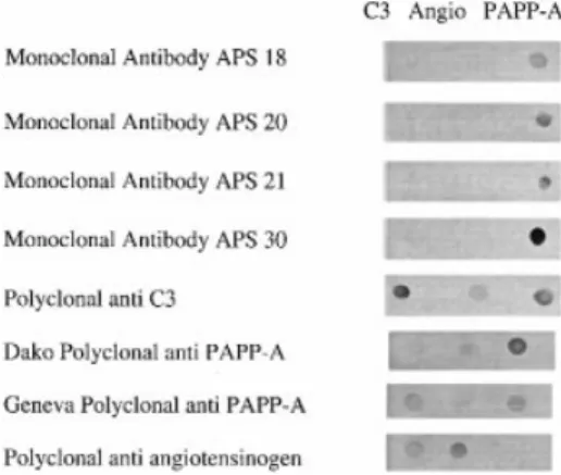
(3) Monoclonal PAPP-A antibodies. ELISA methods for PAPP-A Avidin-coated microplates Modular 8-well flat bottom polystyrene strips (Greiner, France) were coated with 200 µl/well of a solution of avidin (Amersham, France) at 10 µ/ml in PBS (pH 7.2) containing sodium azide (0.65 mg/ml) and incubated for 24 h at ambient temperature in a humid atmosphere. The solution was aspirated and the wells washed twice with 0.9% NaCl containing Tween-20 (0.005% v/v). They were aspirated twice again before being saturated with 200 µl of phosphate buffer (50 mM, pH 7.2) containing BSA (0.1% w/v) and sodium azide (0.65 mg/ml). After 4 h at ambient temperature the strips were aspirated three times and dried in an open channel for 24 h. Then the avidin-coated strips were stored in a sealed bag with a pouch of desiccant. Basic monoclonal ELISA procedure This protocol was used unless stated otherwise in the Results section. The biotinylated mAb (APS-20) was diluted to a final concentration of 5 µg/ml in PBS containing BSA (Fluka, Buchs, Switzerland, 0.1% w/v) and incubated (200 µl/well) in the avidincoated strips for 1 h at 37°C in a dry (Thermostar BMG, Offenburg, Germany) incubator. The strips were washed three times with PBS containing 0.1% (v/v) Tween-20 (PBST) in an automatic washer (Flexiwash, Austria). The standards, made from human pregnancy serum, calibrated against the World Health Organization (WHO) international reference preparation (IRP) for pregnancy-associated proteins 78–610 (Statens Seruminstitut, Denmark) and lyophilized, as well as the early pregnancy sera were added at dilutions ranging between 1:11 and 1:101 (normally 1:26) in PBS–BSA (1% v/v). Incubation was for 1 h at 37°C with shaking (500 r.p.m., Thermostar). The strips were then washed four times with PBST and the peroxidase (HRP)-conjugated mAb, diluted in PBS–BSA (1% v/v), was added at 200 µl/well. Unless stated otherwise, the concentration of the HRP-labelled mAb APS30 was 0.06 µg/ml. Incubation and subsequent washing was as above. The development was performed with o-phenylene diamine (OPD), 0.5–1 mg/ml in 0.1 M citrate/phosphate/perborate substrate buffer at pH 5.0 (Sigma, 200 µl/well). OPD was obtained as 10 mg tablets, also from Sigma. After 10–15 min ’ incubation at ambient temperature in the dark, the enzyme reaction was stopped by the addition of 100 µl sulphuric acid (2.5 M) using the same sequence and intervals as in the substrate addition. After mixing, the optical density was measured at 492 nm using a Titertek Multiscan microplate reader. Polyclonal procedure This was done as described earlier (Bersinger et al., 1995b). Briefly, the polyclonal antiserum, obtained from Dako as an IgG preparation (Cat. A-230), was absorbed in a negative affinity step using a fraction of ,300 kDa from pregnancy serum, obtained by pressure ultrafiltration, as a solid phase. The supernatant of this absorption was used as a passive microplate coating agent (200 µl/well) at a dilution of 1:1500 compared to the original IgG material (e.g. batch 25, coating concentration 2.8 µg/ml). The other steps were as reported (Bersinger et al., 1995b), with the assay buffer being 0.5% (w/v) non-fat milk proteins in PBS (Blotto®, Pierce) and the conjugate a polyclonal antibody coupled to peroxidase (Dako) and used at a 1:500 dilution in Blotto without further treatment. Cross-reactivity in assay (qualitative, indirect procedure) ELISAs were initiated (coating, blocking) as described above. Sample (early pregnancy serum pool) was used at a 1:5 dilution in Blotto. In the detection step, however, the labelled anti-PAPP-. Figure 1. Analysis of purified pregnancy-associated plasma protein A (PAPP-A) on polyacrylamide gradient gel electrophoresis and Western blots. Lane 1, molecular weight standards; lane 2, purified PAPP-A-1, 20 µg; lane 3, Western blot of the gel shown in lane 2, developed with the antibody developed in Geneva (Meisser et al., 1988).. Figure 2. Dot blots. C3 5 human C3 (Calbiochem, La Jolla, CA, USA, 1 µg); Angio 5 human angiotensinogen (Calbiochem, 1.25 µg); PAPP-A 5 purified PAPP-A-1, 3.2 µg. For antiserum sources and dilutions see text. A antibody was replaced by HRP-labelled anti-SP1 (1:2500) or anti-HPL (1:500) IgG conjugates, both obtained from Dako.. Results Purification of PAPP-A antigen and analysis of polyclonal and monoclonal antisera on polyacrylamide gels and blots The protein-stained gradient polyacrylamide gel for the purified PAPP-A-1 preparation is shown in Figure 1, lane 2. At the starting polyacrylamide concentration (4%), PAPP-A was just entering the gel (Rf 5 0.03) and the resulting estimated molecular weight was 400 kDa. Specificity and cross-reactivity of antisera were analysed using the dot blot method; results are shown in Figure 2. The four mAbs APS-18, -20, -21, and -30 as well as two polyclonal preparations were tested. We found that the four mAbs specifically recognized the pure PAPP-A but no reaction was found between any of these antibodies and the C3 complement subunit or angiotensinogen. Both polyclonal anti-PAPP-A reagents, on the other hand, cross-reacted with C3 and, to a lesser extent, with angiotensinogen. Figure 2 also shows that the commercial polyclonal anti-C3 cross-reacted not only 677.
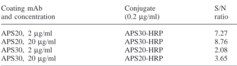
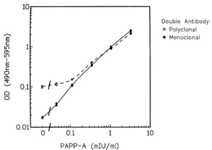
(4) N.A.Bersinger et al.. Table I. Signal-to-noise (S/N) ratios for PAPP-A determination, at 3 mIU/ml, using different monoclonal antibody (mAb) conditions Coating mAb and concentration APS20, APS20, APS30, APS30,. 2 µg/ml 20 µg/ml 2 µg/ml 20 µg/ml. Conjugate (0.2 µg/ml). S/N ratio. APS30-HRP APS30-HRP APS20-HRP APS20-HRP. 7.27 8.76 2.08 3.65. with angiotensinogen but also with purified PAPP-A while polyclonal anti-angiotensinogen recognized C3 but not PAPP-A.. Selection of monoclonal antibodies Standard curves with normal pregnancy serum pools, calibrated to a range from 0.01 to 7.07 WHO mIU/ml, were run using plates coated with Dako polyclonal anti-PAPP-A (see Materials and methods) or with the same four mAbs APS-18, -20, -21, and -30 at 0.25, 1.0, and 4.0 µg/ml as second antibodies (unlabelled). The detecting conjugate was HRP-goat antimouse-IgG (Bio-Rad, dilution of 1:2000 in Blotto). The highest mAb concentration was found to yield a high background signal in the absence of added standard or serum (up to 0.4 optical density for mAb APS-30) without significantly changing the readings at the top of the standard curve when compared to an mAb concentration of 0.25 µg/ml. No differences, except these background variations, were seen between the four mAbs in this experiment. For the subsequent investigations in this study, however, the clones APS-20 and APS-30 were selected based on observations relating to their kinetic performances. In order to decide which of the two above mAbs should be used as solid phase and which as a labelled conjugate, an aliquot of both APS-20 and APS-30 was covalently bound to HRP. Either mAb was used unlabelled for coating at 2 or 20 µg/ml, in 50 mM sodium carbonate buffer, pH 9.6. Standards were run as above, and the other mAb, HRP-labelled, was used as a conjugate at a concentration of 0.2 µg/ml. Signal-to-noise ratios (as optical densities) were calculated for a standard PAPP-A of 3 mIU/ml for all four conditions; there was a considerable difference between the two sequences. The results are shown in Table I. As a consequence of this observation, the subsequent double-monoclonal assays were run exclusively with the mAb APS-20 as an immobilized and the APS-30 as an HRPconjugated phase as described in detail in Materials and methods. Specificity: male sera Six male sera were obtained from the Worker’s Health Centre in Marcoule, France; they were from the routine clinic and devoid of any known pathology. In our ELISAs with both the double-monoclonal (APS20-APS-30) and the double-polyclonal procedure (Dako) in parallel, a serum dilution of 1:11 in the respective buffer systems was used. In the double monoclonal method, all six sera were found to be indistinguishable from the blank readout while with the double polyclonal assay system two sera yielded a positive result; the signal of 678. the other four was below the blank 1 2SD level. In a mixed protocol with unabsorbed (i.e. limited specificity) polyclonal antibody as a solid phase, individual unlabelled mAbs as second antibody, and peroxidase-goat anti-mouse IgG (BioRad, 1:2000), no signal was obtained with either male serum or mAb tested (18, 20, 21, 30), indicating that both APS-20 and -30 individually show sufficient selectivity.. Correlation between monoclonal and polyclonal procedure in normal first trimester pregnancy sera Sera (n 5 134), collected in the antenatal clinic of the University Hospital, Berne, Switzerland, from pregnancies which were precisely dated by ultrasound (crown–rump length) and known to have proceeded to term without complication, were assayed with both the double-monoclonal and the doublepolyclonal procedure as described in Materials and methods. The same calibrators, prepared from pooled pregnancy serum and lyophilized in presence of Superblock® (Pierce) in vials using a Modulyo® system (Edwards, Crawley, UK), were used in both protocols. The correlation curve is shown in Figure 3A. The slope of the curve and thus the mean ratio between the monoclonal and the polyclonal result were near the expected value (i.e. 1.0). No extreme outliers were observed, but the mentioned ratio nevertheless ranged from 0.43 to just under 1.5 in this group. This is illustrated in Figure 3B which shows it as a function of the gestational age. A slight but statistically significant positive correlation (r 5 0.2176; P 5 0.0116) was observed here, indicating that using the monoclonal assay method the normal PAPP-A versus gestational age curve would be slightly steeper than in the polyclonal system. As a low final result would indicate a ‘positive’ (meaning increased risk) in trisomy screening, the relationship between the above monoclonal:polyclonal ratio and the screening value (multiple of median, MoM) was examined as well. The result is shown in Figure 3C. No significant correlation was observed (r 5 –0.0165; P 5 0.8503); the relative outliers (very low or very high ratio on the ordinate) were located near MoM 5 1. Comparison of standard curves: raw data Lyophilized standards, made from pooled late pregnancy serum, calibrated against WHO IRP 78–610 (designated to contain 100 mIU/ml) and lyophilized in 0.5 ml aliquots, were run in up to eight replicates in both monoclonal and polyclonal procedures as described in Materials and methods, using a 1:26 dilution. The examined range was 0.06–2.04 mIU/ml plus blank (final concentration in assay well 2.31–78.5 µIU/ml). The resulting curves are shown in Figure 4 as a doublelogarithmic plot. The two assays show a good overlap for PAPP-A concentrations above 0.3 mIU/ml (original serum level). On the low range part of the standard curve, however, the procedure using mAbs remained linear to concentrations ,0.1 mIU/ml, i.e. it was superior to the absorbed/polyclonal method in terms of functional sensitivity. Detection of PAPP-A in very early pregnancy Serial serum samples were available from nine pregnancies obtained in the in-vitro fertilization (IVF) programme run at the University Hospital in Berne. Such pregnancies, by defini-.
(5) Monoclonal PAPP-A antibodies. Figure 4. Calibration curves (double-logarithmic) obtained with the double-monoclonal in comparison to the double-polyclonal enzymelinked immunosorbent assay (ELISA) procedure.. Figure 3. Correlation of the pregnancy-associated plasma protein A (PAPP-A) concentration values in normal first trimester pregnancy serum obtained in a comparison between the doublemonoclonal and the double-polyclonal (absorbed) microplate enzyme-linked immunosorbent assay (ELISA) procedures (n 5 134). (A) Direct comparison by linear regression analysis (slope 5 0.9568; y intercept 5 0.0082; r 5 0.9668). (B) The ratio of the two values obtained above (monoclonal:polyclonal) plotted against the gestational age. (C) The same ratio, plotted against the polyclonal multiple of median (MoM) which was calculated from 883 previously analysed normal pregnancies.. tion, are characterized by the most precise dating available. Serum was obtained on the day of embryo transfer, thereafter at ~ weekly intervals. PAPP-A was determined with the doublemonoclonal procedure using an 1:11 serum dilution. The result is shown in Figure 5. Up to and including day embryo transfer117 (pregnancy dating 4 weeks 1 5 days), no PAPPA was detected in any of the patients. No sera were routinely taken between days embryo transfer118 and 121 (at which the only available measurement was also negative). Rises in PAPP-A were confirmed from day embryo transfer126, with. Figure 5. Detection of pregnancy-associated plasma protein A (PAPP-A) in very early pregnancy, obtained by in-vitro fertilization (IVF), using double-monoclonal enzyme-linked immunosorbent assay (ELISA). Squares, conventional IVF; circles, intracytoplasmic sperm injections. The arrow indicates the time from when a serum concentration of human chorionic gonadotrophin (HCG) determination would detect the presence of a pregnancy. N.B. Scale is logarithmic.. one pregnancy still being negative on day embryo transfer131 (pregnancy dating 6 weeks 15 days). The increases seemed to be exponential (logarithmic scale).. PAPP-A in fetal serum and maternal urine We have determined PAPP-A in the cord serum (umbilical vein), obtained from seven normal, spontaneous vaginal deliveries at term, using the double-monoclonal assay method. At the dilutions routinely used for serum (1:11 or higher) all fetal samples yielded a result of zero. At a 1:3 dilution, however, PAPP-A values were found to be very low but significantly positive at 0.0073 6 0.0047 (SD) mIU/ml (range 0.0018– 0.0151). Urine from 12 pregnant (including third-trimester) and two non-pregnant (male, female) people were assayed as well, using a low dilution (1:2.6). The result was indistinguishable from the blank in all urine samples. 679.
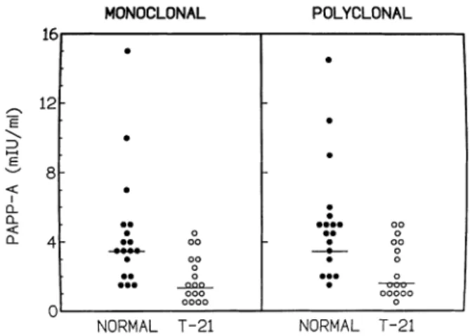
(6) N.A.Bersinger et al.. Figure 6. Pregnancy-associated plasma protein A (PAPP-A) in normal and Down’s syndrome first trimester pregnancies, determined using the monoclonal in comparison to the polyclonal antibody procedure. Closed circles, normal pregnancy (n 5 18); open symbols, trisomy-21 pregnancy (n 5 17). The horizontal bars indicate the medians.. Screening efficiency for fetal trisomy 21 (Down’s Syndrome) A group of first trimester pregnancy sera (gestational age 11 weeks 13 days to 12 weeks 1 6 days), consisting of 18 normal pregnancies and 17 cases of fetal trisomy 21, was examined for its PAPP-A concentration using the doublemonoclonal in comparison to the polyclonal procedure. With the narrow range of gestational age, it was possible to calculate the median as one group for both monoclonal and polyclonal protocols. In normal pregnancy, these medians were 3.49 and 3.53 mIU/ml respectively. For the fetal trisomy population, values of 1.31 and 1.67 mIU/ml were obtained, yielding an average MoM of 0.38 in the monoclonal and 0.47 in the polyclonal assay procedure respectively, for fetal Down’s syndrome. Individual results are presented in Figure 6. Both methods statistically distinguished (P , 0.01) the two populations (Mann–Whitney).. Discussion We have observed significant differences, in terms of specificity and sensitivity, between PAPP-A assay protocols using monoclonal and polyclonal antibodies. PAPP-A, in contrast to pregnancy-specific β1-glycoprotein (SP1) and HCG, has been suggested to be produced not only in the placenta but also by a maternal source (Bischof, 1984; Sjoberg et al., 1984) which could be hormonally stimulated during pregnancy to release amounts of PAPP-A far above the values that had been observed in non-pregnant individuals. Various intriguing observations for PAPP-A (Bersinger et al., 1997), such as a reduced retroplacental concentration (compared with the periphery), its increase in serum concentration at the end of the third trimester, or its slow decrease after parturition had never been satisfactorily explained. Heterogeneity in protein and/or carbohydrate structure could be a reason. The production of monoclonal antibodies could have selected epitopes that are absent on PAPP-A outside of pregnancy, i.e. the increased specificity does not necessarily mean that the observed cross-reactivities with polyclonal 680. antisera are artefactual. Contamination with SP1 antigen might well have been present; this protein is structurally unrelated to PAPP-A but very abundant, leading to the presence of antiSP1 antibodies in polyclonal anti-PAPP-A antisera (Bersinger et al., 1995b; Bersinger, 1996). On the other hand, there may be a biological link between PAPP-A and haptoglobin (Bueler and Bersinger, 1989) and certainly for pro-MBP for which such an association has been demonstrated (Oxvig et al., 1993). The polyclonal antibody preparations tested here all recognize the pro-MBP epitopes and, as a consequence, are cross-reacting with PAPP-A, the complement C3 subunit, and angiotensinogen. Pro-MBP itself could not be tested by the Western blot technique since the available preparation had been obtained by immunopurification on an immobilized monoclonal antibody which would still be present to some extent in the final product and therefore leading to a direct reaction with the second antibody in the absence of pro-MBP antigen. The same would have been the case for PAPP-A-2 which was also obtained by mAb immunopurification. The tested anti-PAPP-A mAbs, on the other hand, do not show such a cross-reaction in the dot blot with C3 or angiotensinogen; and this might be an advantage for the investigation of the structure and function of PAPP-A at research level. Clinically speaking this advantage is somehow less obvious since no difference in first trimester maternal serum Down’s syndrome screening efficiency was found between the polyclonal and the monoclonal antibody-based assay method for PAPP-A (Figure 6); therefore, when combined in a routine screening programme with Fβ-HCG and maternal age, good Down’s syndrome detection rates would be achieved with either PAPPA assay protocol (Brambati et al., 1994; Krantz et al., 1996; Wald et al., 1996; Wheeler and Sinosich, 1998). The slightly reduced sensitivity observed with the polyclonal antibody procedure (Figure 4) would not be relevant for Down’s syndrome screening performance since even in this pathological situation the serum PAPP-A concentrations are expected to be .0.3 mIU/ml. Only sera from the rarely encountered fetal trisomies 18 and 13 would yield results of ø0.1 mIU/ml, and we would therefore expect a superiority of the monoclonal over the polyclonal antibody assay procedure in the screening for viable trisomies other than Down’s syndrome. In trisomy 18 (in contrast to trisomy 21), serum PAPP-A values are still significantly reduced (and this to a higher extent than observed for HCG) in the second trimester, but again largely above the sensitivity limit of the polyclonal assay procedure (Bersinger et al., 1999). It is thus in the theoretical (clinically rare) situation of a very early pregancy (,8 weeks amenorrhoea) trisomy 21 screen that an increased sensitivity would be required. This may be biochemically justified (Bersinger et al., 1995a) but is clinically irrelevant as no routine antenatal care is provided at this stage of gestation since the abnormality assessment by ultrasound is not informative before 10–11 weeks. The advantage of using monoclonal antibodies against PAPP-A is thus not primarily in short-term clinical investigations (such as the usefulness of this marker in various pregnancy pathologies) but in fundamental scientific investigations aiming to explain the various equivocal findings (Bersinger et al., 1997) on the non-placental presence and.
(7) Monoclonal PAPP-A antibodies. biological role of PAPP-A. In the future, however, large-scale prospective clinical Down’s syndrome screening studies will be required and in this context the mAbs have the advantage of being an unlimited and homogeneous source of reagent. Such a project, covering most large centres in Switzerland, is now being launched. In conclusion, we believe that the use of these highly specific monoclonal antibodies will significantly contribute to the elucidation of the biological role of PAPP-A and, as a direct practical consequence, of its clinical usefulness in large screening programmes for the detection of abnormal human pregnancy.. Acknowledgement Part of this research project was funded by a financial contribution from CIS-Bio International, France.. References Berry, E., Aitken, D.A., Crossley, J.A. et al. (1997) Screening for Down’s syndrome: changes in marker levels and detection rates between first and second trimesters. Br. J. Obstet. Gynaecol., 104, 811–817. Bersinger, N.A., Brizot, M.L., Johnson, A. et al. (1994) First trimester maternal serum pregnancy-associated plasma protein A and pregnancy-specific β1glycoprotein in fetal trisomies. Br. J. Obstet. Gynaecol., 101, 970–974. Bersinger, N.A., Marguerat, P., Pescia, G. et al. (1995a) Pregnancy-associated plasma protein A (PAPP-A): measurement by highly sensitive and specific enzyme immunoassay, importance of first-trimester serum determinations, and stability studies. Reprod. Fertil. Dev., 7, 1419–1423. Bersinger, N.A., Zakher, A., Huber, U. et al. (1995b) A sensitive immunoassay for pregnancy-associated plasma protein A (PAPP-A): a possible first trimester method of screening for Down syndrome and other trisomies. Arch. Gynecol. Obstet., 256, 185–192. Bersinger, N.A. (1996) De´termination de la PAPP-A du se´rum maternel par ELISA pour la de´tection de la trisomie foetale au premier trimestre. Reprod. Hum. Horm., 7, 429–434. Bersinger, N.A., Altermatt, H.J., Birkha¨user, M.H. et al. (1997) Non-placental production of pregnancy-associated plasma protein A (PAPP-A): old and new evidence. Early Preg. Biol. Med., 3, 96–101. Bersinger, N.A., Leporrier, N., Herrou, M. et al. (1999) Maternal serum pregnancy-associated plasma protein A (PAPP-A) but not pregnancyspecific β1-glycoprotein (SP1) is a useful second trimester marker for fetal trisomy 18. Prenat. Diag., 19, 537–541. Bischof, P. (1984) Placental proteins: pregnancy-associated plasma protein A. Contrib. Gynecol. Obstet., 12, 41–73. Bischof, P. and Meisser, A. (1989) Immunological heterogeneity of pregnancyassociated plasma protein A (PAPP-A). Effects on the radioimmunoassay of PAPP-A. Br. J. Obstet. Gynaecol., 96, 870–875. Brambati, B., Tului, L., Bonacchi, I. et al. (1994) Serum PAPP-A and free β-hCG are first-trimester screening markers for Down syndrome. Prenat. Diag., 14, 1043–1047. Bueler, M.R. and Bersinger, N.A. (1989) Antiserum to pregnancy-associated plasma protein A (PAPP-A) recognises human haptoglobin. Br. J. Obstet. Gynaecol., 96, 867–869. Haddow, J.E., Palomaki, G.E., Knight, G.J. et al. (1998) Screening of maternal serum for fetal Down’s syndrome in the first trimester. N. Engl. J. Med., 338, 955–961. . Krantz, D.A., Larsen, J.W., Buchanan, P.D. et al. (1996) First-trimester Down syndrome screening: free β-human chorionic gonadotropin and pregnancyassociated plasma protein A. Am. J. Obstet. Gynaecol., 174, 612–616. Lin, T.M., Halbert, S.P., Kiefer, D. et al. (1974) Characterisation of four human pregnancy-associated plasma proteins. Am. J. Obstet. Gynecol., 118, 223–226. . Meisser, A., Geinoz, A. and Bischof, P. (1988) In vitro effects of pregnancyassociated plasma protein A: Artifacts due to heparin. Biol. Reprod., 39, 373–378. Muller, F., Cuckle, H., Teisner, B. et al. (1993) Serum PAPP-A levels are depressed in women with fetal Down’s syndrome in early pregnancy. Prenat. Diag., 13, 633–636. Oxvig, C., Sand, O., Kristensen, T. et al. (1993) Circulating human pregnancy-. associated plasma protein A is disulfide-bridged to the proform of eosinophil major basic protein. J. Biol. Chem., 268, 12243–12246. Oxvig, C., Sand, O., Kristensen, T. et al. (1994) Isolation and characterisation of circulating complex between human pregnancy-associated plasma protein A and proform of eosinophil major basic protein. Biochim. Biophys Acta, 1201, 415–423. Sinosich, M.J., Davey, M.W., Ghosh, P. et al. (1982) Specific inhibition of human granulocyte elastase by human pregnancy-associated plasma protein A. Biochem. Int., 5, 777–786. . Sjoberg, J., Wahlstrom, T. and Seppala, M. (1984) Pregnancy-associated plasma protein A in the human endometrium is dependent on the effect of progesterone. J. Clin. Endocrinol. Metab., 58, 359–362. Spencer, K., Aitken, D.A., Crossley, J.A. et al. (1994) First trimester biochemical screening for trisomy 21: the role of free beta hCG, alpha fetoprotein and pregnancy associated plasma protein A. Ann. Clin. Biochem., 31, 447–454. Wheeler, D.M. and Sinosich, M.J. (1998) Prenatal screening in the first trimester of pregnancy. Prenat. Diag., 18, 537–543. Wald, N., Stone, R., Cuckle, H.S. et al. (1992) First trimester concentrations of pregnancy-associated plasma protein A and placental protein 14 in Down’s syndrome. Br. Med. J., 305, 28. . Wald, N.J., George, L., Smith, D. et al. (1996) Serum screening for Down’s syndrome between 8 and 14 weeks of pregnancy. Br. J. Obstet. Gynaecol., 103, 407–412. Received on December 30, 1998; accepted on March 31, 1999. 681.
(8)
Figure
Documents relatifs
Figure 3 - Mediation analysis between maternal educational attainment (exposure), maternal smoking during pregnancy (potential mediator) and offspring birthweight for
Cet article suit une méthodologie incluant l’analyse d’un certain nombre de projets d’amélioration réalisés et publiés en éducation, touchant particulièrement
The exposed group consisted of 319 pregnant women exposed to sub diaphragmatic ionizing radiations for diagnostic purposes, during the first trimester of pregnancy, and the
The mAb also identified by western blot sCD14 (53 and 58 kDa) in milk and blood and sCD14 (47 kDa) in a lysate of macrophages obtained from involuted bovine mammary gland
Reactive treponemal serology (regardless of nontreponemal test reactivity) along with characteristic late manifestations of congenital syphilis d in a child whose mother was known
Il n’a pas encore étudié toutes les leçons jusqu’à Unit 7 lesson 6 : Vous pouvez encore le faire d’ici septembre 2020.. Vous n’arrivez pas à vous repérer sur le site
Il n’a pas encore étudié toutes les leçons jusqu’à Unit 7 lesson 6 : Vous pouvez encore le faire d’ici septembre 2020.. Vous n’arrivez pas à vous repérer sur le site
Comme je l’ai rappelé aux enfants cette semaine, continuer à faire les exercices, étudier le vocabulaire, relire à haute voix les verbes irréguliers sur le fichier Fun.. si on
