Review
High altitude-induced pulmonary oedema
Marco Maggiorini
⁎
Intensive Care Unit, Department of Internal Medicine, University Hospital, Rämistrasse 100, CH-8091 Zürich, Switzerland Received 6 June 2006; received in revised form 27 June 2006; accepted 3 July 2006
Available online 12 July 2006 Time for primary review 12 days
Abstract
Almost one mountain trekker or climber out of two develops several symptoms of high altitude illness after a rapid ascent (N300 m/day)
to an altitude above 4000 m. Individual susceptibility is the most important determinant for the occurrence of high altitude pulmonary
oedema (HAPE). Symptoms associated with HAPE are incapacitating fatigue, chest tightness, dyspnoea at the slightest effort, orthopnoea,
and cough with due to haemoptysis in an advanced stage of the disease pink frothy sputum. The hallmark of HAPE is an excessively elevated
pulmonary artery pressure (mean pressures of 35 and 55 mm Hg), which precedes the development of pulmonary oedema. Elevated
pulmonary capillary pressure and protein- as well as red blood cell-rich oedema fluid without signs of inflammation in its early stage are
characteristic findings. Furthermore, decreased fluid clearance from the alveoli may contribute to this non-cardiogenic pulmonary oedema.
Immediate descent or supplemental oxygen and nifedipine are recommended until descent is possible. Susceptible individuals can prevent
HAPE by slow ascent: an average gain of altitude not exceeding 400 m/day above an altitude of 2500 m. If progressive high altitude
acclimatization is not possible, a prophylaxis with nifedipine should be recommended.
© 2006 European Society of Cardiology. Published by Elsevier B.V. All rights reserved.
Keywords: High altitude pulmonary oedema; Capillary pressure; Hypoxic pulmonary vasoconstriction; Trans-epithelial Na transport; Nifedipine; Tadalafil; Dexamethasone
1. Introduction
Two forms of high altitude illness can be distinguished: a
cerebral form called acute mountain sickness (AMS) and a
pulmonary form called high altitude pulmonary oedema
(HAPE). Altitude, the rate of ascent, and individual
suscep-tibility in particular are the major determinants of AMS and
HAPE in mountaineers and trekkers. At an intermediate altitude
such as in Colorado, the prevalence of AMS among visitors is
estimated at 25%
[1]
. Among trekkers in the Himalayas and
mountaineers in the Alps ascending at a rate of
N600 m/day, the
prevalence of AMS at altitudes between 4000 m and 5600 m is
30–60%
[2–8]
. In contrast to AMS, HAPE is less frequent. The
estimated incidence of HAPE in visitors to ski resorts in the
Rocky Mountains of Colorado is 0.01
–0.1%
[9]
. In a general
alpine mountaineering population, the prevalence of HAPE is
b0.2%
[10]
. The HAPE incidence among trekkers in the
Himalayas and climbers in the Alps ascending at a rate of
N600 m/day is around 4%
[3,11]
. In the alpine setting, when an
altitude of 4559 m was reached within 22 h, the incidence
increased to 7% in mountaineers without a history of
radio-graphically documented HAPE and to 62% in mountaineers
with such a history
[12]
. In an unselected population of Indian
soldiers, airlift to an altitude of 5500 m was associated with a
HAPE incidence of up to 15%
[13]
.
2. Clinical presentation
2.1. Clinical examination
HAPE presents within 2
–5 days after arrival at high altitude
[13
–15]
. It is rarely observed below altitudes of 2500
–3000 m
and after 1 week of acclimatization at a particular altitude.
Early symptoms of HAPE include exertional dyspnoea, cough,
and suddenly reduced exercise performance. As pulmonary
oedema progresses, orthopnoea, breathlessness at rest, and
gurgling in the chest develop, cough worsens, and pink frothy
⁎ Tel.: +41 44 255 22 04; fax: +41 44 255 31 81. E-mail address:klinmax@usz.unizh.ch.
0008-6363/$ - see front matter © 2006 European Society of Cardiology. Published by Elsevier B.V. All rights reserved. doi:10.1016/j.cardiores.2006.07.004
sputum reveals overt pulmonary oedema
[13
–15]
. The clinical
examination shows cyanosis, tachypnoea, tachycardia, and
frequently body temperature
N37.5 °C
[16]
. Râles are discrete
at the beginning, typically located over the middle lung fields
[13–15]
. Often, there is a discrepancy between the minor
findings at auscultation compared with the widespread disease
on the chest radiograph
[17]
(
Table 1
). In advanced cases, signs
of concomitant severe AMS with ataxia and decreased levels
of consciousness
– signs of high altitude cerebral oedema –
may develop
[18,19]
(
Table 1
).
2.2. Chest radiography and laboratory analyses
Chest radiographs and CT-scans of early HAPE show a
patchy, peripheral distribution of oedema as shown in
Fig. 1
.
The radiographic appearance of HAPE is more
homoge-neous and diffuses in advanced cases and during recovery
[20]
. The results of arterial blood gas, radiographic score,
and AMS score obtained in 19 adults with HAPE at 4559 m
(
Table 1
) demonstrate that HAPE may develop with nearly
no symptoms of AMS (6/19) and that the extension of
pulmonary infiltrates does correlate with the impairment of
gas exchange. In advanced cases of HAPE observed at an
altitude of 4559 m, arterial PO
2likely drops below the
35 mm Hg mark.
There are no characteristic findings in common laboratory
examinations with the exception of moderately elevated
C-reactive protein (b100 mg/l)
[13,15,21]
. In the early stage of
HAPE broncho-alveolar lavage (BAL) reveals a protein- and
red blood cell-rich oedema fluid without signs of
inflamma-tion
[22]
, whereas in a more advanced stage
pro-inflamma-tory mediators and granulocytes add to the initial changes
[15,23]
. Autopsies showed diffuse pulmonary oedema with
bloody foamy fluid present in the airways and signs of
inflammation involving the alveoli and the capillaries
[24,25]
.
Table 1
Clinical and radiographic findings in adults without and with HAPE
HAPE− (n=120) HAPE+ (n = 30)
AMS− (n=87) AMS+ (n = 33) AMS− (n=9) AMS+ (n = 21)
Rales+/++ (%) 7 (8) 5 (15) 3 (33) 8 (38)
Body temperature (°C) 36.8 (36.6–36.9) 37.2 (37.0–37.4)a 37.1 (36.9–37.4)b 37.7 (37.5–37.9)a,c Clin. AMS score 1.9 (1.6–2.3) 4.9 (4.4–5.5)a 2.7 (1.3–4.0) 7.3 (6.4–8.3)a,c
Rad. score 0.3 (0.2–0.5) 0.3 (0.1–0.6) 6.7 (3.5–9.9)b 7.1 (5.3–8.8)c
PaO2 45 (43–46) 40 (38–42)a 37 (32–42)b 33 (30–35)c
PaCO2 26 (25–27) 28 (27–29) 27 (25–29) 27 (25–28)
AaDO2 5.2 (3.9–6.4) 7.1 (5.1–7.1) 12.1 (7.3–16.9)b 15.6 (12.4–18.4)c
Mean (95% confidence intervals) of clinical (clin.) and radiographic (rad.) scores, arterial (a) PO2, PCO2, and the alveolar–arterial difference for oxygen
(AaDO2) in 60 adults examined after ascent to 4559 m and a stay for 3 consecutive days. A total of 150 examinations were performed, and in 30 of them chest
radiography was compatible with the diagnosis of HAPE.
These results were obtained in collaboration with P. Bärtsch and O. Oelz.
a
pb0.01 vs. AMS− in the HAPE−/+ groups.
b
pb0.01 vs. AMS− in the HAPE− group.
c
pb0.01 vs. AMS+ in the HAPE− group.
Fig. 1. Chest radiograph and CT-scan in a mountaineer with HAPE. Radiograph of a male patient with HAPE showing patchy distributed infiltrates over the whole lung (A). The CT-thorax of the same patient shows a patchy distribution of oedema, localized predominately around the right hilus (B). (These illustrations were kindly provided by Dr. H. Fischer, Regional Hospital Visp, Switzerland.)
2.3. Right heart catheter studies
Since the first hemodynamic measurements performed in
patients with HAPE admitted to hospital we know that HAPE is
associated with elevated pulmonary artery pressure
[14,26
–29]
.
In a prospective hemodynamic evaluation of HAPE-susceptible
adults performed after rapid ascent to 4559 m within 24 h, mean
pulmonary artery pressure increased to 38 mm Hg (range 28–
42 mm Hg)
[30]
(
Fig. 2
). In those who developed pulmonary
oedema during that occasion, mean pulmonary artery pressure
was 42 mm Hg (range 36–51 mm Hg). Moreover, all these
studies consistently show that in HAPE, left ventricular filling
pressures, as assessed by the measurement of pulmonary
oc-cluded pressure (wedge pressure), right atrial pressure, and
cardiac output are normal
[28–30]
. Thus, hemodynamic
eval-uations in HAPE clearly indicate that the development of
pulmonary hypertension within hours after rapid exposure to
high altitude is a hallmark of this disease. This is further
supported by those studies indicating that HAPE is prevented or
treated by the use of pulmonary vasodilators
[31
–33]
.
3. Pathophysiology
3.1. Exaggerated hypoxic pulmonary vasoconstriction
Oxygen sensors located in the pulmonary vasculature detect
the drop of alveolar oxygen tension and lead to vasoconstriction
of small pulmonary arteries
[34,35]
and pulmonary veins
[36]
.
The response of smooth-muscle cells in the pulmonary
vas-culature to acute hypoxia begins within seconds and involves
inhibition of voltage-dependent potassium channels, membrane
depolarization, and calcium entry through
L-type calcium
chan-nels
[35,37]
. Moreover, hypoxia up-regulates transient receptor
potential channels, leading to additional calcium entry through
receptor and store-operated calcium-channels
[35]
. Whether a
constitutively decreased mRNA expression of
voltage-depen-dent potassium channels or an acquired transcriptional defect of
the voltage-dependent potassium channels protein expression is
at the origin of HAPE susceptibility remains to be determined.
Exaggerated hypoxic pulmonary vasoconstriction has been
attributed to an increased susceptibility of the pulmonary
circulation
– sustained elevation of cytoplasmic calcium
con-centration
– to sympathetic activity and/or high levels of
en-dothelin-1. Increased sympathetic activity and elevated
norepinephrine plasma levels have been found in individuals
with AMS and HAPE
[19,38–40]
. Rapid exposure to 4559 m
almost doubles plasma endothelin-1 levels
[41]
, the highest
values being measured in individuals with HAPE
[42]
. Both
intensity of sympathetic activity
[40]
and plasma endothelin-1
levels are positively correlated with systolic pulmonary artery
pressure
[41,42]
.
Endothelium-mediated vasodilatation is crucial for the
con-trol of pulmonary vasoconstriction. Hypoxia-induced
endo-thelial dysfunction resulting in an impaired
endothelium-dependent vasodilatation in the systemic circulation
[43]
and
an impaired nitric oxide production in the lung
[22,44,45]
could be another mechanism leading to elevated pulmonary
artery pressure in HAPE-susceptible individuals. In fact, upon
acute exposure to hypoxia, exhaled nitric oxide concentrations
[44,45]
and nitrite/nitrate concentrations in the BAL fluid
[22]
tend to decrease in individuals prone to HAPE, whereas they
increase in those resistant to the condition. Moreover, in
sus-ceptible individuals the prophylactic intake of tadalafil, a
phosphodiesterase-5 inhibitor, prevents high altitude
pulmo-nary hypertension and HAPE
[33]
.
Taken together, the results of all these studies indicate that
an imbalance between hypoxia-mediated vasoconstriction and
impaired nitric oxide bioavailability is the provable
mecha-nism behind the elevated pulmonary artery pressure in
HAPE-susceptible individuals. Whether ethnic differences between
Caucasians
[46]
and Japanese
[47]
for endothelial nitric oxide
polymorphism may also contribute to HAPE susceptibility
remains to be established.
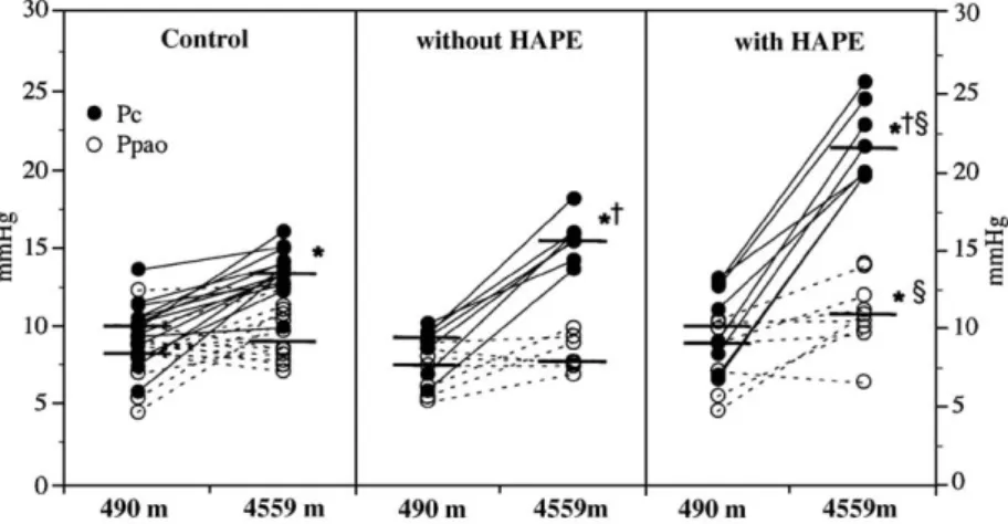
3.2. Elevated pulmonary capillary pressure
During hemodynamic measurements performed in
HAPE-susceptible and non-HAPE-susceptible adults at 4559 m, we
estimated the pulmonary capillary pressure using the arterial
occlusion method
[30]
, which most likely measures pressures
in vessels close to 100
μm in diameter
[48]
and demonstrated
that the pulmonary capillary pressure is elevated in HAPE.
Pulmonary capillary pressure was on average 16 mm Hg
(range 14
–18 mm Hg) in HAPE-susceptible subjects without
pulmonary oedema and 22 mm Hg (range 20
–26 mm Hg) in
those who developed HAPE
[30]
(
Fig. 3
). This result suggests
that in adults, the pulmonary capillary pressure threshold value
for oedema formation is 20 mm Hg, which is in keeping with
previous experimental observations in dogs indicating a PO
2-independent critical capillary pressure of 17 to 24 mm Hg,
above which the lungs continuously gain weight
[49,50]
.
Fig. 2. Changes in mean pulmonary artery pressure from low to high altitude. Individual mean pulmonary artery pressures (Ppa) measured at 4559 m in HAPE-resistant (control) and HAPE-prone (susceptible) adults [30]. The closed dots indicate mean Ppa in individuals without radiographic evidence of HAPE. The open dots indicate those individual subjects who developed HAPE during the 2 days' stay at 4559. The horizontal bars (—) indicate median Ppa value for each group of subjects. ⁎pb0.01 vs. 490 m,†pb0.01 vs. control,
There are two possible mechanisms leading to an elevated
pulmonary capillary pressure in subjects susceptible to
HAPE: a heterogeneous distribution of pulmonary blood
flow within the pulmonary vascular bed
[51,52]
or a hypoxic
constriction occurring at the level of the pulmonary veins
[36,53]
. A heterogeneous distribution of blood flow within
the pulmonary circulation causing regional over-perfusion of
capillaries, i.e. in areas with the least arterial vasoconstriction
[51]
, is suggested by the results of a recent study obtained
using a functional magnetic resonance imaging technique
(arterial spin labelling) in a small number of volunteers
exposed to hypoxia, indicating an increased pulmonary
blood flow heterogeneity in HAPE-susceptible individuals
[52]
. Non-uniformly distributed blood flow in hypoxia was
also found using the fluorescent microspheres technique in
pigs
[54]
and dogs
[55]
. Non-homogeneous distribution of
blood flow could be caused by uneven distribution of
alveolar ventilation, hence hypoxic vasoconstriction
[56]
or
heterogeneous oxygen sensing within smooth muscle cells of
the pulmonary vascular tree
[57–59]
. On the other hand there
is good evidence that pulmonary veins contract in response
to hypoxia
[36,60,61]
, increasing the resistance downstream
of the region of fluid filtration
[62]
, which suggests that
HAPE could develop even in the absence of a heterogeneous
distribution of pulmonary blood flow within the pulmonary
vascular bed. Moreover, markedly increased pulmonary
artery pressure in hypoxia may also cause transvascular
leakage of small arterioles
[63]
. However, the patchy
distribution of pulmonary infiltrates on chest radiographs
and CT scans of the lungs found in individuals with HAPE
(
Fig. 1
) strongly support the heterogeneous distribution of
elevated capillary pressures within the permeable region of
the pulmonary circulation, which in summary is likely to rely
on an unevenly distributed hypoxic vasoconstriction in either
pulmonary arteries or veins, or both.
3.3. High-permeability type of oedema
Broncho-alveolar lavage (BAL) performed in
HAPE-susceptible adults within a day after ascent to 4559 m revealed
elevated red blood cell counts and serum-derived protein
concentration in BAL fluid
[22]
. The number of red blood
cells/μl and the albumin concentration was higher in those
individuals with HAPE at the time of BAL than in those who
developed it within the next 24 h. The threshold for the
increase in albumin and red blood cells was at a systolic
pulmonary artery pressure of approximately 35 mm Hg and
60 mm Hg, respectively (
Fig. 4
). The number of alveolar
macrophages/
μl and neutrophils/μl and the concentration of
the pro-inflammatory mediators interleukin-1 (IL-1), TNF-
α,
Fig. 4. Relationship between systolic pulmonary artery pressure and BAL red blood cell count and albumin concentration. Individual broncho-alveolar lavage (BAL) red blood cell and albumin concentration plotted against systolic pulmonary artery pressure (sPpa) at high altitude (4559 m). The figure shows that the threshold sPpa for the appearance in the BAL fluid of albumin was 35 mm Hg and that for red blood cells was 60 mm Hg[22]. Fig. 3. Changes in pulmonary capillary pressure and pulmonary artery occlusion pressure upon ascent to 4559 m. Individual pulmonary capillary pressure (Pc) and pulmonary artery occluded pressure (Ppao = wedge pressure), assessed using the arterial occlusion technique, in controls, and HAPE-susceptible subjects without and with pulmonary oedema[30]. The Pc is indicated by the filled dots and Ppao values by the open dots. The figure shows that in subjects who develop HAPE, the Pc was higher than 19 mm Hg and that the increase in Ppao, although significant, is minimal. The horizontal bars (—) indicate median Ppa value for each group of subjects. ⁎pb0.01 vs. 490 m,†pb0.01 vs. control,§pb0.01 vs. HAPE-susceptible adults without
IL-8, thromboxane, prostaglandin E
2, and leukotriene B
4(LTB
4), was not increased. These results are in line with studies
showing that in rabbit lungs, elevated pulmonary vascular
pressure causes injury to both the alveolar epithelial and the
capillary endothelial cells, resulting in a protein- and red blood
cell-rich lung oedema fluid
[64–66]
(
Fig. 5
). Thus, HAPE in its
early stage is a high pressure-mediated permeability type of
pulmonary oedema.
BAL fluid examination in adults with advanced HAPE show
also elevated levels of pro-inflammatory cytokines and LTB
4[23,67]
, suggesting secondary inflammation to the
high-pres-sure injury to the blood
–gas barrier and/or lung oedema
for-mation. Elevated concentrations of pro-inflammatory cytokines
found in patients with cardiogenic pulmonary oedema
[68,69]
support this concept. A release of pro-inflammatory cytokines
[69]
continuing for several days after normalization of the
pulmonary artery pressure may be the origin of a prolonged
respiratory failure described in some individuals
[70]
.
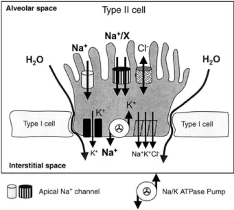
3.4. Reduced fluid clearance from the alveolar space
Studies performed in cell cultures and rats exposed to
hypoxia indicate that hypoxia inhibits the activity and the
expression of alveolar epithelial cell sodium (Na
+)
transpor-ters, particularly the apical membrane epithelial Na
+channel
(ENaC) and the basolateral membrane Na
+/K
+-ATPase, and
hence the Na
+transport and associated alveolar fluid clearance
across the alveolar epithelial membrane
[71
–74]
(
Fig. 6
).
Since alveolar epithelium is not accessible in humans, nasal
epithelium, which has Na
+transporters that are similar to those
of the alveolar epithelium, is used to estimate alveolar
epithe-lium Na
+transport activity
[75]
. Accordingly, hypoxia was
found to inhibit nasal epithelial Na
+transport in both
HAPE-resistant and -susceptible mountaineers
[76,77]
. Moreover, at
low altitude, HAPE-susceptible adults present a lower activity
of the ENaC compared to HAPE-resistant individuals
[76–
78]
, suggesting a possible contribution of ENaC to the
patho-physiology of HAPE.
β
2-Receptor agonists have been shown to stimulate alveolar
epithelial Na
+and fluid transport in rats exposed to hypoxia
[74]
and pulmonary oedema reabsorption in patients with acute
respiratory distress syndrome
[79]
. The prophylactic inhalation
of a high dose (2 × 125
μg) of salmeterol decreased the incidence
of HAPE from 74% to 33%
[78]
. Thus, it is possible that a
decreased activity of Na
+transporters, particularly the ENaC,
across the alveolar epithelial membrane will be part of the
pathophysiologic mechanism of HAPE. On the other hand, one
cannot exclude that the effect of aerosolized salmeterol
pro-phylaxis may be attributed to other actions of the drug
[80,81]
.
Treatment with a
β
2-agonist may cause vasodilatation by an
increase in nitric oxide production
[82]
, inhibition of endothelial
cell contraction, and reduction in intercellular gaps
[83
–85]
.
Moreover,
β
2-agonists also have a clear anti-inflammatory
ef-fect by reducing neutrophil influx and degranulation and the
accumulation of TNF-α in the alveolar airspaces
[86]
. Thus, to
really test the role of Na
+transporters in HAPE, more specific
drugs are needed.
4. Factors contributing to lung oedema formation
4.1. AMS and hypoxemia
AMS is not a precondition for the development of HAPE.
This is suggested by epidemiological studies indicating a
7- to 8-fold higher incidence of AMS than HAPE
[3,11]
Fig. 6. Alveolar epithelial cell fluid reabsorbing mechanism. Alveolar epithelial apical and basolateral membrane ion channels and exchangers involved in active transepithelial sodium and water absorption. There is an active reabsorption of sodium; water and chloride follow passively. Acute hypoxia reduces alveolar fluid clearance by inhibition of apical sodium entry pathways and basolateral Na+/K+-ATPase activity.
Fig. 5. Mechanism of pulmonary capillary leak in HAPE. Elevated pulmonary capillary pressure (Pc) cause progressive distension of the vessel wall leading to opening of endothelial and epithelial gaps through which first proteins and later red blood cells leak into the alveolar space.
and by the observation that HAPE may develop even in the
absence of AMS
[17]
(
Table 1
). On the other hand, it is likely
that severe AMS may be a risk factor for HAPE. This is
suggested by studies indicating that individuals with severe
AMS have a low PaO
2(
Table 1
)
[87,88]
and/or a low hypoxic
ventilatory drive. A low hypoxic ventilatory drive is known to
possibly increase susceptibility to HAPE
[89
–91]
; however,
the considerable overlap between HAPE-susceptible and
-resistant individuals suggests that it is at best permissive but
not compulsory regarding susceptibility to HAPE.
4.2. Airway infections
It is conceivable that any process enhancing the
permeabil-ity of the alveolar-capillary barrier decreases the pulmonary
capillary pressure threshold above which pulmonary oedema
develops. Increased lung fluid accumulation during hypoxic
exposure after priming rats with endotoxins or viruses
[92]
and
the reported association of preceding viral infections
(predom-inantly of the upper respiratory tract) and HAPE in children
visiting Colorado
[93]
support this concept. Thus, a variable
pulmonary capillary permeability between high altitude
expo-sures could tentatively explain why in HAPE-susceptible
indi-viduals the reoccurrence rate of pulmonary oedema after rapid
ascent to high altitude is not 100%.
4.3. Congenital anomalies
Restriction of the pulmonary vascular bed cross-sectional
area may also contribute to increase pulmonary artery pressure
upon exposure to high altitude. This theory is supported by
reports indicating that congenital anomalies of the large
pul-monary arteries
[94,95]
and pulmonary embolism
[96,97]
are
associated with an increased risk to develop HAPE even at
altitudes below 3000 m. Moreover, small lungs relative to
body size have also been retained as a possible risk factor for
HAPE
[56,98]
.
At risk for HAPE at a moderate altitude are also patients
with congenital cardiac shunts
[99]
and/or pre-existing
pulmo-nary hypertension
[100]
. A right–left shunt across a patent
foramen ovale may exacerbate high altitude hypoxemia and
hence lead to HAPE
[101]
. Thus, in patients who have
deve-loped HAPE at altitudes below 3000 m, echocardiography is
recommended to exclude pulmonary hypertension and a
con-genital anomaly.
4.4. Exercise
Strenuous exercise may also contribute to increasing
pul-monary capillary pressure and hence the risk of HAPE. In fact,
there is evidence that strenuous exercise causes subclinical
permeability oedema with high red blood cells and protein
concentrations that may last for more than a day at high altitude
[102]
. This may be caused by uneven distribution of blood
flow across the pulmonary vascular bed
[56]
and/or elevated
pulmonary vascular pressures
[103,104]
. In normoxia and
hypoxia, strenuous exercise causes pulmonary blood flow and
pulmonary vascular pressures to increase by a large extent, the
increase in vascular pressure being essentially related to the
upstream transmission of increased left atrial pressure, and the
increase in pulmonary vascular resistance being less important
[103,104]
. In HAPE-susceptible adults, exercise increases
pul-monary artery pressure and pulpul-monary artery occluded
pres-sure (wedge prespres-sure) more than in HAPE-resistant individuals
[105]
, which could be at least in part attributed to an impaired
left ventricular filling because of the dilation of the right
ventricle and bulging of the septum toward the left side
[106]
.
5. Prevention
5.1. Slow ascent
Slow ascent is the major measure of prevention that is
effective even in susceptible individuals. In contrast to AMS,
there are no studies prospectively investigating the incidence of
HAPE according to the rate of ascent. Indirect evidence has
come from the observation that even subjects who developed
HAPE more than once upon rapid ascent in the Alps
suc-cessfully reached altitudes up to 7000 m when the average
daily ascent rate above 2000 m does not exceed 350–400 m/
day
[107]
. Climbers with any symptoms of AMS or beginning
HAPE should be advised not to ascend further and to avoid
vigorous exercise during the first days of exposure to altitudes
above 3000 m, since exercise may enhance or cause pulmonary
oedema
[102,105]
. Furthermore, susceptibility to HAPE may
be increased during and shortly after infection
[93]
.
5.2. Drug prevention
Prevention of an excessive rise in pulmonary artery
pres-sure is the standard for the prevention of HAPE in individuals
with a positive history of HAPE when slow ascent is not
possible. The calcium channel blocker nifedipine acts as a
vasodilator on both the pulmonary and the systemic
circu-lation, although at high altitude with sympathetic activation the
systemic vasodilatory effect is negligible. 20 mg nifedipine of
the slow-release formulation taken every 8 h starting 24 h
before ascent to 4559 m and continued until descent decreased
the incidence of HAPE from 63% to 10%. Recently, these
results could be reproduced using 10 mg tadalafil bid, a
phosphodiesterase-5 inhibitor
[33]
. The incidence of HAPE
was 74% in the placebo and 10% in the tadalafil group.
However, it should be underlined that both nifedipine and
tadalafil are not effective in preventing AMS
[33,91]
, and that
in some susceptible individuals phosphodiesterase-5 inhibitors
may possibly exacerbate AMS by unknown mechanism
[108]
.
No other significant side effects were reported for either drug
[32,33]
. Thus, a pulmonary vasodilator should be given for
HAPE prevention only, starting with the ascent and ending
when acclimatization is completed. If AMS is present despite
pulmonary vasodilator prophylaxis, additional acclimatization
or AMS prophylaxis with acetazolamide is recommended
[109,110]
. Whether acetazolamide prophylaxis prevents HAPE
is yet unknown, but recent results suggest that this could be the
case. In fact, in animals exposed to acute hypoxia,
acetazol-amide inhibited hypoxic pulmonary vasoconstriction
[111,112]
.
The use of the
β
2-agonist salmeterol has been suggested as
an alternative for the prophylaxis of HAPE in susceptible
adults. Salmeterol inhaled at the high dose of 125
μg bid
during rapid ascent to 4559 m followed by a two-night stay
decreased the incidence of HAPE from 74% to 33%
[78]
, thus
slightly less than a pulmonary vasodilator, suggesting that
preventing an excessive increase in pulmonary artery pressure
is possibly more effective. Therefore, the routine use of
sal-meterol for HAPE prophylaxis cannot be recommended until a
clinical trial proves equivalence between salmeterol and a
pulmonary vasodilator.
Interestingly, recent preliminary data indicate that
prophy-laxis with dexamethasone, which has been proven effective in
the prevention and treatment of AMS
[113,114]
, prevents
HAPE in susceptible adults when taken 1 day prior to ascent
and continued during ascent and stay at 4559 m
[33]
.
Sur-prisingly, in this study we found that dexamethasone
sig-nificantly attenuated the increase in pulmonary artery pressure
at high altitude, its effect being comparable to that observed in
a second group of HAPE-susceptible participants receiving
tadalafil. This effect can tentatively be explained by a
dexa-methasone-mediated stimulation of cGMP production in
hypoxia
[115]
, an increase in the activity of nitric oxide
synthase
[116]
, and a favourable modulation of the increased
sympathetic activity in these individuals
[38,40,117]
.
How-ever, other mechanisms may also account for the effect of
dexamethasone such as an improvement of the alveolar
trans-epithelial Na
+and water transport
[118]
, tightening of the
pulmonary capillary endothelium
[119]
possibly by inhibition
of hypoxia-induced inflammation
[120]
, and improvement of
surfactant production
[121,122]
. Although prophylaxis with
dexamethasone for individuals susceptible to HAPE and AMS
appears attractive, before general recommendation can be
given further studies are needed to determine the minimal
effective dose, its best route of administration (topical vs.
systemic) and its safety profile in the setting of mountaineering.
6. Treatment
Immediate improvement of oxygenation either by
supple-mental oxygen, hyperbaric treatment
[123,124]
, or by rapid
descent is the treatment of choice for HAPE. For the
moun-taineer in a remote area without medical care, descent has first
priority, while the tourist with HAPE visiting a high altitude
plateau in the Andes, Himalayas, or Rocky Mountains may
stay at altitude if medical facilities are available. If it takes a
few days in a remote area to reach lower altitude, treatment
with nifedipine is strongly recommended. In mountaineers
with HAPE at 4559 m, treatment with 20 mg slow-release
nifedipine taken every 6 h led to a persistent relief of
symp-toms, improvement of gas exchange, and radiographic
clearance over an observational period of 34 h
[31]
. In this
study, nifedipine therapy was not associated with hypotension.
To date, there are no clinical trials on the use of more selective
pulmonary vasodilators such as sildenafil or other
phospho-diesterase-5 inhibitors in this setting. In an area where medical
infrastructure and assistance are available, vasodilatory
treat-ment is not strictly necessary because with bed-rest and
sup-plemental oxygen for 24 to 48 h, relief of symptoms is
achieved within hours and complete clinical recovery within
several days while staying at the same altitude
[125]
. Whether
the combined treatment of bed-rest, supplemental oxygen, and
nifedipine or other vasodilator is superior to bed-rest and
oxygen alone has not yet been investigated. In adults with
advanced HAPE, intermittent, continuous, positive
end-ex-piratory airway pressure has been shown to improve SaO
2by
10–20%
[126,127]
; however, one should be aware that it
might cause high altitude cerebral oedema by increasing
cen-tral venous pressure
[128]
.
7. Summary
HAPE develops in non-acclimatized mountaineers after
rapid ascent to altitudes above 2500 m. Besides rapid ascent,
individual susceptibility is the major risk factor, with the
occurrence in individuals with a previous HAPE episode
being 60–70% after ascent to 4559 m within 24 h. HAPE
usually develops within the first 4–5 days at altitude and
presents with cough, dyspnoea, and tachycardia, and in its
advanced stage with orthopnoea and pink sputum. Chest
radiography reveals patchy distributed pulmonary infiltrates.
Laboratory exams show severe hypoxemia and, in its late
stage, a slightly elevated c–reactive protein plasma level.
HAPE is a non-cardiogenic type of pulmonary oedema
most probably caused by excessively elevated pulmonary
artery pressure and pulmonary capillary pressure that lead to a
permeability type of pulmonary oedema. In its early stage
pulmonary oedema fluid is rich in red blood cells, and the
albumin concentration is elevated. Pro-inflammatory
media-tors are found only in an advanced stage, suggesting secondary
inflammation. Impaired alveolar epithelial Na
+transport, and
hence alveolar fluid clearance, may add to the accumulation of
oedema in the alveoli. A heterogeneous distribution of hypoxic
pulmonary vasoconstriction with consequent over-perfusion
of unprotected pulmonary capillaries and/or a hypoxic
con-striction of pulmonary veins are the possible mechanisms
leading to elevated pulmonary capillary pressure. Congenital
anomalies of the pulmonary circulation, restriction of the
pul-monary vascular bed, and strenuous exercise may further add
to increased pulmonary capillary pressure. Preceding or
con-comitant infection may favour HAPE development, increasing
pulmonary capillary permeability.
For the prevention of HAPE, slow ascent (
b400 m/day) is
strongly recommended. If this is not possible, prophylaxis with
vasodilators such as nifedipine or tadalafil has been shown to be
effective. Recently, in a small randomised, placebo-controlled
trial, dexamethasone taken 24 h before ascent prevented
excessive elevation of pulmonary artery pressure and HAPE. In
easily accessible areas, HAPE has been successfully treated
with supplemental oxygen and bed-rest, followed by a descent
to lower altitude. In more remote areas, the use of nifedipine
and oxygen are strongly recommended.
References
[1] Montgomery AB, Millis J, Luce JM. Incidence of acute mountain sickness at intermediate altitude. JAMA 1989;261:732–4. [2] Hackett PH, Rennie D, Levine HD. The incidence, importance, and
prophylaxis of acute mountain sickness. Lancet 1976;2(7996):1149–54. [3] Maggiorini M, Bühler B, Walter M, Oelz O. Prevalence of acute
mountain sickness in the Swiss Alps. BMJ 1990;301:853–5. [4] Ziaee V, Yunesian M, Ahmadinejad Z, Halabchi F, Kordi R,
Alizadeh R, et al. Acute mountain sickness in Iranian trekkers around Mount Damavand (5671 m) in Iran. Wilderness Environ Med 2003 (Winter);14:214–9.
[5] Basnyat B, Lemaster J, Litch JA. Everest or bust: a cross sectional, epidemiological study of acute mountain sickness at 4243 meters in the Himalayas. Aviat Space Environ Med 1999;70:867–73. [6] Schneider M, Bernasch D, Weymann J, Holle R, Bartsch P. Acute
mountain sickness: influence of susceptibility, preexposure, and ascent rate. Med Sci Sports Exerc 2002;34:1886–91.
[7] Gaillard S, Dellasanta P, Loutan L, Kayser B. Awareness, prevalence, medication use, and risk factors of acute mountain sickness in tourists trekking around the Annapurnas in Nepal: a 12-year follow-up. High Alt Med Biol 2004;5:410–9.
[8] Pesce C, Leal C, Pinto H, Gonzalez G, Maggiorini M, Schneider M, et al. Determinants of acute mountain sickness and success on Mount Aconcagua (6962 m). High Alt Med Biol 2005;6:158–66. [9] Sophocles Jr AM. High-altitude pulmonary edema in Vail, Colorado,
1975–1982. High Alt Med Biol 1986;144:569–73.
[10] Hochstrasser J, Nanzer A, Oelz O. Altitude edema in the Swiss Alps. Observations on the incidence and clinical course in 50 patients 1980–1984. Schweiz Med Wochenschr 1986;116:866–73. [11] Hackett PH, Rennie D. Rales, peripheral edema, retinal hemorrhage
and acute mountain sickness. Am J Med 1979;67:214–8.
[12] Bärtsch P, Vock P, Maggiorini M, Franciolli M, Fretz C, Schobersberger W, et al. Respiratory symptoms, radiographic and physiologic correlations at high altitude. In: Decker, editor. Phyladelphia. Hypoxia: The Adaptation: JR Sutton, G Coates, JE Remmers; 1990.
[13] Singh I, Roy SB. High altitude pulmonary edema: clinical, hemo-dynamic, and pathologic studies. In: Command UARaD, editor. Biomedicine of high terrestrial elevation problems; 1969. p. 108–20. Washington D.C.
[14] Hultgren NH, Lopez CE, Lundberg E, Miller H. Physiologic studies of pulmonary edema at high altitude. Circulation 1964;29:393–408. [15] Kobayashi T, Koyama S, Kubo KMF, Kusama S. Clinical features of patients with high altitude pulmonary edema in Japan. Chest 1987;92:814–21.
[16] Maggiorini M, Bärtsch P, Oelz O. Association between raised body temperature and acute mountain sickness: a cross sectional study. BMJ 1997;315:403–4.
[17] Vock P, Fretz C, Franciolli M, Bärtsch P. High altitude pulmonary edema: findings at high altitude chest radiography and physical examination. Radiology 1989;170:661–6.
[18] Houston CS, Dickinson J. Cerebral form of high-altitude illness. Lancet 1975;2:758–61.
[19] Koyama S, Kobayashi T, Kubo K, Fukushima M, Yoshimura K, Shibamoto T, et al. The increased sympathoadrenal activity in patients with high altitude pulmonary edema is centrally mediated. Jpn J Med 1988;27:10–6.
[20] Vock P, Brutsche MH, Nanzer A, Bärtsch P. Variable radiomorphologic data of high altitude pulmonary edema. Chest 1991;100:1306–11.
[21] Kleger G-R, Bärtsch P, Vock P, Heilig B, Roberts LJ, Ballmer PE. Evidence against an increase in capillary permeability in subjects exposed to high altitude. J Appl Physiol 1996;81:1917–23. [22] Swenson S, Maggiorini M, Mongovin S, Gibbs S, Greve I, Maierbaurl
H, et al. High altitude pulmonary edema is a non-inflammatory high permeability leak of the alveolar–capillary barrier. JAMA 2002;287:2226–35.
[23] Schoene R, Hackett PH, Hederson WR, Sage EH, Chow M, Roach RC, et al. High altitude pulmonary edema. Characteristics of lung lavage fluid. J Am Med Assoc 1986;256:63–9.
[24] Mosso A. Fisiologia dell'uomo sulle alpi. Milano: Fratelli Treves, Editori 1889.
[25] Nayak NC, Roy S, Narayanan TK. Pathologic features of altitude sickness. Am J Pathol 1964;45:381–91.
[26] Roy BS, Guleria JS, Khanna PK, Manchanda SC, Pande JN, Subba PS. Haemodynamic studies in high altitude pulmonary edema. Br Heart J 1969;31:52–8.
[27] Penaloza D, Sime F. Circulatory dynamics during high altitude pulmonary edema. Am J Cardiol 1969;23:368–78.
[28] Kronenberg RG, Safar P, Wright F, Noble W, Wahrenbrock E, Hickey R, et al. Pulmonary artery pressure and alveolar gas exchange in men during acclimatization to 12,470 ft. J Clin Invest 1971;50:827–37. [29] Hultgren HN, Grover RF, Hartley LH. Abnormal circulatory
res-ponses to high altitude in subjects with a previous history of high-altitude pulmonary edema. Circulation 1971;44:759–70.
[30] Maggiorini M, Mélot C, Pierre S, Pfeiffer F, Greve I, Sartori C, et al. High altitude pulmonary edema is initially caused by an increase in capillary pressure. Circulation 2001;103:2078–83.
[31] Oelz O, Maggiorini M, Ritter M, Waber U, Jenni R, Vock P, et al. Nifedipine for high altitude pulmonary oedema. Lancet 1989;2:1241–4. [32] Bärtsch P, Maggiorini M, Ritter M, Noti C, Vock P, Oelz O. Prevention of high altitude pulmonary edema by nifedipine. N Engl J Med 1991;325:1284–9.
[33] Maggiorini M, Brunner-La Rocca H-P, Bärtsch P, Fischler M, Böhm T, Bloch KE, et al. Dexamethasone and tadalafil prophylaxis prevents both excessive pulmonary constriction and high altitude pulmonary edema in susceptible subjects. Eur Respir J 2004;24(Suppl 28):S110. [34] Weir EK, Lopez-Barneo J, Buckler KJ, Archer SL. Acute
oxygen-sensing mechanisms. N Engl J Med 2005;353:2042–55.
[35] Murray F, Insel PA, Yuan JX. Role of O(2)-sensitive K(+) and Ca(2+) channels in the regulation of the pulmonary circulation: potential role of caveolae and implications for high altitude pulmonary edema. Respir Physiol Neurobiol 2006;151:192–208.
[36] Gao Y, Raj JU. Role of veins in regulation of pulmonary circulation. Am J Phys 2005 (Feb);288:L213–26.
[37] Remillard CV, Yuan JX. High altitude pulmonary hypertension: role of K+and Ca2+channels. High Alt Med Biol 2005;6:133–46.
[38] Johnson TS, Rock PB, Young JB, Fulco CS, Trad LA. Hemodynamic and sympathoadrenal responses to altitude in humans: effect of dexamethasone. Aviat Space Environ Med 1988;59:208–12. [39] Bärtsch P, Shaw S, Franciolli M, Gnädinger MP, Weidmann P. Atrial
natriuretic peptide in acute mountain sickness. J Appl Physiol 1988;65:1929–37.
[40] Duplain H, Vollenweider L, Delabays A, Nicod P, Bartsch P, Scherrer U. Augmented sympathetic activation during short-term hypoxia and high-altitude exposure in subjects susceptible to high-altitude pulmonary edema. Circulation 1999;99:1713–8.
[41] Goerre S, Wenk M, Bärtsch P, Lüscher TF, Niroomand F, Hohenhaus E, et al. Endothelin-1 in pulmonary hypertension associated with high altitude exposure. Circulation 1995;90:359–64.
[42] Sartori C, Vollenweider L, Loffler BM, Delabays A, Nicod P, Bartsch P, et al. Exaggerated endothelin release in high-altitude pulmonary edema. Circulation 1999;99:2665–8.
[43] Berger MM, Hesse C, Dehnert C, Siedler H, Kleinbongard P, Bardenheuer HJ, et al. Hypoxia impairs systemic endothelial function in individuals prone to high-altitude pulmonary edema. Am J Respir Crit Care Med 2005;172:763–7.
[44] Duplain H, Sartori C, Lepori M, Egli M, Allemann Y, Nicod P, et al. Exhaled nitric oxide in high-altitude pulmonary edema: role in the regulation of pulmonary vascular tone and evidence for a role against inflammation. Am J Respir Crit Care Med 2000;162:221–4. [45] Busch T, Bartsch P, Pappert D, Grunig E, Hildebrandt W, Elser H,
et al. Hypoxia decreases exhaled nitric oxide in mountaineers sus-ceptible to high-altitude pulmonary edema. Am J Respir Crit Care Med 2001;163:368–73.
[46] Weiss J, Haefeli WE, Gasse C, Hoffmann MM, Weyman J, Gibbs S, et al. Lack of evidence for association of high altitude pulmonary edema and polymorphisms of the no pathway. High Alt Med Biol 2003;4:355–66.
[47] Droma Y, Hanaoka M, Ota M, Katsuyama Y, Koizumi T, Fujimoto K, et al. Positive association of the endothelial nitric oxide synthase gene polymorphisms with high-altitude pulmonary edema. Circulation 2002;106:826–30.
[48] Hakim TS, Kelly S. Occlusion pressures vs. micropipette pressures in the pulmonary circulation. J Appl Physiol 1989;67:1277–85. [49] Homik LA, Bshouty Z, Light RB, Younes M. Effect of alveolar
hypoxia on pulmonary fluid filtration in in situ dog lungs. J Appl Physiol 1988;65:46–52.
[50] Drake RE, Smith JH, Gabel JC. Estimation of the filtration coefficient in intact dog lungs. Am J Physiol 1980;238:H430–8.
[51] Hultgren NH. High altitude pulmonary edema. In: Staub N, editor. Lung water and solute exchange. New York: Marcel Dekker; 1978. p. 437–64.
[52] Hopkins SR, Garg J, Bolar DS, Balouch J, Levin DL. Pulmonary blood flow heterogeneity during hypoxia and high altitude pulmonary edema. Am J Respir Crit Care Med 2004;14:14.
[53] Hillier SC, Graham JA, Hanger CC, Godbey PS, Glenny RW, Wagner Jr WW. Hypoxic vasoconstriction in pulmonary arterioles and venules. J Appl Physiol 1997;82:1084–90.
[54] Hlastala MP, Lamm WJ, Karp A, Polissar NL, Starr IR, Glenny RW. Spatial distribution of hypoxic pulmonary vasoconstriction in the supine pig. J Appl Physiol 2004;96:1589–99.
[55] Lamm WJ, Starr IR, Neradilek B, Polissar NL, Glenny RW, Hlastala MP. Hypoxic pulmonary vasoconstriction is heterogeneously distrib-uted in the prone dog. Respir Physiol Neurobiol 2004;144:281–94. [56] Podolsky A, Eldridge MW, Richardson RS, Knight DR, Johnson EC,
Hopkins SR, et al. Exercise-induced Va/Q inequality in subjects with prior high-altitude pulmonary edema. J Appl Physiol 1996;81:922–32. [57] Walker BR. Evidence for uneven distribution ofL-type calcium channels
in rat pulmonary circulation. Am J Physiol 1995;269:H2051–6. [58] Weir EK, Reeve HL, Cornfield DN, Tristani-Firouzi M, Peterson DA,
Archer SL. Diversity of response in vascular smooth muscle cells to changes in oxygen tension. Kidney Int 1997;51:462–6.
[59] Yang XR, Lin MJ, Yip KP, Jeyakumar LH, Fleischer S, Leung GP, et al. Multiple ryanodine receptor subtypes and heterogeneous ryanodine receptor-gated Ca2+stores in pulmonary arterial smooth muscle cells.
Am J Physiol Lung Cell Mol Physiol 2005;289:L338–48.
[60] Raj JU, Chen P. Micropuncture measurement of microvascular pressures in isolated lamb lungs during hypoxia. Circ Res 1986;59:398–404.
[61] Zhao Y, Packer CS, Rhoades RA. Pulmonary vein contracts in response to hypoxia. Am J Physiol 1993;265:L87–92.
[62] Mitzner W, Sylvester JT. Hypoxic vasoconstriction and fluid filtration in pig lungs. J Appl Physiol 1981;51:1065–71.
[63] Whayne Jr TF, Severinghaus JW. Experimental hypoxic pulmonary edema in the rat. J Appl Physiol 1968;25:729–32.
[64] West JB, Tsukimoto K, Mathieu-Costello O, Prediletto R. Stress failure in pulmonary capillaries. J Appl Physiol 1991;70:1731–42. [65] Bachofen H, Schurch S, Weibel ER. Experimental hydrostatic
pulmonary edema in rabbit lungs. Barrier lesions. Am Rev Respir Dis 1993;147:997–1004.
[66] Bachofen H, Schurch S, Michel RP, Weibel ER. Experimental hydrostatic pulmonary edema in rabbit lungs. Morphology. Am Rev Respir Dis 1993;147:989–96.
[67] Kubo K, Hanaoka M, Yamaguchi S, Hayano T, Hayasaka M, Koizumi T, et al. Cytokines in bronchoalveolar lavage fluid in patients with high altitude pulmonary edema at moderate altitude in Japan. Thorax 1996;51:739–42.
[68] Schutte H, Lohmeyer J, Rosseau S, Ziegler S, Siebert C, Kielisch H, et al. Bronchoalveolar and systemic cytokine profiles in patients with ARDS, severe pneumonia and cardiogenic pulmonary oedema. Eur Respir J 1996;9:1858–67.
[69] De Pasquale CG, Arnolda LF, Doyle IR, Grant RL, Aylward PE, Bersten AD. Prolonged alveolocapillary barrier damage after acute cardiogenic pulmonary edema. Crit Care Med 2003;31:1060–7. [70] Zimmerman GA, Crapo RO. Adult respiratory distress syndrome
secondary to high altitude pulmonary edema. High Alt Med Biol 1980;133:335–7.
[71] Mairbaurl H, Schwobel F, Hoschele S, Maggiorini M, Gibbs S, Swenson ER, et al. Altered ion transporter expression in bronchial epithelium in mountaineers with high-altitude pulmonary edema. J Appl Physiol 2003;95:1843–50.
[72] Wodopia R, Ko HS, Billian J, Wiesner R, Bartsch P, Mairbaurl H. Hypoxia decreases proteins involved in epithelial electrolyte transport in a549 cells and rat lung. Am J Physiol Lung Cell Mol Physiol 2000;279:L1110–9. [73] Planes C, Escoubet B, Blot-Chabaud M, Friedlander G, Farman N,
Clerici C. Hypoxia downregulates expression and activity of epithelial sodium channels in rat alveolar epithelial cells. Am J Respir Cell Mol Biol 1997;17:508–18.
[74] Vivona ML, Matthay M, Chabaud MB, Friedlander G, Clerici C. Hypoxia reduces alveolar epithelial sodium and fluid transport in rats: reversal by beta-adrenergic agonist treatment. Am J Respir Cell Mol Biol 2001;25:554–61.
[75] Knowles MR, Carson JL, Collier AM, Gatzy JT, Boucher RC. Measurements of nasal transepithelial electric potential differences in normal human subjects in vivo. Am Rev Respir Dis 1981;124:484–90. [76] Mairbaurl H, Weymann J, Mohrlein A, Swenson ER, Maggiorini M, Gibbs JS, et al. Nasal potential difference at high altitude (4559 m): evidence for secretion. Am J Respir Crit Care Med 2003;9:9. [77] Sartori C, Duplain H, Lepori M, Egli M, Maggiorini M, Nicod P, et al.
High altitude impairs nasal transepithelial sodium transport in HAPE-prone subjects. Eur Respir J 2004;23:916–20.
[78] Sartori C, Allemann Y, Duplain H, Lepori M, Egli M, Lipp E, et al. Salmeterol for the prevention of high-altitude pulmonary edema. N Engl J Med 2002;346:1631–6.
[79] Perkins GD, McAuley DF, Thickett DR, Gao F. The beta-agonist lung injury trial (BALTI): a randomized placebo-controlled clinical trial. Am J Respir Crit Care Med 2006;173:281–7.
[80] Groshaus HE, Manocha S, Walley KR, Russell JA. Mechanisms of beta-receptor stimulation-induced improvement of acute lung injury and pulmonary edema. Crit Care 2004;8:234–42.
[81] Matthay MA, Abraham E. Beta-adrenergic agonist therapy as a potential treatment for acute lung injury. Am J Respir Crit Care Med 2006;173:254–5.
[82] Adding LC, Agvald P, Artlich A, Persson MG, Gustafsson LE. Beta-adrenoceptor agonist stimulation of pulmonary nitric oxide produc-tion in the rabbit. Br J Pharmacol 1999;126:833–9.
[83] Khimenko PL, Barnard JW, Moore TM, Wilson PS, Ballard ST, Taylor AE. Vascular permeability and epithelial transport effects on lung edema formation in ischemia and reperfusion. J Appl Physiol 1994;77:1116–21.
[84] Parker JC, Ivey CL. Isoproterenol attenuates high vascular pressure-induced permeability increases in isolated rat lungs. J Appl Physiol 1997;83:1962–7.
[85] McAuley DF, Frank JA, Fang X, Matthay MA. Clinically relevant concentrations of beta2-adrenergic agonists stimulate maximal cyclic adenosine monophosphate-dependent airspace fluid clearance and decrease pulmonary edema in experimental acid-induced lung injury. Crit Care Med 2004;32:1470–6.
[86] Maris NA, de Vos AF, Dessing MC, Spek CA, Lutter R, Jansen HM, et al. Antiinflammatory effects of salmeterol after inhalation of
lipopolysaccharide by healthy volunteers. Am J Respir Crit Care Med 2005;172:878–84.
[87] Bartsch P, Roach R. Acute mountain sickness and high-altitude cerebral edema. In: Hornbein TF, Schoene RB, editors. High altitude: an exploration of human adaptation. New York: Marcel Dekker; 2001. p. 731–76.
[88] Erba P, Anastasi S, Senn O, Maggiorirni M, Bloch KE. Acute mountain sickness is related to nocturnal hypoxemia but not to hypoventilation. Eur Respir J 2004;24:303–8.
[89] Hackett PH, Roach RC, Schoene RB, Harrison GL, Mills Jr WJ. Abnormal control of ventilation in high-altitude pulmonary edema. J Appl Physiol 1988;64:1268–72.
[90] Matsuzawa Y, Fujimoto K, Kobayashi T, Namushi NR, Harada K, Kohno H, et al. Blunted hypoxic ventilatory drive in subjects susceptible to high-altitude pulmonary edema. J Appl Physiol 1989;66:1152–7. [91] Hohenhaus E, Paul A, McCullough RE, Kucherer H, Bartsch P.
Ventilatory and pulmonary vascular response to hypoxia and suscepti-bility to high altitude pulmonary oedema. Eur Respir J 1995;8:1825–33. [92] Carpenter TC, Reeves JT, Durmowicz AG. Viral respiratory infection increases susceptibility of young rats to hypoxia-induced pulmonary edema. J Appl Physiol 1998;84:1048–54.
[93] Durmowicz AG, Noordeweir E, Nicholas R, Reeves JT. Inflamma-tory processes may predispose children to high-altitude pulmonary edema. J Pediatr 1997;130:838–40.
[94] Fiorenzano G, Rastelli V, Greco V, Di Stefano A, Dottorini M. Unilateral high-altitude pulmonary edema in a subject with right pulmonary artery hypoplasia. Respiration 1994;61:51–4.
[95] Hackett PH, Creagh CE, Grover RF, Honigman B, Houston CS, Reeves JT, et al. High altitude pulmonary edema in persons without the right pulmonary artery. N Engl J Med 1980;302:1070–3. [96] Hyers TM, Fowler AA, Wicks AB. Focal pulmonary edema after
massive pulmonary embolism. Am Rev Respir Dis 1981;123:232–3. [97] Nakagawa S, Kubo K, Koizumi T, Kobayashi T, Sekiguchi M. High-altitude pulmonary edema with pulmonary thromboembolism. Chest 1993;103:948–50.
[98] Viswanathan R, Jain SK, Subramanian S, Subramanian TAV, Dua GL, Giri J. Pulmonary edema of high altitude: II. Clinical, aerohemody-namic and biochemical studies in a group with history of pulmonary edema of high altitude. Am Rev Respir Dis 1969;100:334–49. [99] Durmowicz AG. Pulmonary edema in 6 children with Down syndrome
during travel to moderate altitudes. Pediatrics 2001;108:443–7. [100] Naeije R, De Backer D, Vachiery JL, De Vuyst P. High-altitude
pulmonary edema with primary pulmonary hypertension. Chest 1996;110:286–9.
[101] Levine BD, Grayburn PA, Voyles WF, Greene ER, Roach RC, Hackett PH. Intracardiac shunting across a patent foramen ovale may exacerbate hypoxemia in high-altitude pulmonary edema. Ann Intern Med 1991;114:569–70.
[102] Eldridge MW, Braun RK, Yoneda KY, Walby WF. Effects of altitude and exercise on pulmonary capillary integrity: evidence for subclinical high-altitude pulmonary edema. J Appl Physiol 2006;100:972–80. [103] Naeije R, Mélot C, Niset G, Delcroix M, Wagner PD. Improved
arterial oxygenation by a pharmacological increase in chemosensi-tivity during hypoxic exercise in normal subjects. J Appl Physiol 1993;78:1666–71.
[104] Reeves JT, Dempsey JA, Grover RF. Pulmonary circulation during exercise. In: Weir EK, Reeves JT, editors. Pulmonary vascular physiology and pathophysiology. New York: Marcel Dekker; 1989. p. 107–33. [105] Eldridge MW, Podolsky A, Richardson RS, Johnson DH, Knight RD,
Johnson EC, et al. Pulmonary hemodynamic response to exercise in subjects with prior high-altitude pulmonary edema. J Appl Physiol 1996;81:911–21.
[106] Ritter M, Jenni R, Maggiorini M, Grimm J, Oelz O. Abnormal left ventricular diastolic filling patterns in acute hypoxic pulmonary hypertension at high altitude. Am J Noninvasive Cardiol 1993;7:33–8.
[107] Bartsch P, Mairbaurl H, Swenson ER, Maggiorini M. High altitude pulmonary oedema. Swiss Med Wkly 2003;133:377–84.
[108] Ghofrani HA, Reichenberger F, Kohstall MG, Mrosek EH, Seeger T, Olschewski H, et al. Sildenafil increased exercise capacity during hypoxia at low altitudes and at Mount Everest base camp: a randomized, double-blind, placebo-controlled crossover trial. Ann Intern Med 2004;141:169–77.
[109] Greene MK, Kerr AM, McIntosh IB, Prescott RJ. Acetazolamide in prevention of acute mountain sickness: a double-blind controlled cross-over study. Br Med J (Clin Res Ed) 1981;283:811–3. [110] Basnyat B, Gertsch JH, Johnson EW, Castro-Marin F, Inoue Y, Yeh C.
Efficacy of low-dose acetazolamide (125 mg bid) for the prophylaxis of acute mountain sickness: a prospective, double-blind, randomized, placebo-controlled trial. High Alt Med Biol 2003;4:45–52. [111] Hohne C, Krebs MO, Seiferheld M, Boemke W, Kaczmarczyk G,
Swenson ER. Acetazolamide prevents hypoxic pulmonary vasocon-striction in conscious dogs. J Appl Physiol 2004;97:515–21. [112] Berg JT, Ramanathan S, Swenson ER. Inhibitors of hypoxic
pulmonary vasoconstriction prevent high-altitude pulmonary edema in rats. Wilderness Environ Med 2004;15:32–7.
[113] Rock PB, Johnson TS, Larsen RF, Fulco CS, Trad LA, Cymerman A. Dexamethasone as prophylaxis for acute mountain sickness. Effect of dose level. Chest 1989;95:568–73.
[114] Ellsworth AJ, Larson EB, Strickland D. A randomized trial of dexa-methasone and acetazolamide for acute mountain sickness pro-phylaxis. Am J Med 1987;83:1024–30.
[115] Murata T, Hori M, Sakamoto K, Karaki H, Ozaki H. Dexamethasone blocks hypoxia-induced endothelial dysfunction in organ-cultured pulmonary arteries. Am J Respir Crit Care Med 2004;170:647–55. [116] Asoh K, Kumai T, Murano K, Kobayashi S, Koitabashi Y. Effect of
antenatal dexamethasone treatment on Ca2+-dependent nitric oxide
synthase activity in rat lung. Pediatr Res 2000;48:91–5.
[117] Scherrer U, Vollenweider P, Randin D, Jequier E, Nicod P, Tappy L. Suppression of insulin-induced sympathetic activation and vasodila-tion by dexamethasone in humans. Circulavasodila-tion 1993;88:388–94. [118] Matthay MA, Clerici C, Saumon G. Invited review: active fluid
clearance from the distal air spaces of the lung. J Appl Physiol 2002;93:1533–41.
[119] Stelzner TJ, O'Brien RF, Sato K, Weil JV. Hypoxia-induced increases in pulmonary transvascular protein escape in rats. Modulation by glucocorticoids. J Clin Invest 1988;82:1840–7.
[120] Stenmark KR, Davie NJ, Reeves JT, Frid MG. Hypoxia, leukocytes, and the pulmonary circulation. J Appl Physiol 2005;98:715–21. [121] Wang JY, Yeh TF, Lin YC, Miyamura K, Holmskov U, Reid KB.
Measurement of pulmonary status and surfactant protein levels during dexamethasone treatment of neonatal respiratory distress syndrome. Thorax 1996;51:907–13.
[122] Young SL, Silbajoris R. Dexamethasone increases adult rat lung surfactant lipids. J Appl Physiol 1986;60:1665–72.
[123] King JS, Greenlee RR. Successful use of the Gamow hyperbaric bag in the treatment of altitude illness at Mount Everest. J Wilderness Med 1990;1:193–202.
[124] Taber RL. Protocols for the use of portable hyperbaric chamber for the treatment of high altitude disorders. J Wilderness Med 1990;1:181–92. [125] Hultgren HN, Honigman B, Theis K, Nicholas D. High-altitude pulmonary edema at a ski resort. High Alt Med Biol 1996;164:222–7. [126] Schoene RB, Roach RC, Hackett PH, Harrison G, Mills Jr WJ. High altitude pulmonary edema and exercise at 4400 meters on Mount McKinley. Effect of expiratory positive airway pressure. Chest 1985;87:330–3.
[127] Larson EB. Positive airway pressure for high-altitude pulmonary oedema. Lancet 1985;1:371–3.
[128] Oelz O. High altitude cerebral oedema after positive airway pressure breathing at high altitude. Lancet 1983;2:1148.