www.impactjournals.com/oncotarget/ Oncotarget, 2017, Vol. 8, (No. 69), pp: 114393-114413
Microglia at center stage: a comprehensive review about the
versatile and unique residential macrophages of the central
nervous system
Nils Lannes
1, Elisabeth Eppler
2, Samar Etemad
3, Peter Yotovski
1and Luis Filgueira
1 1Albert Gockel, Anatomy, Department of Medicine, University of Fribourg, CH-1700 Fribourg, Switzerland2Pestalozzistrasse Zo, Department of BioMedicine, University of Basel, CH-4056 Basel, Switzerland
3Building 71/218 RBWH Herston, Centre for Clinical Research, The University of Queensland, QLD 4029 Brisbane, Australia
Correspondence to: Luis Filgueira, email: [email protected]
Keywords: microglia; neuroinflammation; neurodegeneration; virus infection; brain cancer
Received: August 23, 2017 Accepted: November 15, 2017 Published: December 11, 2017
Copyright: Lannes et al. This is an open-access article distributed under the terms of the Creative Commons Attribution License 3.0 (CC BY
3.0), which permits unrestricted use, distribution, and reproduction in any medium, provided the original author and source are credited.
ABSTRACT
Microglia cells are the unique residential macrophages of the central nervous system (CNS). They have a special origin, as they derive from the embryonic yolk sac and enter the developing CNS at a very early stage. They play an important role during CNS development and adult homeostasis. They have a major contribution to adult neurogenesis and neuroinflammation. Thus, they participate in the pathogenesis of neurodegenerative diseases and contribute to aging. They play an important role in sustaining and breaking the blood-brain barrier. As innate immune cells, they contribute substantially to the immune response against infectious agents affecting the CNS. They play also a major role in the growth of tumours of the CNS. Microglia are consequently the key cell population linking the nervous and the immune system. This review covers all different aspects of microglia biology and pathology in a comprehensive way.
INTRODUCTION
Microglia cells belong to the innate immune
system and are the resident tissue macrophages of the
central nervous system (CNS), including the brain, the
spinal cord, the retina and the olfactory bulb. The term
“microglia” was coined by Del Rio-Hortega in the first
half of the 20th century [1]. More recently, the embryonic
yolk sac macrophages have been identified as microglia
precursors that migrate into the CNS [2]. Microglia
have a multitude of functions, including support of CNS
development and synaptogenesis, sustaining homeostasis
and structure, contribution to an immune response against
infectious agents and participation in adult neurogenesis,
neuroinflammation, degenerative diseases, stroke, trauma
and regeneration [3]. Resting ramified microglia build a
dense steady-state network of dynamic and reactive cells
throughout the nervous tissue that they monitor, scan and
control [4]. They interact with all different cell types of
the CNS, including neurons [5, 6] and oligodendrocytes
[7]. They are important phagocytes and essential for the
development of the CNS, as they eliminate apoptotic
neurons, produce growth factors and contribute to function
and structural organisation of the nerve tissue [8, 9]. Later
in life, microglia play a major role in adult neurogenesis
[10] and in remodelling of the CNS, especially by
contributing to synapse structure and function [11, 12].
Various danger signals can activate microglia, including
intrinsic factors derived from cellular and tissue damage,
like stroke [13–15] and traumatic injury [16], as well as
extrinsic inflammatory factors like cytokines or microbial
products [17, 18]. Thereby, microglia responses may be
very diverse with a broad range between the two opposite
functions, i.e. pro-inflammatory and pro-regeneration
responses [19–26]. Microglia often change shape
upon activation and may even become migratory [27].
However, activated microglia are the major players in
neuroinflammation with possible induction of substantial
damage to CNS function and structure. In addition,
microglia play an important role in various CNS diseases,
including encephalitis [28], Alzheimer’s [29–31] and
Parkinson’s disease [32], multiple sclerosis (MS) [33],
amyotrophic lateral sclerosis (ALS) [34] and even in
mental conditions [35]. Microglia help to sustain the
blood-brain barrier (BBB) and upon activation are often
responsible for its functional and structural disruption
[36, 37] with consecutive invasion of various immune cell
types into the CNS [38–41], including blood monocytes
[42], granulocytes [43] and lymphocytes [44]. In addition,
chronically activated microglia may also be responsible
for age-related cognitive and structural decline of the
brain [45]. Microglia play also a major role in the immune
response against infectious agents that invade the CNS,
including viruses [46, 47], bacteria [48] and parasites [49].
Microglia have also a major contribution to development
and growth of primary brain tumours, including glioma
and glioblastoma, as well as tumour metastasis [50]. Due
to the emerging importance of microglia in health and
disease of the CNS, various in silico, in vitro and in vivo
research models have been developed [51], which resulted
in the generation of plenty of new recent knowledge [52]
that is to be translated into various promising therapeutic
approaches [53]. This review covers all the issues
mentioned above, but focusing on immune, infection and
inflammatory perspectives. It includes up-to date review
articles for important subtopics that are not covered in
detail and original research articles for emerging topics.
It also emphasizes gaps of knowledge, raises important
research related question and suggests areas where more
research work may be required.
Origin of microglia and their role in brain
development, synapsogenesis and adult
neurogenesis
Microglia are the tissue macrophages of the CNS
and serve initially its development and subsequently its
homeostasis [54, 55]. The origin of microglia has only
recently been discovered [2, 56]. Specialized precursor
yolk sac macrophages [57–59] migrate at an early
embryonic stage into the developing CNS within a limited
time frame, once the cardiovascular development has
started [60] and before the blood-brain barrier is closed
[61]. Expression of transcription factor PU.1 is essential
and defines the microglia lineage, which separates them
from other tissue macrophages [62, 63]. Most studies have
been done in mouse models, where microglia populate
the developing CNS at around E8.5 to E9.5, even before
astrocytes and oligodendrocytes emerge [64, 65]. More
recently, also zebrafish has been used as a model to study
microglia in the embryo, where colonisation takes place
around 48hpf [66–70]. Much less is known about early
microglia colonisation of the CNS in humans [71, 72].
Expression of the colony-stimulating factor 1 receptor
(CD115, M-CSFR, c-fms) [73] and the corresponding
ligands CSF-1 (M-CSF) and/or IL-34 [74] are essential for
the maintenance and expansion of microglia [73, 75]. In
addition, GM-CSF and its receptor, as well as neurotrophins
sustain survival and proliferation of microglia [76–78].
Initially, neuronal cells provide these factors, before later
emerging astrocytes also contribute to their production.
However, little is known about spatial expression patterns
and the regulation of these factors in the CNS.
Microglia have multiple functions in the developing
CNS [8], as they contribute to (1) elimination of apoptotic
cells and preventing oversupply of neurons, (2) support
of neurogenesis, migration and differentiation of neurons,
(3) axon growth and synaptogenesis, (4) generation and
maturation of astrocytes and oligodendrocytes and (5)
angiogenesis (Figure 1). The main mechanisms for these
functions are phagocytosis and cell-to-cell communication
through direct intercellular contacts or via soluble mediators,
which often are still not well understood at cellular and
molecular level and which require more future research.
Microglia are attracted to and accumulate in
locations of cell death where they engulf apoptotic cells
during neurogenesis and migration, differentiation and
positioning of the newly generated neurons [66, 79, 80].
Microglia phagocytosis function will be covered in more
detail further down. During the early phase of development,
the density of microglia is rather low, which is compensated
by their increased mobility [81]. Depending on timing
and developmental processes, there is a corresponding
dynamic spatial patterning of microglia [82–84]. First, they
accumulate around areas with proliferation of neuronal
precursor cells. Then they line up along the developing
axons in the white mater. Later, when neurons have formed
the relevant functional structures and have built their wiring
connections, supported by astroglia and oligodendroglia,
microglia density increases, migration decreases and the
cells become highly ramified and remain in their preferred
spatial area, whereby as described for the mouse, they are
not uniformly shaped across the brain regions [85]. There
is a steady-state condition with dying microglia being
replaced through proliferation of remaining cells [86].
Of note, the areas covered by ramified microglia do not
overlap, in comparison with the area covered by astrocytes
[87]. Only little is known about the factors and molecular
mechanisms controlling colonisation, migration and settling
of the cells, although α5β1 integrin and fibronectins,
as well as γ-secretase, seem to play an important role
[88, 81]. In addition, little is known about the molecular
mechanisms of the interaction between microglia and
neurons. However, CX3CL1 (fractalkine) that is expressed
on neurons and its receptor CX3CR1 that is expressed on
microglia mediate the intercellular communication [5, 89,
90]. Of note, absence or functional deficiency of microglia
or the interaction between microglia and neurons result in
oversupply of newly generated neurons and accumulation
of apoptotic cells in the developing brain leading to
functional and structural brain deficiencies [9, 91–96].
As soon as neurons have found their appropriate
location, they grow their dendrites and axon, and
synaptogenesis emerges, in which microglia play an
important role [9, 11, 12, 97–99]. Microglia are essential
for the pruning of synapsis, synaptic maturation and
the subsequent synaptic communication [4]. In the case
of glutamatergic excitatory neurons, pre- and
post-synaptic structures are supported and controlled by the
interaction between the neurons, astrocytes and the
microglia forming a quad-partite synapse [100, 101].
Interestingly, complement factors C1 and C3, as well as
the corresponding receptor CR3 (also termed CD11b),
which is a β2-integrin, play an important role in microglia
mediated synapse modification [102], as such that
astrocyte-mediated activation of C1q, the initiator of the
classical complement cascade, has been suggested to result
in downstream activation of C3b, which then deposits on
neurites, thus “tagging” synapse for elimination [103].
Microglia link the immune system with the nerve
system, but also respond to endocrine events. In that
respect, immune-related or endocrine pathologies may
affect microglia functions during development, leading
to subsequent functional or even mental disturbance [104,
105]. For mammals, these disturbances can happen during
the foetal phase in the course of maternal inflammation
or infections, or subsequently because of neonatal
systemic immune activation and inflammation [106–109]
or stressful social events [110, 111]. Interestingly, it
has recently also been proposed that a pathological gut
microbiome may somehow act on microglia and their
function, resulting in certain mental diseases [112, 113].
However, more neuroimmunology research is needed
to get better understanding of long-term influence of
microglia deficiencies and pathologies during development
on post-natal and adult mental health.
After the embryonic stage, there is a natural
turnover of bone marrow derived monocytes that find
their way through the blood circulation into the meninges,
the perivascular space of the CNS and across the choroid
plexus into the cerebral liquor space, from where these
monocytes may be quickly recruited into the CNS if
required [114–118]. CCR2 expressing monocytes have
thereby been identified as a key population that immigrate
into the CNS [119], integrate within the microglia
network and may no longer be separated from the resident
microglia population.
Microglia also contributes substantially to
adult neurogenesis [10, 120–123], which takes place
in the dentate gyrus for the hippocampus [124], the
subventricular zone for the olfactory system [125, 126]
and in the hypothalamus for the neuro-endocrine system
[127]. Life-long adult neurogenesis is required for the
function of memory, olfaction, neuroendocrine system
and possibly others. As for the development of the CNS,
in adult neurogenesis, enhanced or reduced generation
of new neurons and their integration may substantially
influence the brain function [124]. Neurogenesis is
influenced by microglia [128] either by producing
corresponding supporting or suppressing factors, or by
eliminating new neurons [129], as well as by sustaining
new neurons alive. Thereby, microglia may be influenced
in their function by local [130] and systemic factors [131],
including fractalkine (CX3CL1) [132], TLR9 [133] and
Wnt signalling [134], LPS [135, 136], progranulin [137],
allergic reactions [138], systemic inflammation [139],
Figure 1: Schematic drawing of important functions of microglia during development of the central nervous system.
In the upper blue area, the contribution of microglia to neurogenesis and synaptogenesis is depicted, including neuronal differentiation and elimination of apoptotic neurons, neuronal migration and axonal growth. The blue arrow indicates the direction of neuronal development. In the middle green area, the contribution of microglia to astrocytogenesis and oligodendrocytogenesis is depicted. In the lower red area, the contribution of microglia to angiogenesis is depicted.
vaccines [140] and vaccination [141], dietary factors
[142], exercise [143] and aging [144]. Recently, there are
also first experimental attempts to control the influence
of microglia on adult neurogenesis via drug therapies,
including with indomethacin [145] and minocycline [146].
However, more research is required to better understand
the cellular and molecular mechanism of how microglia
and systemic immune events influence adult neurogenesis.
Phenotype and function of microglia
Upon their discovery, microglia have been identified
and differentiated from other CNS-associated myeloid
cell populations morphologically through histology.
Thereby complex shape and distribution patterns have
been described, depending on animal species, location in
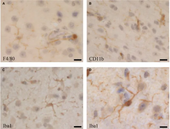
the CNS and activation stage [87]. Despite their different
origin by early immigration from the yolk sac, microglia
share many markers with other macrophage populations,
such as the blood-derived perivascular, choroid plexus
and leptomeningeal macrophages, including F4/80
(Figure 2A), CD11b (Figure 2B) [118], but as microglia
usually represent the majority of immune cells in the
CNS, some of those markers are routinely used for
their characterization [147–149]. Iba1 (ionized calcium
binding adaptor molecule 1), which is highly conserved
in mammals, has been useful as a specific marker for the
detection of microglia, since its discovery [150, 151], as
it is not expressed in blood monocytes, but often also in
blood-derived tissue macrophages and dendritic cells [118,
152, 153]. Thus, to distinguish microglia from
blood-derived immigrated macrophage populations, and from
blood monocytes, reduced expression of the common
leukocyte antigen CD45 as a marker has been suggested,
although CD45 is upregulated in activated microglia [152].
More recently, a plenitude of membrane proteins has been
identified in microglia. CX3CR1 (fractalkine receptor) is
one important functional membrane protein, as its ligand
(fractalkine, CX3CL1) is expressed on neurons [5, 90]
and astrocytes [154]. CX3CR1 has been widely used as
a marker for flow cytometry and immune histochemistry,
as well as for developing a crucial CX3CR1-GFP mouse
model, which is functionally very close to the Iba1-GFP
model [155]. However, when it comes to identifying
newly immigrated monocytic cells into the CNS and
to differentiate them from resident microglia, reliable
markers are still missing.
The transcriptome analysis of mouse microglia
has revealed a unique signature for the freshly isolated
brain-derived cells, whereas the cultured cells display
properties of activated microglia [156]. In addition to
some transcription factors (Rhox5, Cebpe, E2f6, Hoxc6,
Phf17, Ppargc1b), several membrane proteins have been
identified in microglia that are unique and not expressed in
other macrophages, including the ion transporters Slco4a1,
Slc30a5, Mcoln3, the lipid-metabolism associated cell
Figure 2: Microglia cells visualised by immune histochemistry in the mouse brain using antibodies directed against.
(A) F4/80 (Serotec, Kidlington, UK, clone Cl:A3-1), (B) CD11b (BD Pharmingen, San Diego, USA, cat. no. 553308), and (C and D) Iba1 (Abcam, UK, cat. no. ab5076). Bars: (A and C) 25 µm, (B) 20 µm, (D) 40 µm.
membrane molecules Lrp8, Lpcat3, Stab1, Pap2c, and
the putative efflux cell membrane receptor Mfsd10.
Unfortunately, reliable antibodies against these proteins,
for specific histological detection of microglia in the CNS,
are still missing. Similar work has also been done with
human brain-derived microglia, which shows substantial
similarities to the mouse microglia under physiological
resting conditions [157]. However, activated microglia and
cells from aged individuals show significant differences
between mouse and human, as well as depending on
location and condition [158, 159], making comparison of
transcriptome studies quite difficult [160].
Microglia build a 3-dimensional network in the
CNS and they communicate also through hemichannels
and gap junctions [161, 162]. The hemichannels allow
secretion and uptake of glutamate and ATP, factors that
are essential in the communication with neurons and
astrocytes. The gap junctions allow microglia to react as a
syncytium. However, the extent of such connections and
their relevance need further investigation, also in respect
of possible pharmacological treatment by using functional
modulators or blockers.
Microglia are the professional phagocytes of the
CNS. They are able to sense and take up extracellular
material, like cell debris, apoptotic cells, as well as
tumour cells and microbes. Consequently, they contribute
substantially to the function and structure of the CNS.
They express a variety of sensing and binding receptors
on their surface membrane [163]. Phagocytosis is
essential for the control of newly generated neurons
during development and adult neurogenesis, as well as
for synapse homeostasis. However, they are able to engulf
whole or parts of neurons, which may become fatal if
phagocytosis gets out of control and if live, functional
neurons are eliminated [164–166]. Microglia use a variety
of receptors for the recognition of molecules, particles
and cells that they engulf [167]. Sialic acid binding
immunoglobulin-like lectins (Siglecs) are important
regulatory receptors expressed on microglia and binding
to sialated ligands on neurons or CNS tumor cells [168–
171]. Siglecs signaling modulates activation of microglia
and thus also phagocytosis activity. Although they also
serve as binding receptors, signaling through SIRPα
(signal-regulatory protein alpha; CD172a), complement
receptor 3 (CR3; CD11b), LRP (low-density lipoprotein
receptor-related protein; CD90.2) and TREM2 (protein
triggering receptor expressed on myeloid cells-2) also
modulates phagocytosis by microglia, indicating that live
neurons can control phagocytosis through expression
of corresponding ligands [165, 172–179]. On the other
hand, activated microglia secret inflammatory cytokines
or other mediators that are able to regulate expression of
those ligands on neurons, which may eliminate live and
functional neurons and induce disease. Consequently,
fine-tuning of the balance between eat-me and
don’t-eat-me signals, in the interaction between microglia and
neurons, controls for whether neurons are engulfed and
eliminated, or not [164]. Interestingly, there seem to be sex
and age differences, when it comes to microglia functions,
including phagocytosis, although the mechanisms are not
well understood [180, 181]. For instance, in experimental
autoimmune encephalomyelitis (EAE) mouse model
for MS, which may present with reduced relapses
during pregnancy, estrogen was found to promote
anti-inflammatory, protective and regenerative microglia [182,
183]. Further, in experimental stroke, smaller infarcts and
anti-inflammatory microglia were observed in female, but
not in male mice [184]. Thus, phagocytosis by microglia is
crucial for function and structure of the CNS, as well as in
many pathologies. Therefore, more research on this topic
is required in the future.
Continuous and sufficient blood supply to the CNS is
essential and disruption of perfusion results in subsequent
damage of function and structure [185, 186]. Regional
perfusion of the CNS is tightly regulated at capillary
level depending on oxygen and energy needs by the local
neurons, which control regional perfusion indirectly via
astrocytes. However, the molecular mechanisms for the
regional perfusion control are still not well understood.
Most important is also the tight separation of the CNS
tissue and intercellular space from the intravascular space,
which is given by the blood-brain-barrier (BBB) [187,
188]. The BBB is important to keep serum proteins (e.g.
complement system, antibodies, etc.) and a plenitude of
soluble factors (e.g. cytokines, microbial products, etc.)
in the blood circulation out of the CNS tissue, as many
of those factors induce immediate and strong activation
of microglia that results in devastating neuroinflammation
[189, 190]. In mouse, the CNS vascularisation starts at
about E8 by endothelial cells forming a capillary network,
at around the same time of microglia colonisation, but
before astrocytes emerge. However, CNS pericytes that
stabilise the endothelial capillaries emerge around the
same time [191]. Yet unknown neuronal factors and
the pericytes control differentiation of CNS capillary
endothelial cells and thus the formation of the first line
of the BBB by increasing the tight connections between
endothelial cells and by forcing them to establish
limited, CNS-specific transcytosis. The functional
capillary system, forming the internal part of the BBB,
is composed of a continuous layer of endothelial cells
interconnected through tight junctions and enclosing the
lumen containing the blood. A distinct continuous basal
lamina surrounds the endothelial cells in which pericytes
are embedded without forming a continuous layer [192].
The basal lamina and the pericytes form the perivascular
space which may also contain immune cells, including
macrophages, often referred to as perivascular microglia,
and lymphocytes. The perivascular space is peripherally
completely covered by endfoot processes of the astrocytes
forming the glia limitans, a second, external closure of
the BBB interconnected through yet unknown adhesion
molecules [193, 194]. Occasionally, also processes of
microglia also contribute to the glia limitans layer, called
the juxtavascular microglia. Whereas the astrocytes
control the transport of nutrients, oxygen and other
molecules between the blood vessels and the neurons, the
role of the perivascular and juxtavascular microglia is not
well understood. However, they may monitor the interface
between the first inner and second outer line of the BBB,
and consequently mediate between the blood and the CNS
environment, and vice-versa. Most important, activated
microglia opens up the BBB by releasing inflammatory
factors, resulting in enhanced neuroinflammation, by
allowing serum components and immune cells entering
the CNS tissue [36, 195, 196]. As most recently described
[197] in healthy condition, the endothelial BBB is closed
and enforced by claudin (CLDN) 5 and . In inflammation,
the endothelial part of the BBB downregulates CLDN5
and opens towards the perivascular space, while the
astrocytes of the glia limitans upregulate CLDN1,
CLDN4, and junctional adhesion molecule A (JAM-A),
and forms an enhanced second barrier, composed of
reactive astrocytes with tight junctions containing CLDN1,
CLDN4 and JAM-A subunits. Enhancement of the second
barrier has been attributed to local microglia secreting
IL-1β, a driver cytokine of lesion pathogenesis in multiple
sclerosis and the corresponding EAE model. In addition
to inflammatory diseases of the CNS, stroke and trauma
of the CNS are extreme cases of the breakdown of the
BBB with subsequent substantial activation of microglia
[37, 198, 199]. Activated perivascular microglia may
also be able to control repair of the damaged BBB [200,
201], as well as to induce or support angiogenesis, which
is important in CNS tumours [202, 203] and vascular
pathologies of the retina [204–207]. First experimental
therapeutic approaches have been suggested [208], but
more research is required. Substantial research of this
topic has used live animal imaging [209, 210], often the
retina model, as the blood vessels of the eye are easily
monitored. However, more in vivo research about the
influence of microglia on the BBB would be needed and
new models ought to be developed.
Activation of microglia and their role in
neuroinflammation, neurodegenerative
conditions, mental diseases, aging and gender
Under physiological conditions, ramified, resting
microglia provides a neuroprotective environment
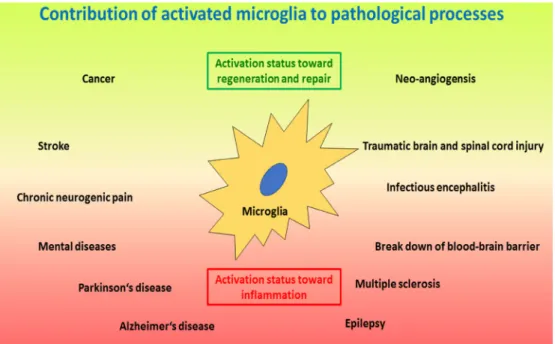
[211, 212]. However, most CNS pathologies, as well as
regenerative efforts, include activation of microglia with
corresponding inflammatory events (Figure 3) [213, 214].
Activated, inflammatory microglia are thus neurotoxic
and kill neurons by engulfing them or releasing various
neurotoxic molecules and factors, including reactive
oxygen species (ROS), glutamate, Fas-ligand, tumour
necrosis factor α (TNFα) and others [215–218]. On the
other hand, activated neuroprotective microglia may
secrete neurotrophins that support neuroregeneration
[219]. Of note, microglia do not act on their own, but
they coordinate their action with astroglia [27, 220].
Recently, a new type of microglia has been described
under pathological conditions named the ¨dark microglia¨,
due to their characteristic dark appearance in electron
microscopy because of ultrastructural changes, which
are proposed to reflect oxidative stress in a particularly
hyperactive subset of microglia [221]. Morphologically,
microglia activation results in increased dynamics of the
cell processes that are extended and retracted according
to corresponding signals. In addition, the cells may give
up their home location and migrate towards the area of
action close by, where they may accumulate and may form
a protective enclosure around the pathological or damaged
area usually seen around damaged CNS tissue after trauma
or amyloid-β plaques in Alzheimer’s disease [222, 223].
Similar to other tissue macrophages, microglia change
upon activation the pattern of surface proteins and secrete
various soluble factors [224]. In addition, phagocytosis is
upregulated upon activation. Depending on continuous
presence or absence of the cause for the activation, or on
clearance of the problem, microglia may either display the
inflammatory properties and remain chronically activated,
or they may change towards a protective phenotype and
function where they support tissue repair and restoration
of structure and function of the CNS [20, 225, 226].
Interestingly, severe local or systemic events, external
to the CNS, like systemic inflammation or bacterial
infection with sepsis can open and cross the BBB and
subsequently activate microglia [18, 227] and interfere
with ongoing CNS processes [228]. On the other hand,
activation of microglia and neuroinflammation may also
result in the break-down of the BBB and corresponding
leakage of complement into the CNS, which results in
enhanced activation of microglia and neuroinflammation
[229]. However, chronic neuroinflammation may result in
neurodegenerative diseases.
Microglia express various pattern recognition
receptors (PPRs) for sensing endogenous
danger-associated molecular patterns (DAMPs: e.g. heat shock
proteins) [230] and exogenous pathogen-associated
molecular patterns (PAMPs: microbial proteins,
saccharides, lipids, RNA and DNA) [231]. Toll-like
receptors (TLRs) are such PPRs, transmit danger signals
and strongly activate microglia in the context of CNS
damage or infection [232, 233]. Interestingly, galectin-3
(Gal3) secreted by activated microglia is also a ligand
of TLR4, which may result in chronic activation of
microglia [234]. Amyloid-β, which is found in the context
of Alzheimer’s disease, as well as prions also activate
microglia [235]. It has also been shown that chromogranin
A, released by stressed neurons and recognized by
scavenger receptors on microglia results in their activation
[217]. Inflammatory activation signals in microglia are
then integrated in the inflammasome [236], which results
in activation of transcription factors for the transcription
of inflammatory genes [237]. Microglia activation can
be such a deleterious and destructive event by harming
structure and function of the CNS that many regulatory
mechanisms ought to be built in this process, of which few
have recently been discovered, including TREM2 [238,
239]. Interestingly, upregulation of macrophage
colony-stimulating factor receptor (CD115, M-CSFR; c-fms) on
microglia makes them rather neuroprotective [240], as
well as signalling through P2X7 receptor [241]. Like for
most immune cells, microglia respond to metabolic and
energy-related events and activation results in metabolic
reprogramming of the cells [242], which interestingly
differs between different activation stages [23], which
allows accordingly more detailed differentiation between
microglia activation stages. In addition, chronic activated
microglia may have substantial epigenetic changes of their
chromatin, which may then be difficult to reverse [243].
Trauma of the CNS is a frequent pathology,
including traumatic brain (TBI) and spinal cord (SCI)
injury. Severity of the damage can range from very
minute, e.g. in contusion, to substantial tissue damage
resulting in deleterious loss of function, including
paresis, paralysis and hemi- or tetraplegia. Microglia are
immediately activated in such an event [26] and they try
at first to enclose the damage and minimise the spread
of it [223]. However, strong activation of microglia
may damage viable neighbouring neurons [244]. In the
course of the response to injury, microglia contribute
first to cleaning debris [245] and later to tissue repair,
usually resulting in non-functional scar tissue [21, 27].
Interestingly, minor recurrent trauma like concussion
during sport (boxing, soccer, etc.), or injury in a distal
CNS region, may also activate microglia in otherwise
healthy tissue and result on a longer term in substantial
mental disease or chronic pain [246, 247]. One has also
to consider that injury breaks down the BBB and allows
immigration of blood derived immune cells, including
macrophages and lymphocytes that contribute to the acute
inflammatory reaction and subsequent tissue repair [16,
248]. Various factors known to contribute to induction
and regulation of neuroinflammation, as well as to tissue
repair are involved in the course of CNS injury, including
CX3CR1-CX3CL1 that mediate communication between
microglia and neurons or astrocytes [249]. However, the
detailed molecular mechanisms and the extent of the role
of microglia in CNS trauma and repair are still not well
understood and need further future research [250].
CNS ischemia and stroke are events with similar
tissue responses seen in traumatic injury of the CNS,
although the cause is different. Correspondingly, microglia
reacts very similarly to tissue damage and similarly
supports tissue repair mechanisms [13–15, 251–254],
also including immigration of peripheral blood leucocytes
[255, 256]. Most important is of course to consider
Figure 3: Schematic drawing of important processes where activated microglia play a crucial role.
The lower red area depicts pathologies induced and/or sustained by microglia activated along the inflammatory and neurotoxic pathway, including neurodegenerative and autoimmune-like diseases, as wells as neurogenic pain, infectious encephalitis and break-down of the blood-brain barrier. The upper green area depicts microglia activated along the regenerative pathway where the focus is on tissue repair, including injury and repair, as well as tumour growth and angioneogenesis. Of note, microglia activation status can be different at different locations at the same time. In addition, microglia activation status may change from one polarized status to the other polarized status over time for certain pathologies; e.g. in infections at first the inflammatory status is required to eliminate the infectious agent, whereas later a regenerative status will support repair of damage caused by the infection.the underlying systemic metabolic and inflammatory
condition, usually contributing to the cardiovascular
disease and subsequent stroke, which may also influence
microglia response to ischemia and tissue damage and
which increases the complexity of the pathology of stroke
[257, 258].
Activated microglia driving chronic
neuroinflammation have also been shown to
substantially contribute to aging of the CNS [259, 260],
epilepsy [261], chronic neuropathic pain [262], mental
diseases [35, 263, 264] and neurodegenerative diseases,
including Alzheimer’s disease [222], Parkinson’s
disease [265], amyotrophic lateral sclerosis (ALS)
[34] and multiple sclerosis [33]. Aging goes in parallel
with systemic chronic activation of the immune system
and polarization towards a low-level inflammatory
status [266, 267]. This process also affects the CNS
and thus microglia [259, 268], which interferes with
CNS homeostasis, especially adult neurogenesis
[269], the function and structure of myelination
[270] and synapses, as well as the BBB. Mental and
neurodegenerative diseases have then probably to be
seen as focal processes of age-related activated and
dysfunctional microglia [45, 271–274]). However, better
understanding of the molecular and cellular mechanisms
in the activation of microglia and the regulation of
inflammatory processes in the CNS is required and
more research is therefore needed in these areas of
neuroimmunology.
The role of microglia in viral encephalitis and in
brain tumours
Virus infections are the most frequent reason for
encephalitis. A variety of viruses affect the CNS and
induce encephalitis, including flaviviruses such as Dengue
[275, 276], hepatitis C [277], Japanese encephalitis [278,
279], West Nile [44] and Zika [280–282], as well as
herpes viruses such as herpes simplex [283], varicella
Zoster [284] and Kaposi’s sarcoma associated herpes
virus [285], as well as HIV [286, 287]. All the viruses
mentioned above may infect microglia [276–278, 280,
281, 288], whereas flaviviruses and herpes viruses, but
not HIV [289], may also infect neurons and/or astroglia.
Infected neurons and astroglia usually respond with
inflammatory signals that activate surrounding microglia.
Infected microglia are usually also activated and may
substantially contribute to the anti-viral immune response
in the CNS, which results in the recruitment of blood
monocytes and T-lymphocytes and in the clearance of the
virus. Virus-dependent neuroinflammation often results in
the local break-down of the BBB. However, damage of
the BBB may not be the initial event for the transmission
of the virus from the blood to the CNS tissue and cells,
but other mechanisms can be involved. E.g. in the case of
Japanese encephalitis, somehow astrocytes and microglia
are first infected, before the BBB opens up [290]. Thus,
the virus could either get into the CNS via infected
capillary endothelial cells which release infectious viral
particles towards the CNS, or via infected leucocytes
(in HIV) [291] that enter into the CNS and transmit
the virus to susceptible microglia [292]. There is also a
broad variety of reactivity between different individuals
in humans, resulting in a broad spectrum between low
and severe neuroinflammation and neuronal infection
and cell death. Correspondingly, the infection outcome
may vary between asymptomatic and fatal consequences,
with a broad spectrum between minor mental disorder
and severe cognitive deficiencies. Viral infection of the
developing CNS results usually in severe malformation
which may even result in a non-viable foetus and thus
lead to abortion, as it has been recently documented
for Zika [282, 293]. Viral infections of microglia may
also interfere with their function, which may result in
enhanced, uncontrolled inflammation and/or immune
deficiency, of which the mechanisms are still not well
understood. Fortunately, vaccines have been successfully
established for some viruses, e.g. Japanese encephalitis,
which prevents infection in the first instance. However,
for many of the mentioned viruses, there is still no vaccine
available and substantial more research is required to
better understand the role of microglia in corresponding
viral encephalitis.
Glioma, especially glioblastoma, is the most
frequent primary brain tumour [50]. Therefore, glioma
is covered here, representing the principal mechanisms
of microglia in CNS primary tumours and metastases.
Gliomas develop from genetically aberrant glia cell
precursors and are usually monoclonal [294]. Microglia
play a role in establishing a growing glioma tumour, and
certainly help glioma to grow together with its vascular
supply [295–297]. The vascular bed of gliomas has to
be considered different from the functional BBB [298]
and allows substantial immigration of CCR2 + blood
derived monocytes, as well as regulatory/suppressive
lymphocytes and other leucocytes into the tissue, due to
CCL2 production of the tumour [299, 300]. Interestingly,
there is a more immune-suppressive, tumour growth
promoting population of residential microglia, as well
as an inflammatory, monocytes-derived macrophage
population [202]. Together, in the interaction with the
tumour cells, they provide an optimal environment
for survival and growth of glioma, by preventing an
effective anti-tumour immune response, generating
space in the healthy tissue and providing growth factors
[301–303]. However, future therapies have probably to
target, in addition to the tumour cells, also microglia
and macrophages, as well as the vascular cells [304].
In addition to mouse and human in vitro models, the
zebrafish in vivo model has become very helpful to
investigate the role and interaction of microglia with
glioma [305].
Microglia research models
Microglia research still relies heavily on in vivo [51]
and ex vivo cellular and tissue models [241], including
mouse [32, 70, 209, 306, 307], rat [308] and zebrafish
[70, 309], as these cells are unique and very different from
other tissue macrophages and bone marrow derived cells.
However, for research in humans, two in vitro approaches
have been successfully envisaged: (1) isolation of primary
microglia from surgical specimens of the brain [310] or
post-mortem tissue samples [147, 278, 311, 312], or (2)
differentiation of microglia-like cells from embryonic stem
cells [313], induced pluripotent stem cells (iPS) [314–
316], bone marrow stem cells or blood monocytes [317–
319]. Although all those in vitro human models have their
limitations, they have so far been very useful to investigate
cellular and molecular mechanisms related to cellular
structure and function in the context of inflammation,
infection and immune response. Recently, microglia from
different models have been better characterized using
modern transcriptomic methods [320] and compared,
indicating that all cultured cells used for in vitro models
are probably at an activated stage [147], and only in
vivo models represent physiological resting microglia.
However, it may be possible in the future to also develop
better in vitro models of resting microglia [321] once
the in situ tissue conditions and molecular and cellular
properties are better known. Interestingly, microglia
depletion models [322] that can also be replenished with
different kinds of modified cells [241] have more recently
been developed. It is thus expected that more new models
will be developed in the near future in microglia research,
adding new knowledge and new aspects of the very
versatile and important CNS macrophages, the microglia.
CONCLUSIONS
Microglia are thus unique cells of the central nervous
system, linking it to the immune system. They participate
in the biology and pathology of the CNS from early on
through-out development and later in CNS homeostasis.
Their origin and many of their functions have only recently
been discovered, but much more is still not known.
Consequently, microglia will remain for the years to come
on the centre stage of research in neuroimmunology.
Abbreviations
Central nervous system (CNS), induced pluripotent
stem cells (iPS), multiple sclerosis MS), Chemokine ligand
(CCL), Chemokine receptor (CCR), tumour necrosis
factor alpha (TNFα), macrophage colony-stimulating
factor receptor (CD115, M-CSFR; c-fms), reactive oxygen
species (ROS), blood-brain barrier (BBB), human immune
deficiency virus (HIV), interleukin (IL), complement
receptor 3 (CR3, CD11b).
CONFLICTS OF INTEREST
The authors declare no conflicts of interest.
REFERENCES
1. del Rio-Hortega P. The microglia. Lancet. 1939; 1:1023–26. 2. Ginhoux F, Greter M, Leboeuf M, Nandi S, See P,
Gokhan S, Mehler MF, Conway SJ, Ng LG, Stanley ER, Samokhvalov IM, Merad M. Fate mapping analysis reveals that adult microglia derive from primitive macrophages. Science. 2010; 330:841–45.
3. Casano AM, Peri F. Microglia: multitasking specialists of the brain. Dev Cell. 2015; 32:469–77.
4. Salter MW, Beggs S. Sublime microglia: expanding roles for the guardians of the CNS. Cell. 2014; 158:15–24. 5. Sheridan GK, Murphy KJ. Neuron-glia crosstalk in health
and disease: fractalkine and CX3CR1 take centre stage. Open Biol. 2013; 3:130181.
6. Vecino E, Rodriguez FD, Ruzafa N, Pereiro X, Sharma SC. Glia-neuron interactions in the mammalian retina. Prog Retin Eye Res. 2016; 51:1–40.
7. Peferoen L, Vogel D, Breur M, Gerritsen W, Dijkstra C, Amor S. Do stressed oligodendrocytes trigger microglia activation in pre-active MS lesion? Glia. 2013; 61:S164– 164.
8. Frost JL, Schafer DP. Microglia: Architects of the Developing Nervous System. Trends Cell Biol. 2016; 26:587–97.
9. Mosser CA, Baptista S, Arnoux I, Audinat E. Microglia in CNS development: shaping the brain for the future. Prog Neurobiol. 2017; 149-150:1–20.
10. Gemma C, Bachstetter AD. The role of microglia in adult hippocampal neurogenesis. Front Cell Neurosci. 2013; 7:229.
11. Nisticò R, Salter E, Nicolas C, Feligioni M, Mango D, Bortolotto ZA, Gressens P, Collingridge GL, Peineau S. Synaptoimmunology - roles in health and disease. Mol Brain. 2017; 10:26.
12. Schafer DP, Rosen AR, Lehrman E, Heller C, Stevens B. Pruning CNS synapses: an active role for glia and the complement cascade. J Neurochem. 2013; 125:65.
13. Anttila JE, Whitaker KW, Wires ES, Harvey BK, Airavaara M. Role of microglia in ischemic focal stroke and recovery: focus on Toll-like receptors. Prog Neuropsychopharmacol Biol Psychiatry. 2017; 79:3–14.
14. Guruswamy R, ElAli A. Complex Roles of Microglial Cells in Ischemic Stroke Pathobiology: New Insights and Future Directions. Int J Mol Sci. 2017; 18:18.
15. Xiong XY, Liu L, Yang QW. Functions and mechanisms of microglia/macrophages in neuroinflammation and neurogenesis after stroke. Prog Neurobiol. 2016; 142:23– 44.
16. David S, Greenhalgh AD, Kroner A. Macrophage and microglial plasticity in the injured spinal cord. Neuroscience. 2015; 307:311–18.
17. Bieber K, Autenrieth SE. Insights how monocytes and dendritic cells contribute and regulate immune defense against microbial pathogens. Immunobiology. 2015; 220:215–26.
18. Hoogland IC, Houbolt C, van Westerloo DJ, van Gool WA, van de Beek D. Systemic inflammation and microglial activation: systematic review of animal experiments. J Neuroinflammation. 2015; 12:114.
19. Ascoli BM, Géa LP, Colombo R, Barbé-Tuana FM, Kapczinski F, Rosa AR. The role of macrophage polarization on bipolar disorder: identifying new therapeutic targets. Aust N Z J Psychiatry. 2016; 50:618–30.
20. Cherry JD, Olschowka JA, O’Banion MK. Neuroinflammation and M2 microglia: the good, the bad, and the inflamed. J Neuroinflammation. 2014; 11:98. 21. Loane DJ, Kumar A. Microglia in the TBI brain: the good, the
bad, and the dysregulated. Exp Neurol. 2016; 275:316–27. 22. Nakagawa Y, Chiba K. Diversity and plasticity of microglial
cells in psychiatric and neurological disorders. Pharmacol Ther. 2015; 154:21–35.
23. Orihuela R, McPherson CA, Harry GJ. Microglial M1/M2 polarization and metabolic states. Br J Pharmacol. 2016; 173:649–65.
24. Ransohoff RM. A polarizing question: do M1 and M2 microglia exist? Nat Neurosci. 2016; 19:987–91.
25. Tang Y, Le W. Differential Roles of M1 and M2 Microglia in Neurodegenerative Diseases. Mol Neurobiol. 2016; 53:1181–94.
26. Xu H, Wang Z, Li J, Wu H, Peng Y, Fan L, Chen J, Gu C, Yan F, Wang L, Chen G. The Polarization States of Microglia in TBI: A New Paradigm for Pharmacological Intervention. Neural Plast. 2017; 2017:5405104.
27. Gao Z, Zhu Q, Zhang Y, Zhao Y, Cai L, Shields CB, Cai J. Reciprocal modulation between microglia and astrocyte in reactive gliosis following the CNS injury. Mol Neurobiol. 2013; 48:690–701.
28. Terry RL, Getts DR, Deffrasnes C, van Vreden C, Campbell IL, King NJ. Inflammatory monocytes and the pathogenesis of viral encephalitis. J Neuroinflammation. 2012; 9:270. 29. Doens D, Fernández PL. Microglia receptors and their
implications in the response to amyloid β for Alzheimer’s disease pathogenesis. J Neuroinflammation. 2014; 11:48. 30. Leyns CE, Holtzman DM. Glial contributions to
neurodegeneration in tauopathies. Mol Neurodegener. 2017; 12:50.
31. Schlachetzki JC, Hüll M. Microglial activation in Alzheimer’s disease. Curr Alzheimer Res. 2009; 6:554–63. 32. Joers V, Tansey MG, Mulas G, Carta AR. Microglial
phenotypes in Parkinson’s disease and animal models of the disease. Prog Neurobiol. 2017; 155:57–75.
33. Luo C, Jian C, Liao Y, Huang Q, Wu Y, Liu X, Zou D, Wu Y. The role of microglia in multiple sclerosis. Neuropsychiatr Dis Treat. 2017; 13:1661–67.
34. Brites D, Vaz AR. Microglia centered pathogenesis in ALS: insights in cell interconnectivity. Front Cell Neurosci. 2014; 8:117.
35. Réus GZ, Fries GR, Stertz L, Badawy M, Passos IC, Barichello T, Kapczinski F, Quevedo J. The role of inflammation and microglial activation in the pathophysiology of psychiatric disorders. Neuroscience. 2015; 300:141–54.
36. da Fonseca C, Carolina A, Matias D, Garcia C, Amaral R, Geraldo LH, Freitas C. Souza Lima FR. The impact of microglial activation on blood-brain barrier in brain diseases. Front Cell Neurosci. 2014; 8: 362.
37. Dudvarski Stankovic N, Teodorczyk M, Ploen R, Zipp F, Schmidt MH. Microglia-blood vessel interactions: a double-edged sword in brain pathologies. Acta Neuropathol. 2016; 131:347–63.
38. Herz J, Filiano AJ, Smith A, Yogev N, Kipnis J. Myeloid Cells in the Central Nervous System. Immunity. 2017; 46:943–56.
39. Lively S, Schlichter LC. The microglial activation state regulates migration and roles of matrix-dissolving enzymes for invasion. J Neuroinflammation. 2013; 10:75.
40. Lopes Pinheiro MA, Kooij G, Mizee MR, Kamermans A, Enzmann G, Lyck R, Schwaninger M, Engelhardt B, de Vries HE. Immune cell trafficking across the barriers of the central nervous system in multiple sclerosis and stroke. Biochim Biophys Acta. 2016; 1862:461–71.
41. Mracsko E, Javidi E, Na SY, Kahn A, Liesz A, Veltkamp R. Leukocyte invasion of the brain after experimental intracerebral hemorrhage in mice. Stroke. 2014; 45:2107–14.
42. Varvel NH, Neher JJ, Bosch A, Wang W, Ransohoff RM, Miller RJ, Dingledine R. Infiltrating monocytes promote brain inflammation and exacerbate neuronal damage after status epilepticus. Proc Natl Acad Sci USA. 2016; 113:E5665–74.
43. Neumann J, Riek-Burchardt M, Herz J, Doeppner TR, König R, Hütten H, Etemire E, Männ L, Klingberg A, Fischer T, Görtler MW, Heinze HJ, Reichardt P, et al. Very-late-antigen-4 (VLA-4)-mediated brain invasion by neutrophils leads to interactions with microglia, increased ischemic injury and impaired behavior in experimental stroke. Acta Neuropathol. 2015; 129:259–77.
44. Bréhin AC, Mouriès J, Frenkiel MP, Dadaglio G, Desprès P, Lafon M, Couderc T. Dynamics of immune cell recruitment during West Nile encephalitis and identification of a new CD19+B220-BST-2+ leukocyte population. J Immunol. 2008; 180:6760–67.
45. Spittau B. Aging Microglia-Phenotypes, Functions and Implications for Age-Related Neurodegenerative Diseases. Front Aging Neurosci. 2017; 9:194.
46. Das Sarma J. Microglia-mediated neuroinflammation is an amplifier of virus-induced neuropathology. J Neurovirol. 2014; 20:122–36.
47. Furr SR, Marriott I. Viral CNS infections: role of glial pattern recognition receptors in neuroinflammation. Front Microbiol. 2012; 3:201.
48. Barichello T, Generoso JS, Simões LR, Goularte JA, Petronilho F, Saigal P, Badawy M, Quevedo J. Role of Microglial Activation in the Pathophysiology of Bacterial Meningitis. Mol Neurobiol. 2016; 53:1770–81.
49. Morocoima A, Socorro G, Avila R, Hernández A, Merchán S, Ortiz D, Primavera G, Chique J, Herrera L, Urdaneta-Morales S. Trypanosoma cruzi: experimental parasitism in the central nervous system of albino mice. Parasitol Res. 2012; 111:2099–107.
50. Poon CC, Sarkar S, Yong VW, Kelly JJ. Glioblastoma-associated microglia and macrophages: targets for therapies to improve prognosis. Brain. 2017; 140:1548–60.
51. Sieger D, Peri F. Animal models for studying microglia: the first, the popular, and the new. Glia. 2013; 61:3–9. 52. Atallah N, Vasiu R, Boşca AB, Creţu DI, Georgiu C,
Constantin AM, Sovrea AS. Microglia—performers of the 21st century. Rom J Morphol Embryol. 2014; 55:745–65. 53. Peña-Altamira E, Prati F, Massenzio F, Virgili M,
Contestabile A, Bolognesi ML, Monti B. Changing paradigm to target microglia in neurodegenerative diseases: from anti-inflammatory strategy to active immunomodulation. Expert Opin Ther Targets. 2016; 20:627–40.
54. Masuda T, Prinz M. Microglia: A Unique Versatile Cell in the Central Nervous System. ACS Chem Neurosci. 2016; 7:428–34.
55. Tay TL, Savage JC, Hui CW, Bisht K, Tremblay MÈ. Microglia across the lifespan: from origin to function in brain development, plasticity and cognition. J Physiol. 2017; 595:1929–45.
56. Kierdorf K, Erny D, Goldmann T, Sander V, Schulz C, Perdiguero EG, Wieghofer P, Heinrich A, Riemke P, Hölscher C, Müller DN, Luckow B, Brocker T, et al. Microglia emerge from erythromyeloid precursors via Pu.1- and Irf8-dependent pathways. Nat Neurosci. 2013; 16:273–80.
57. Gomez Perdiguero E, Klapproth K, Schulz C, Busch K, Azzoni E, Crozet L, Garner H, Trouillet C, de Bruijn MF, Geissmann F, Rodewald HR. Tissue-resident macrophages originate from yolk-sac-derived erythro-myeloid progenitors. Nature. 2015; 518:547–51.
58. Hoeffel G, Ginhoux F. Ontogeny of Tissue-Resident Macrophages. Front Immunol. 2015; 6:486.
59. Nayak D, Roth TL, McGavern DB. Microglia development and function. Annu Rev Immunol. 2014; 32:367–402. 60. Walls JR, Coultas L, Rossant J, Henkelman RM.
Three-dimensional analysis of vascular development in the mouse embryo. PLoS One. 2008; 3:e2853.
61. Tay TL, Hagemeyer N, Prinz M. The force awakens: insights into the origin and formation of microglia. Curr Opin Neurobiol. 2016; 39:30–37.
62. Goldmann T, Wieghofer P, Jordão MJ, Prutek F, Hagemeyer N, Frenzel K, Amann L, Staszewski O, Kierdorf K, Krueger M, Locatelli G, Hochgerner H, Zeiser R, et al. Origin, fate and dynamics of macrophages at central nervous system interfaces. Nat Immunol. 2016; 17:797–805.
63. Hoeffel G, Chen J, Lavin Y, Low D, Almeida FF, See P, Beaudin AE, Lum J, Low I, Forsberg EC, Poidinger M, Zolezzi F, Larbi A, et al. C-Myb(+) erythro-myeloid progenitor-derived fetal monocytes give rise to adult tissue-resident macrophages. Immunity. 2015; 42:665–78. 64. Minocha S, Valloton D, Arsenijevic Y, Cardinaux JR, Guidi
R, Hornung JP, Lebrand C. Nkx2.1 regulates the generation of telencephalic astrocytes during embryonic development. Sci Rep. 2017; 7:43093.
65. Petrik D, Yun S, Latchney SE, Kamrudin S, LeBlanc JA, Bibb JA, Eisch AJ. Early postnatal in vivo gliogenesis from nestin-lineage progenitors requires cdk5. PLoS One. 2013; 8:e72819.
66. Casano AM, Albert M, Peri F. Developmental Apoptosis Mediates Entry and Positioning of Microglia in the Zebrafish Brain. Cell Reports. 2016; 16:897–906.
67. Lyons DA, Talbot WS. Glial Cell Development and Function in Zebrafish. Cold Spring Harb Perspect Biol. 2015; 7: a020586.
68. Rossi F, Casano AM, Henke K, Richter K, Peri F. The SLC7A7 Transporter Identifies Microglial Precursors prior to Entry into the Brain. Cell Reports. 2015; 11:1008–17. 69. Svahn AJ, Graeber MB, Ellett F, Lieschke GJ, Rinkwitz
S, Bennett MR, Becker TS. Development of ramified microglia from early macrophages in the zebrafish optic tectum. Dev Neurobiol. 2013; 73:60–71.
70. Yu T, Guo W, Tian Y, Xu J, Chen J, Li L, Wen Z. Distinct regulatory networks control the development of macrophages of different origins in zebrafish. Blood. 2017; 129:509–19.
71. Semple BD, Blomgren K, Gimlin K, Ferriero DM, Noble-Haeusslein LJ. Brain development in rodents and humans: identifying benchmarks of maturation and vulnerability to injury across species. Prog Neurobiol. 2013; 106-107:1–16. 72. Smith AM, Dragunow M. The human side of microglia.
Trends Neurosci. 2014; 37:125–35.
73. Elmore MR, Najafi AR, Koike MA, Dagher NN, Spangenberg EE, Rice RA, Kitazawa M, Matusow B, Nguyen H, West BL, Green KN. Colony-stimulating factor 1 receptor signaling is necessary for microglia viability, unmasking a microglia progenitor cell in the adult brain. Neuron. 2014; 82:380–97.
74. Solary E, Droin N. The emerging specificities of interleukin-34. J Leukoc Biol. 2014; 95:3–5.
75. Greter M, Lelios I, Pelczar P, Hoeffel G, Price J, Leboeuf M, Kündig TM, Frei K, Ginhoux F, Merad M, Becher B.
Stroma-derived interleukin-34 controls the development and maintenance of langerhans cells and the maintenance of microglia. Immunity. 2012; 37:1050–60.
76. Koguchi K, Nakatsuji Y, Okuno T, Sawada M, Sakoda S. Microglial cell cycle-associated proteins control microglial proliferation in vivo and in vitro and are regulated by GM-CSF and density-dependent inhibition. J Neurosci Res. 2003; 74:898–905.
77. Salimi K, Moser K, Zassler B, Reindl M, Embacher N, Schermer C, Weis C, Marksteiner J, Sawada M, Humpel C. Glial cell line-derived neurotrophic factor enhances survival of GM-CSF dependent rat GMIR1-microglial cells. Neurosci Res. 2002; 43:221–29.
78. Zhang J, Geula C, Lu C, Koziel H, Hatcher LM, Roisen FJ. Neurotrophins regulate proliferation and survival of two microglial cell lines in vitro. Exp Neurol. 2003; 183:469–81.
79. Svahn AJ, Giacomotto J, Graeber MB, Rinkwitz S, Becker TS. miR-124 Contributes to the functional maturity of microglia. Dev Neurobiol. 2016; 76:507–18.
80. Xu J, Wang T, Wu Y, Jin W, Wen Z. Microglia Colonization of Developing Zebrafish Midbrain Is Promoted by Apoptotic Neuron and Lysophosphatidylcholine. Dev Cell. 2016; 38:214–22.
81. Smolders SM, Swinnen N, Kessels S, Arnauts K, Smolders S, Le Bras B, Rigo JM, Legendre P, Brône B. Age-specific function of α5β1 integrin in microglial migration during early colonization of the developing mouse cortex. Glia. 2017; 65:1072–88.
82. Riquier AJ, Sollars SI. Microglia density decreases in the rat rostral nucleus of the solitary tract across development and increases in an age-dependent manner following denervation. Neuroscience. 2017; 355:36–48.
83. Schafer DP, Stevens B. Microglia Function in Central Nervous System Development and Plasticity. Cold Spring Harb Perspect Biol. 2015; 7:a020545.
84. Swinnen N, Smolders S, Avila A, Notelaers K, Paesen R, Ameloot M, Brône B, Legendre P, Rigo JM. Complex invasion pattern of the cerebral cortex bymicroglial cells during development of the mouse embryo. Glia. 2013; 61:150–63.
85. Gordon S, Plüddemann A. Tissue macrophages: heterogeneity and functions. BMC Biol. 2017; 15:53. 86. Askew K, Li K, Olmos-Alonso A, Garcia-Moreno F, Liang
Y, Richardson P, Tipton T, Chapman MA, Riecken K, Beccari S, Sierra A, Molnár Z, Cragg MS, et al. Coupled Proliferation and Apoptosis Maintain the Rapid Turnover of Microglia in the Adult Brain. Cell Reports. 2017; 18:391–405.
87. Walker FR, Beynon SB, Jones KA, Zhao Z, Kongsui R, Cairns M, Nilsson M. Dynamic structural remodelling of microglia in health and disease: a review of the models, the signals and the mechanisms. Brain Behav Immun. 2014; 37:1–14.
88. Kemmerling N, Wunderlich P, Theil S, Linnartz-Gerlach B, Hersch N, Hoffmann B, Heneka MT, de Strooper B, Neumann H, Walter J. Intramembranous processing by γ-secretase regulates reverse signaling of ephrin-B2 in migration of microglia. Glia. 2017; 65:1103–18.
89. Arnoux I, Audinat E. Fractalkine Signaling and Microglia Functions in the Developing Brain. Neural Plast. 2015; 2015:689404.
90. Paolicelli RC, Bisht K, Tremblay ME. Fractalkine regulation of microglial physiology and consequences on the brain and behavior. Front Cell Neurosci. 2014; 8:129. 91. Bergon A, Belzeaux R, Comte M, Pelletier F, Hervé
M, Gardiner EJ, Beveridge NJ, Liu B, Carr V, Scott RJ, Kelly B, Cairns MJ, Kumarasinghe N, et al. CX3CR1 is dysregulated in blood and brain from schizophrenia patients. Schizophr Res. 2015; 168:434–43.
92. Derecki NC, Cronk JC, Lu Z, Xu E, Abbott SB, Guyenet PG, Kipnis J. Wild-type microglia arrest pathology in a mouse model of Rett syndrome. Nature. 2012; 484:105–09. 93. Hellwig S, Brioschi S, Dieni S, Frings L, Masuch A, Blank T, Biber K. Altered microglia morphology and higher resilience to stress-induced depression-like behavior in CX3CR1-deficient mice. Brain Behav Immun. 2016; 55:126–37.
94. Lehmann ML, Cooper HA, Maric D, Herkenham M. Social defeat induces depressive-like states and microglial activation without involvement of peripheral macrophages. J Neuroinflammation. 2016; 13:224.
95. Schafer DP, Heller CT, Gunner G, Heller M, Gordon C, Hammond T, Wolf Y, Jung S, Stevens B. Microglia contribute to circuit defects in Mecp2 null mice independent of microglia-specific loss of Mecp2 expression. eLife. 2016; 5:5.
96. VanRyzin JW, Yu SJ, Perez-Pouchoulen M, McCarthy MM. Temporary Depletion of Microglia during the Early Postnatal Period Induces Lasting Sex-Dependent and Sex-Independent Effects on Behavior in Rats. eNeuro. 2016; 3:3.
97. Squarzoni P, Oller G, Hoeffel G, Pont-Lezica L, Rostaing P, Low D, Bessis A, Ginhoux F, Garel S. Microglia modulate wiring of the embryonic forebrain. Cell Reports. 2014; 8:1271–79.
98. Squarzoni P, Thion MS, Garel S. Neuronal and microglial regulators of cortical wiring: usual and novel guideposts. Front Neurosci. 2015; 9:248.
99. Wake H, Moorhouse AJ, Jinno S, Kohsaka S, Nabekura J. Resting microglia directly monitor the functional state of synapses in vivo and determine the fate of ischemic terminals. J Neurosci. 2009; 29:3974–80.
100. Macht VA. Neuro-immune interactions across development: A look at glutamate in the prefrontal cortex. Neurosci Biobehav Rev. 2016; 71:267–80.
101. Mayhew J, Beart PM, Walker FR. Astrocyte and microglial control of glutamatergic signalling: a primer
on understanding the disruptive role of chronic stress. J Neuroendocrinol. 2015; 27:498–506.
102. Zabel MK, Kirsch WM. From development to dysfunction: microglia and the complement cascade in CNS homeostasis. Ageing Res Rev. 2013; 12:749–56.
103. Stevens B, Allen NJ, Vazquez LE, Howell GR, Christopherson KS, Nouri N, Micheva KD, Mehalow AK, Huberman AD, Stafford B, Sher A, Litke AM, Lambris JD, et al. The classical complement cascade mediates CNS synapse elimination. Cell. 2007; 131:1164–78.
104. Arcuri C, Mecca C, Bianchi R, Giambanco I, Donato R. The Pathophysiological Role of Microglia in Dynamic Surveillance, Phagocytosis and Structural Remodeling of the Developing CNS. Front Mol Neurosci. 2017; 10:191. 105. Buschert J, Sakalem ME, Saffari R, Hohoff C,
Rothermundt M, Arolt V, Zhang W, Ambrée O. Prenatal immune activation in mice blocks the effects of environmental enrichment on exploratory behavior and microglia density. Prog Neuropsychopharmacol Biol Psychiatry. 2016; 67:10–20.
106. Claypoole LD, Zimmerberg B, Williamson LL. Neonatal lipopolysaccharide treatment alters hippocampal neuroinflammation, microglia morphology and anxiety-like behavior in rats selectively bred for an infantile trait. Brain Behav Immun. 2017; 59:135–46.
107. Fernández de Cossío L, Guzmán A, van der Veldt S, Luheshi GN. Prenatal infection leads to ASD-like behavior and altered synaptic pruning in the mouse offspring. Brain Behav Immun. 2017; 63:88–98.
108. Lee JH, Espinera AR, Chen D, Choi KE, Caslin AY, Won S, Pecoraro V, Xu GY, Wei L, Yu SP. Neonatal inflammatory pain and systemic inflammatory responses as possible environmental factors in the development of autism spectrum disorder of juvenile rats. J Neuroinflammation. 2016; 13:109.
109. Mattei D, Ivanov A, Ferrai C, Jordan P, Guneykaya D, Buonfiglioli A, Schaafsma W, Przanowski P, Deuther-Conrad W, Brust P, Hesse S, Patt M, Sabri O, et al. Maternal immune activation results in complex microglial transcriptome signature in the adult offspring that is reversed by minocycline treatment. Transl Psychiatry. 2017; 7:e1120. 110. Wang CY, Cheng CW, Wang WH, Chen PS, Tzeng SF.
Postnatal Stress Induced by Injection with Valproate Leads to Developing Emotional Disorders Along with Molecular and Cellular Changes in the Hippocampus and Amygdala. Mol Neurobiol. 2016; 53:6774–85.
111. Wang HT, Huang FL, Hu ZL, Zhang WJ, Qiao XQ, Huang YQ, Dai RP, Li F, Li CQ. Early-Life Social Isolation-Induced Depressive-Like Behavior in Rats Results in Microglial Activation and Neuronal Histone Methylation that Are Mitigated by Minocycline. Neurotox Res. 2017; 31:505–20.
112. Erny D, Hrabě de Angelis AL, Jaitin D, Wieghofer P, Staszewski O, David E, Keren-Shaul H, Mahlakoiv T,
Jakobshagen K, Buch T, Schwierzeck V, Utermöhlen O, Chun E, et al. Host microbiota constantly control maturation and function of microglia in the CNS. Nat Neurosci. 2015; 18:965–77.
113. Sherwin E, Rea K, Dinan TG, Cryan JF. A gut (microbiome) feeling about the brain. Curr Opin Gastroenterol. 2016; 32:96–102.
114. Aguzzi A, Barres BA, Bennett ML. Microglia: scapegoat, saboteur, or something else? Science. 2013; 339:156–61. 115. Asheuer M, Pflumio F, Benhamida S, Dubart-Kupperschmitt
A, Fouquet F, Imai Y, Aubourg P, Cartier N. Human CD34+ cells differentiate into microglia and express recombinant therapeutic protein. Proc Natl Acad Sci USA. 2004; 101:3557–62.
116. Barr CM, Manning J, Lewis CA, Rossi FM, Krieger C. Submyeloablative conditioning with busulfan permits bone marrow-derived cell accumulation in a murine model of Alzheimer’s disease. Neurosci Lett. 2015; 588:196–201. 117. Jin N, Gao L, Fan X, Xu H. Friend or Foe? Resident
Microglia vs Bone Marrow-Derived Microglia and Their Roles in the Retinal Degeneration. Mol Neurobiol. 2017; 54:4094–112.
118. Prinz M, Priller J, Sisodia SS, Ransohoff RM. Heterogeneity of CNS myeloid cells and their roles in neurodegeneration. Nat Neurosci. 2011; 14:1227–35.
119. Mildner A, Schmidt H, Nitsche M, Merkler D, Hanisch UK, Mack M, Heikenwalder M, Brück W, Priller J, Prinz M. Microglia in the adult brain arise from Ly-6ChiCCR2+ monocytes only under defined host conditions. Nat Neurosci. 2007; 10:1544–53.
120. De Lucia C, Rinchon A, Olmos-Alonso A, Riecken K, Fehse B, Boche D, Perry VH, Gomez-Nicola D. Microglia regulate hippocampal neurogenesis during chronic neurodegeneration. Brain Behav Immun. 2016; 55:179–90. 121. Ribeiro Xavier AL, Kress BT, Goldman SA, Lacerda
de Menezes JR, Nedergaard M. A Distinct Population of Microglia Supports Adult Neurogenesis in the Subventricular Zone. J Neurosci. 2015; 35:11848–61. 122. Sato K. Effects of Microglia on Neurogenesis. Glia. 2015;
63:1394–405.
123. Sierra A, Beccari S, Diaz-Aparicio I, Encinas JM, Comeau S, Tremblay ME. Surveillance, phagocytosis, and inflammation: how never-resting microglia influence adult hippocampal neurogenesis. Neural Plast. 2014; 2014:610343.
124. Pistikova A, Brozka H, Stuchlik A. Adult neurogenesis in the hippocampus from a perspective of discrimination and generalization: a hypothesis. Physiol Res. 2017; 66:441–48. 125. Lim DA, Alvarez-Buylla A. The Adult Ventricular-Subventricular Zone (V-SVZ) and Olfactory Bulb (OB) Neurogenesis. Cold Spring Harb Perspect Biol. 2016; 8:8. 126. Shigemoto-Mogami Y, Hoshikawa K, Goldman JE,
Sekino Y, Sato K. Microglia enhance neurogenesis and oligodendrogenesis in the early postnatal subventricular zone. J Neurosci. 2014; 34:2231–43.