HAL Id: hal-02494096
https://hal.umontpellier.fr/hal-02494096
Submitted on 28 Feb 2020
HAL is a multi-disciplinary open access archive for the deposit and dissemination of sci-entific research documents, whether they are pub-lished or not. The documents may come from teaching and research institutions in France or abroad, or from public or private research centers.
L’archive ouverte pluridisciplinaire HAL, est destinée au dépôt et à la diffusion de documents scientifiques de niveau recherche, publiés ou non, émanant des établissements d’enseignement et de recherche français ou étrangers, des laboratoires publics ou privés.
A single amino acid substitution (H451Y) in Leishmania
calcium-dependent kinase SCAMK confers high
tolerance and resistance to antimony
Baptiste Vergnes, Elodie Gazanion, Cédric Mariac, Miléna Du Manoir,
Lauriane Sollelis, Jose-Juan Lopez-Rubio, Yvon Sterkers, Anne-Laure Bañuls
To cite this version:
Baptiste Vergnes, Elodie Gazanion, Cédric Mariac, Miléna Du Manoir, Lauriane Sollelis, et al.. A single amino acid substitution (H451Y) in Leishmania calcium-dependent kinase SCAMK confers high tolerance and resistance to antimony. Journal of Antimicrobial Chemotherapy, Oxford University Press (OUP), 2019, 74 (11), pp.3231-3239. �10.1093/jac/dkz334�. �hal-02494096�
Title: A single amino acid substitution (H451Y) in Leishmania calcium-dependent kinase
1
SCAMK confers high tolerance and resistance to antimony.
2
Running title: Leishmania tolerance and resistance
3 4
Authors: Baptiste VERGNES1*, Elodie GAZANION1, Cédric MARIAC2, Miléna DU MANOIR1,
5
Lauriane SOLLELIS1, José-Juan LOPEZ-RUBIO1, Yvon STERKERS1,3, Anne-Laure BAÑULS1
6 7
Contact Information: 1 MIVEGEC, IRD, CNRS, Univ. Montpellier, Montpellier, France; 2 DIADE,
8
IRD, Univ. Montpellier, Montpellier, France; 3 Department of Parasitology-Mycology, Faculty
9
of Medicine, University Hospital Center of Montpellier, Univ. Montpellier, Montpellier, 10
France. 11
Present address: Lauriane Sollelis, Wellcome Centre for Molecular Parasitology, Institute of
12
Infection, Immunity & Inflammation, College of Medical, Veterinary and Life Sciences, 13
University of Glasgow, Glasgow, UK 14
*Corresponding author: Baptiste Vergnes Ph.D., UMR MIVEGEC (IRD, CNRS, Univ.
15
Montpellier), 911 Avenue Agropolis, 34394 Montpellier Cedex 5, France. Phone number: 16
+33(0)467416308. Email address: baptiste.vergnes@ird.fr
17
A final version of this manuscript has been published, please cite:
Baptiste Vergnes, Elodie Gazanion, Cédric Mariac, Miléna Du Manoir, Lauriane Sollelis, José-Juan Lopez-Rubio, Yvon Sterkers, Anne-Laure Bañuls, A single amino acid
substitution (H451Y) in Leishmania calcium-dependent kinase SCAMK confers high tolerance and resistance to antimony, Journal of Antimicrobial Chemotherapy, Volume 74, Issue 11, November 2019, Pages 3231–3239, https://doi.org/10.1093/jac/dkz334
Synopsis:
18
Background: For almost a century, antimonials remain the first-line drugs for the treatment
19
of leishmaniasis. However, little is known about their mode of action and clinical resistance 20
mechanisms. 21
Objectives: We have previously shown that Leishmania nicotinamidase (PNC1) is an essential
22
enzyme for parasite NAD+ homeostasis and virulence in vivo. Here, we found that parasites 23
lacking pnc1 gene (Dpnc1) are hypersusceptible to the active form of antimony (SbIII) and used 24
these mutant parasites to better understand antimony mode of action and resistance. 25
Methods: SbIII-resistant WT and Dpnc1 parasites were selected in vitro by stepwise selection
26
method. NAD(H)/NADP(H) dosages and quantitative RT-PCR experiments were performed to 27
explain the susceptibility differences observed between strains. WGS and a marker-free 28
CRISPR/Cas9 base editing approach were used to identify and validate the role of a new 29
resistance mutation. 30
Results: NAD+ depleted Dpnc1 parasites are highly susceptible to SbIII and this phenotype can
31
be rescued by NAD+ precursor of trypanothione precursors supplementation. Dpnc1 parasites 32
can become resistant to SbIII by unknown mechanism. WGS revealed a unique amino acid 33
substitution (H451Y) in an EF-hand domain of an orphan calcium-dependent kinase recently 34
named SCAMK. When introduced into a wild type reference strain by base editing, the H451Y 35
mutation allows Leishmania parasites to survive at extreme concentrations of SbIII, 36
potentiating the rapid emergence of resistant parasites. 37
Conclusions: These results establish that Leishmania SCAMK is a new central hub of antimony
38
mode of action and resistance development and uncover the importance of drug tolerance 39
mutations in the evolution of parasite drug resistance. 40
Introduction
41
Leishmania are protozoan parasites transmitted by sandflies that are responsible for a wide 42
spectrum of human infections ranging from the life-threatening visceral disease to disfiguring 43
mucosal and cutaneous forms1. Treatment of leishmaniasis is limited to four main drugs
44
(antimonials, miltefosine, amphotericin B and paromomycin). Alone or in combination, 45
antimonials have been the mainstay of anti-Leishmania therapy worldwide for over 70 years. 46
However, our knowledge of antimony mode of action is still partial and the emergence of 47
parasite resistance to antimony in the Indian subcontinent during the last decades has 48
necessitated the use of alternative medications. The active form of antimony (SbIII) is known 49
to target the Leishmania redox potential by inducing rapid thiols efflux and inhibiting the 50
NADPH-dependent trypanothione reductase (TR) that maintains the main parasite thiol, 51
trypanothione, in its reduced form2 (Figure 1a). Leishmania resistance to SbIII is generally
52
mediated by drug uptake reduction through the aquaglyceroprotein 1 (AQP1) transporter 53
mutations or down-expression, or by drug efflux/sequestration through the concomitant 54
overexpression of genes involved in the parasite thiol metabolism and of the gene encoding 55
the ATP-binding cassette transporter ABC-C3 (MRPA)3,4 (Figure 1a). Other transporters of the
56
ABC superfamily also interfere with antimonials drug accumulation, including ABC-C7 (PRP1)5,
57
ABC-I36, ABC-I47 or ABC-G28. In addition, it has been shown that a number of intracellular
58
proteins can also modulate the susceptibility of parasites to SbIII when they are overexpressed 59
in experimental strains or clinical isolates. These include heat shock proteins (Hsp239,
60
Hsp8310), kinase (MAPK111), serine/threonine phosphatase (LinJ.12.061012) or proteins with
61
still unknown functions (ARM569, ARM589, P29913). Overall, these results illustrate that
62
Leishmania parasites have multiple strategies to acquire drug resistance and highlight the 63
difficulty to define a conserved molecular marker of antimony resistance. 64
In a previous work14, we showed that Leishmania parasites are auxotrophic for NAD+
65
and rely on a salvage pathway to recycle NAD+ precursors from their environment (Figure 1a). 66
The nicotinamidase PNC1 is a central enzyme of this pathway that controls NAD+ homeostasis 67
by hydrolysing nicotinamide (NAm) to nicotinic acid (NA). Leishmania infantum parasites in 68
which the pnc1 gene was disrupted (Dpnc1) are depleted in NAD+ and cannot cause durable 69
infections in mice, making of this enzyme an attractive target for drug development14,15.
70
NAD(H), and its derived phosphorylated forms NADP(H), are essential cofactors required for 71
energy-producing pathways and antioxidant defence in all living cells. They are further key 72
metabolites involved in host-pathogen interactions16. Here, we used NAD+ depleted Dpnc1
73
mutant parasites to better understand SbIII mode of action and the mechanisms leading to 74
resistance to this drug. We found that intracellular NAD+ levels can directly modulate the 75
susceptibility of parasites to SbIII and identified the first mutation conferring antimony 76
tolerance and resistance in Leishmania. 77
Materials and Methods
78
Strains and cultures
79
The pnc1 null mutant (Dpnc1) was previously generated by targeted gene replacement in the 80
L. infantum (MHOM/MA/67/ITMAP-263) strain14. CRISPR/Cas9 genome edition experiments
81
were performed in the L. major “Friedlin” reference strain (MHOM/IL/80/Friedlin, 82
LEM3171)17. All strains and the derived mutants were maintained as promastigote forms in
83
SDM79 medium supplemented with 10% FBS, penicillin/streptomycin and hemin (5 mg/L) 84
(complete medium). Potassium antimonyl tartrate trihydrate (SbIII), L-Glutathione reduced 85
(GSH), N-Acetyl-L-cysteine (NAC), nicotinic acid (NA) and MTT were all purchased from Sigma-86
Aldrich. SbIII-resistant parasites were generated by a stepwise approach starting with a drug 87
concentration corresponding to the EC50 for that strain. Growth curve and EC50 were
88
determined starting from an inoculum of 106 parasites/mL in 5 mL of complete medium,
89
unless otherwise stated. Parasite density was determined daily (growth kinetics) or after 3 90
days of incubation with the drug (EC50 determination) using a flow cytometer14 or by
91
measuring the OD at 600 nm (for graphical convenience, OD values were multiplied by 1000). 92
All EC50 values were obtained with GraphPad Prism v5 software using a sigmoidal
dose-93
response model with variable slope. Cell viability assays on edited parasites were performed 94
using a MTT test. In this assay, the yellow tetrazolium MTT dye was reduced to insoluble 95
formazan crystals (purple color) in living cells using NADH as reducing agent. Briefly, 100µl of 96
parasite cultures were mixed with 10µL of a MTT solution (10 mg/mL in PBS) and incubated 97
4h at 26°C. The reaction was stopped with 100 µL of lysis solution (50% isopropanol/10% SDS) 98
and the plates were incubated for an additional 30 min with gentle shaking in the dark. The 99
change in colour from yellow to purple was read at an absorbance of 600 nm. 100
NAD(H)/NADP(H) quantification
101
Total (NAD+ plus NADH) or individual (NAD+ and NADH) dinucleotides were quantified using 102
the bioluminescent NAD/NADH-Glo™ Assay (Promega) following manufacturer’s instructions. 103
This kit uses a proluciferin substrate to produce a light signal proportional to the amount of 104
NAD and/or NADH present in the samples. Total (NADP+ plus NADPH) and individual (NADP+ 105
and NADPH) phosphorylated forms were quantified similarly using the NADP/NADPH-Glo™ 106
Assay kit (Promega). Data are expressed as the mean value (±SD) of Relative Luminescence 107
Units (RLU) obtained for 106 parasites and after 60 min of incubation.
108
Quantitative RT-PCR analyses (qRT-PCR)
109
All primers used in this study were designed with the primer3Plus software and are listed in 110
table S1. Total RNA was extracted from parasite cultures in the exponential phase of growth 111
using the RNeasy+ Mini Kit (Qiagen) with an additional treatment with turbo DNA-free DNase 112
(Thermo Fischer Scientific). Two µg of total RNA was transcribed into complementary DNA 113
(cDNA) using the Superscript reverse transcriptase III (Invitrogen) and oligo(dT)12-18 primers 114
(Thermo Fischer Scientific), according to the manufacturer’s instructions. Diluted cDNA (1/10) 115
was used for the qPCR reactions in 10 µL final volume with SYBR Green I Master on a 116
LightCycler480 (Roche). The relative expression of each gene was determined from two 117
biological replicates with the 2-ΔΔCT method and the GAPDH gene as reference, using the
118
LightCycler480 software. 119
CRISPR/Cas9 genome editing and off-target mutation analysis
120
The marker-free nucleotide editing approach developed in P. Falciparum18 was adapted to
121
introduce the H451Y mutation in the L. major reference strain. The 20nt single guide RNA 122
(sgRNA) sequence TTTGATCGACTCCGAGCACT (the targeted nucleotide is underlined) was 123
based on the LmjF.33.1710 orthologue gene sequence. Plasmids were constructed as 124
described in17. Briefly, a donor DNA sequence that consists of an 880 bp-long intragenic region
125
of LmjF.33.1710 gene was generated by PCR fusion using the modified primer pairs 126
Lm1F/Lm1R and Lm2F/Lm2R (Table S1) to create the C1351T mutation and a “shield” silent 127
mutation (G1356T) in the protospacer adjacent motif (PAM) to prevent further Cas9-mediated 128
cleavage (Figure 2f). The donor DNA sequence was cloned in the pLS5 plasmid digested with 129
KpnI and XbaI. To obtain the 20nt sgRNA sequence surrounded by the 15nt adaptors necessary 130
for In-Fusion® cloning, the two oligonucleotides Seed_F and Seed_R were annealed by 131
incubation in boiling water for 2min followed by gentle cooling in room-temperature water 132
for 2h. The sgRNA sequence and adaptors were then cloned in the BsgI-digested pLS5-donor 133
DNA plasmid using the In-Fusion HD cloning kit (Clonetech). pLS5-donor DNA plasmids with 134
and without the sgRNA sequence were transfected in the L. major Friedlin strain that harbours 135
the pTCAS9 plasmid17 to generate edited (Lm_H451Y) and control (Lm_ctrl) cell lines,
respectively. After transfection and selection with hygromycin and puromycin, gene edition 137
was checked by PCR amplification and Sanger sequencing using the 138
LmiSCAMK_F/LmiSCAMK_R primer pair (Table S1). Once heterozygosity was confirmed in the 139
mutated locus, parasites were cloned by limiting dilution. A parasite clone containing the 140
homozygous desired and shield mutations (named Lm_H451Y) was selected for subsequent 141
phenotype analyses. To confirm the absence of off-target mutations, the genomes of Lm_ctrl 142
and Lm_H451Y strains were sequenced (see below) and off-target candidates (up to five 143
mismatches allowed) were identified using the Protospacer Workbench software suite19. For
144
each candidate, the vicinity (within a 21nt window) to specific INDELs of the edited cell lines 145
was assessed as described in Vasquez et al.20 (Table S2).
146
WGS and analyses
147
Parasite genomic DNA was extracted using the QIAamp DNA Mini Kit (Qiagen). A paired-end 148
sequencing library for Dpnc1 parasites was prepared with the Nextera DNA Sample 149
Preparation Kit (Illumina) and sequenced on an Illumina HiSeq 2500 (2x250 bp) apparatus. 150
DNA samples from the KO-SbR, Lm_ctrl, Lm_H451Y and Lm_H451Y-SbR strains were sheared 151
using a Bioruptor Pico sonication device (Diagenode) to yield »400 bp fragments. Libraries 152
were constructed for WGS as previously described21. Paired-end sequencing (2 x 150 bp) was
153
performed on an Illumina MiSeq apparatus with the MiSeq Reagent Kit V2. The presence of 154
SNPs and INDELs was determined using the EuPathDB Galaxy platform22 and the workflow
155
pipeline for “Variant Calling, paired-end sequencing” and pre-loaded reference genomes for 156
the L. infantum JPCM5 and L. major Friedlin strains (TritrypDB build 29). Filtered Variant Call 157
Format (VCF) files corresponding to SNPs and INDELs with an impact on coding sequences 158
were compared between the Dpnc1 and KO-SbR libraries. Specific SNPs/INDELs detected in 159
KO-SbR (16 SNPs and 15 INDELs) were manually verified with the IGV 2.4.8 visualization tool, 160
and eliminated if already present in the Dpnc1 parental line. After this verification step, only 161
three specific SNPs were retained for analysis (Table S3). The same strategy was used to 162
compare SNPs/INDELs in the Lm_ctrl, Lm_H451Y and Lm_H451Y-SbR libraries and only SNPs 163
and INDELS with read depth >2 were kept for further analysis (Table S4). The CNV-seq pipeline 164
was used to identify CNVs potentially associated with drug resistance23. This pipeline identifies
165
localized regions in which the read depth normalized across the length of the chromosome 166
differs significantly between samples. For Dpnc1 and KO-SbR pairwise comparisons, a 3kb 167
window size was chosen, but similar results were obtained with smaller and larger window 168
sizes (Table S5). For Lm_H451Y and Lm_H451Y-SbR pairwise comparisons, the sliding window 169
size was automatically determined (4265 bp). Input hits files were derived from the Binary 170
Alignment Map (BAM) files generated by the EupathDB Variant Calling pipeline. 171
Accession number
172
All sequencing data have been submitted to the European Nucleotide Archive (ENA) and are 173
available under the accession number PRJEB27329. 174
Results
175
Dpnc1 parasites are highly susceptible to SbIII
176
When grown in SDM79 medium, Dpnc1 promastigotes had similar growth rates than wild type 177
(WT) parasites (Figure 1b). However, they are about ten-fold more susceptible to SbIII (EC50
178
Dpnc1 = 2. 51 µg/mL; EC50 WT = 20.92 µg/mL) (Figure 1c). This phenotype could be rescued by
179
NA supplementation in the growth medium (Figure 1c). NA supplementation also induced a 180
dose-dependent increase of both NAD(H) and NADP(H) content in Dpnc1 parasites where 181
these pools are significantly depleted (Figure 1d). Conversely, it did not have any effect on 182
NAD(H) and NADP(H) content in WT parasites (Figure 1d). Individual dinucleotides 183
measurements showed that Dpnc1 parasites were specifically depleted in NAD+ and NADPH 184
forms (Figure 1e). As the Leishmania redox homeostasis is also maintained by de novo 185
biosynthesis of reduced trypanothione (Figure 1a), we tested whether supplementation with 186
trypanothione precursors could restore the physiological SbIII susceptibility in Dpnc1 187
parasites. Supplementation with N-acetyl-cysteine (NAC) or reduced glutathione (GSH) 188
decreased the susceptibility of Dpnc1 parasites to SbIII (Figure 1f). Altogether, these results 189
established a direct connection between Leishmania NAD+ metabolism, SbIII toxicity and thiol 190
metabolism (Figure 1a). qRT-PCR assays further indicated that known genes involved in SbIII 191
mode of action and resistance were similarly transcribed in Dpnc1 and WT parasites, except 192
for GSH1, the rate-limiting step in glutathione biosynthesis, for which the transcript level was 193
increased 2-fold in Dpnc1 (Figure 1g). 194
Dpnc1 parasites resistant to SbIII show a single amino acid substitution in the SCAMK gene
195
To better understand the link between NAD+ homeostasis and SbIII susceptibility, we 196
tried to generate SbIII-resistant WT and Dpnc1 parasites (named WT-SbR and KO-SbR, 197
respectively) by stepwise selection in the presence of increasing concentrations of the drug. 198
We could select KO-SbR parasites that proliferated in the presence of up to 320 µg/mL of SbIII 199
(EC50 = 309 µg/mL), but not at higher concentrations (Figure 2a). In WT-SbR parasites (EC50
200
>1000µg/mL), MRPA was ten-fold upregulated as compared with WT parasites (Figure 2b) 201
which is in agreement with the known model of antimony resistance3,4 and the presence of
202
an extrachromosomal amplicon of 15 kb bearing MRPA gene in the WT-SbR strain (data not 203
shown). Conversely, qRT-PCR analysis showed a modest, but paradoxical overexpression of 204
the genes encoding the AQP1 transporter and MRPA efflux-pump in KO-SbR parasites (Figure 205
2b). Moreover, the levels of NAD+, NADH, NADP+ and NADPH were similar in KO-SbR and 206
Dpnc1 parasites (Figure 2c). As drug resistance in Leishmania is often associated with gene 207
copy number variations (CNVs) or SNPs24, we sequenced the genomes of Dpnc1 and KO-SbR
208
parasites by Illumina paired-end sequencing. As expected, no read aligned to the pnc1 gene, 209
confirming its deletion in the both strains (Figure S1). All CNVs detected in KO-SbR mutants 210
were located in coding telomeric or intergenic regions (Table S5). The look for non-211
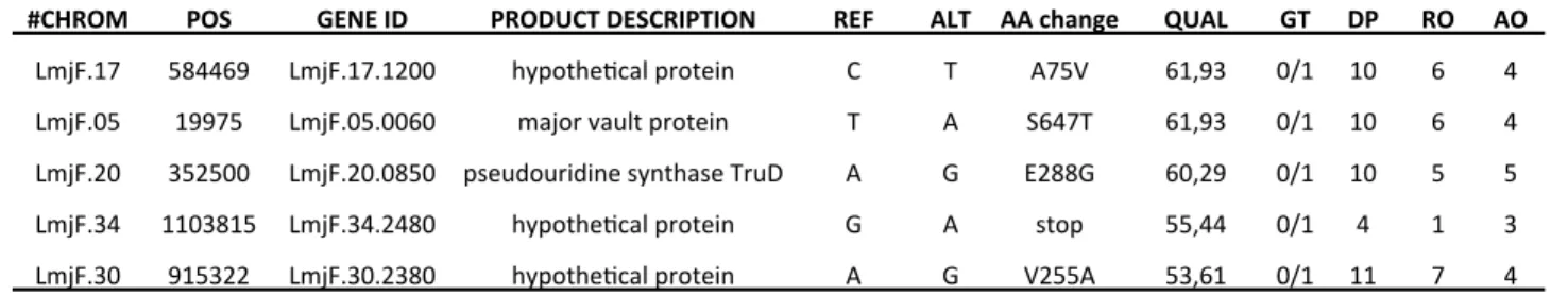
synonymous SNPs only present in the KO-SbR strain identified a single homozygous mutation 212
(C1351T) leading to the H451Y substitution in a putative protein kinase gene (LinJ.33.1810) 213
(Table S3). The fixation of the H451Y mutation in KO-SbR parasites was confirmed by PCR and 214
Sanger sequencing (Figure 2d). Interestingly, another point mutation (E629K) in the C-215
terminus of the LinJ.33.1810 gene was already described in a L. infantum SbIII-resistant strain 216
bearing MRPA amplification25. This single copy gene defines a new kinase family recently
217
named SCAMK26 that is specific to Metakinetoplastina protists and functionally related to the
218
calcium-dependent protein kinases (CDPKs) present in plants and protozoans27. CDPKs have
219
been widely studied in apicomplexan parasites where they are considered to be important 220
signal transducers involved in parasite motility, invasion or host cell egress28. Specifically, Ca2+
221
binding to the EF-hand domains of CDPKs induces a global conformational change of the 222
protein, resulting in the activation of the kinase activity. The H residue in position 451, which 223
is mutated in the KO-SbR strain, is located within the calcium-binding site of the third EF-hand 224
domain of Leishmania SCAMK, and is conserved across kinetoplastids (Figure 2e). 225
The H451Y mutation generates SbIII tolerant Leishmania parasites
226
To gain insight into the role of the H451Y mutation in SbIII resistance, we used the 227
CRISPR/cas9 genome editing technology17 with a marker-free nucleotide editing approach18
228
to introduce the H451Y mutation in the Leishmania major WT “Friedlin” reference strain. L. 229
major parasites expressing Cas9 from an episomal plasmid were transfected with a second 230
plasmid bearing an sgRNA complementary to the L. major SCAMK orthologue (LmjF.33.1710), 231
and a donor sequence including the desired mutation (C1351T) and a shield mutation 232
(G1356T) in the PAM motif (Lm_H451Y strain) (Figure 2f). As a control, we transfected 233
parasites with the same plasmid backbone, but without the sgRNA (Lm_ctrl strain) (Figure 2g). 234
After selection and cloning, we confirmed the presence of the H451Y mutation (Figure 2g) and 235
the absence of off-target mutations (Table S2) by Sanger and WGS, respectively. Phenotype 236
analyses indicated that Lm_ctrl and Lm_H451Y strains had similar growth rates (Figure 3a). 237
The SbIII susceptibility of both strains was compared using a MTT-based assay test and an 238
inoculum of 106 parasites/mL. As shown in figure 3b, Lm_H451Y parasites are not more
239
resistant than controls. Surprisingly, Lm_H451Y (but not Lm_ctrl) parasites exposed to the 240
highest drug concentrations resumed growth after seven days in culture (Figure 3c). Analysis 241
of the growth curves obtained starting from a higher parasite inoculum (4.106 parasites/mL)
242
and using cytocidal concentrations of SbIII showed that Lm_H451Y parasites can survive and 243
slowly proliferate in the presence of 1000 µg/mL of SbIII, which is the limit of SbIII solubility in 244
the culture medium (Figure 3d). In a parallel experiment, the viability of edited parasites 245
exposed to maximal SbIII concentrations has been confirmed and quantified by MTT-based 246
assays performed at days 3 and 5 of culture (Figure 3e). Light-microscopy examination after 247
exposure to the maximal concentration of SbIII revealed the presence of a heterogenous 248
population of dead and unstressed promastigote forms in Lm_H451Y cultures, and the 249
absence of surviving parasites in Lm_ctrl cultures (Figures 3f and S2). In agreement, we 250
detected intact high molecular weight genomic DNA in all tested conditions for Lm_H451Y 251
parasites, but only up to 250 µg/mL of SbIII for Lm_ctrl parasites (Figure 3g). The ability of a 252
microorganism to survive lethal concentrations of a drug while keeping similar EC50 than
253
susceptible strains is characteristic of drug tolerant/persistent microorganisms29. This
254
phenomenon is known to facilitate drug resistance acquisition in bacteria but remains largely 255
understudied in protozoan parasites30. To confirm the role of the H451Y mutation in
256
Leishmania SbIII tolerance, we transiently exposed parasites to various bolus doses of SbIII for 257
two days, and followed their growth after drug withdrawal. The growth of Lm_ctrl parasites 258
was delayed and they could survive after transient exposure to SbIII concentrations up to 400 259
µg/mL (Figure 3h). Conversely, Lm_H451Y parasites recovered within 10 days post-bolus, 260
whatever the concentration of SbIII used (Figure 3h). The toxicity of most anti-leishmanial 261
drugs has been partly attributed to induction of reactive oxygen species (ROS) formation and 262
consequently to oxidative damage. Therefore, we carried out similar growth curve 263
experiments in the presence of miltefosine, amphotericin B, the oxidative stress inducer 264
menadione, or H2O2 (Figure S3). We did not detect any difference between Lm_H451Y and
265
Lm_ctrl parasites in all tested conditions, suggesting that the tolerance phenotype induced by 266
the H451Y mutation is specific to SbIII mode of action and does not involve a generic response 267
to oxidative stress. Next, we tested whether Lm_H451Y parasites were prone to rapidly 268
develop resistance to SbIII. As expected, highly resistant Lm_H451Y parasites (named 269
Lm_H451Y-SbR) were selected after only five passages (P5) in the presence of 1000 µg/mL of 270
SbIII (EC50 >1000 µg/mL) and this resistance phenotype remained stable up to P15 (Figure 3i).
271
Genome sequencing of Lm_H451Y-SbR parasites cultured for 10 passages in the presence of 272
1000 µg/mL of SbIII did not reveal any new CNV compared with the Lm_H451Y parental cell 273
line. Moreover, non-synonymous SNPs specific to Lm_H451Y-SbR were all heterozygous and 274
only concerned hypothetical proteins or factors not known to be involved in SbIII resistance 275
(Table S4). These results corroborate our initial observations in the L. infantum KO-SbR mutant 276
and show that the H451Y mutation is necessary and sufficient to generate SbIII-tolerant 277
parasites that can rapidly evolve into resistant parasites in the presence of drug pressure. 278
Discussion
279
Drug resistance in eukaryotic microorganisms shares many similarities with antibiotic 280
resistance in bacteria3. Our knowledge about drug resistance mechanisms in Leishmania (and
281
other protozoan parasites) mainly results from experiments using growth inhibitory 282
measurements that determine the lowest drug concentration needed to inhibits parasite 283
growth by 50% (EC50). Mechanisms of drug action and resistance patterns can be however
284
different when measured at dosages that kill the parasite, not just inhibit its growth31.
285
Moreover, these approaches do not allow the identification of parasite subpopulations with 286
variable levels of resistance. In bacteria or fungi, drug tolerant subpopulations (also known as 287
“persisters”) can survive at lethal drug concentrations while being still sensitive to cytostatic 288
effect of the drug. The mechanisms leading to persisters formation are diverse and can include 289
genetic mutations that increase the proportion of persister cells within a population32,33.
290
Importantly, persisters are known to cause treatment failure due to relapsing infections and 291
behave as an evolutionary reservoir of drug resistance30,34,35. The phenomenon of drug
292
tolerance/persistence in protozoan parasites and its link with parasite drug resistance and 293
treatment failure have been poorly studied to date. In trypanosomatids, the ability of drug-294
sensitive parasites to survive a lethal and prolonged drug exposure has been recently 295
described in Trypanosoma cruzi36 and Leishmania37. However, these phenotypes have been
296
attributed to clearly distinct mechanisms involving a metabolic dormancy state and a genetic 297
preadaptation to resistance, respectively. 298
In this study, we report that (i) Leishmania NAD+ depletion led to SbIII hypersusceptibility that 299
is rescued by NAD+ precursor or trypanothione precursors supplementation; (ii) NAD+ 300
depleted parasites can develop SbIII resistance through a single amino acid substitution in a 301
metakinetoplastid-specific kinase; (iii) this mutation makes Leishmania parasites tolerant to 302
high concentrations of SbIII and facilitates the emergence of resistant parasites without 303
additional genomic modification. Although these findings have been obtained in mutants 304
selected under laboratory conditions in vitro, they provide novel insights into the mode of 305
action of antimony and establish that the orphan Leishmania kinase SCAMK is a new central 306
actor of SbIII mode of action and resistance. Because the H451Y mutation is precisely located 307
within the calcium-binding site of the third EF-hand domain of Leishmania SCAMK, we can 308
speculate that it should directly impair Ca2+ binding and the associated conformational change
required for the enzymatic activation of CDPKs. The study of the structural consequences of 310
the H451Y mutation should help us in understanding how this orphan kinase integrates 311
calcium signaling to control the lethal effect of SbIII and could result in new therapeutic 312
applications. We were unable to analyse the tolerance phenotype of edited parasites in the 313
intracellular amastigote stage because the L. major strain used in this study is a reference 314
strain that lost its virulence due to repeated passages in vitro. Further work is therefore 315
required to determine whether this mutation can lead to treatment failure and persistent 316
infection in vivo and to assess whether it is harmful or whether it can be spread in natural 317
populations. Nevertheless, the correlation between Leishmania SCAMK gene polymorphisms 318
and antimony treatment failure in clinical isolates could constitute the first step towards the 319
identification of a new molecular marker of antimony tolerance/resistance in the field. 320
Acknowledgements: We thank the MGX platform (Montpellier, France) and Dr Mallorie Hide
321
for HiSeq sequencing, and the UMR DIADE (Dynadiv team) for library preparation and MiSeq 322
sequencing at CIRAD (Montpellier, France). We are also thankful to Elisabetta Andermarcher 323
for assistance in editing the manuscript. 324
Funding: This work was supported by the Institut de Recherche pour le Développement (IRD),
325
the Centre National de la Recherche Scientifique (CNRS), the French Ministry of Research and 326
the Centre Hospitalier Universitaire of Montpellier institutional fundings. LS, JJLR, and YS were 327
supported by the Laboratoire d’Excellence (LabEx) ParaFrap (the French Parasitology Alliance 328
for Health Care, grant number ANR-11- LABX-0024). 329
References
330
1. Burza S, Croft SL, Boelaert M. Leishmaniasis. The Lancet 2018; 392: 951–70.
331
2. Wyllie S. Dual action of antimonial drugs on thiol redox metabolism in the human pathogen
332
Leishmania donovani. J Biol Chem 2004; 279: 39925–32. 333
3. Fairlamb AH, Gow NAR, Matthews KR et al. Drug resistance in eukaryotic microorganisms.
334
Nat Microbiol 2016; 1: 16092. 335
4. Ponte-Sucre A, Gamarro F, Dujardin J-C et al. Drug resistance and treatment failure in
336
leishmaniasis: A 21st century challenge. PLoS Negl Trop Dis 2017; 11: e0006052–24. 337
5. Leprohon P, Légaré D, Ouellette M. Intracellular localization of the ABCC proteins of
338
Leishmania and their role in resistance to antimonials. Antimicrob Agents Chemother 2009; 339
53: 2646–9.
340
6. Arcari T, Manzano JI, Gamarro F. ABCI3 is a new mitochondrial ABC transporter from
341
Leishmania major involved in susceptibility to antimonials and infectivity. Antimicrob Agents 342
Chemother 2017; 61: 113–8. 343
7. Manzano JI, Garcia-Hernandez R, Castanys S et al. A new ABC half-transporter in Leishmania
344
major is involved in resistance to antimony. Antimicrob Agents Chemother 2013; 57: 3719–30. 345
8. Perea A, Manzano JI, Castanys S et al. The LABCG2 transporter from the protozoan parasite
346
Leishmania is involved in antimony resistance. Antimicrob Agents Chemother 2016; 60: 3489– 347
96. 348
9. Tejera Nevado P, Bifeld E, Höhn K et al. A telomeric cluster of antimony resistance genes on
349
chromosome 34 of Leishmania infantum. Antimicrob Agents Chemother 2016; 60: 5262–75. 350
10. Vergnes B, Gourbal B, Girard I et al. A proteomics screen implicates HSP83 and a small
351
kinetoplastid calpain-related protein in drug resistance in Leishmania donovani clinical field 352
isolates by modulating drug-induced programmed cell death. Mol Cell Proteomics 2007; 6: 88– 353
101. 354
11. Garg M, Goyal N. MAPK1 of Leishmania donovani modulates antimony susceptibility by
355
downregulating P-glycoprotein efflux pumps. Antimicrob Agents Chemother 2015; 59: 3853– 356
63. 357
12. Gazanion E, Fernandez-Prada C, Papadopoulou B et al. Cos-Seq for high-throughput
358
identification of drug target and resistance mechanisms in the protozoan parasite Leishmania. 359
Proc Natl Acad Sci USA 2016; 113: E3012–21. 360
13. Choudhury K, Zander D, Kube M et al. Identification of a Leishmania infantum gene
361
mediating resistance to miltefosine and SbIII. Int J Parasitol 2008; 38: 1411–23. 362
14. Gazanion E, Garcia D, Silvestre R et al. The Leishmania nicotinamidase is essential for
363
NAD(+) production and parasite proliferation. Mol Microbiol 2011; 82: 21–38. 364
15. Michels PAM, Avilán L. The NAD(+) metabolism of Leishmania, notably the enzyme
365
nicotinamidase involved in NAD(+) salvage, offers prospects for development of anti-parasite 366
chemotherapy. Mol Microbiol 2011; 82: 4–8. 367
16. Mesquita I, Varela P, Belinha A et al. Exploring NAD(+) metabolism in host-pathogen
368
interactions. Cell Mol Life Sci 2016; 73: 1225–36. 369
17. Sollelis L, Ghorbal M, MacPherson CR et al. First efficient CRISPR-Cas9-mediated genome
370
editing in Leishmania parasites. Cell Microbiol 2015; 17: 1405–12. 371
18. Ghorbal M, Gorman M, MacPherson CR et al. Genome editing in the human malaria
372
parasite Plasmodium falciparum using the CRISPR-Cas9 system. Nat Biotechnol 2014; 32: 819– 373
21. 374
19. MacPherson CR, Scherf A. Flexible guide-RNA design for CRISPR applications using
375
Protospacer Workbench. Nat Biotechnol 2015; 33: 805–6. 376
20. Vasquez JJ, Wedel C, Cosentino RO et al. Exploiting CRISPR-Cas9 technology to investigate
377
individual histone modifications. Nucleic Acids Res 2018; 46: e106. 378
21. Mariac C, Scarcelli N, Pouzadou J et al. Cost-effective enrichment hybridization capture of
379
chloroplast genomes at deep multiplexing levels for population genetics and phylogeography 380
studies. Mol Ecol Resour 2014; 14: 1103–13. 381
22. Aurrecoechea C, Barreto A, Basenko EY et al. EuPathDB: the eukaryotic pathogen genomics
382
database resource. Nucleic Acids Res 2017; 45: D581–91. 383
23. Xie C, Tammi MT. CNV-seq, a new method to detect copy number variation using
high-384
throughput sequencing. BMC Bioinformatics 2009; 10: 80–9. 385
24. Leprohon P, Fernandez-Prada C, Gazanion E et al. Drug resistance analysis by next
386
generation sequencing in Leishmania. Int J Parasitol Drugs Drug Resist 2015; 5: 26–35. 387
25. Brotherton MC, Bourassa S, Leprohon P et al. Proteomic and genomic analyses of
388
antimony resistant Leishmania infantum mutant. PLoS ONE 2013; 8: e81899. 389
26. Chen F, Zhang L, Lin Z et al. Identification of a novel fused gene family implicates
390
convergent evolution in eukaryotic calcium signaling. BMC Genomics 2018; 19: 306. 391
27. Harper JF, Harmon A. Plants, symbiosis and parasites: a calcium signalling connection. Nat
392
Rev Mol Cell Biol 2005; 6: 555–66. 393
28. Billker O, Lourido S, Sibley LD. Calcium-dependent signaling and kinases in apicomplexan
394
parasites. Cell Host Microbe 2009; 5: 612–22. 395
29. Fisher RA, Gollan B, Helaine S. Persistent bacterial infections and persister cells. Nat Rev
396
Microbiol 2017; 15: 453–64. 397
30. Cohen NR, Lobritz MA, Collins JJ. Microbial persistence and the road to drug resistance.
398
Cell Host Microbe 2013; 13: 632–42. 399
31. Roepe PD. To kill or not to kill, that is the question: cytocidal antimalarial drug resistance.
400
Trends Parasitol 2014; 30: 130–5. 401
32. Michiels JE, Van den Bergh B, Verstraeten N et al. Molecular mechanisms and clinical
402
implications of bacterial persistence. Drug Resist Updat 2016; 29: 76–89. 403
33. Balaban NQ, Helaine S, Lewis K et al. Definitions and guidelines for research on antibiotic
404
persistence. Nat Rev Microbiol 2019; doi: 10.1038/s41579-019-0196-3. [Epub ahead of print] 405
406
34. Delarze E, Sanglard D. Defining the frontiers between antifungal resistance, tolerance and
407
the concept of persistence. Drug Resist Updat 2015; 23: 12–9. 408
35. Levin-Reisman I, Ronin I, Gefen O et al. Antibiotic tolerance facilitates the evolution of
409
resistance. Science 2017; 355: 826–30. 410
36. Sánchez-Valdéz FJ, Padilla A, Wang W et al. Spontaneous dormancy protects Trypanosoma
411
cruzi during extended drug exposure. eLife 2018; 7: 833. 412
37. Dumetz F, Cuypers B, Imamura H et al. Molecular preadaptation to antimony resistance in
413
Leishmania donovani on the Indian subcontinent. mSphere 2018; 3: e00548–17. 414
Figure legends
415
Figure 1. Dpnc1 parasites are highly susceptible to SbIII. (a) Schematic representation of the
416
cross-talk between Leishmania NAD+ metabolism, trivalent antimony (SbIII) mode of action 417
and thiol metabolism. NA, nicotinic acid; NAm, nicotinamide; NR, nicotinamide riboside; 418
NAMN, nicotinic acid mononucleotide; NAAD, nicotinic acid dinucleotide; NADK, NAD+ kinase; 419
PPP, pentose phosphate pathway; G6PDH, glucose-6-phosphate dehydrogenase; AQP1, 420
aquaglyceroporin 1; TR, trypanothione reductase; T(SH)2, reduced trypanothione; T(S)2, 421
oxidized trypanothione; TXN1, tryparedoxin 1; MRPA, ABCC3 transporter; Glu, glutamate; Cys, 422
cysteine; Orn, ornithine; GSH1, gamma-glutamylcysteine synthetase; GSH, glutathione; Spd, 423
spermidine; GSpdS, glutathionylspermidine synthetase; Gspd, glutathionylspermidine; TryS, 424
trypanothione synthase. (b) Growth curves of L. infantum WT and Dpnc1 promastigotes in 425
SDM79 medium supplemented with NA (1, 10 and 100 µM). Data are representative of three 426
independent experiments. (c) Effect of SbIII on the growth kinetics of L. infantum WT and 427
Dpnc1 parasites (supplemented or not with 1, 10 and 100 µM NA). Data are the mean ± SD of 428
two biological replicates and are representative of at least five independent experiments. (d) 429
Intracellular NAD(H) (left) and NADP(H) (right) levels measured in L. infantum WT (black bars) 430
and Dpnc1 (white bars) parasites after 2 days of culture in the presence of increasing 431
concentrations of NA (quantification of parallel series of cultures as described in Methods 432
section). Values are expressed as Relative Luminescence Units (RLU) for 106 parasites and are
433
the mean ± SD of triplicate measurements. (e) Relative quantification of NAD+, NADH, NADP+ 434
and NADPH in WT and Dpnc1 parasites after 2 days of growth. Data are the mean ± SD of 435
duplicate measurements and are representative of two independent experiments. (f) Effect 436
of GSH and N-acetyl cysteine (NAC) supplementation on the growth kinetics of L. infantum WT 437
and Dpnc1 parasites in the presence of increasing concentrations of SbIII. Data are the mean 438
± SD of duplicate measurements and are representative of three independent experiments. 439
(g) Relative gene expression levels of candidate genes involved in SbIII mode of
440
action/resistance in Dpnc1 parasites relative to the WT strain (see above for abbreviations). 441
The fold-change cut-offs (0.5 and 1.5) are represented by dashed lines. 442
Figure 2. SbIII-resistant Dpnc1 parasites show a single amino acid substitution (H451Y) in
443
SCAMK gene. (a) SbIII susceptibility of L. infantum WT-SbR and KO-SbR parasites. Growth in
the presence of increasing concentrations of SbIII was monitored at 72h by measuring the 445
culture OD at 600 nm. Data are the mean ± SD of duplicate measurements and are 446
representative of three independent experiments. (b) Relative quantification of transcript 447
abundance in WT-SbR relative to WT parasites (left panel) and in KO-SbR relative to Dpnc1 448
parasites (right panel). The fold-change cut-offs (0.5 and 1.5) are represented by dashed lines. 449
(c) Quantification of NAD+, NADH, NADP+ and NADPH levels in KO-SbR parasites relative to
450
Dpnc1 parental line after 2 days of growth. Dotted line, ratio = 1. Data are the mean ± SD RLU 451
ratio values obtained from two biological replicates, each measured in duplicate. (d) 452
Comparison of the LinJ.33.1810 gene sequences in Dpnc1 (top) and KO-SbR (bottom) parasites 453
showing the C to T substitution (asterisk) in KO-SbR parasites. (e) Schematic representation of 454
the functional domains present in the protein encoded by the LinJ.33.1810 gene using the 455
ScanProsite tool (https://prosite.expasy.org/scanprosite/). Lower panel: alignment of the
EF-456
hand domain 3 from LinJ.33.1810 orthologues in L. major (LmjF.33.1710), L. donovani 457
(LdBPK_331810.1), L. braziliensis (LbrM.33.1980), Trypanosoma brucei (Tb927.2.1820) and 458
Trypanosoma cruzi (TcCLB.510257.130). The black arrow indicates the conserved H residue in 459
position 451. The calcium-binding site of EF hand 3 (EF-loop 3) is underlined. (f) Position of 460
the sgRNA sequence and the protospacer adjacent motif (PAM) in the LmjF.33.1710 sequence 461
used to introduce the H451Y mutation in L. major with the CRISPR/Cas9 gene editing system. 462
The partial donor DNA sequence illustrates the presence of the targeted and shield mutations. 463
(g) Left: schematic representation of the plasmids used to generate the Lm_ctrl and
464
Lm_H451Y strains. Right: chromatograms corresponding to part of the LmjF.33.1710 gene 465
sequence (Sanger method). The targeted and shield mutations are indicated by a red and a 466
green asterisk, respectively. 467
Figure 3. The H451Y mutation generates SbIII tolerant and resistant Leishmania parasites.
468
(a) Growth curves of Lm_ctrl and Lm_H451Y parasites in SDM79 medium. Data are the mean
469
± SD of two biological replicates and are representative of at least three independent 470
experiments. (b) SbIII susceptibility of Lm_ctrl and Lm_H451Y parasites. Growth in the 471
presence of increasing concentrations of SbIII (25, 50, 100, 200, 400, 600 µg/mL) was 472
monitored at 72h by MTT-based assay. Data are the mean ± SD of two biological replicates 473
and representative of two independent experiments. (c) Same growth curves experiments as 474
in (b) showing that Lm_H451Y parasites exposed to the highest SbIII concentrations restart to 475
growth after 7 days. (d) Growth curves of Lm-ctrl (left) and Lm_H451Y (right) parasites seeded 476
at 4.106 parasites/mL and exposed to cytocidal concentrations of SbIII (250, 500 and 1000
477
µg/mL) for eight days. Data are the mean ± SD of duplicate OD measurements and are 478
representative of three independent experiments. (e) Quantification of Lm_ctrl and 479
Lm_H451Y parasites viability using MTT-based assays. Parasites were seeded at 4.106
480
parasites/mL and exposed to high concentrations of SbIII (100, 250, 500, 1000 µg/ml) for 3 481
and 5 days. Data are expressed as OD 600 nm values and are the mean ± SD of two biological 482
replicates. (f) Light-microscopy images of Lm_ctrl (top panel) and Lm_H451Y (bottom panel) 483
parasites exposed to 1000 µg/mL of SbIII for 3 days (EVOS FL inverted microscope and x20 484
magnification). (g) Integrity of genomic DNA extracted from 5 mL of parasite cultures seeded 485
at 4.106 parasites/mL after 3 days of culture in the presence of different SbIII concentrations
486
(0, 250, 500 and 1000 µg/mL). MW: GeneRuler 1kb DNA ladder. (h) Lm_ctrl (left) and 487
Lm_H451Y (right) parasites were incubated with increasing concentrations of SbIII (100 to 800 488
µg/mL) for 48h. Parasites were then centrifuged, washed three times with PBS and grown in 489
drug-free medium for ten days. The experiment timeline is shown at the bottom. Parasite 490
density (OD) was checked daily. Data are the mean ± SD of two duplicate measurements and 491
are representative of two independent experiments. (i) Growth inhibition assay of Lm_H451Y 492
parasites cultured with 1000 µg/mL SbIII for 1 (P1), 5 (P5), 10 (P10) and 15 (P15) consecutive 493
passages. 494
(b) (f) (g) (c) (d) 0 2 4 6 0 5000 10000 15000 20000 25000 days Pa ra si te co u n ts WTWT+NA1 WT+NA10 WT+NA100 KO KO+NA1 KO+NA10 KO+NA100
Figure 1
(a) 0 1 10 100 0 1 10 100 0 2×1005 4×1005 6×1005 8×1005 NA supplementation (µM) RL U/ 1 0 6 p a ra si te s NAD(H) - day 2 WT KO 0 1 10 100 0 1 10 100 0 1×1005 2×1005 3×1005 NA supplementation (µM) RL U/ 1 0 6 p a ra si te s NADP(H) - day 2 WT KO 0 1 10 100 0 1 10 100 0 2×1005 4×1005 6×1005 8×1005 NA supplementation (µM) RL U/ 1 0 6 p a ra si te s NAD(H) - day 3 WT KO 0 1 10 100 0 1 10 100 0 1×1005 2×1005 3×1005 NA supplementation (µM) RL U/ 1 0 6 p a ra si te s NADP(H) - day 3 WT KO 0 1 10 100 0 1 10 100 0 2×1005 4×1005 6×1005 8×1005 NA supplementation (µM) RL U/ 1 0 6 p a ra si te s NAD(H) - day 4 WT KO 0 1 10 100 0 1 10 100 0 1×1005 2×1005 3×1005 NA supplementation (µM) RL U/ 1 0 6 p a ra si te s NADP(H) - day 4 WT KO 0 1 10 100 0 1 10 100 0 2×1005 4×1005 6×1005 8×1005 NA supplementation (µM) RL U/ 1 0 6 p a ra si te s NAD(H) - day 2 WT KO 0 1 10 100 0 1 10 100 0 1×1005 2×1005 3×1005 NA supplementation (µM) RL U/ 1 0 6 p a ra si te s NADP(H) - day 2 WT KO 0 1 10 100 0 1 10 100 0 2×1005 4×1005 6×1005 8×1005 NA supplementation (µM) RL U/ 1 0 6 p a ra si te s NAD(H) - day 3 WT KO 0 1 10 100 0 1 10 100 0 1×1005 2×1005 3×1005 NA supplementation (µM) RL U/ 1 0 6 p a ra si te s NADP(H) - day 3 WT KO 0 1 10 100 0 1 10 100 0 2×1005 4×1005 6×1005 8×1005 NA supplementation (µM) RL U/ 1 0 6 p a ra si te s NAD(H) - day 4 WT KO 0 1 10 100 0 1 10 100 0 1×1005 2×1005 3×1005 NA supplementation (µM) RL U/ 1 0 6 p a ra si te s NADP(H) - day 4 WT KO 0 2×10 04 4×10 04 6×10 04 1×10 05 2×10 05 3×10 05 4×10 05 NAD+ NADH NADP+ NADPH NAD+ NADH NADP+ NADPH RLU/106 parasites WT KO (e) NA NA NAm NR NAm NR NAMNNAAD NAD+ NADP+
NADH NADPH T(S)2 T(SH)2 SbIII SbIII SbIII-T(S)2 TR Pnc1 NADK PPP (G6PDH) AQ P1 NA D+ -co ns um in g en zy m es MRPA SbIII intracellular vesicle TXN1 ROOH ROH + H2O ? ? ? Glu + Cys GSH1 GSH Orn. Spd GSpd T(SH)2 ODC TryS GSpdS Thiol metabolism
NAD+ metabolism SbIII mode of action
0 10 20 30 40 50 0 50 100 150 SbIII (µg/mL) Re la tiv e g ro wt h (%) KO KO+GSH (2.5mM) KO+GSH (5mM) KO+NAC (2.5mM) KO+NAC (5mM) WT 0 10 20 30 40 50 0 20 40 60 80 100 SbIII (µg/mL) Re la tiv e g ro wt h (%) WT KO KO+NA1 KO+NA10 KO+NA100 0 1 2 3 AQP1 MRPA TR GSH1 ODC GSpdS TryS TXN1 G6PDH NADK Normalized ratio