Received: January 21, 2015; Revised: March 1, 2015; Accepted: May 22, 2015
© The Author 2015. Published by Oxford University Press. All rights reserved. For Permissions, please e-mail: journals.permissions@oup.com. doi:10.1093/jnci/djv171
First published online June 12, 2015 Review
1 of 10
review
review
Integrated Akt/PKB Signaling in Immunomodulation
and Its Potential Role in Cancer Immunotherapy
Gongda Xue, Alfred Zippelius
*
, Andreas Wicki
*
, Mario Mandalà,
Fengyuan Tang, Daniela Massi, Brian A. Hemmings
Affiliations of authors: Department of Mechanisms of Cancer, Friedrich Miescher Institute for Biomedical Research, 4058 Basel, Switzerland (GX, FT, BAH); Department of Biomedicine, University Hospital Basel, 4031 Basel, Switzerland (GX, AZ, AW); Unit of Clinical and Translational Research, Department of Oncology and Hematology, Papa Giovanni XXIII Hospital, 24100 Bergamo, Italy (MM); Division of Pathological Anatomy, Department of Surgery and Translational Medicine, University of Florence, 50134 Florence, Italy (DM).
* Authors contributed equally to this work.
Correspondence to: Gongda Xue, PhD, Department of Biomedicine, University Hospital Basel, 4031 Basel, Switzerland (e-mail: gongda.xue@unibas.ch).
Abstract
T cell development and maturation involve a variety of defined and coordinated developmental stages under the control
of a variety of signaling networks. They function as the major mediator in cell-based immunity that defends against
pathogen infections and executes immune surveillance against tumor cells. Protein kinase B (PKB, also called Akt) is central
to multiple signaling pathways and transduces extracellular signals to dictate cellular responses towards proliferation,
migration, anti-apoptosis, and maintenance of metabolic homeostasis. Although the prosurvival function of PKB was
thought to be responsible for most of the functions regulated by PKB, emerging evidence has started to dissect its role
in immunomodulation. More importantly, hyperactivation of PKB in cancer stroma frequently occurs in patients treated
clinically with targeted cancer therapies, where it acts as a key mediator involved in the trapping of host immune cells in
the vicinity of tumors, which supports cancer cell invasion and the escape of cancer cells from host immune surveillance.
Encouragingly, recent studies have shown that inhibition of PKB improves the recognition of cancer cells by the host
immune system, indicating a potential clinical strategy to rekindle the suppressed host immune response through the
specific targeting of PKB. In this review, we explore how PKB signaling contributes to T cell development and cellular
immune responses and discuss the mechanistic roles that PKB plays in the creation of immunosuppressive conditions and
the escaping of immune recognition in the microenvironment of cancer.
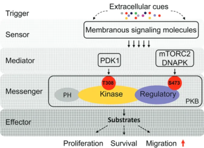
Introduction to PKB
PKB is a serine/threonine kinase of the AGC protein kinase
subfamily (
1
). It comprises three highly sequence-identical
iso-forms in mammals, PKBα, PKBβ, and PKBγ, that are ubiquitously
expressed in all cell types with slight differences in tissue
dis-tribution (
2
). In response to a variety of stimuli including growth
factors and hormones that trigger rapid activation of lipid kinase
phosphoinositide 3 kinase (PI3K), the secondary messenger
mol-ecule Phosphatidylinositol (
3
,
4
,
5
)-trisphosphate (PIP3) is
gener-ated (
3
), which in turn recruits PKB to the plasma membrane
where it is phospho-activated by phosphoinositide-dependent
kinase-1 (PDK1) (on Thr308). Maximal kinase activity of PKB is
achieved through mammalian target of rapamycin complex 2
(mTORC2)– or DNA-dependent protein kinase
(DNAPK)–medi-ated phosphorylation on Ser473 (
1
,
4
). Therefore, PKB is a direct
functional downstream target of PI3K under the control of PDK1
and mTORC2 or DNAPK (
Figure 1
). Genetic studies in knockout
mice have indicated a primary function of PKB as a promoter
of cell proliferation and survival (
5
,
6
). Consistent with its
physi-ological role in development, PKB has been found to be
hyper-activated in many types of cancer. Inhibition of PKB activity not
review
review
only attenuates tumor growth (
7
,
8
) but also local invasion and
metastasis (
9
) resulting from crosstalk with other oncogenic
signaling such as mitogen-activated protein kinase (MAPK) (
10
),
transforming growth factor-β (TGF-β) (
11
,
12
), vascular
endothe-lial growth factor (VEGFR) (
13
), and (ephrin) Eph (
14
). In this
regard, PKB is an attractive target for cancer therapies and
sev-eral promising inhibitors of PKB are currently being validated
in clinical trials including MK-2206 (
15
,
16
), Perifosin (
17–21
), and
RX-0201 (
22
).
PKB-Regulated T Cell Development
PKB at the β-Selection Checkpoint
T cell development can broadly be split into four steps:
1) com-mon lymphoid progenitors (CLPs) derived from bone-marrow
hematopoietic cells migrate to the thymus where they act as
early T cell progenitors (ETPs); 2) ETPs then autonomously
undergo four stages of transition from double-negative
expres-sion of CD4/CD8 (DN, CD4
-CD8
-) to double-positive expression
(DP, CD4
+CD8
+). This represents the maturation stage of
thy-mocytes, and these cells express low levels of pre-T cell
recep-tor (TCR); 3) mature “single-positive” cells are then selected
by MHC-induced positive and negative selection and
4) subse-quently emigrate to the periphery in response to
sphingosine-1-phosphate (S1P) stimulation for final maturation (
Figure 2
).
At the first key checkpoint (the transition from DN3 to DN4
cells before CD4 and CD8 are simultaneously expressed), ETPs
undergo rearrangement of the β-chain of the pre-TCR (
23
,
24
), a
fundamental event to prime the structural properties for pairing
with pre-TCR α-chain on the surface of the thymocytes, a
pro-cess defined as β-selection. A number of early studies showed
that Notch activity plays a crucial role in determining which
thymocytes with functional pre-TCRs enter into the DN4 stage
(
25–27
). Importantly, this prosurvival effect of Notch signaling is
abolished upon PI3K/PKB inhibition, and expression of
consti-tutively active PKB efficiently rescues Notch loss–induced
apop-tosis by bypassing PI3K signaling, indicating a stringent role
for PKB in the control of pre-T cell survival (
28
). A recent study
uncovered that Hes1, the downstream target of Notch signaling,
mediates elevated PKB signaling through targeting phosphatase
and tensin homolog (PTEN) (
29
). Similarly, DN3 cells with PKB
depletion undergo apoptosis in response to pre-TCR stimulation
(
30
). In fact, downregulation of PKB activity leads to substantial
developmental blockage at the DN3 stage, indicating the failure
of differentiation driven by Notch signaling (
31
). In line with the
role of PKB in promoting β-selection, some PKB targets such
as Glycogen synthase kinase 3 (GSK3)/T-cell factor 1
(TCF-1)/β-catenin (
32–34
) and cAMP response element-binding protein
(CREB) (
35
) were also shown to participate in signaling
regu-lation at this checkpoint. This raises the question of whether
these events are also dependent on PKB activity, which needs
future investigation.
PKB Activation During Thymic Selection and
Maturation
When thymocytes pass β-selection and enter the DP (CD4
+CD8
+)
stage, characterized by low-level expression of pre-TCR, the cells
need to vigorously proliferate. Previous studies showed that this
was enabled by activated PKB that upregulated levels of Bcl-XL,
a member of the anti-apoptotic Bcl-2 family (
36
,
37
). DP
thymo-cytes that moderately recognize self-peptide-major
histocom-patibility complex (MHC) complexes survive and are induced
to differentiate (positive selection), whereas those that
recog-nize self-peptide-MHC complexes with high affinity/avidity
are induced toward cell death (negative selection) to eliminate
potentially toxic T cells. These two programs of thymic selection
are coupled with TCR-regulated cell survival through PKB, which
selectively modulates an apoptotic effector downstream of TCR
signaling, the orphan nuclear receptor Nur77, and thus drives
naïve T cell maturation (
38
).
Following functional postselection of thymocytes expressing
either CD4 or CD8 protein, these single-positive (SP, CD4
+CD8
-or CD4
-CD8
+) thymocytes migrate to the periphery where they
further develop into different functional T cell populations. S1P
signaling is required to ensure that only mature SP thymocytes
are permitted to enter the periphery. Loss of S1P receptor 1
(S1P
1) expression on mature SP thymocytes blocks the thymic
egress of these cells because of inhibition of the chemotactic
activity mediated by S1P/S1P
1signaling (
39
). Interestingly,
PKB-dependent phosphorylation of S1P
1is required for S1P
1-mediated
chemotaxis (
40
). In addition, S1P
1expression is transcriptionally
regulated by Krüppel-like factor 2 (KLF2) which is also required
for cell trafficking (
41
) and whose expression is stringently
con-trolled by forkhead box O (FoxO) family member FoxO1, a
well-characterized phospho-target of PKB (
42
,
43
). Therefore, PKB/
FoxO1/KLF2 signaling is likely central in determining a complete
mature state of the thymocytes that egress from the thymus at
the late stage of T cell development. C-C chemokine receptor 7
(CCR7) and CCR9 were also shown to determine the fate of ETPs
homing to thymus (
44
,
45
); however, it is not clear whether this
early step in T cell development is also regulated by PKB/FoxO1
signaling.
Owing to the global importance of PKB in supporting cell
proliferation and survival and overcoming apoptosis, it is not
surprising that PKB acts as a gatekeeper to secure the smooth
transition through these developmental checkpoints. This role
of PKB is unlikely to be restricted only to T cell development.
A few studies have already shown the similar functions for PKB
in other types of immune cells such as B cells (
46–48
), dendritic
cells (
49–52
), macrophages (
53
,
54
), and neutrophils (
55
). Thus,
PKB is crucial in regulating immune cell development.
Figure 1. Molecular mechanism of protein kinase B (PKB) signaling transduction. Extracellular signals such as chemokines, cytokines, and growth factors bind to their membrane receptors and activate corresponding axes. Intracellular media-tors such as PDK1 and mTORC2 kinases are simultaneously activated and trans-duce messages to the central hub PKB. As a “tertiary” messenger, PKB directs cell fate through phospho-regulation of its substrates, most of which tran-scriptionally control cell proliferation, survival and migration. DNAPK = DNA-dependent protein kinase; mTORC2 = mammalian target of rapamycin complex 2; PDK1 = phosphoinositide-dependent kinase 1.
review
review
PKB-Dependent Regulation of T Cell
Functionality
The Impact of PKB on CD8
+T Cell Metabolism and
Response
mTOR-directed involvement of PI3K/PKB signal
transduc-tion has been shown to be pivotal to glucose metabolism (
56
).
Upon stimulation, the fate of CD8
+T cells (effector or memory)
is determined by cytokines, particularly interleukin 2 (IL-2).
Previously it was shown that stimulation of T cells with anti-CD3
and anti-CD28 induces glucose uptake and aerobic glycolysis
(the Warburg effect) in a PKB-dependent but IL-2–independent
manner (
57
). In primary CD8
+T cells with depleted TCR
signal-ing, attenuated glucose metabolism through reduced glycolysis
is triggered by decreased expression of glucose transporter 1
(Glut1) (
58
), whose expression level and stabilized membrane
translocation require PKB activity (
59
,
60
). PKB-regulated
glu-cose metabolism was further supported by follow-up studies
that showed that constitutive activation of PKB may override
the influence of cytokine stimulation on Glut1 activation (
61
,
62
).
Consistently, upon reactivation of memory CD8
+T cells, the
metabolic switch from basal fatty acid oxidation and
mitochon-drial oxidative phosphorylation to aerobic glycolysis and lipid
synthesis, needed for rapid acquisition of effector functionality
such as the production of IFNγ, is regulated by PKB rather than
mTORC1 (
63
). Thus, PKB is a crucial signaling cascade
responsi-ble for elevated metabolic rate in T cells.
The activities of many transcriptional factors that are crucial
for determining cell fate and behavior are post-translationally
modulated by PKB (
9
). A recent study showed that inhibition of
PKB decreases mRNA levels of several key molecules responsible
for T cell effector functions, including granzymes, perforin, Fas
ligand, INFγ, and cytokine receptors such as IL-12R, indicating
that TCR-mediated transcriptional programs that determine
effector or memory cell differentiation are strictly dependent
on PKB (
64
). This seems to be in agreement with a recent study
showing that conversion of CD8
+T cells from memory to effector
state requires PKB activation (
63
).
CD8
+T cells responding to stimuli follow three distinct
phases: clonal expansion, contraction, and establishment of
memory. As the metabolic and survival regulator of T cells, it
is not surprising that PKB dictates the magnitude of CD8
+T
cell memory through controlling dynamic alterations in
prolif-eration and apoptotic rates via its downstream mediator FoxO
family members (
65
). Once the infecting pathogens are cleared,
autonomous apoptosis occurs in the majority of
pathogen-spe-cific effector CD8
+T cells through the PKB/FoxO axis, whereas
a small percentage will develop into memory CD8
+T cells for
rapid response to secondary stimulation in the future (
66
). This
is similar to the role of PKB during thymic selection, where TCR
priming leads to massive cell death in absence of PKB
signal-ing. Taken together, these data support a concept that PKB is an
essential regulator along the immunometabolic signaling
path-way in T cells, but with differentially defined functional
speci-ficity in different T cell subpopulations in response to diverse
stimuli.
PKB Regulates CD4
+T Cell Differentiation
Peripheral naïve CD4
+T cells mature and differentiate in response
to antigenic stimulation into distinct functional subsets with
specific roles in the regulation of the immune response. The fate
of the cells is determined by individual stimuli, most of which
Figure 2. Key steps and checkpoints during T cell development. APC = antigen-presenting cell; CLP = common lymphoid progenitor; CTL = cytotoxic T lymphocytes; DN = double negative; DP = double positive; HSC = hematopoietic stem cell; MPP = multipotent progenitor; SP = single positive; Th = helper T cells; Treg = regulatory T cells.review
review
are from interleukin family members. Each specialized CD4
+T
cell lineage exhibits functionally dominant transcriptional
pro-grams driven by unique pools of transcription factors such as
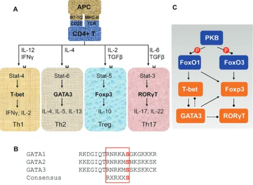
T-box binding protein (T-bet), GATA family member GATA3,
fork-head box P3 (FoxP3), and RAR-related orphan receptor gamma 3
(RORγ3), representing T helper cell 1 (Th1), Th2, regulatory T cell
(Treg), and Th17 lineages, respectively (
Figure 3A
). PKB activity is
tightly associated with the differentiation state of CD4
+T cells,
and numerous studies have shown that it contributes
substan-tially to the regulation of signaling events at transcriptional and
translational levels, under both physiological and pathological
settings (
67–69
). In the scenario of global control, PKB-mediated
direct phosphorylation of FoxO1 and FoxO3a leads to
cytoplas-mic sequestration, thus inhibiting their DNA binding activity
(
70
). In T cells, FoxO1 and FoxO3a transcriptionally activate T-bet
(
71
,
72
) and FoxP3 (
73–76
), which inversely associate with PKB
activity.
In most cases, it is the expression ratio among the
transcrip-tion factors, rather than the overexpression or suppression
of a single protein, that determines the differentiation fate of
CD4
+T cell lineage. This obviously raises the question of how
this balance of transcription factors is maintained. Recent
stud-ies have started to uncover the potential mechanisms. Under
inflammatory conditions, elevated TGF-β signaling promotes
Treg cell functionality through promotion of FoxP3 but restrains
Th17 phenotype by decreasing levels of RAR-related orphan
receptor gamma t (RORγt) (
77
). Opposing expression patterns
of FoxP3 and RORγt in the same cell population are possibly, at
least partly, because of a physical inhibitory interaction (
78
,
79
).
When extracellular conditions favor a transition to Th17
dif-ferentiation, IL-6 may counteract FoxP3-mediated inhibition
of RORγt. Interestingly, a recent study also showed that PKB/
mTORC1-mediated activation of ribosomal protein S6 kinase
2 (S6K2) promotes RORγt nuclear translocation through direct
coupling (
80
). Moreover, to enhance the maintenance of Treg
differentiation, expression of GATA3 maintains high levels of
FoxP3 but inhibits T-bet and RORγt through as yet undefined
mechanisms (
81
). By contrast, to maintain a Th1 phenotype the
functionality of GATA3 can be physically repressed by T-bet by
directly interfering with its DNA binding capacity (
82
). While it
is not yet clear whether PKB can directly affect the function of
GATA3, studies have shown that PKB can directly phosphorylate
and functionally influence GATA1 (
83
) and GATA2 (
84
). Given the
high degree of sequence similarity in the DNA-binding domain
adjacent to the distal zinc-finger motif (
85
) among GATA family
members (
Figure 3B
), it would be interesting to investigate if PKB
can influence Th2 cell differentiation directly through GATA3 by
bypassing FoxO. Therefore, under the global surveillance of PKB/
FoxO signaling axis, a precisely regulated interplay between the
transcriptional signature molecules seems to be the dominating
factor that determines the differentiation of CD4
+T cell lineages
(
Figure 3C
).
The Role of PKB in Escaping
Immunosurveillance in Cancer
PKB Controls the Expression of Chemokines
The mTOR/PI3K/PKB signaling pathway is highly deregulated
in human diseases, such as cancer, which require an
aber-rant demand of metabolic rate. The surrounding environment
of tumors, the cancer stroma, is the fundamental resource for
nutrient supply to cancer cells. Cancer stroma consists of a
vari-ety of cell types, including cancer-associated fibroblasts (CAF),
endothelial cells, myeloid cells, lymphocytes, and pericytes (
86
),
many of which are actively recruited to the cancer environment
and utilized by cancer cells to satisfy the increased metabolic
demand. Consistent with the pivotal role of PKB during immune
cell development, extensive studies have revealed that PKB
stimulates aerobic glycolysis in many types of cancer cells (
87–
90
) (comprehensively reviewed by Ward et al. [
91
]). Intriguingly,
PKB-regulated cancer cell metabolism can be further enhanced
Figure 3. Regulation of CD4+ T cell differentiation. A) CD4+ T cells are differentiated into four main lineages with distinct transcriptional programs in response to stimulation of different chemokines. B) Sequence alignment of human GATA family members. C) Protein kinase B (PKB)–mediated regulation of the transcriptional determinants towards terminal differentiation of CD4+ T cells. APC = antigen presenting cell; MHC = major histocompatibility complex; TCR = T cell receptor.
review
review
when cancer cells encounter disturbing situations such as a
stiffened extracellular matrix (
92
), indicating that cancer cells
are capable of autonomous adaption to their niche environment
through PKB-promoted metabolism.
CAFs and endothelial cells are responsible for tumor
initia-tion and establishment of tumor vascular network essential for
local invasiveness and nutrient/oxygen supply (
93
,
94
). It is now
becoming evident that the immune cells in the vicinity of the
cancer niche also markedly impact aspects of cancer biology,
including tumor cell metastasis to distant organs, cancer
angio-genesis, cancer immune regulation, and overcoming metabolic
stress (
95
). Directional trafficking of immune cells to the cancer
niche is a chemotactic process triggered mainly by chemokines
secreted from cancer cells in a gradient-dependent manner (
96
),
which results in the entire tumor niche acting as a core
regula-tor that modulates survival, invasion, and immune-suppression
(
97
).
Although PI3K is a key regulator of chemoattractants (such
as PIP3) (
98
), PKB signaling can potently control cell migration
in different settings (
9
). Inflammatory conditions in the
vicin-ity of developing tumors, particularly chronic inflammation, are
positively associated with tumor progression. Pivotal pathways
that regulate tumor-associated inflammation, such as the tumor
necrosis factor (TNF)/IκB kinase (IKK)/nuclear factor κB (NF-κB)
axis, can be induced and enhanced through
tumor-express-ing molecules, whose activation, in turn, promotes malignant
transformation of tumor cells. Upregulation of
tumor-associ-ated inflammatory factors is often orchestrtumor-associ-ated by
transcrip-tional events driven by NF-κB signaling, which is tightly under
the control of crosstalk between PKB and the IKK signalosome
(
99–102
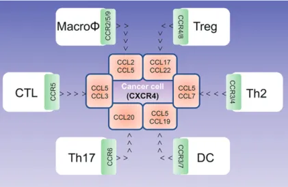
). By pairing to CCR6, C-C chemokine ligand 20 (CCL20)
has been shown to be a key mediator in many inflammatory
diseases including cancer (
103–105
). Migration of Th17 cells
towards developing tumors is driven by CCL20, and this requires
PKB activation. Mechanistically, the transcriptional regulation
of CCL20 is mediated through nuclear translocation and
acti-vation of NF-κB (
106–108
), a transcriptional complex negatively
regulated by its endogenous inhibitor IκB, which is degraded by
PKB-mediated phosphorylation. Similarly, PKB-mediated
activa-tion of NF-κB regulates many chemokines during chemotactic
migration of cells such as dendritic cells via CCL19 (
109
); CTL
cells, Th2 cells, and macrophages via CCL5 (
110
); and Treg cells
via CCL17 and CCL22 (
111
,
112
). The chemokine-mediated
immune cell migration to the cancer niche is summarized in
Figure 4
. Undoubtedly, most of these chemoattractants are also
expressed by different subsets of immune cells that
coopera-tively stimulate local inflammation in cancer stroma. It is also
known that PKB-regulated NF-κB transcriptionally activates
Snail, an epithelial-mesenchymal transition inducer that
pro-motes cell migration in a context-dependent manner (
113
,
114
).
Snail is capable of upregulating pro-inflammatory cytokines
such as IL-1, IL-6, and IL-8, which enhance the chemotactic
migration of both immune cells and metastatic cancer cells
(
115
). Moreover, activation of Snail also elevates levels of CCL2
in melanoma (
116
). PKB activates Snail by phospho-inhibition of
GSK3, an upstream inhibitory kinase that prevents the nuclear
translocation of Snail (
117
).
Likewise, the expression of chemokine receptors may also
be controlled by PKB. For example, in prostate cancer with
PTEN loss, ablation of PKB results in a sizable decrease in C-X-C
chemokine receptor 4 (CXCR4) at both the transcriptional and
translational levels, which consequently inhibits metastatic
spread by disrupting the CCL12/CXCR4 network (
118
). As well
as regulating chemokines, NF-κB is also thought to regulate
chemokine receptors such as CCR5 and CCR7 (
119
). Activation
of chemokine and cytokine signaling can mediate a
feed-for-ward signaling loop that enhances intracellular PKB activity, a
mechanism that has been widely demonstrated in the
signal-ing network of receptor tyrosine kinase/PI3K/PKB in cancer. In
a xenograft model of breast cancer, CCL5 secreted from
mesen-chymal stem cells activated PKB signaling in circulating tumor
cells to promote extravasation from the circulation and
colo-nization at distal organs (
120
). Therefore, PI3K/PKB activation
and upregulation of inflammatory chemokines and receptors
can reciprocally control cell-cell communication in the cancer
environment through modulation of chemotactic migration.
In fact, the functional interaction between PKB and IκB/NF-κB
has been shown to create a protumoral microenvironment at
inflammatory sites where particularly chronic inflammation
persistently occurs (
121
). The resulting inflammatory responses
Figure 4. Chemokine-mediated immune cell trafficking to tumor vicinity. Individual chemokines expressed by cancer cells are highlighted in pink boxes, and the cor-responding chemokine receptors expressed by immune cells are highlighted in the green boxes. Note: Many chemokines are also expressed by different immune cells peritumorally or intratumorally to regulate local inflammation and cell-cell interaction. Because of its complex nature, this aspect is not reflected in the simplified car-toon. Open arrows indicate migratory direction. CCL = C-C chemokine ligand; CCR = C-C chemokine receptor; CTL = cytotoxic T lymphocytes; CXCR = C-X-C chemokine receptor; DC = dendritic cells; MacroΦ = macrophage; Th = T helper cell; Treg = regulatory T cells.
review
review
can augment tumor cell growth and metastasis through a
com-bination of accelerated cell cycle, increased genomic instability,
and cytoskeleton remodeling-induced motility.
PKB Supports Tumor Cells to Escape from
Immunosurveillance
For several decades, the infiltration and accumulation of
immune cells in the tumor vicinity was thought to be
detri-mental to cancer cells. However, all types of immune cells are
found in different tumors, regardless of the cancer type, origin,
and residing organs. These immune cells differentially impact
tumor development by mediating the interactions between
can-cer cells and the environment. Clinical trial data has shown that
infiltration of CD8
+T cells, natural killer (NK) cells, and Th1 cells
within tumors is associated with a good prognosis in most types
of cancer, whereas infiltration of Th2, Treg, Th17 cells,
mac-rophages, and neutrophils is associated with poor prognosis
(
122–126
). This implies that certain immune cell types may have
a tumor-promoting role during cancer development. Indeed,
tumor cells that survive intrinsic and/or extrinsic stress,
includ-ing host immune defense, neglect the immune detection and
gain tolerance (
127
).
Despite the impacts of myeloid-derived suppressor cells
(MDSC) and Treg-mediated T cell anergy (
128
,
129
), several
mech-anisms for the escape from host immunosurveillance by cancer
cells have been suggested, including impaired
antigen-present-ing activity, unfavorable cytokine and chemokine secretion, and
direct suppression of immune cell function by interaction of
inhibitory molecules on cancer cell surface with immune cell
surface markers (
130
,
131
). These immune resistance
mecha-nisms may arise from local immunological stress, whereby
selected cancer cells that survive the innate immune response
undergo stress-induced genetic or epigenetic modifications
to adapt to the tumor microenvironment and evade immune
detection. This organized process, also called cancer
immu-noediting (
132
), mechanistically mimics the clonal evolution of
cancer cells that underlies the acquisition of resistance to
thera-peutic drugs, which typically results in more aggressive tumors.
Thus, it is evident that the inflammatory factors expressed by
both immune cells and tumor cells in a PKB-regulated manner,
can contribute to create immunosuppressive conditions.
Cell-cell interaction-triggered T cell dysfunction is mainly
mediated through inhibitory molecular pairing between
pro-grammed death ligands 1 and 2 (PD-L1/2) (
133
). These members
of the B7 protein family are expressed on the surface of
infiltrat-ing cells and cancer cells, and their receptor, programmed death
1 (PD-1), is expressed on the surface of activated T cells,
mac-rophages, dendritic cells, B cells, and NK cells (
134
). Under
physi-ological conditions, when PD-1 binds to their ligands, negative
signals are delivered into the T cells that attenuate their
activi-ties by inhibiting TCR-mediated proliferation to prevent tissue
damage from unfavorable immune responses (
135
). This
mech-anism is appropriated by cancer cells to directly induce T cell
apoptosis through a similar interaction. Interestingly,
expres-sion of PD-L1 in tumor cells is tightly associated, or regulated
by PKB. In human gliomas, PTEN loss leads to increased
immu-noresistance through upregulation of PD-L1 in a PKB activation–
dependent manner (
136
). Selective inhibition of PKB with small
molecule inhibitor (Akt inhibitor III) was shown to decrease the
expression level of PD-L1, which was mediated through
PKB-regulated mTORC1/S6K1 signaling. This is in agreement with the
finding that interferons may also activate PI3K/PKB/mTOR/S6K
pathway (
137
), though the molecular mechanisms underlying
PD-L1 upregulation need further investigation. Similarly, this
PKB-dependent PD-L1 expression pattern is also observed in
colorectal cancer (
138
).
Further evidence for the role of PKB in regulation of PD-L1
come from the investigation of PD-L1 expression in
triple-negative human breast cancer, where specifically targeting
PKB activity with the pharmacological inhibitor MK-2206 was
shown to substantially downregulate PD-L1 at the
transcrip-tional level (
139
). Independent studies also showed a potential
mechanism for PD-L1 downregulation at the translational level
by PKB in both breast and prostate cancer (
140
), mutant
BRAF-harboring melanomas (
141
), and pancreatic cancer (
142
). Clearly,
the molecular mechanisms explaining how PKB mediates PD-L1
expression are the missing piece of the puzzle. This links to an
even more complex situation in which each PKB isoform may
have a different impact, as many studies have shown
oppos-ing roles of PKB isoforms in drivoppos-ing tumor metastasis in
differ-ent types of cancer (
9
). Nonetheless, the emerging data clearly
shows that PKB actively participates in tumor cell–mediated
immunosuppression, at least through two distinct paths via
promoting chemokine/cytokine expression and upregulating
PD-L1 expression. Therefore, targeting the PKB signaling node
may enable reviving of the host immune response in clinical
cancer therapy.
T Cell Functionality Upon PKB Inhibition in the
Cancer Niche
Despite the robust inhibitory effect on cancer cell proliferation
and glucose uptake, evidence from recent studies also suggested
that inhibition of PKB restored and enhanced physiological
functionalities of T cells in the tumor microenvironment. In a
mouse model of adoptive cell therapy, pharmacological
inhibi-tion of PKB with an allosteric inhibitor (AKT inhibitor VIII)
repro-grammed the transcriptional events in CD8
+tumor-infiltrating
lymphocytes (TIL) into phenotypic memory cells (
143
). In
addi-tion, this phenotype was coupled with improved survival of
transferred antitumor TIL in vivo and an enhanced antitumor
effect in the mouse model. This in vivo observation was further
supported in a grafted myeloma mouse model, in which
inhibi-tion of PKB with the same inhibitor blocked the differentiainhibi-tion
of CD8
+T cells, improved the expansion of CD8
+T cells, and was
associated with superior antitumor effects in the mice (
144
).
Moreover, inhibition of the PKB pathway with MK-2206
selec-tively suppressed Treg proliferation and enhanced the
antitu-mor effect of a tuantitu-mor-specific vaccine in a mouse model (
145
).
Thus, these findings further support the concept that targeting
PKB enables tumor-specific lymphocytes to exert antitumor
immunity in immunotherapy for advanced cancer.
Conclusions and Perspectives
Emerging evidence highlights the importance of the tumor
microenvironment as a critical regulator that determines the
outcome of clinic cancer therapy. This impact is
fundamen-tally because of the complex network of cell-cell interactions
between tumor cells, immune cells, and the extracellular
matrix. Therapeutic strategies that block the intercellular
com-munication of this network have shown promising clinical
benefit to the patients, and cancer immunotherapy strategies
aimed at reviving functional immunosurveillance, in particular
in combination with other targeted therapies, have become an
effective approach in cancer clinic. For example, recent clinical
review
review
studies with PD-1– and PD-L1–blocking antibodies highlight the
substantial therapeutic strategy in cancer patients with
incur-able tumors or relapsed tumors post-therapy (
146
,
147
). In
addi-tion, blocking CCL22 inhibits the trafficking of CCR4-expressing
Treg cells to the metastatic lesion (
148
). Targeting tumorigenic
inflammation by inhibition of CCL2 restrains CCR2-expressing
immune cells, such as monocytes in bone marrow (
149
), which
decreased the inflammatory risk from recruited
tumor-associ-ated macrophages (
150
) and led to suppression of metastasis in
syngeneic mouse models of metastatic breast cancer.
Hyperactivation of PKB may represent a fundamental
hall-mark of cancer that contributes to resistance to both
chemo-therapy and radiochemo-therapy (
151
,
152
). As described above, CCL22
is under transcriptional regulation of NK-κB, which is
acti-vated by PKB-mediated degradation of IκB. CCL2 synthesis is
regulated by the Snail transcription factor, which is negatively
controlled by GSK3, a direct substrate of PKB. Thus,
downregula-tion of PKB activity can theoretically interfere with the CCL22/
CCR4 and CCL2/CCR2 axes to block tumor-directed trafficking
of immunosuppressive Treg and monocytes. The mechanisms
of PKB-promoted immune evasion of cancer cells have been
demonstrated by increased resistance to CD8
+T cell–mediated
apoptosis (
145
,
153
,
154
), overriding death receptor signaling
(
155
), strengthening energetic metabolism (
156
), and enforcing
the functionality of immunosuppressive Treg cells (
157
,
158
).
Therefore, inhibition of PI3K/PKB can principally suppress the
tumor-driven immunosuppressive effect through remodeling of
the entire tumor microenvironment.
Current cancer therapeutic strategies invariably induce drug
resistance in the clinic. Among many potential mechanisms,
the disruption of negative feedback loops and compensatory
activation of other oncogenic signaling pathways are two major
causes. In melanoma patients bearing an activating mutation
on BRAF (V600E), inhibition of mutant BRAF with vemurafenib
or dabrafenib dramatically reduces MAPK activity and improves
median response duration (
159
). Vemurafenib therapy not only
inhibits tumor growth through tumor intrinsic mechanisms but
also decreases the population of immunosuppressive myeloid
derived suppressor cells (MDSCs) (
160
) and increases levels of
tumor infiltration of CD8
+T cells (
161
). However, this
immunore-sponsive phenotype is diminished at the time of tumor
repro-gression, and a comparative study implies that the intratumoral
T cell infiltration is dependent on tumor sensitivity to
vemu-rafenib (
162
). Importantly, at this resistance developing stage,
inhibition of mutant BRAF spontaneously strengthens PI3K/PKB
signaling through various mechanisms, as well as NF-κB (
163
),
which coincides with reduced recognition of melanoma cells by
immune cells. Given the regulatory role of PKB in production of
inflammatory factors, it is foreseeable that PKB will prove to be a
contributor to immunounresponsivess in melanomas resistant
to vemurafenib therapy. Thus, a combinatory approach
target-ing oncogenic signaltarget-ing while stimulattarget-ing immune response is
emerging as a more effective strategy (
164
).
As well as its critical role in chemotactic migration and
chemokine/cytokine-mediated cell-cell interaction, PKB also
participates in cellular adhesion to the extracellular matrix,
which in parallel further enhances cell survival and
migra-tory potential driven by local immune tolerance. This is largely
attributable to the impacts on the activity of
adhesion-associ-ated molecules including focal adhesion kinase (
165
), EphA2
(
14
), Ron (
166
,
167
), Zyxin (
168
), CD34 (
169
), and CTNND2 (
170
),
et al. Encouragingly, emerging data show improved antitumor
response of T cells by inhibiting PKB signaling (
171
). Given
the broad impact of the PKB cascade on many aspects during
cancer development, functional dissection of its emerging role
in antagonizing immunosurveillance is likely to contribute
sub-stantially to cancer immunotherapeutic strategies.
Funding
The research project was funded by the Swiss National Science
Foundation 31-130838 (to BAH and GX).
Notes
The Swiss National Science Foundation (the sponsor of the
study) was not involved in the design of the study, the writing of
the article, or the decision to submit the article for publication.
References
1. Pearce LR, Komander D, Alessi DR. The nuts and bolts of AGC protein kinases. Nat Rev Mol Cell Biol. 2010;11(1):9–22.
2. Fayard E, Xue G, Parcellier A, et al. Protein kinase B (PKB/Akt), a key mediator of the PI3K signaling pathway. Curr Top Microbiol Immunol. 2010;346:31–56. 3. Engelman JA, Luo J, Cantley LC. The evolution of
phosphatidylinosi-tol 3-kinases as regulators of growth and metabolism. Nat Rev Genet. 2006;7(8):606–619.
4. Cybulski N, Hall MN. TOR complex 2: a signaling pathway of its own. Trends Biochem Sci. 2009;34(12):620–627.
5. Dummler B, Hemmings BA. Physiological roles of PKB/Akt isoforms in devel-opment and disease. Biochem Soc Trans. 2007;35(Pt 2):231–235.
6. Scheid MP, Woodgett JR. PKB/AKT: functional insights from genetic models. Nat Rev Mol Cell Biol. 2001;2(10):760–768.
7. Fruman DA, Rommel C. PI3K and cancer: lessons, challenges and opportuni-ties. Nat Rev Drug Discov. 2014;13(2):140–156.
8. Vivanco I, Sawyers CL. The phosphatidylinositol 3-Kinase AKT pathway in human cancer. Nat Rev Cancer. 2002;2(7):489–501.
9. Xue G, Hemmings BA. PKB/Akt-dependent regulation of cell motility. J Natl Cancer Inst. 2013;105(6):393–404.
10. Carracedo A, Ma L, Teruya-Feldstein J, et al. Inhibition of mTORC1 leads to MAPK pathway activation through a PI3K-dependent feedback loop in human cancer. J Clin Invest. 2008;118(9):3065–3074.
11. Xue G, Restuccia DF, Lan Q, et al. Akt/PKB-mediated phosphorylation of Twist1 promotes tumor metastasis via mediating cross-talk between PI3K/ Akt and TGF-beta signaling axes. Cancer Discov. 2012;2(3):248–259.
12. Zhang L, Zhou F, ten Dijke P. Signaling interplay between transforming growth factor-beta receptor and PI3K/AKT pathways in cancer. Trends Bio-chem Sci. 2013;38(12):612–620.
13. Kitamura T, Asai N, Enomoto A, et al. Regulation of VEGF-mediated angio-genesis by the Akt/PKB substrate Girdin. Nat Cell Biol. 2008;10(3):329–337. 14. Miao H, Li DQ, Mukherjee A, et al. EphA2 mediates ligand-dependent
inhibi-tion and ligand-independent promoinhibi-tion of cell migrainhibi-tion and invasion via a reciprocal regulatory loop with Akt. Cancer Cell. 2009;16(1):9–20.
15. Yap TA, Yan L, Patnaik A, et al. First-in-man clinical trial of the oral pan-AKT inhibitor MK-2206 in patients with advanced solid tumors. J Clin Oncol. 2011;29(35):4688–4695.
16. Yap TA, Yan L, Patnaik A, et al. Interrogating Two Schedules of the AKT Inhib-itor MK-2206 in Patients with Advanced Solid Tumors Incorporating Novel Pharmacodynamic and Functional Imaging Biomarkers. Clin Cancer Res. 2014;20(22):5672–5685.
17. Orlowski RZ. Novel agents for multiple myeloma to overcome resistance in phase III clinical trials. Semin Oncol. 2013;40(5):634–651.
18. Guidetti A, Carlo-Stella C, Locatelli SL, et al. Phase II Study of Perifosine and Sorafenib Dual-Targeted Therapy in Patients with Relapsed or Refractory Lymphoproliferative Diseases. Clin Cancer Res. 2014;20(22):5641–5651. 19. Richardson PG, Wolf J, Jakubowiak A, et al. Perifosine plus bortezomib and
dexamethasone in patients with relapsed/refractory multiple myeloma pre-viously treated with bortezomib: results of a multicenter phase I/II trial. J Clin Oncol. 2011;29(32):4243–4249.
20. Bendell JC, Nemunaitis J, Vukelja SJ, et al. Randomized placebo-controlled phase II trial of perifosine plus capecitabine as second- or third-line therapy in patients with metastatic colorectal cancer. J Clin Oncol. 2011;29(33):4394– 4400.
21. Ghobrial IM, Roccaro A, Hong F, et al. Clinical and translational studies of a phase II trial of the novel oral Akt inhibitor perifosine in relapsed or relapsed/refractory Waldenstrom’s macroglobulinemia. Clin Cancer Res. 2010;16(3):1033–1041.
22. Pal SK, Reckamp K, Yu H, et al. Akt inhibitors in clinical development for the treatment of cancer. Expert Opin Investig Drugs. 2010;19(11):1355–1366. 23. Levelt CN, Carsetti R, Eichmann K. Regulation of thymocyte development
matura-review
review
tion to the CD4+8+ stage are highly correlated in individual thymocytes. J Exp Med. 1993;178(6):1867–1875.
24. Levelt CN, Ehrfeld A, Eichmann K. Regulation of thymocyte development through CD3. I. Timepoint of ligation of CD3 epsilon determines clonal dele-tion or inducdele-tion of developmental program. J Exp Med. 1993;177(3):707–716. 25. Duncan AW, Rattis FM, DiMascio LN, et al. Integration of Notch and Wnt sign-aling in hematopoietic stem cell maintenance. Nat Immunol. 2005;6(3):314– 322.
26. Huang EY, Gallegos AM, Richards SM, et al. Surface expression of Notch1 on thymocytes: correlation with the double-negative to double-positive transi-tion. J Immunol. 2003;171(5):2296–2304.
27. Ciofani M, Schmitt TM, Ciofani A, et al. Obligatory role for cooperative sign-aling by pre-TCR and Notch during thymocyte differentiation. J Immunol. 2004;172(9):5230–5239.
28. Ciofani M, Zuniga-Pflucker JC. Notch promotes survival of pre-T cells at the beta-selection checkpoint by regulating cellular metabolism. Nat Immunol. 2005;6(9):881–888.
29. Wong GW, Knowles GC, Mak TW, et al. HES1 opposes a PTEN-dependent check on survival, differentiation, and proliferation of TCRbeta-selected mouse thymocytes. Blood. 2012;120(7):1439–1448.
30. Juntilla MM, Wofford JA, Birnbaum MJ, et al. Akt1 and Akt2 are required for alphabeta thymocyte survival and differentiation. Proc Natl Acad Sci U S A. 2007;104(29):12105–12110.
31. Mao C, Tili EG, Dose M, et al. Unequal contribution of Akt isoforms in the double-negative to double-positive thymocyte transition. J Immunol. 2007;178(9):5443–5453.
32. Gounari F, Aifantis I, Khazaie K, et al. Somatic activation of beta-catenin bypasses pre-TCR signaling and TCR selection in thymocyte development. Nat Immunol. 2001;2(9):863–869.
33. Xu Y, Banerjee D, Huelsken J, et al. Deletion of beta-catenin impairs T cell development. Nat Immunol. 2003;4(12):1177–1182.
34. Goux D, Coudert JD, Maurice D, et al. Cooperating pre-T-cell receptor and TCF-1-dependent signals ensure thymocyte survival. Blood. 2005;106(5):1726– 1733.
35. Grady GC, Mason SM, Stephen J, et al. Cyclic adenosine 5’-monophosphate response element binding protein plays a central role in mediating prolifera-tion and differentiaprolifera-tion downstream of the pre-TCR complex in developing thymocytes. J Immunol. 2004;173(3):1802–1810.
36. Jones RG, Parsons M, Bonnard M, et al. Protein kinase B regulates T lympho-cyte survival, nuclear factor kappaB activation, and Bcl-X(L) levels in vivo. J Exp Med. 2000;191(10):1721–1734.
37. Suzuki A, Yamaguchi MT, Ohteki T, et al. T cell-specific loss of Pten leads to defects in central and peripheral tolerance. Immunity. 2001;14(5):523–534. 38. Masuyama N, Oishi K, Mori Y, et al. Akt inhibits the orphan nuclear receptor
Nur77 and T-cell apoptosis. J Biol Chem. 2001;276(35):32799–32805.
39. Matloubian M, Lo CG, Cinamon G, et al. Lymphocyte egress from thymus and peripheral lymphoid organs is dependent on S1P receptor 1. Nature. 2004;427(6972):355–360.
40. Lee MJ, Thangada S, Paik JH, et al. Akt-mediated phosphorylation of the G protein-coupled receptor EDG-1 is required for endothelial cell chemotaxis. Mol Cell. 2001;8(3):693–704.
41. Carlson CM, Endrizzi BT, Wu J, et al. Kruppel-like factor 2 regulates thymo-cyte and T-cell migration. Nature. 2006;442(7100):299–302.
42. Rena G, Guo S, Cichy SC, et al. Phosphorylation of the transcription fac-tor forkhead family member FKHR by protein kinase B. J Biol Chem. 1999;274(24):17179–17183.
43. Ouyang W, Li MO. Foxo: in command of T lymphocyte homeostasis and toler-ance. Trends Immunol. 2011;32(1):26–33.
44. Zlotoff DA, Sambandam A, Logan TD, et al. CCR7 and CCR9 together recruit hematopoietic progenitors to the adult thymus. Blood. 2010;115(10):1897–1905. 45. Calderon L, Boehm T. Three chemokine receptors cooperatively regulate
homing of hematopoietic progenitors to the embryonic mouse thymus. Proc Natl Acad Sci U S A. 2011;108(18):7517–7522.
46. Calamito M, Juntilla MM, Thomas M, et al. Akt1 and Akt2 promote peripheral B-cell maturation and survival. Blood. 2010;115(20):4043–4050.
47. Werner M, Hobeika E, Jumaa H. Role of PI3K in the generation and survival of B cells. Immunol Rev. 2010;237(1):55–71.
48. Szydlowski M, Jablonska E, Juszczynski P. FOXO1 transcription factor: a criti-cal effector of the PI3K-AKT axis in B-cell development. Int Rev Immunol. 2014;33(2):146–157.
49. van de Laar L, van den Bosch A, Boonstra A, et al. PI3K-PKB hyperactivation augments human plasmacytoid dendritic cell development and function. Blood. 2012;120(25):4982–4991.
50. van de Laar L, Buitenhuis M, Wensveen FM, et al. Human CD34-derived myeloid dendritic cell development requires intact phosphatidylinositol 3-kinase-protein kinase B-mammalian target of rapamycin signaling. J Immunol. 2010;184(12):6600–6611.
51. Mattioli B, Giordani L, Quaranta MG, et al. Leptin exerts an anti-apoptotic effect on human dendritic cells via the PI3K-Akt signaling pathway. FEBS Lett. 2009;583(7):1102–1106.
52. Park D, Lapteva N, Seethammagari M, et al. An essential role for Akt1 in dendritic cell function and tumor immunotherapy. Nat Biotechnol. 2006;24(12):1581–1590.
53. Arranz A, Doxaki C, Vergadi E, et al. Akt1 and Akt2 protein kinases differ-entially contribute to macrophage polarization. Proc Natl Acad Sci U S A. 2012;109(24):9517–9522.
54. Busca A, Saxena M, Iqbal S, et al. PI3K/Akt regulates survival during differ-entiation of human macrophages by maintaining NF-kappaB-dependent expression of antiapoptotic Bcl-xL. J Leukoc Biol. 2014;96(6):1011–1022. 55. Chen J, Tang H, Hay N, et al. Akt isoforms differentially regulate neutrophil
functions. Blood. 2010;115(21):4237–4246.
56. Shimobayashi M, Hall MN. Making new contacts: the mTOR network in metabolism and signalling crosstalk. Nat Rev Mol Cell Biol. 2014;15(3):155–162. 57. Frauwirth KA, Riley JL, Harris MH, et al. The CD28 signaling pathway
regu-lates glucose metabolism. Immunity. 2002;16(6):769–777.
58. Rathmell JC, Vander Heiden MG, Harris MH, et al. In the absence of extrinsic signals, nutrient utilization by lymphocytes is insufficient to maintain either cell size or viability. Mol Cell. 2000;6(3):683–692.
59. Barthel A, Okino ST, Liao J, et al. Regulation of GLUT1 gene transcription by the serine/threonine kinase Akt1. J Biol Chem. 1999;274(29):20281–20286. 60. Rathmell JC, Fox CJ, Plas DR, et al. Akt-directed glucose metabolism can
pre-vent Bax conformation change and promote growth factor-independent sur-vival. Mol Cell Biol. 2003;23(20):7315–7328.
61. Wieman HL, Wofford JA, Rathmell JC. Cytokine stimulation promotes glu-cose uptake via phosphatidylinositol-3 kinase/Akt regulation of Glut1 activ-ity and trafficking. Mol Biol Cell. 2007;18(4):1437–1446.
62. Jacobs SR, Herman CE, Maciver NJ, et al. Glucose uptake is limiting in T cell activation and requires CD28-mediated Akt-dependent and independent pathways. J Immunol. 2008;180(7):4476–4486.
63. Gubser PM, Bantug GR, Razik L, et al. Rapid effector function of memory CD8+ T cells requires an immediate-early glycolytic switch. Nat Immunol. 2013;14(10):1064–1072.
64. Macintyre AN, Finlay D, Preston G, et al. Protein kinase B controls transcrip-tional programs that direct cytotoxic T cell fate but is dispensable for T cell metabolism. Immunity. 2011;34(2):224–236.
65. Sullivan JA, Kim EH, Plisch EH, et al. FOXO3 regulates CD8 T cell memory by T cell-intrinsic mechanisms. PLoS Pathog. 2012;8(2):e1002533.
66. Kim EH, Suresh M. Role of PI3K/Akt signaling in memory CD8 T cell differen-tiation. Front Immunol. 2013;4:20.
67. Sauer S, Bruno L, Hertweck A, et al. T cell receptor signaling con-trols Foxp3 expression via PI3K, Akt, and mTOR. Proc Natl Acad Sci U S A. 2008;105(22):7797–7802.
68. Haxhinasto S, Mathis D, Benoist C. The AKT-mTOR axis regulates de novo differentiation of CD4+Foxp3+ cells. J Exp Med. 2008;205(3):565–574. 69. Crellin NK, Garcia RV, Levings MK. Altered activation of AKT is required for
the suppressive function of human CD4+CD25+ T regulatory cells. Blood. 2007;109(5):2014–2022.
70. Coffer PJ, Burgering BM. Forkhead-box transcription factors and their role in the immune system. Nat Rev Immunol. 2004;4(11):889–899.
71. Kerdiles YM, Stone EL, Beisner DR, et al. Foxo transcription factors control regulatory T cell development and function. Immunity. 2010;33(6):890–904. 72. Rao RR, Li Q, Gubbels Bupp MR, et al. Transcription factor Foxo1 represses
T-bet-mediated effector functions and promotes memory CD8(+) T cell dif-ferentiation. Immunity. 2012;36(3):374–387.
73. Harada Y, Harada Y, Elly C, et al. Transcription factors Foxo3a and Foxo1 cou-ple the E3 ligase Cbl-b to the induction of Foxp3 expression in induced regu-latory T cells. J Exp Med. 2010;207(7):1381–1391.
74. Ouyang W, Beckett O, Ma Q, et al. Foxo proteins cooperatively control the differentiation of Foxp3+ regulatory T cells. Nat Immunol. 2010;11(7):618–627. 75. Merkenschlager M, von Boehmer H. PI3 kinase signalling blocks Foxp3
expression by sequestering Foxo factors. J Exp Med. 2010;207(7):1347–1350. 76. Ouyang W, Liao W, Luo CT, et al. Novel Foxo1-dependent transcriptional
pro-grams control T(reg) cell function. Nature. 2012;491(7425):554–559.
77. Nakano K, Higashi T, Hashimoto K, et al. Antagonizing dopamine D1-like receptor inhibits Th17 cell differentiation: preventive and therapeutic effects on experimental autoimmune encephalomyelitis. Biochem Biophys Res Commun. 2008;373(2):286–291.
78. Zhou L, Lopes JE, Chong MM, et al. TGF-beta-induced Foxp3 inhibits T(H)17 cell differentiation by antagonizing RORgammat function. Nature. 2008;453(7192):236–240.
79. Ichiyama K, Yoshida H, Wakabayashi Y, et al. Foxp3 inhibits RORgammat-mediated IL-17A mRNA transcription through direct interaction with ROR-gammat. J Biol Chem. 2008;283(25):17003–17008.
80. Kurebayashi Y, Nagai S, Ikejiri A, et al. PI3K-Akt-mTORC1-S6K1/2 axis con-trols Th17 differentiation by regulating Gfi1 expression and nuclear translo-cation of RORgamma. Cell Rep. 2012;1(4):360–373.
81. Wohlfert EA, Grainger JR, Bouladoux N, et al. GATA3 controls Foxp3(+) regula-tory T cell fate during inflammation in mice. J Clin Invest. 2011;121(11):4503– 4515.
82. Hwang ES, Szabo SJ, Schwartzberg PL, et al. T helper cell fate speci-fied by kinase-mediated interaction of T-bet with GATA-3. Science. 2005;307(5708):430–433.
83. Kadri Z, Maouche-Chretien L, Rooke HM, et al. Phosphatidylinositol 3-kinase/Akt induced by erythropoietin renders the erythroid differentia-tion factor GATA-1 competent for TIMP-1 gene transactivadifferentia-tion. Mol Cell Biol. 2005;25(17):7412–7422.
review
review
84. Menghini R, Marchetti V, Cardellini M, et al. Phosphorylation of GATA2 by Akt increases adipose tissue differentiation and reduces adipose tissue-related inflammation: a novel pathway linking obesity to atherosclerosis. Circulation. 2005;111(15):1946–1953.
85. Ho IC, Tai TS, Pai SY. GATA3 and the T-cell lineage: essential functions before and after T-helper-2-cell differentiation. Nat Rev Immunol. 2009;9(2):125–135. 86. Tlsty TD, Coussens LM. Tumor stroma and regulation of cancer development.
Annu Rev Pathol. 2006;1:119–150.
87. Ran C, Liu H, Hitoshi Y, et al. Proliferation-independent control of tumor gly-colysis by PDGFR-mediated AKT activation. Cancer Res. 2013;73(6):1831–1843. 88. Elstrom RL, Bauer DE, Buzzai M, et al. Akt stimulates aerobic glycolysis in
cancer cells. Cancer Res. 2004;64(11):3892–3899.
89. Coloff JL, Macintyre AN, Nichols AG, et al. Akt-dependent glucose metabo-lism promotes Mcl-1 synthesis to maintain cell survival and resistance to Bcl-2 inhibition. Cancer Res. 2011;71(15):5204–5213.
90. Sun Q, Chen X, Ma J, et al. Mammalian target of rapamycin up-regulation of pyruvate kinase isoenzyme type M2 is critical for aerobic glycolysis and tumor growth. Proc Natl Acad Sci U S A. 2011;108(10):4129–4134.
91. Ward PS, Thompson CB. Metabolic reprogramming: a cancer hallmark even warburg did not anticipate. Cancer Cell. 2012;21(3):297–308.
92. Schrader J, Gordon-Walker TT, Aucott RL, et al. Matrix stiffness modulates proliferation, chemotherapeutic response, and dormancy in hepatocellular carcinoma cells. Hepatology. 2011;53(4):1192–1205.
93. Ohlund D, Elyada E, Tuveson D. Fibroblast heterogeneity in the cancer wound. J Exp Med. 2014;211(8):1503–1523.
94. Karagiannis GS, Poutahidis T, Erdman SE, et al. Cancer-associated fibroblasts drive the progression of metastasis through both paracrine and mechanical pressure on cancer tissue. Mol Cancer Res. 2012;10(11):1403–1418.
95. Hanahan D, Coussens LM. Accessories to the crime: functions of cells recruited to the tumor microenvironment. Cancer Cell. 2012;21(3):309–322. 96. Condeelis J, Singer RH, Segall JE. The great escape: when cancer cells hijack
the genes for chemotaxis and motility. Annu Rev Cell Dev Biol. 2005;21:695–718. 97. McAllister SS, Weinberg RA. The tumour-induced systemic environment
as a critical regulator of cancer progression and metastasis. Nat Cell Biol. 2014;16(8):717–727.
98. Sotsios Y, Ward SG. Phosphoinositide 3-kinase: a key biochemical signal for cell migration in response to chemokines. Immunol Rev. 2000;177:217–235. 99. Oeckinghaus A, Hayden MS, Ghosh S. Crosstalk in NF-kappaB signaling
pathways. Nat Immunol. 2011;12(8):695–708.
100. Kane LP, Shapiro VS, Stokoe D, et al. Induction of NF-kappaB by the Akt/PKB kinase. Curr Biol. 1999;9(11):601–604.
101. Bai D, Ueno L, Vogt PK. Akt-mediated regulation of NFkappaB and the essen-tialness of NFkappaB for the oncogenicity of PI3K and Akt. Int J Cancer. 2009;125(12):2863–2870.
102. Dan HC, Cooper MJ, Cogswell PC, et al. Akt-dependent regulation of NF-{kappa}B is controlled by mTOR and Raptor in association with IKK. Genes Dev. 2008;22(11):1490–1500.
103. Hirota K, Yoshitomi H, Hashimoto M, et al. Preferential recruitment of CCR6-expressing Th17 cells to inflamed joints via CCL20 in rheumatoid arthritis and its animal model. J Exp Med. 2007;204(12):2803–2812.
104. Yamazaki T, Yang XO, Chung Y, et al. CCR6 regulates the migration of inflam-matory and regulatory T cells. J Immunol. 2008;181(12):8391–8401.
105. Ito M, Teshima K, Ikeda S, et al. MicroRNA-150 inhibits tumor invasion and metastasis by targeting the chemokine receptor CCR6, in advanced cutane-ous T-cell lymphoma. Blood. 2014;123(10):1499–1511.
106. Battaglia F, Delfino S, Merello E, et al. Hypoxia transcriptionally induces macrophage-inflammatory protein-3alpha/CCL-20 in primary human mononuclear phagocytes through nuclear factor (NF)-kappaB. J Leukoc Biol. 2008;83(3):648–662.
107. Li X, Syrovets T, Simmet T. The serine protease plasmin triggers expression of the CC-chemokine ligand 20 in dendritic cells via Akt/NF-kappaB-depend-ent pathways. J Biomed Biotechnol. 2012;2012:186710.
108. Harant H, Eldershaw SA, Lindley IJ. Human macrophage inflammatory pro-tein-3alpha/CCL20/LARC/Exodus/SCYA20 is transcriptionally upregulated by tumor necrosis factor-alpha via a non-standard NF-kappaB site. FEBS Lett. 2001;509(3):439–445.
109. Pietila TE, Veckman V, Lehtonen A, et al. Multiple NF-kappaB and IFN regula-tory factor family transcription factors regulate CCL19 gene expression in human monocyte-derived dendritic cells. J Immunol. 2007;178(1):253–261. 110. Wickremasinghe MI, Thomas LH, O’Kane CM, et al. Transcriptional
mecha-nisms regulating alveolar epithelial cell-specific CCL5 secretion in pulmo-nary tuberculosis. J Biol Chem. 2004;279(26):27199–27210.
111. Mizukami Y, Kono K, Kawaguchi Y, et al. CCL17 and CCL22 chemokines within tumor microenvironment are related to accumulation of Foxp3+ regulatory T cells in gastric cancer. Int J Cancer. 2008;122(10):2286–2293. 112. Sekiya T, Miyamasu M, Imanishi M, et al. Inducible expression of a Th2-type
CC chemokine thymus- and activation-regulated chemokine by human bronchial epithelial cells. J Immunol. 2000;165(4):2205–2213.
113. Wu Y, Deng J, Rychahou PG, et al. Stabilization of snail by NF-kappaB is required for inflammation-induced cell migration and invasion. Cancer Cell. 2009;15(5):416–428.
114. Maier HJ, Schmidt-Strassburger U, Huber MA, et al. NF-kappaB promotes epithelial-mesenchymal transition, migration and invasion of pancreatic carcinoma cells. Cancer Lett. 2010;295(2):214–228.
115. Zhou C, Liu J, Tang Y, et al. Inflammation linking EMT and cancer stem cells. Oral Oncol. 2012;48(11):1068–1075.
116. Kudo-Saito C, Shirako H, Takeuchi T, et al. Cancer metastasis is accelerated through immunosuppression during Snail-induced EMT of cancer cells. Can-cer Cell. 2009;15(3):195–206.
117. Zhou BP, Deng J, Xia W, et al. Dual regulation of Snail by GSK-3beta-mediated phosphorylation in control of epithelial-mesenchymal transition. Nat Cell Biol. 2004;6(10):931–940.
118. Conley-LaComb MK, Saliganan A, Kandagatla P, et al. PTEN loss mediated Akt activation promotes prostate tumor growth and metastasis via CXCL12/ CXCR4 signaling. Mol Cancer. 2013;12(1):85.
119. Hopken UE, Foss HD, Meyer D, et al. Up-regulation of the chemokine receptor CCR7 in classical but not in lymphocyte-predominant Hodgkin disease cor-relates with distinct dissemination of neoplastic cells in lymphoid organs. Blood. 2002;99(4):1109–1116.
120. Karnoub AE, Dash AB, Vo AP, et al. Mesenchymal stem cells within tumour stroma promote breast cancer metastasis. Nature. 2007;449(7162):557–563. 121. Elinav E, Nowarski R, Thaiss CA, et al. Inflammation-induced cancer:
cross-talk between tumours, immune cells and microorganisms. Nat Rev Cancer. 2013;13(11):759–771.
122. Fridman WH, Pages F, Sautes-Fridman C, et al. The immune contexture in human tumours: impact on clinical outcome. Nat Rev Cancer. 2012;12(4):298– 306.
123. Ruffell B, DeNardo DG, Affara NI, et al. Lymphocytes in cancer develop-ment: polarization towards pro-tumor immunity. Cytokine Growth Factor Rev. 2010;21(1):3–10.
124. Pollard JW. Tumour-educated macrophages promote tumour progression and metastasis. Nat Rev Cancer. 2004;4(1):71–78.
125. Qian BZ, Pollard JW. Macrophage diversity enhances tumor progression and metastasis. Cell. 2010;141(1):39–51.
126. Gregory AD, Houghton AM. Tumor-associated neutrophils: new targets for cancer therapy. Cancer Res. 2011;71(7):2411–2416.
127. Drake CG, Jaffee E, Pardoll DM. Mechanisms of immune evasion by tumors. Adv Immunol. 2006;90:51–81.
128. Zheng Y, Zha Y, Gajewski TF. Molecular regulation of T-cell anergy. EMBO Rep. 2008;9(1):50–55.
129. Nagaraj S, Gabrilovich DI. Tumor escape mechanism governed by myeloid-derived suppressor cells. Cancer Res. 2008;68(8):2561–2563.
130. Vesely MD, Kershaw MH, Schreiber RD, et al. Natural innate and adaptive immunity to cancer. Annu Rev Immunol. 2011;29:235–271.
131. de Visser KE, Eichten A, Coussens LM. Paradoxical roles of the immune sys-tem during cancer development. Nat Rev Cancer. 2006;6(1):24–37.
132. Schreiber RD, Old LJ, Smyth MJ. Cancer immunoediting: integrating immuni-ty’s roles in cancer suppression and promotion. Science. 2011;331(6024):1565– 1570.
133. Keir ME, Butte MJ, Freeman GJ, et al. PD-1 and its ligands in tolerance and immunity. Annu Rev Immunol. 2008;26:677–704.
134. Greenwald RJ, Freeman GJ, Sharpe AH. The B7 family revisited. Annu Rev Immunol. 2005;23:515–548.
135. Chen DS, Irving BA, Hodi FS. Molecular pathways: next-generation immuno-therapy--inhibiting programmed death-ligand 1 and programmed death-1. Clin Cancer Res. 2012;18(24):6580–6587.
136. Parsa AT, Waldron JS, Panner A, et al. Loss of tumor suppressor PTEN func-tion increases B7-H1 expression and immunoresistance in glioma. Nat Med. 2007;13(1):84–88.
137. Kaur S, Uddin S, Platanias LC. The PI3’ kinase pathway in interferon signal-ing. J Interferon Cytokine Res. 2005;25(12):780–787.
138. Song M, Chen D, Lu B, et al. PTEN loss increases PD-L1 protein expression and affects the correlation between PD-L1 expression and clinical param-eters in colorectal cancer. PLoS One. 2013;8(6):e65821.
139. Mittendorf EA, Philips AV, Meric-Bernstam F, et al. PD-L1 expression in triple-negative breast cancer. Cancer Immunol Res. 2014;2(4):361–370.
140. Crane CA, Panner A, Murray JC, et al. PI(3) kinase is associated with a mechanism of immunoresistance in breast and prostate cancer. Oncogene. 2009;28(2):306–312.
141. Atefi M, Avramis E, Lassen A, et al. Effects of MAPK and PI3K pathways on PD-L1 expression in melanoma. Clin Cancer Res. 2014;20(13):3446–3457. 142. Zhang Y, Zhang J, Xu K, et al. PTEN/PI3K/mTOR/B7-H1 signaling pathway
regulates cell progression and immuno-resistance in pancreatic cancer. Hepatogastroenterology. 2013;60(127):1766–1772.
143. Crompton JG, Sukumar M, Roychoudhuri R, et al. Akt inhibition enhances expansion of potent tumor-specific lymphocytes with memory cell charac-teristics. Cancer Res. 2015;75(2):296–305.
144. van der Waart AB, van de Weem NM, Maas F, et al. Inhibition of Akt signal-ing promotes the generation of superior tumor-reactive T cells for adoptive immunotherapy. Blood. 2014;124(23):3490–3500.
145. Abu-Eid R, Samara RN, Ozbun L, et al. Selective Inhibition of Regulatory T Cells by Targeting the PI3K-Akt Pathway. Cancer Immunol Res. 2014;2(11):1080– 1089.
146. Ansell SM, Lesokhin AM, Borrello I, et al. PD-1 Blockade with Nivolumab in Relapsed or Refractory Hodgkin’s Lymphoma. N Engl J Med. 2014; In press. 147. Herbst RS, Soria JC, Kowanetz M, et al. Predictive correlates of response
to the anti-PD-L1 antibody MPDL3280A in cancer patients. Nature. 2014;515(7528):563–567.