HAL Id: hal-02446276
https://hal-amu.archives-ouvertes.fr/hal-02446276
Submitted on 3 Mar 2020
HAL is a multi-disciplinary open access
archive for the deposit and dissemination of sci-entific research documents, whether they are pub-lished or not. The documents may come from teaching and research institutions in France or abroad, or from public or private research centers.
L’archive ouverte pluridisciplinaire HAL, est destinée au dépôt et à la diffusion de documents scientifiques de niveau recherche, publiés ou non, émanant des établissements d’enseignement et de recherche français ou étrangers, des laboratoires publics ou privés.
experimental ingestion
Mustapha Fellag, Ahmed Loukil, Jamal Saad, Hubert Lepidi, Feriel Bouzid,
Fabienne Bregeon, Michel Drancourt
To cite this version:
Mustapha Fellag, Ahmed Loukil, Jamal Saad, Hubert Lepidi, Feriel Bouzid, et al.. Translocation of Mycobacterium tuberculosis after experimental ingestion. PLoS ONE, Public Library of Science, 2019, 14 (12), pp.e0227005. �10.1371/journal.pone.0227005�. �hal-02446276�
Translocation of Mycobacterium tuberculosis
after experimental ingestion
Mustapha Fellag1,2☯, Ahmed LoukilID2☯, Jamal Saad1,2, Hubert Lepidi2, Fe´riel Bouzid2¤,
Fabienne Bre´geon2‡, Michel DrancourtID2‡*
1 IHU Me´ diterrane´e Infection, Marseille, France, 2 Aix-Marseille Univ., IRD, MEPHI, IHU Me´diterrane´e Infection, Marseille, France
☯These authors contributed equally to this work.
¤ Current address: Universite´ de Gafsa, Faculte´ des Sciences de Gafsa, Campus Universitaire Sidi Ahmed Zarrouk, Gafsa, Tunisia
‡ These authors also contributed equally to this work
*michel.drancourt@univ-amu.fr
Abstract
Human tuberculosis is a life-threatening infection following the inhalation of Mycobacterium tuberculosis, while the closely related bacteria Mycobacterium bovis and Mycobacterium canettii are thought to be transmitted by ingestion. To explore whether M. tuberculosis could also infect individuals by ingestion, male BALBc mice were fed 2 x 10⁶CFUs of M. tubercu-losis Beijing or phosphate-buffered saline as a negative control, over a 28-day experiment. While eight negative control mice remained disease-free, M. tuberculosis was identified in the lymph nodes and lungs of 8/14 mice and in the spleens of 4/14 mice by microscopy, PCR-based detection and culture. Whole-genome sequencing confirmed the identity of the inoculum and the tissue isolates. In these genetically identical mice, the dissemination of M. tuberculosis correlated with the results of the culture detection of four intestinal bacteria. These observations indicate that ingested M. tuberculosis mycobacteria can translocate, notably provoking lymphatic tuberculosis.
Introduction
“The Lu¨beck disaster” was a dramatic episode of involuntary quasi-experimental deadly pul-monary tuberculosis following the ingestion ofMycobacterium tuberculosis [1]. It took place in Lu¨beck, Germany in 1929–1930, and a total of 251 neonates were given an oral vaccination with bacille Calmette-Gue´rin (BCG) that was inadvertently contaminated with various inocula of the virulentM. tuberculosis Kiel strain [1]. Subsequently, 228 (90%) neonates developed clinical tuberculosis, which presented as pharyngeal, abdominal and lymphatic tuberculosis in the vast majority of neonates; pulmonary tuberculosis developed in 13% of neonates, and 72 (28.7%) neonates died [1]. This carefully documented episode illustrated the potential forM. tuberculosis to cause deadly lymphatic and pulmonary tuberculosis after its ingestion under
specific inoculation conditions in a pediatric population. Nevertheless, this episode did not further draw the attention of doctors, as it appeared to be an accident that did not reflect the
a1111111111 a1111111111 a1111111111 a1111111111 a1111111111 OPEN ACCESS
Citation: Fellag M, Loukil A, Saad J, Lepidi H,
Bouzid F, Bre´geon F, et al. (2019) Translocation of
Mycobacterium tuberculosis after experimental
ingestion. PLoS ONE 14(12): e0227005.https:// doi.org/10.1371/journal.pone.0227005
Editor: Selvakumar Subbian, Rutgers Biomedical
and Health Sciences, UNITED STATES
Received: July 24, 2019 Accepted: December 9, 2019 Published: December 30, 2019
Copyright:© 2019 Fellag et al. This is an open access article distributed under the terms of the
Creative Commons Attribution License, which permits unrestricted use, distribution, and reproduction in any medium, provided the original author and source are credited.
Data Availability Statement: All relevant data are
within the paper. All genome sequences files are available in EBI under the following accession numbers: PRJEB31649-1, PRJEB31649-2, LR536056.1 and PRJEB31649-4.
Funding: This work was supported by the French
Government under the Investissements d’avenir (Investments for the Future) program managed by the Agence Nationale de la Recherche (ANR, fr: National Agency for Research), (reference: Me´diterrane´e Infection 10-IAHU-03). This work was supported by Re´gion Le Sud (Provence Alpes
natural history of pulmonary tuberculosis, which is considered to be an airborne-transmitted infection [2,3].
More recently, the emergence of primary lymph node tuberculosis due toMycobacterium canettii in the Horn of Africa [4] and the re-emergence of primary pulmonary tuberculosis due toMycobacterium bovis in Europe [5] has been observed. While the digestive route of con-tamination (foodborne disease) is suspected forM. canettii [4], ingestion is also the proven route of contamination for some cases ofM. bovis tuberculosis in patients [6,7].
These clinical observations directed our attention to question whether ingestedM. tubercu-losis could disseminate from the digestive tract to induce extra-digestive tubercutubercu-losis. Indeed,
it was not possible to directly derive the behavior ofM. tuberculosis from that of M. canettii,
which is evolutionary distant fromM. tuberculosis and encodes a *1.6% larger genome,
offer-ing a broader potential for transmission routes. As the question of translocation ofM. tubercu-losis was explored in very few and ancient experimental works in animals, results remain
unclear [8–10]. We derived a mouse model of ingestedM. canettii that we previously set up
[11] to question whether ingestedM. tuberculosis may induce systemic dissemination. Here we
demonstrate the possible dissemination ofM. tuberculosis after contamination by the oral
route which raises the question of the role ofM. tuberculosis translocation in the natural
his-tory of tuberculosis.
Materials and methods
Ethics statement
The experimental protocol, registered by the “Ministère de l’Enseignement Supe´rieur et de la Recherche” under reference no 235 2015092415474605, was approved by the Institutional Ani-mal Care and Use Committee of Aix-Marseille University “C2EA-14”, France. The mice were handled according to the rules of De´cret N˚ 2013–118, Fe´vrier 7, 2013, France. All procedures on animals were performed in accordance with European law and agreed with Animal Research: Reporting In Vivo Experiments’ (ARRIVE Guidelineshttp://www.nc3rs.org.uk). Mice were housed in individual plastic cages in a ventilated pressurized cabinet (A-BOX 160; Noroit, Reze´, France) with free access to sterile water and food. To limit the stress of animals, the environment was enriched with litter and cardboard tunnels. The behavior of the mice was observed daily for any signs of discomfort or distress (ruffled coat, hunched posture, lethargy). All efforts were made to limit the suffering of animals. The animals were sacrificed by cervical dislocation preceded by full general anesthesia (Sevoflurane). Then, the samples were collected post-mortem. All experiments were performed in a biosafety level 3 laboratory of the Institut Hospitalier Universitaire (IHU), Marseille, France.
M. tuberculosis culture conditions and the preparation of bacterial inocula
M. tuberculosis Beijing strain was cultured in Middlebrook 7H10 (Becton Dickinson, Le Pont
de Claix, France) supplemented with 10% oleic acid-albumin-dextrose catalase (OADC) (Bec-ton Dickinson) for 30 days. The colonies were suspended in PBS, vigorously vortexed for 10 min using 3 mm sterile glass beads (Sigma-Aldrich, Saint-Quentin- Fallavier, France) and passed 10 times through a 25 G needle to disperse the clustered cells. The mycobacterial sus-pensions were then calibrated at 107CFU/mL using an optical density of 580 nm (Cell Density Meter; Fisher Scientific, Illkirch, France) and were confirmed by counting the mycobacteria after Ziehl-Neelsen staining.
Coˆte d’Azur) and European funding FEDER PA 0000320 PRIMI. The funders had no role in study design, data collection and analysis, decision to publish, or preparation of the manuscript.
Competing interests: The authors have declared
Mice infection protocol
A total of 22 eight-week-old BALB/CBYJ male mice (Charles River Laboratories, L’Arbresle, Lyon, France) weighing between 22 g and 24 g were housed in individual plastic cages (four to six animals per cage) placed in an isolator with free access to water and a standard diet. The mice were randomly allocated to the control group (n = 8) or to theM. tuberculosis-infected
group (n = 14). Digestive inoculation was performed using 200μL sterile PBS or mycobacterial suspension (equivalent to 2x10⁶CFU) through sterile, single-use flexible feeding tubes for mouse oral gavage (Instech Laboratories, Inc., Plymouth Meeting, PA USA) as previously described [11], this method of gavage infection does not cause pain and does not require the use of anesthesia. To assess any possibility of procedure-induced lung contamination, two challenged mice were sacrificed just after inoculation, and their lungs and organs were sam-pled for PCR and culture analyses. The other mice were transferred to a safety cabinet with free access to food and water.
Animal follow-up and tissue samplings
The animals were carefully monitored and weighed daily to assess their general condition and to look for endpoints. The endpoints were defined by the existence of at least one of the follow-ing signs: noisy breathfollow-ing spontaneously audible by an experimenter standfollow-ing near the cage, abdominal movements, head movements accompanying breathing, prostration and sluggish-ness, lack of feeding with a weight loss greater than or equal to 20% of their initial weight. A series of 4 infected mice were euthanized at 7 days, 14 days and 28 days p.i. A series of 2 control mice were euthanized at the same time points. The following organs were sampled: lungs, liver, spleen, lymph nodes, axillary lymph nodes, and kidneys. In the case of macroscopic abnormalities, an imprint slide of the organ was directly performed. Fresh organs were cut, and the inside surface of the tissue section was dabbed against a clean glass slide. Fresh stool was sampled directly from the mouse distal colon after laparotomy and intestine dissection. The samples were stored at –20˚C for further analyses.
Pathological examination and FISH
For each sacrificed mouse, the liver, lungs, kidneys, heart, and lymph nodes were fixed with 4% buffered formalin and were embedded in paraffin. Sections (3μm) of these specimens were obtained for routine hematoxylin-eosin-saffron staining. Granulomas were defined as collections of ten or more macrophages and lymphocytes within the organs. The specific detection ofM. tuberculosis in the lung and lymph node tissues was performed on imprint
slides combining FISH and Ziehl-Neelsen staining as previously described with some modifi-cations [12]. Briefly, the imprint slides were heat-fixed at 90˚C for 30 min and were flooded with 4% formaldehyde (Sigma-Aldrich, Saint-Quentin-Fancy, France) for 30 min; then, the slides were submitted for FISH and Ziehl-Neelsen staining. Slides were incubated with 10 mg/ mL of lysozyme solution for 30 minutes at 37˚C with (Sigma-Aldrich, Saint-Quentin-Fancy, France) and 5μg/mL of proteinase K (Sigma-Aldrich) for 5 min at 37˚C. After washing with distilled water, slides were covered with a hybridization solution containing the red/orange fluorescent oligonucleotide probe (5'- Alexa-555-AGCGGGGTGATGTCAACCCAG-3') targetingM. tuberculosis complex rpoB gene (10 μmol/L) and incubated at 65˚C for 10 minutes
then at 37˚C overnight. Finally, slides were serially washed with saline-sodium citrate (SSC) buffer, stained with a cold Ziehl-Neelsen staining (Kit Quick-TB, RAL DIAGNOSTICS, Mar-tillac, France) and mounted with ProLong Diamond Antifade (Fisher Scientific) containing 4’,6-diamidino-2-phenylindole (DAPI). Microscopic observation was performed using a 100X oil immersion objective of a Leica DMI6000 fluorescence microscope.
PCR-based experiments
Molecular PCR-based assays were used to confirm the identification of cultured colonies and to quantify theM. tuberculosis load into crushed organs as previously described with
modifica-tions [11]. Briefly, for positive cultures, the colonies were inactivated in 150μL PBS by incuba-tion at 56˚C for three hours with 150μL G2 lysis buffer and 15 μL proteinase K (20 mg/mL); then, the colonies were broken with glass powder using a FastPrep instrument (MP Biomedical Europe, Illkirch, France) at a speed of 6.5 m/s for 90 s. The DNA was extracted using an EZ1 DNA Tissue Kit (Qiagen, Hilden, Germany). Quantitative real-time PCR (qPCR) was per-formed using the CFX96 thermocycler (Bio-Rad, Hercules, CA, USA). The qPCR reagents, primers and probes were incorporated as previously described [13]. The quantification ofM. tuberculosis in culture-positive tissues was performed with molecular detection by measuring
the DNA copy number of mycobacteria divided by the number of described [11]. The tissue DNA was extracted as follows: aliquots of 150μL were incubated overnight at 56˚C with 150μL of G2 buffer mixed with 15 μL proteinase K (20 mg/mL). After two cycles of mechanical lysis (45 s), the total DNA was extracted using the EZ1 DNA Tissue Kit. DNA extraction from stools was performed using a QIAamp1 DNA Stool Kit (Qiagen). Two qPCR assays were per-formed targeting theM. tuberculosis internal transcribed spacer (ITS) [13] and the housekeep-ing mouse hydroxyl-methylbilane synthase gene (HMBS) for normalization as previously
described [14]. A standard curve ofM. tuberculosis ITS qPCR was generated using the
extracted DNA from 3-fold serial dilutions ofM. tuberculosis Beijing suspensions (107CFUs/ mL to 1 CFU/mL).
Bacterial culture
Frozen organs were thawed, lymph node material was suspended in 250μL of sterile PBS, and the other organs were suspended in 500μL and were mechanically grounded. The cultures were inoculated and were incubated in a Middlebrook 7H10—OADC (Becton Dickinson) for two months at 37˚C. When the colonies were visualized, the gelose surface was carefully sam-pled, and the material was processed for matrix-assisted laser desorption/ionization mass spec-trometry (MALDI-TOF-MS) (Microflex, Brucker Daltonics1) and real-time PCR analysis.
Genome sequencing
The total DNA was extracted using an InstaGen matrix (Bio-Rad, Marnes-la-Coquette, France) from theM. tuberculosis inoculum and from one isolate made from one cervical
lymph node, one mesenteric lymph node and one lung. A total of 200μL of IntsaGen matrix was added to the pellet of each isolation and was incubated at 56˚C for 30 min; then, glass beads were added. We vortexed at a high speed for 10 seconds, and we placed the tube in a 100˚C heat block for 10 min. Next, a mechanical treatment by a FastPrep BIO 101 instrument (Qbiogene Strasbourg, France) was performed at maximum speed (6.5 m/sec) for 45 seconds to stain the membrane. Finally, the tube was centrifuged for 15 min, and the DNA in the supernatant was ready for sequencing. The DNA extracted was quantified by a Qubit assay with a High Sensitivity Kit (Life Technologies, Carlsbad, CA, USA), and 0.2μg/μL of DNA was sequenced by an Illumina MiSeq (Illumina Inc., San Diego, USA). The DNA was fragmented and amplified by limited PCR (12 cycles) to introduce dual-index barcodes and sequencing adapters. After purification with AMPure XP beads (Beckman Coulter Inc., Fullerton, CA, USA), the libraries were normalized and pooled for sequencing on a MiSeq instrument. Paired-end sequencing and automated cluster generation with dual indexed 2× 251 bp reads. The total data obtained was 10.3 Gb from a 564 k/mm2cluster density with cluster passing quality control filters of 96.1%. The genomic reads of each isolate were assembled using
SPAdes software [15] and were annotated using [16]. The Total Genotyping Solution forM. tuberculosis online tool was used for the identification of lineages and sublineages using the
KvarQ script [17]. Using the same tool,in silico IS6110 insertion and spoligotyping were
detected. The identification results were further supported by TB Profiler and Mykrobe Pre-dictor-TB [18,19]. The genome sequences have been deposited into EBI under the following accession numbers: PRJEB31649-1 (Mycobacterium tuberculosis inoculum), PRJEB31649-2
(Mycobacterium tuberculosis cervical lymph node isolate), LR536056.1 (Mycobacterium tuber-culosis mesenteric lymph node isolate) and PRJEB31649-4 (Mycobacterium tubertuber-culosis lung
isolate).
Statistics
The data were input into Microsoft Excel for Office 365 to estimate the means and standard deviations. A chi-square statistical test was performed using RStudio-R software (https://www. rstudio.com), and a P value < 0.05 was considered significant.
Results
Ingested
M. tuberculosis infects the digestive tract in mice
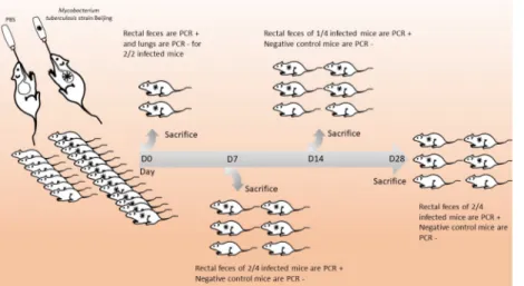
A total of 14 male BALBc mice were inoculated with theM. tuberculosis Beijing strain by the
gastric route after the inoculum’s resistance to acidity and biliary salts was verified in the pres-ence of 8 negative control mice given sterile phosphate-buffered saline (PBS). After two mice were sacrificed four hours post-challenge to verify the absence of inhaledM. tuberculosis, the
12 remaining mice were apparently healthy and showed no sign of pain and no weight loss was observed over the 28-day experiment. In these 12 mice,M. tuberculosis was detected by
PCR in the rectal feces collected from the sacrificed mice at day 7 (2/4 mice), day 14 (1/4) and day 28 (2/4), confirming that ingestedM. tuberculosis was transmitted and persisted along the
entire digestive tract of the challenged mice (Fig 1).
Ingested
M. tuberculosis infects the lymph nodes and lungs of mice
Enlarged cervical, tracheobronchial and mediastinal lymph nodes were observed in 3/4 mice sacrificed at day 7, in 2/4 mice sacrificed at day 14 and in 2/4 mice sacrificed at day 28. Macro-scopic changes were observed with variable degrees of congestion in the lungs of all infected
Fig 1. Infection of the digestive tract in a mouse model of ingestedM. tuberculosis. https://doi.org/10.1371/journal.pone.0227005.g001
mice, while the livers, spleens, kidneys and digestive tracts had a normal appearance in all mice. Accordingly, pathological examination found no detectable granulomas, inflammatory infiltrates or necrotic damages in any organ. These observations agree with previous observa-tions in the experimental models of pulmonary tuberculosis after tracheal instillation and aero-sols [20,21]. However, microscopic observations confirmed the dissemination ofM.
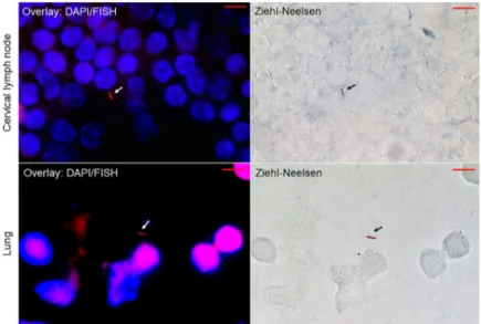
tuberculosis in the lung and lymph node tissues on post-infection day 28, with the
mycobacte-ria appearing as Ziehl-Neelsen-positive rods that were fluorescing in red due to the use of fluo-rescentin situ hybridization (FISH) staining [12] (Fig 2,S1 Fig). These mycobacteria were confirmed to beM. tuberculosis and were quantified by quantitative real-time PCR (qPCR)
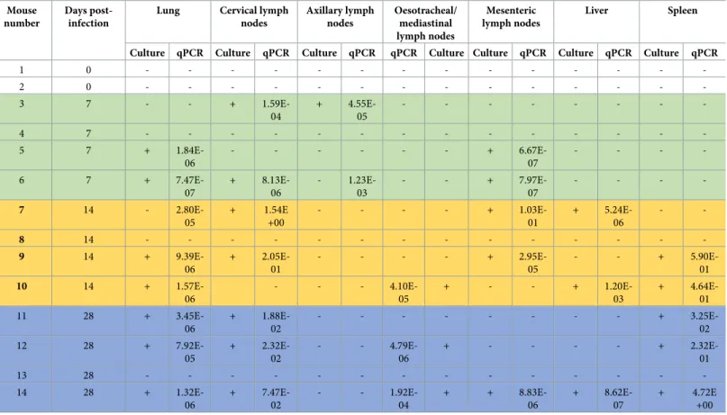
(Table 1,S1 Table). The spleen and cervical lymph nodes exhibited the highest load of myco-bacteria after 14 days and 28 days of infection. The mycomyco-bacterial load was 5± 0.9 mycobacte-ria/10 spleen cells and 3± 9 mycobacteria/10 lymph node cells at day 14 post-infection and was 4± 26 mycobacteria/10 spleen cells and 0.6 ± 0.3 mycobacteria/10 lymph node cells at day 28 post-infection. In the lungs, the highestM. tuberculosis burden was measured at day 14
post-infection, and the lowest burden was measured at day 7 post-infection; the mycobacterial loads were 11± 13 and 1.2 ± 0.7 mycobacteria/106cells, respectively. All the samples collected immediately after gavage in the inoculated and control animals remained negative according to the culture and PCR analyses, except for the digestive tract samples. Finally, the viability of detectedM. tuberculosis mycobacteria was assessed by culture (Table 1) as follows: on day 7,
M. tuberculosis was cultured from the lungs, the cervical and mesenteric lymph nodes and the
stool in two out of four mice. Additionally, one mouse showed anM. tuberculosis-positive
axil-lary lymph node. At day 14,M. tuberculosis was cultured from 3 of the 4 sacrificed mice; from
the lungs, stools, spleens, cervical and mesenteric lymph nodes and livers of two of these three mice; and from the mediastinal and tracheobronchial lymph nodes of one of these three mice. In the last group of 4 mice sacrificed at 28 days post-infection,M. tuberculosis was cultured
from the lungs, spleen and cervical lymph nodes of three mice and in the oesotracheal lymph
Fig 2. Microscopic images of imprint slides prepared from lung and cervical lymph nodes ofMycobacterium tuberculosis-infected mice at day 28 post-infection with combined FISH, DAPI and Ziehl-Neelsen staining. For fluorescentin situ hybridization (FISH), the slides were observed using a red channel of a Leica DMI6000 microscope
under a 100 X oil-immersion objective. The images were captured in the same microscopic field using a Hamamatsu Orca AG camera (Hamamatsu Photonics, Herrsching-am-Ammersee, Germany) for FISH-positive mycobacteria (left images, white arrows) and a DFC425 C Digital Microscope Camera (Leica Microsystemes, Nanterre, France) for Ziehl-Neelsen-positive mycobacteria (right images, black arrows). Scale bar = 5μm.
nodes and livers of two mice. In one mouse, the culture wasM. tuberculosis-positive in the
mediastinal and mesenteric lymph nodes, in the liver and in the stool.M. tuberculosis was not
cultured in any of the eight negative control mice (Table 1). Comparing the whole genome sequence of the inoculum with that derived from the lymph node and lung isolates indicated the same 2.2.1 sublineage in these four isolates. Core-genome analysis showed that the four strains were grouped into a unique cluster with the referenceM. tuberculosis lineage 2.2.1,
sharing the same position as IS6110 and the same spoligotyping pattern. These observations
confirmed the translocation of viableM. tuberculosis beyond the lymphatic system to several
organs, including the lungs.
Discussion
IngestedM. tuberculosis disseminated to lymph nodes and the lungs in the experimental
labo-ratory animals as was verified by theM. tuberculosis-negative state of the negative controls, the
concordant results obtained by the independent methods of observation and the reproducibil-ity of the results. The fact thatM. tuberculosis mycobacteria was not retrieved in the lungs in
animals sacrificed within four hours post-challenge, demonstrate that the observations here reported do not result from the mere inhalation of the mycobacteria. Pulmonary infection observed in this experimental model was characterized by congestive lungs containing viable and culturableM. tuberculosis. These observations matched those issued from previously
reported experimental mouse models. In particular, we found no significant difference
Table 1. Culture result and microbial load dataafor organs sampled from mice challenged with
M. tuberculosis.
Mouse number
Days post-infection
Lung Cervical lymph nodes Axillary lymph nodes Oesotracheal/ mediastinal lymph nodes Mesenteric lymph nodes Liver Spleen
Culture qPCR Culture qPCR Culture qPCR qPCR Culture Culture qPCR Culture qPCR Culture qPCR
1 0 - - - -2 0 - - - -3 7 - - + 1.59E-04 + 4.55E-05 - - - -4 7 - - - -5 7 + 1.84E-06 - - - + 6.67E-07 - - - -6 7 + 7.47E-07 + 8.13E-06 - 1.23E-03 - - + 7.97E-07 - - - -7 14 - 2.80E-05 + 1.54E +00 - - - - + 1.03E-01 + 5.24E-06 - -8 14 - - - -9 14 + 9.39E-06 + 2.05E-01 - - - - + 2.95E-05 - - + 5.90E-01 10 14 + 1.57E-06 - - - 4.10E-05 + - - + 1.20E-03 + 4.64E-01 11 28 + 3.45E-06 + 1.88E-02 - - - + 3.25E-02 12 28 + 7.92E-05 + 2.32E-02 - - 4.79E-06 + - - - - + 2.32E-01 13 28 - - - -14 28 + 1.32E-06 + 7.47E-02 - - 1.92E-04 + + 8.83E-06 + 8.62E-07 + 4.72E +00 a
qPCR data are the number of mycobacteria/mouse cell.
(P = 0.336) in terms of overall four-week post-infection survival andM. tuberculosis
dissemi-nation in the lungs of the challenged mice [21–24]. It has been reported that no granulomas were observable before 30 days post-infection inM. tuberculosis-infected mice; this is in line
with our observations [21]. This indicates that the model reported here is valuable and pro-duces a gross pathology indistinguishable from that previously reported in male mice that were experimentally infected withM. tuberculosis. However, mice challenged by the digestive
route developed more diseased lymph nodes earlier in the infection than those in previously reported aerosol models [25]. These results suggest thatM. tuberculosis translocated from the
digestive tract to the lungs through the lymphatic system, then followed by a progressive clear-ance ofM. tuberculosis by the mouse immune system. Altogether, our observations are in
agreement with the previously reported observations that indicate that the route ofM. tubercu-losis inoculation may govern the clinical form of tubercutubercu-losis.
Here, we challenged genetically homogeneous mice to bypass the genetic control of the infection [26]. We observed that only half of the mice had a lung dissemination ofM. tubercu-losis despite all challenged mice having the same genetic background (BALBc male mice).
Considering that digestive tract microbiota is part of the individual [27], we suspected that subtle differences in the digestive tract microbiota could correlate with the dissemination of
M. tuberculosis by the lymphatic route. Indeed, the mouse stool microbiota was shown to be
altered afterM. tuberculosis aerosol experimental infection, by metagenomics analyses [28]. Also, altered gut microbiota was observed in patients infected withM. tuberculosis [29] along with the excretion of viable mycobacteria in the stool [30]. In some populations, the epidemiol-ogy of tuberculosis is currently shifting from pulmonary to lymph node tuberculosis. As an example in Tunisia, increasing the extra-pulmonary/pulmonary tuberculosis ratio from 19.6% in 1996 to 32.6% in 2007 was driven by lymph node tuberculosis, the second most common form that causes 23% of cases [26]. Likewise, infections ofM. bovis and M. canettii from the
digestive route promote a predominantly lymphatic form of infection [11]. These observations and the ones reported here suggest that the current epidemiological shift of tuberculosis reported in some countries may be partially driven by the changing modes of contamination in populations, from the classic airborne transmission to digestive transmission. This hypothe-sis warrants further field studies including that of the digestive tract microbiota by culturomics [31].
Conclusion
The observations reported here enrich the literature to indicate that ingestedM. tuberculosis
promotes lymphatic and pulmonary tuberculosis. The experimental observations reported here fully agree with the clinical observations made during the “Lu¨beck disaster”, where almost all the neonates contaminated by the oral route withM. tuberculosis developed
lymphadenopa-thies; this was also the most salient clinical feature in this episode [1].
These observations may change our common view of the natural history of tuberculosis as an exclusively air-borne infection. In particular, the experimental results here reported ques-tion the potential of recirculaques-tion of viableM. tuberculosis, otherwise routinely detected in the
stools of pulmonary tuberculosis patients [30].
Translocation is therefore a common trait of theM. tuberculosis complex, previously
observed forM. bovis [6,7] and forM. canettii [11]. These observations suggest that transloca-tion was shared by the common ancestor of theM. tuberculosis complex and was conserved
along with the co-evolution of tuberculosis andHomo sapiens [32,33]. In the late Pleistocene, hunter-gatherer populations ofH. sapiens were very thinly spread [34] and unlikely to reach the critical mass necessary to allow tuberculosis evolution from an environmental ancestor.
Pleistocene hunter-gatherers could possibly have contracted tuberculosis from environmental sources such as infected meat and other animal products in a first step, then following an oral-fecal route during the first times of sedentarization. Indeed,M. tuberculosis is routinely
cul-tured in the stools of patients with pulmonary tuberculosis, confirming the viability ofM. tuberculosis in the digestive tract of these patients [30]; viability known to be preserved for months inM. tuberculosis-contaminated soil [35].
Whether oral route contamination still translates in the actual epidemiology of tuberculosis in some populations, in particular in populations known to be dually exposed to tuberculosis and enteric pathogens with a human reservoir such as typhoid fever, warrants further investi-gations. As part of such investigations, culturomic investigations of the digestive tract micro-biota may provide clues to the epidemiological shift from pulmonary to lymphatic
tuberculosis, which is currently being observed in some populations.
Supporting information
S1 Fig. Microscopic images of imprint slides prepared from lung and cervical lymph nodes ofMycobacterium tuberculosis-infected mice at day 28 post-infection with combined FISH,
DAPI and Ziehl-Neelsen staining. The slides were observed using a Leica DMI6000 scope under a 100 X oil-immersion objective. The images were captured in the same micro-scopic field for FISH-positive mycobacteria (white arrows) and Ziehl-Neelsen-positive mycobacteria (black arrows). Scale bar = 5μm.
(TIF)
S1 Table. Quantification data ofM. tuberculosis load per organ cells.
(XLSX)
Acknowledgments
The authors acknowledge Olga Cusack for her expertise in editing the manuscript.
Author Contributions
Conceptualization: Fabienne Bre´geon, Michel Drancourt.
Investigation: Mustapha Fellag, Ahmed Loukil, Jamal Saad, Hubert Lepidi, Fe´riel Bouzid. Methodology: Mustapha Fellag, Ahmed Loukil, Jamal Saad, Hubert Lepidi, Fe´riel Bouzid,
Fabienne Bre´geon.
Supervision: Michel Drancourt.
Validation: Fabienne Bre´geon, Michel Drancourt.
Writing – original draft: Mustapha Fellag, Ahmed Loukil, Jamal Saad, Hubert Lepidi, Fe´riel Bouzid, Fabienne Bre´geon, Michel Drancourt.
Writing – review & editing: Fabienne Bre´geon, Michel Drancourt.
References
1. Fox GJ, Orlova M, Schurr E. Tuberculosis in Newborns: The Lessons of the “Lu¨beck Disaster” (1929– 1933). PLOS Pathogens. 2016; 12: e1005271.https://doi.org/10.1371/journal.ppat.1005271PMID:
26794678
2. Escombe AR, Oeser C, Gilman RH, Navincopa M, Ticona E, Martı´nez C, et al. The Detection of Air-borne Transmission of Tuberculosis from HIV-Infected Patients, Using an In Vivo Air Sampling Model. Clin Infect Dis. 2007; 44: 1349–1357.https://doi.org/10.1086/515397PMID:17443474
3. Turner RD, Bothamley GH. Cough and the Transmission of Tuberculosis. J Infect Dis. 2015; 211: 1367–1372.https://doi.org/10.1093/infdis/jiu625PMID:25387581
4. Aboubaker Osman D, Bouzid F, Canaan S, Drancourt M. Smooth Tubercle Bacilli: Neglected Opportu-nistic Tropical Pathogens. Front Public Health. 2016; 3.https://doi.org/10.3389/fpubh.2015.00283
PMID:26793699
5. Evans JT, Smith EG, Banerjee A, Smith RM, Dale J, Innes JA, et al. Cluster of human tuberculosis caused by Mycobacterium bovis: evidence for person-to-person transmission in the UK. The Lancet. 2007; 369: 1270–1276.https://doi.org/10.1016/S0140-6736(07)60598-4
6. Smith RMM, Drobniewski F, Gibson A, Montague JDE, Logan MN, Hunt D, et al. Mycobacterium bovis Infection, United Kingdom. Emerg Infect Dis. 2004; 10: 539–541.https://doi.org/10.3201/eid1003. 020819PMID:15109433
7. Grange JM. Mycobacterium bovis infection in human beings. Tuberculosis. 2001; 81: 71–77.https://doi. org/10.1054/tube.2000.0263PMID:11463226
8. Calmette, M.A. The intestinal origin of pulmonary tuberculosis, and the mechanism of tuberculous infec-tion. Reviews. Agriculture and Fisheries Annual Reports of Proceedings under the Diseases of Animal Actes, for the year 1905. 1905.
9. Pierce C, Dubos RJ, Middlebrook G. Infection of Mice with Mammalian Tubercle Bacilli Grown in Tween-Albumin Liquid Medium. Journal of Experimental Medicine. 1947; 86: 159–174.https://doi.org/ 10.1084/jem.86.2.159PMID:19871664
10. Lefford M.J. Diseases in mice and rats. In Kubica GP and Wayne LG (ed), The mycobacteria, New York. Pp. 947–977. 1984.
11. Bouzid F, Bre´geon F, Lepidi H, Donoghue HD, Minnikin DE, Drancourt M. Ready Experimental Translo-cation of Mycobacterium canettii Yields Pulmonary Tuberculosis. Infect Immun. 2017; 85.https://doi. org/10.1128/IAI.00507-17PMID:28923895
12. Loukil A, Kirtania P, Bedotto M, Drancourt M. FISHing Mycobacterium tuberculosis Complex by Use of a rpoB DNA Probe Bait. Journal of Clinical Microbiology. 2018; 56: e00568–18.https://doi.org/10.1128/ JCM.00568-18PMID:30068538
13. Bruijnesteijn Van Coppenraet ES, Lindeboom JA, Prins JM, Peeters MF, Claas ECJ, Kuijper EJ. Real-time PCR assay using fine-needle aspirates and tissue biopsy specimens for rapid diagnosis of myco-bacterial lymphadenitis in children. J Clin Microbiol. 2004; 42: 2644–2650.https://doi.org/10.1128/JCM. 42.6.2644-2650.2004PMID:15184446
14. Ding S, Chi MM, Scull BP, Rigby R, Schwerbrock NMJ, Magness S, et al. High-fat diet: bacteria interac-tions promote intestinal inflammation which precedes and correlates with obesity and insulin resistance in mouse. PLoS ONE. 2010; 5: e12191.https://doi.org/10.1371/journal.pone.0012191PMID:20808947
15. Bankevich A, Nurk S, Antipov D, Gurevich AA, Dvorkin M, Kulikov AS, et al. SPAdes: A New Genome Assembly Algorithm and Its Applications to Single-Cell Sequencing. Journal of Computational Biology. 2012; 19: 455–477.https://doi.org/10.1089/cmb.2012.0021PMID:22506599
16. Seemann T. Prokka: rapid prokaryotic genome annotation. Bioinformatics. 2014; 30: 2068–2069.
https://doi.org/10.1093/bioinformatics/btu153PMID:24642063
17. Steiner A, Stucki D, Coscolla M, Borrell S, Gagneux S. KvarQ: targeted and direct variant calling from fastq reads of bacterial genomes. BMC Genomics. 2014; 15: 881. https://doi.org/10.1186/1471-2164-15-881PMID:25297886
18. Coll F, McNerney R, Preston MD, Guerra-Assunc¸ão JA, Warry A, Hill-Cawthorne G, et al. Rapid deter-mination of anti-tuberculosis drug resistance from whole-genome sequences. Genome Med. 2015; 7: 51.https://doi.org/10.1186/s13073-015-0164-0PMID:26019726
19. Coll F, McNerney R, Guerra-Assunc¸ão JA, Glynn JR, Perdigão J, Viveiros M, et al. A robust SNP bar-code for typing Mycobacterium tuberculosis complex strains. Nature Communications. 2014; 5: 4812.
https://doi.org/10.1038/ncomms5812PMID:25176035
20. Dormans J, Burger M, Aguilar D, Hernandez-Pando R, Kremer K, Roholl P, et al. Correlation of viru-lence, lung pathology, bacterial load and delayed type hypersensitivity responses after infection with dif-ferent Mycobacterium tuberculosis genotypes in a BALB/c mouse model. Clin Exp Immunol. 2004; 137: 460–468.https://doi.org/10.1111/j.1365-2249.2004.02551.xPMID:15320894
21. Jeon B-Y, Kwak J, Hahn M-Y, Eum S-Y, Yang J, Kim S-C, et al. In vivo characteristics of Korean Beijing
Mycobacterium tuberculosis strain K1 in an aerosol challenge model and in the Cornell latent
tuberculo-sis model. Journal of Medical Microbiology. 2012; 61: 1373–1379.https://doi.org/10.1099/jmm.0. 047027-0PMID:22820694
22. Dibbern J, Eggers L, Schneider BE. Sex differences in the C57BL/6 model of Mycobacterium
23. Medina E, North RJ. Resistance ranking of some common inbred mouse strains to Mycobacterium
tuberculosis and relationship to major histocompatibility complex haplotype and Nramp1 genotype.
Immunology. 1998; 93: 270–274.https://doi.org/10.1046/j.1365-2567.1998.00419.xPMID:9616378
24. Dunn PL, North RJ. Virulence ranking of some Mycobacterium tuberculosis and Mycobacterium bovis strains according to their ability to multiply in the lungs, induce lung pathology, and cause mortality in mice. Infect Immun. 1995; 63: 3428–3437. PMID:7642273
25. Chackerian AA, Alt JM, Perera TV, Dascher CC, Behar SM. Dissemination of Mycobacterium
tuberculo-sis is influenced by host factors and precedes the initiation of T-cell immunity. Infect Immun. 2002; 70:
4501–4509.https://doi.org/10.1128/IAI.70.8.4501-4509.2002PMID:12117962
26. Smaoui S, Mezghanni MA, Hammami B, Zalila N, Marouane C, Kammoun S, et al. Tuberculosis lymph-adenitis in a southeastern region in Tunisia: Epidemiology, clinical features, diagnosis and treatment. International Journal of Mycobacteriology. 2015; 4: 196–201.https://doi.org/10.1016/j.ijmyco.2015.04. 004PMID:27649866
27. Lagier J-C, Dubourg G, Million M, Cadoret F, Bilen M, Fenollar F, et al. Culturing the human microbiota and culturomics. Nat Rev Microbiol. 2018; 540–550.https://doi.org/10.1038/s41579-018-0041-0PMID:
29937540
28. Winglee K, Eloe-Fadrosh E, Gupta S, Guo H, Fraser C, Bishai W. Aerosol Mycobacterium tuberculosis Infection Causes Rapid Loss of Diversity in Gut Microbiota. PLOS ONE. 2014; 9: e97048.https://doi. org/10.1371/journal.pone.0097048PMID:24819223
29. Luo M, Liu Y, Wu P, Luo D-X, Sun Q, Zheng H, et al. Alternation of Gut Microbiota in Patients with Pul-monary Tuberculosis. Front Physiol. 2017; 8: 822.https://doi.org/10.3389/fphys.2017.00822PMID:
29204120
30. El Khe´chine A, Henry M, Raoult D, Drancourt M. Detection of Mycobacterium tuberculosis complex organisms in the stools of patients with pulmonary tuberculosis. Microbiology (Reading, Engl). 2009; 155: 2384–2389.https://doi.org/10.1099/mic.0.026484–0
31. Lagier J-C, Hugon P, Khelaifia S, Fournier P-E, Scola BL, Raoult D. The Rebirth of Culture in Microbiol-ogy through the Example of Culturomics To Study Human Gut Microbiota. Clinical MicrobiolMicrobiol-ogy Reviews. 2015; 28: 237–264.https://doi.org/10.1128/CMR.00014-14PMID:25567229
32. Comas I, Coscolla M, Luo T, Borrell S, Holt KE, Kato-Maeda M, et al. Out-of-Africa migration and Neo-lithic coexpansion of Mycobacterium tuberculosis with modern humans. Nat Genet. 2013; 45: 1176– 1182.https://doi.org/10.1038/ng.2744PMID:23995134
33. Comas I, Hailu E, Kiros T, Bekele S, Mekonnen W, Gumi B, et al. Population Genomics of
Mycobacte-rium tuberculosis in Ethiopia Contradicts the Virgin Soil Hypothesis for Human Tuberculosis in
Sub-Saharan Africa. Curr Biol. 2015; 25: 3260–3266.https://doi.org/10.1016/j.cub.2015.10.061PMID:
26687624
34. Stewart JR, Stringer CB. Human evolution out of Africa: the role of refugia and climate change. Science. 2012; 335: 1317–1321.https://doi.org/10.1126/science.1215627PMID:22422974
35. Ghodbane R, Mba Medie F, Lepidi H, Nappez C, Drancourt M. Long-term survival of tuberculosis com-plex mycobacteria in soil. Microbiology. 2014; 160: 496–501.https://doi.org/10.1099/mic.0.073379-0