HAL Id: hal-03020718
https://hal.sorbonne-universite.fr/hal-03020718
Submitted on 24 Nov 2020
HAL is a multi-disciplinary open access
archive for the deposit and dissemination of sci-entific research documents, whether they are pub-lished or not. The documents may come from teaching and research institutions in France or abroad, or from public or private research centers.
L’archive ouverte pluridisciplinaire HAL, est destinée au dépôt et à la diffusion de documents scientifiques de niveau recherche, publiés ou non, émanant des établissements d’enseignement et de recherche français ou étrangers, des laboratoires publics ou privés.
plasticity of PTEN
Larissa Kotelevets, Barbara Trifault, Eric Chastre, Mark Scott
To cite this version:
Larissa Kotelevets, Barbara Trifault, Eric Chastre, Mark Scott. Post-translational regulation and conformational plasticity of PTEN. Cold Spring Harbor Perspectives in Medicine, 2020, 10 (11). �hal-03020718�
Post-translational regulation and conformational plasticity of PTEN
Larissa Kotelevets1,2, Barbara Trifault3,4,5, Eric Chastre1,2¶ and Mark G.H. Scott3,4,5¶ 1Institut National de la Santé et de la Recherche Médicale, UMR S 938, Centre de
Recherche Saint-Antoine, Paris, France;
2Sorbonne Université, UFR de Médecine, Site Saint-Antoine, Paris, France 3Université de Paris, Paris, France;
4Institut Cochin, Inserm, U1016, Paris, France; 5CNRS, UMR8104, Paris, France;
¶Corresponding authors:
[email protected] Address:
Institut Cochin, INSERM U1016, CNRS UMR8104, Université de Paris 27, Rue du Faubourg Saint Jacques, 75014 Paris, France
[email protected] Address:
Centre de Recherche Saint-Antoine, Inserm UMR S 938, Hôpital Saint-Antoine, Bâtiment Kourilsky, 34 rue Crozatier, 75012 Paris, France
Abstract
PTEN is a tumour suppressor that is frequently downmodulated in human cancer. PTEN inhibits the PI3K/AKT pathway through its lipid phosphatase activity. Multiple PI3K/AKT-independent actions of PTEN, protein-phosphatase activities and functions within the nucleus have also been described. PTEN therefore regulates many cellular processes including cell proliferation, survival, genomic integrity, polarity, migration and invasion. Even a modest decrease in the functional dose of PTEN may promote cancer development. Understanding the molecular and cellular mechanisms that regulate PTEN protein levels and function, and how these may go awry in cancer contexts, is therefore key to fully understanding the role of PTEN in tumourigenesis. Here, we discuss current knowledge on post-translational control and conformational plasticity of PTEN, as well as therapeutic possibilities towards re-establishment of PTEN tumour suppressor activity in cancer.
Introduction
PTEN (phosphatase and tensin homolog deleted on chromosome ten) is a ubiquitously expressed tumour suppressor that is frequently deregulated in human cancers. Three different groups initially identified the PTEN gene in 1997 (Li and Sun 1997; Li et al. 1997; Steck et al. 1997). The gene is located on chromosome 10q23, a region that displays loss of heterozygosity (LOH) in several cancers (Teng et al. 1997). Following on from this, PTEN mutations were documented in the germline of patients with a group of rare autosomal dominant syndromes, collectively termed as the PTEN hamartoma tumour syndromes (PHTS) (Yehia et al. 2019). These disorders are characterized by the presence of multiple hamartomas and increased cancer predisposition, therefore providing direct evidence for the deregulation of PTEN in cancer. In vivo studies in mice have also analysed the importance of Pten in cancer development. Whereas homozygous deletion of Pten provokes early embryonic death,
Pten+/- mice develop various cancers demonstrating that Pten is haploinsufficient for
suppressing cancer (Di Cristofano et al. 1998; Stambolic et al. 1998; Suzuki et al. 1998; Podsypanina et al. 1999). Furthermore, studies using a series of mice engineered to carry different combinations of wild-type, null and hypomorphic Pten alleles, have shown that even subtle reductions in Pten levels are sufficient to promote cancer susceptibility (Trotman et al. 2003; Alimonti et al. 2010). These findings suggest a “continuum model” of PTEN tumour suppression, whereby modest alterations in the functional dose of PTEN may promote tumourigenesis without mutation or loss of even one allele. In light of these studies, understanding the molecular and cellular mechanisms that regulate PTEN protein levels and function, and how these may go awry in cancer settings, is key to fully understanding the role of PTEN in tumourigenesis.
The PTEN protein comprises 403 amino acids (Fig. 1a). The crystal structure of human PTEN, using PTEN truncated at its termini and an internal loop, revealed the existence of an N-terminal phosphatase domain (amino acids 7-185) and a C-terminal C2 domain involved in membrane binding (amino acids 186-351) (Lee et al. 1999). PTEN also contains a short N-terminal PIP2-binding motif (amino acids 6-15) involved in membrane targeting, and a regulatory C-terminal tail (amino acids 352-403) that contains two phosphorylation site clusters and a PDZ binding motif. In addition to this 403 amino acid form of PTEN, several N-terminally extended isoforms expressed at lower levels, translated from upstream alternative start codons, have recently been
described: PTEN-O, PTEN-N, PTEN-M/PTEN- and PTEN-L/PTEN- (Malaney et al. 2017). The N-terminal extensions affect their subcellular localizations and the first discovered isoform PTEN-L, with a 173 amino acid N-terminal extension including a signal peptide, is secreted and enters into neighbouring cells (Hopkins et al. 2013).
PTEN is a phosphatase that can dephosphorylate both protein and lipid substrates. It can dephosphorylate protein substrates on serine/threonine and tyrosine residues. The protein phosphatase activity of PTEN is however weak, and determining what the physiological protein substrates of PTEN are remains an important area of investigation. Several potential protein substrates of PTEN have been proposed and include FAK (Gu et al. 1998), IRS1 (Shi et al. 2014) and Dvl2 (Shnitsar et al. 2015). PTEN can also autodephosphorylate threonine residues contained within its regulatory carboxy-terminal region (Raftopoulou et al. 2004; Tibarewal et al. 2012). The lipid phosphatase function of PTEN serves to negatively regulate the proproliferative PI3K/AKT pathway by dephosphorylating the lipid signalling intermediate PIP3 that is generated by PI3Ks (Maehama and Dixon 1999) (Fig. 1b). PIP3 is considered the major physiological substrate for PTEN. PIP3 serves to recruit proteins, such as AKT, containing pleckstrin homology domains, to the membrane. By counterbalancing the PI3K/AKT pathway the lipid phosphatase activity of PTEN therefore serves to limit cell proliferation, survival, migration and invasiveness (Stambolic et al. 1998; Chalhoub and Baker 2009). When PTEN function is impaired or lost, PIP3 levels are not properly regulated leading to hyperactivation of the PI3K/AKT pathway, cell transformation and tumourigenesis (Suzuki et al. 1998; Podsypanina et al. 1999; Di Cristofano et al. 1998; Leslie and Downes 2004).
A number of phosphatase-independent biological actions of PTEN have also been described in the cytoplasm and nucleus that rely on protein-protein interactions. For example, PTEN directly binds tumour suppressor p53 in the nucleus, increasing its stability and transcriptional activity (Freeman et al. 2003). Nuclear PTEN also interacts with the APC/C (anaphase-promoting complex) and promotes its association with CDH1 (CDC20 homolog 1) to enhance the tumour-suppressive activity of the APC-CDH1 complex in a phosphatase-independent fashion (Song et al. 2011). The isolated membrane-targeting PTEN C2 domain is also able to mimic effects of full-length PTEN on inhibition of glioma cell migration (Raftopoulou et al. 2004; Lima-Fernandes et al. 2011) and glandular morphogenesis in 3D colorectal cancer cell systems (Javadi et al. 2017). Mechanisms controlling PTEN subcellular
compartmentalization are therefore key in spatially controlling both phosphatase-dependent and -inphosphatase-dependent functions.
PTEN levels and function are subject to tight regulation at the transcriptional, post-transcriptional and post-translational levels (Salmena et al. 2008; Leslie and Foti 2011; Song et al. 2012; Lee et al. 2018) (Fig. 2). Initially believed to be a constitutively active phosphatase, pleiotropic post-translational modifications and protein-protein interactions are now known to calibrate PTEN function (Keniry and Parsons 2008; Salmena et al. 2008; Wang and Jiang 2008; Leslie and Foti 2011; Song et al. 2012; Kotelevets et al. 2018; Lee et al. 2018). These post-translational mechanisms control PTEN conformation, phosphatase activity and association with protein complexes, drive subcellular compartmentalization and impact protein stability. Studying how these mechanisms contribute to the loss of PTEN function in cancer is therefore essential. With this knowledge in hand it may be possible to develop strategies with the goal of restoring or enhancing PTEN tumour suppressor activity in cancer contexts. Here, we discuss post-translational control and conformational plasticity of PTEN, and implications in human cancer.
PTEN Regulation
PTEN expression and function are tightly regulated at the transcriptional, post-transcriptional and post-translational levels (Salmena et al. 2008; Leslie and Foti 2011; Song et al. 2012; Lee et al. 2018) (Fig. 2). In addition to genetic loss or somatic mutations in human cancers, disorder in these regulatory molecular mechanisms controlling PTEN expression and function can lead to a spectrum of different loss-of-function PTEN protein categories and/or levels, which contribute to tumourigenesis in diverse manners (Lee et al. 2018). Epigenetic and transcriptional silencing as well as dysregulation of miRNAs and competitive endogenous RNA (ceRNA) systems have all been shown to suppress PTEN expression (Fig. 2). Regulation of PTEN at the post-translational level via dynamic post-post-translational modifications (PTMs) and protein-protein interactions calibrates PTEN function. Dysregulation of these non-genomic mechanisms can again lead to repression of PTEN function (Leslie and Foti 2011). Mechanisms of post-translational control of PTEN are discussed below.
Post-translational modifications of PTEN
PTEN function is calibrated by multiple PTMs summarized in Fig. 3a and Table 1. Aberrant regulation of mechanisms controlling these PTMs can lead to inhibited PTEN function. This point of control on PTEN also provides therapeutic avenues to explore with the aim of re-establishing or enhancing PTEN function.
Phosphorylation
A key PTM regulating PTEN function is phosphorylation. Phosphorylation controls PTEN conformation, activity, stability and subcellular targeting. PTEN is phosphorylated on serine/threonine clusters contained in its regulatory C-terminal tail (Fig. 3a: Ser362, Thr366, Ser370, Ser380, Thr382, Thr383 and Ser385). Ser380, Thr382, Thr383 and Ser385 are phosphorylated to high stoichiometry by casein kinase 2 (CK2) (Torres and Pulido 2001). CK2 also phosphorylates Ser370 and this promotes subsequent phosphorylation of Thr366 and Ser362 by glycogen synthase kinase 3 (GSK3) (Al-Khouri et al. 2005; Maccario et al. 2007). Phosphorylation of the Ser380, Thr382, Thr383, Ser385 cluster promotes a "closed", less active but stable form of PTEN that has increased conformational compaction, reduced interaction with membrane-anchored PDZ-domain-containing proteins, such as MAGI-2 and MAGI-3, and decreased plasma membrane targeting (Vazquez et al. 2000; Torres and Pulido
2001; Vazquez et al. 2001; Das et al. 2003; Bolduc et al. 2013) (Fig. 3b). This "closed" conformation of PTEN is more stable due to it displaying reduced access to E3 ubiquitin ligases and therefore being less targeted for proteasomal-mediated degradation (Maccario et al. 2010). An intramolecular interaction of the phosphorylated C-terminal tail with basic residues located in the N-terminal PIP2-binding motif, the catalytic and C2 domains, maintains PTEN in its "closed" form, blocking the active site (Odriozola et al. 2007; Rahdar et al. 2009). Mutation of these C-terminal phosphorylation sites disrupts the intramolecular interaction leading to an "open" conformation of PTEN, which displays enhanced plasma membrane targeting and increased activity (Fig. 3b). In human T cell acute lymphoblastic leukaemia (T-ALL) cells PTEN is inactivated by hyperphosphorylation on C-terminal phosphorylation sites resulting from CK2 overexpression and hyperactivation (Silva et al. 2008). This leads to decreased PTEN lipid phosphatase activity and constitutive hyperactivation of the PI3K/AKT pathway. CK2 inhibition using selective CK2 inhibitors, re-established PTEN function to impair PI3K/AKT signalling in T-ALL cells.
Another mechanism leading to enhanced PTEN C-terminal tail phosphorylation, is via loss of protein phosphatase 2A (PP2A)-mediated dephosphorylation of PTEN (Nakahata et al. 2014). The adaptor protein N-myc downstream-regulated gene (NDGR2) is a PTEN-binding protein that recruits PP2A to PTEN (Nakahata et al. 2014). NGDR2 is frequently downregulated in T-ALL, leading to enhanced PTEN phosphorylation and increased PI3K/AKT signalling. PTEN is also thought to be capable of auto-dephosphorylation of Thr366 and Thr383 with implications for glioma cell migration/invasion (Raftopoulou et al. 2004; Tibarewal et al. 2012). Phosphorylation of PTEN in the C-terminal tail region can also lead to changes in cytonuclear distribution of PTEN. For example, phosphorylation of PTEN at Thr398 by the kinase ataxia telangiectasia mutated (ATM) promotes its nuclear export and sensitizes cells to DNA damage (Bassi et al. 2013). Polo-like kinase 1 (PLK1), which regulates cell cycle-related processes, phosphorylates PTEN on Ser380 enhances PTEN accumulation on chromatin, and normal mitotic progression (Choi et al. 2014). A phospho-deficient PTEN Ser380Ala mutant promotes enhanced mitotic exit. Interestingly, PTEN dephosphorylates and destabilizes PLK1 (Zhang et al. 2016). Outwith the regulatory C-terminal tail of PTEN, Ser229/Thr232 and Thr319/Thr321 in the C2 domain are phosphorylated by Rho-associated kinase 1 (ROCK1), promoting enhanced lipid phosphatase activity, polarized distribution of PTEN in neutrophils and
chemotaxis (Li et al. 2005). Genotoxic agents activate ATM, which besides Thr398, can also phosphorylate PTEN at Ser113 to trigger nuclear translocation and autophagy (Chen et al. 2015).
PTEN is also phosphorylated by tyrosine kinases. PTEN function is inhibited by phosphorylation on tyrosine residues by Src family kinases (Koul et al. 2002; Lu et al. 2003; Fenton et al. 2012). The oncoprotein Src phosphorylates PTEN in the C2 domain at Tyr240 and Tyr315, leading to decreased stability and also likely decreased PTEN binding to cellular membranes. Interestingly, trastuzumab, used in the treatment of ERBB2-overexpressing breast cancers, induces PTEN membrane targeting and phosphatase activity by reducing PTEN tyrosine phosphorylation via Src inhibition (Nagata et al. 2004). FGFR2 and FGFR3, but not EGFR or PDGFR, also phosphorylate PTEN on Tyr240. Tyr240 phosphorylation is associated with resistance to EGFR inhibitors in patients with glioblastoma multiforme and a shortened overall survival (Fenton et al. 2012). This process might be independent of PTEN phosphatase activity but related to its targeting to specific subcellular compartments. Accordingly, in response to DNA damage, pTyr240-PTEN binds to chromatin through interaction with Ki-67 and facilitates the recruitment of RAD51 to promote DNA repair and thus resistance to ionizing radiation therapy (Ma et al. 2019). Tyr336 in the PTEN C2 domain is phosphorylated by FAK (Tzenaki et al. 2015) and Rak (Yim et al. 2009), leading to increased phosphatase activity and protein stability of PTEN. Increased PTEN stability in the presence of Rak is due to decreased binding of the E3 ubiquitin ligase NEDD4-1 to PTEN. In terms of tyrosine phosphatases, SHP-1 has been shown to dephosphorylate PTEN in Src transfected cells, restoring PTEN stability (Lu et al. 2003).
Ubiquitination
Several ubiquitin E3 ligases are implicated in the ubiquitination of PTEN, which has implications for PTEN stability and subcellular localization. Polyubiquitination leads to PTEN degradation via the ubiquitin-proteasome system, whereas monoubiquitination on Lys13 and Ly289 promotes its nuclear import. The HECT-domain ubiquitin E3 ligase NEDD4-1 was the first ubiquitin E3 ligase identified for PTEN (Wang et al. 2007). NEDD4-1 is implicated in both polyubiquitination and monoubiquitination of PTEN (Trotman et al. 2007; Wang et al. 2007). Depletion of Nedd4-1 in mice, however, found no change in Pten protein stability in vivo, suggesting
that there is some redundancy and other E3 ubiquitin ligases are likely also implicated in the regulation of PTEN proteosomal degradation (Fouladkou et al. 2008). In line with this, several other E3 ubiquitin ligases have been implicated in PTEN ubiquitination and increased PTEN turnover. These include, WWP2 the NEDD4-like protein family E3 ligase (Maddika et al. 2011), the RING-domain X-linked inhibitor of apoptosis protein (XIAP) (Van Themsche et al. 2009), the chaperone-assisted E3 ligase C-terminus of Hsc70-interacting protein (CHIP) (Ahmed et al. 2012), and RING finger protein 146 (RNF146) (Li et al. 2015). Non-degradative polyubiquitination by RFP, negatively regulates PTEN activity, without affecting PTEN stability (Lee et al. 2013). Very interestingly, non-degradative Lys27-linked polyubiquitination of PTEN by the E3 ubiquitin ligase WWP1 was recently shown to suppress PTEN oligomerization (see protein partners), plasma membrane recruitment, and tumour-suppressive functions both in vitro and in vivo (Lee et al. 2019). Genetic deletion or pharmacological inhibition of WWP1 provoked PTEN reactivation and tumour suppressive activity. This study therefore indicates a potential therapeutic strategy to reactivate PTEN for cancer treatment.
Several deubquitinases (DUBs) have also been identified for PTEN. These include ubiquitinspecific proteases10 (Sun et al. 2018), 11 (Park et al. 2019) and -13 (Zhang et al. 20-13) (USP-10, USP-11 and USP--13), as well as OTU-deubiquitinase (OTUD3) (Yuan et al. 2015) that are all implicated in PTEN deubiquitination and stabilization. In addition, ubiquitin-specific protease-7 (USP7 or HAUSP) is involved in the reversal of PTEN monoubiquitination, leading to nuclear exclusion of PTEN (Song et al. 2008).
SUMOylation
Small ubiquitin-like modifier (SUMO) protein can be conjugated to Lys254 and Lys266 in the C2 domain of PTEN (Huang et al. 2012; Bassi et al. 2013; Wang et al. 2014). SUMOylation on Lys266 in the CBR3 loop of PTEN promotes plasma membrane binding via electrostatic interactions (Huang et al. 2012). This enhanced membrane targeting of SUMOylated PTEN leads to decreased PI3K/AKT signalling, suppression of anchorage-independent cell growth and tumour growth in vivo. PTEN SUMOylation on Lys254, enhances its nuclear retention (Bassi et al. 2013). This promotes the nuclear function of PTEN in DNA repair mechanisms. Following genotoxic stress, SUMO-PTEN is rapidly excluded from the nucleus in an ATM
kinase-dependent manner (phosphorylation of Thr398 in the C-terminal tail of PTEN) (Bassi et al. 2013). Cells lacking nuclear PTEN were found to be hypersensitive to DNA damage. Finally, cross-talk between SUMOylation and ubiquitination exists on PTEN. PTEN SUMOylation is increased by the SUMO E3 ligase PIASx, resulting in decreased polyubiquitination, increased PTEN stability, leading to inhibition of the PI3K/AKT pathway, inhibition of cell proliferation and tumour suppression (Wang et al. 2014).
Acetylation
Following growth factor receptor stimulation, the histone acetylase p300/CBP-associated factor (PCAF) acetylates PTEN on Lys125 and Lys128, which are both located within the catalytic pocket (Okumura et al. 2006). Acetylation on these residues negatively regulates PTEN lipid phosphatase activity leading to enhanced AKT signalling. PTEN can also be acetylated by p300-CREB-binding protein (CBP) on Lys402, located in its C-terminal PDZ domain-binding motif (Ikenoue et al. 2008). Acetylation here does not impact catalytic activity but instead affects PTEN interaction with PDZ-domain containing proteins. The deacetylase sirtuin 1 (SIRT1) is mainly responsible for PTEN deacetylation (Ikenoue et al. 2008). In Sirt1-knockout cells, PTEN is hyperacetylated and excluded from the nucleus, indicating that acetylation modulates PTEN subcellular localization. It was also recently demonstrated that PTEN acetylation at Lys163, following inhibition of Histone Deacetylase 6 (HDAC6), switched PTEN into an "open" conformation that was targeted to the plasma membrane (Meng et al. 2016). This resulted in the inhibition of cell proliferation, migration and invasion, as well as xenograft tumour growth. This suggests that HDAC inhibitors may be clinically relevant in tumours expressing wild-type PTEN.
Oxidation
As with other protein tyrosine phosphatases PTEN contains a catalytic site cysteine nucleophile that is susceptible to oxidation. Reactive oxygen species (ROS) promote the oxidation of Cys124, which leads to the formation of an intramolecular disulphide bond with Cys71, suppressing PTEN phosphatase activity (Lee et al. 2002). The oxidation of PTEN is reversible. Endogenous ROS produced in activated cells promotes transient oxidation of PTEN leading to inactivation of a fraction of cellular PTEN, an increase in PIP3 levels, and activation of AKT (Leslie et al. 2003; Kwon et
al. 2004). This suggests localized control of a subcellular pool of PTEN by oxidation. Thioredoxin-interacting protein (TXNIP) regulates reversible PTEN oxidation by maintaining sufficient thioredoxin NADPH activity to reductively reactivate oxidized PTEN and counterbalance PI3K/AKT signalling (Hui et al. 2008). Oxidation of PTEN-binding partners also influences PTEN activity. For example, the oncoprotein DJ-1 binds PTEN and inhibits its catalytic activity (Kim et al. 2009). Oxidation of DJ-1 increases its affinity for binding PTEN, leading to a more profound decrease in PTEN activity. In contrast two other PTEN-binding partners peroxidase peroxiredoxin 1 (Prdx1) (Cao et al. 2009) and apoptosis-inducing factor (AIF) (Shen et al. 2015) protect PTEN from oxidation-induced inactivation via direct interaction.
S-Nitrosylation
Low concentrations of nitric oxide (NO) lead to S-nitrosylation of PTEN (SNO-PTEN) at Cys83 (Numajiri et al. 2011). This event results in inhibition of PTEN catalytic activity and enhanced AKT signalling, demonstrating that Cys83 is an important site for redox regulation of PTEN. In addition to the effect on lipid phosphatase activity, S-nitrosylation of PTEN also promotes PTEN protein degradation via the ubiquitin-proteasome system through NEDD4-1-mediated ubiquitination (Kwak et al. 2010). Depletion of the E3 ubiquitin ligase Parkin contributes to AMPK-mediated activation of endothelial nitric oxide synthase (eNOS), increased levels of reactive oxygen species, and a concomitant increase in oxidized NO levels (Gupta et al. 2017). This promotes S-nitrosylation of PTEN and its ubiquitination. Together, these studies highlight the influence of PTEN S-nitrosylation in supporting AKT signalling, cell survival and proliferation.
Methylation
PTEN is methylated on Lys313 by the oncogenic protein methyltransferase SMYD2, which has been proposed to lead to negative regulation of PTEN activity and increased PI3K/AKT signalling (Nakakido et al. 2015). DNA double-strand breaks (DSBs) also increase NSD2-mediated di-methylation of PTEN at Lys349, which is recognized by the tudor domain of 53BP to recruit PTEN to DNA damage sites, allowing for efficient repair of DSBs (Zhang et al. 2019). Interestingly, inhibiting NSD2-mediated PTEN methylation sensitizes cancer cells to combination treatment with PI3K inhibitor and DNA-damaging agents.
Ribosylation
PTEN is also a substrate for tankyrases: tankyrase1 (TNKS1) and tankyrase2 (TNKS2) ribosylate PTEN at Glu40/Glu150 in the phosphatase domain and Asp326 in the C2 domain (Li et al. 2015). PTEN ribosylation promotes the recognition of PTEN by the E3 ubiquitin ligase, RNF146, leading to subsequent PTEN ubiquitination and degradation. TNKS levels were found to negatively correlate with PTEN levels in human colon carcinomas. Simultaneous knockdown of TNKS1/2 in colorectal cancer cells resulted in inhibition of tumour growth in PTEN-expressing but not PTEN-depleted cells, indicating that targeting TNKS may only be effective in wild-type PTEN contexts. Taken together, these data support the idea to explore the development of tankyrase inhibitors to restore PTEN function.
Protein-protein interactions
PTEN interacts with a number of protein partners that influence its subcellular localization, activity and stability. These PTEN partners include integral plasma membrane proteins, adaptor proteins, transport proteins, and proteins that influence PTEN PTMs. PTEN monomers can also oligomerize, a requirement for full PTEN activation at the plasma membrane. A list of some of these PTEN partners, where they bind on PTEN, and their effects on PTEN function are shown in Table 2, and discussed below.
Negative regulators of PTEN function include PIP3-dependent Rac exchanger protein (PREX2) (Fine et al. 2009; Hodakoski et al. 2014), shank-interacting protein-like 1 (SIPL1) (He et al. 2010; De Melo et al. 2014), -mannosidase 2C1 (MAN2C1) (He et al. 2011), DJ1 (Kim et al. 2009; Das et al. 2011), BMI (Fan et al. 2009), and Protein Phosphatase 1 Regulatory subunit 10 (PNUTS) (Kavela et al. 2013). These negative regulators all inhibit PTEN function through diverse mechanisms leading to enhanced PI3K/AKT signalling in cells, and therefore potentially represent therapeutic targets.
A number of partners affect PTEN stability by protecting PTEN from ubiquitination by E3 ligases. For example, both the regulatory subunit of PI3K, p85, in a homodimeric form (Cheung et al. 2015), and the G protein-coupled receptor (GPCR) Melanocortin receptor 1 (MC1R) (Cao et al. 2013) protect PTEN from proteasomal degradation by E3 ubiquitin ligase WWP2. Interestingly,
cancer-associated mutants of MC1R or p85 lead to decreased PTEN binding and increased PI3K-AKT signalling. The Rak tyrosine kinase (Table 1) interacts and phosphorylates PTEN on Tyr336, and this decreases binding of PTEN to the E3 ubiquitin ligase NEDD4-1 (Yim et al. 2009). Discs Large MAGUK Scaffold 1 (DLG1) a PDZ-domain containing scaffold binds the PDZ-binding motif of PTEN to promote PTEN stability and decreased PI3K/AKT signalling (Adey et al. 2000; Valiente et al. 2005). DLG1 also interacts with APC and therefore may serve as a platform to bring PTEN and APC in proximity, providing a functional network of tumour suppressor activities (Sotelo et al. 2012).
Protein interaction partners of PTEN can also regulate its subcellular localization. Plasma membrane targeting of PTEN by these partners localizes PTEN to membrane-localized PIP3, resulting in decreased PI3K/AKT signalling. A number of PDZ-domain containing proteins bind the PDZ-binding motif of PTEN and target it to the plasma membrane/junctional complexes. These include 1, 2, MAGI-3, MAST1, MAST2, MASTMAGI-3, PAR-3 and NHERF1, with functional consequences for PTEN recruitment into high molecular weight complexes, inhibition of PI3K/AKT signalling, regulation of epithelial cell polarity, stabilization of adherens junctions and suppression of invasiveness (Wu et al. 2000; Tolkacheva et al. 2001; Kotelevets et al. 2005; Valiente et al. 2005; Takahashi et al. 2006; Wu et al. 2007; Chastre et al. 2009). Neural endopeptidase (NEP) (Sumitomo et al. 2004), the GPCR Sphingosine-1-Phosphate Receptor 2 (S1PR2) (Sanchez et al. 2005), the Cystic Fibrosis Transmembrane Conductance Regulator (CFTR) (Riquelme et al. 2017), and the motor protein Myosin V (van Diepen et al. 2009) also all target PTEN to the plasma membrane towards membrane-localized PIP3. The RhoA effector, RhoA associated protein kinase (ROCK), interacts with and phosphorylates PTEN (Table 1), promoting its membrane recruitment and activation (Li et al. 2005). ROCK activation also promotes the association of the multifunctional adaptor proteins -arrestin1 (-arr1) and -arrestin2 (-arr2) with PTEN (Lima-Fernandes et al. 2011). -arrs recruit PTEN to the plasma membrane following GPCR/Rho/ROCK stimulation and enhance its lipid phosphatase activity. However, during glioma cell migration -arrs bind the C2 domain of PTEN to inhibit its lipid-phosphatase-independent anti-migratory function. -arr1 also binds the PTEN C2 domain as part of a plasma membrane-associated regulatory complex incorporating the Cdc42 GTPase-activating protein ARHGAP21 and Cdc42
(Javadi et al. 2017). This complex regulates Cdc42-dependent mitotic spindle formation and lumen formation in 3D cultures of colorectal cancer cells. Disruption of this protein network provokes mitotic spindle misorientation and abnormal multilumen that are evocative of colorectal cancer.
PTEN also has function in the nucleus, including enhancing both tumour suppressor p53 function (Freeman et al. 2003) and the tumour-suppressive activity of the APC-CDH1 complex in a phosphatase-independent fashion (Song et al. 2011). Nuclear translocation of PTEN occurs through various mechanisms including interaction with the Major Vault Protein (Chung et al. 2005; Minaguchi et al. 2006) and the nuclear transport receptor Importin-11 (Chen et al. 2017). Importin-11 protects PTEN from cytoplasmic degradation by NEDD4-1, and there is correlative loss of both Importin-11 and PTEN in lung tumours.
Finally, PTEN monomers can oligomerize at the plasma membrane, adopting an active conformation with full lipid phosphatase activity against PIP3 (Papa et al. 2014). Catalytically inactive cancer-associated PTEN mutants can heterodimerize with wild-type PTEN and act in a dominant-negative fashion to inhibit wild-type catalytic activity. A subsequent study demonstrated that PTEN can form homodimers in vitro and furnished a possible structural model for the complex (Heinrich et al. 2015). The C-terminal tail of PTEN functions to stabilize the homodimer and C-tail phosphorylation interferes with this stabilization.
Probing PTEN conformation
The combination of PTEN post-translational modifications and protein partner interactions occurring at a given moment in time will give rise to a particular conformation of PTEN. To rapidly probe changes in PTEN conformation dynamics in live cells, we recently developed an intramolecular bioluminescence energy transfer (BRET)-based biosensor of PTEN (Lima-Fernandes et al. 2014). For this, PTEN is sandwiched between the energy donor Renilla luciferase (Rluc) and the energy acceptor yellow fluorescent protein (YFP) (Fig. 4a). The energy transfer between donor and acceptor can be measured in live cells following the addition of coelenterazine, the substrate for Rluc (Fig. 4a and b). In cells expressing the biosensor a strong basal BRET signal was detected indicating molecular proximity of the donor and acceptor molecules (Fig. 4c). As the biosensor is unimolecular, BRET signals remain constant over a wide range of Rluc concentrations (Lima-Fernandes et al. 2014; Misticone et al.
2016) (Fig. 4c). This is particularly useful in situations where, for example, a mutant of PTEN is less stable, as a change in BRET will only reflect a conformational change, allowing conformational readout comparisons of cancer-associated PTEN mutants that may be less stable than wild-type PTEN. BRET from the biosensor can also be detected at “endogenous expression” levels of PTEN (Lima-Fernandes et al. 2014).
The biosensor can be used in different modes to probe PTEN conformational change occurring at the cellular and molecular levels (Fig. 4d). In a structure-function mode we demonstrated that combined mutation of key C-terminal tail phosphorylation sites (Ser380, Thr382, Thr383 and Ser385) implicated in PTEN intramolecular interaction and control of PTEN plasma membrane targeting/cellular activity, results in a decreased BRET signal (Fig. 4e), compared to baseline BRET, indicative of conformational rearrangement (Lima-Fernandes et al. 2014). This change is consistent with the "opening" of the PTEN molecule upon dephosphorylation of its C-terminal tail with increased membrane targeting and indicates that PTEN conformational changes can be followed in live cells using the biosensor. These carboxy-terminal regulatory sites are phosphorylated by the serine/threonine kinase CK2. CK2 overexpression has been documented in several types of cancer, leading to phospho-dependent PTEN inhibition and enhanced PI3K/AKT pathway activation (Silva et al. 2008). CK2 inhibitors have been shown to re-establish PTEN signalling and restrain the PI3K pathway (Silva et al. 2008). Negative BRET shifts (Fig. 4e) in the biosensor were also obtained following pharmacological inhibition of CK2 using either TBB or CX-4945, which is in development for clinical use (Chon et al. 2015). The BRET shifts obtained using CX-4945 paralleled decreased phosphorylation of the regulatory C-terminal residues (Misticone et al. 2016). In contrast to the results obtained with the CK2 inhibitors, overexpression of CK2 provoked a significant increase above baseline BRET of the biosensor (Lima-Fernandes et al. 2014) (Fig. 4e). This indicates that both positive and negative modification of a signalling pathway impacting PTEN function can be detected using the biosensor. Use of the biosensor, in live cells, therefore validates the model of a PTEN switch between a "closed/less active" cytoplasmic state and an "open/active" conformation that is targeted to the plasma membrane. The significance of PTEN membrane localization is underscored by studies demonstrating that certain cancer-associated mutants display normal catalytic activity in vitro, but display defective plasma membrane targeting (Nguyen et al. 2015). This impaired
plasma membrane localization leads to a PTEN-null phenotype of these mutants in cells.
The biosensor can also be used to probe conformational change that occurs in cancer-associated mutants: Lys13 is mutated to Glu (K13E) in spontaneous cancer (Duerr et al. 1998) and Lys289 to Glu (K289E) is associated with Cowden syndrome (Trotman et al. 2007). These amino acids are both located on unstructured regions of PTEN and are major monoubiquitination sites that are important for PTEN nuclear import. Introduction of the K13E/K289E mutations, which renders PTEN defective in nuclear import, provoked a BRET change signifying conformational change. The biosensor can therefore detect conformational changes associated with changes in different post-translational modifications of PTEN (i.e. phosphorylation and ubiquitination).
PTEN-protein partner interactions and the effect of upstream intracellular signalling proteins on PTEN conformation can also be detected using the biosensor. Co-expression of the biosensor with active RhoA and the molecular scaffold -arrestin2, both proteins known to activate PTEN, promoted changes in PTEN conformation (Lima-Fernandes et al. 2014; Misticone et al. 2016). Previously characterized or candidate PTEN protein partners that may form part of the PTEN interactome, as well as signalling proteins that may impinge on PTEN function can therefore be rapidly assessed for their effects on PTEN conformation in live cells by coexpression with the biosensor.
The biosensor can also be used to report real-time changes in PTEN conformational change occurring in kinetic experiments in live cells, following incubation with either physiological ligands or therapeutic agents that target cell surface receptors. We found that the lipid sphingosine 1-phosphate (S1P) changed the BRET signal of the biosensor in cells expressing the GPCR S1PR2, but not in cells expressing the S1PR1, in agreement with previous studies that S1PR2, but not S1PR1, activates PTEN (Sanchez et al. 2005). Using this kinetic approach we also identified several other GPCRs that elicited PTEN conformational rearrangement upon ligand incubation. The ERBB2-targeting antibody, trastuzumab, used in the treatment of ERBB2-overexpressing breast cancers, induces PTEN membrane targeting and lipid phosphatase activity by provoking a decrease in PTEN tyrosine phosphorylation via Src inhibition (Nagata et al. 2004). We showed that trastuzumab elicited a time-dependent change in BRET that coincided with enhanced PTEN lipid phosphatase
activity and decreased PI3K/AKT signalling. This series of experiments therefore provides proof of principle that the biosensor can detect real-time changes in PTEN conformation downstream of cell surface receptor targeting and therapeutic agents.
The different BRET signatures obtained using the biosensor indicate that PTEN can adopt a spectrum of different conformations, depending on changes in post-translational regulation mechanisms, and that the biosensor provides a means of identifying additional signalling contexts that influence PTEN function. As the biosensor can be used as a rapid, direct and sensitive reporter to detect potential changes in PTEN localization and activity, it could be used as readout for drug discovery screening of small molecule libraries to identify chemicals that enhance or restore PTEN-dependent pathways. The identification of such chemical agents would help pave the way towards reactivating PTEN tumour suppressor as a potential cancer treatment. Indeed, proof of principle supporting this concept comes from a recent study demonstrating that pharmacological inhibition of WWP1 provokes PTEN reactivation and tumour suppressive activity, uncovering a potential therapeutic strategy to reactivate PTEN for cancer treatment (Lee et al. 2019).
Concluding remarks
PTEN acts as a tumour suppressor via its lipid phosphatase activity to inhibit the PI3K/AKT oncogenic pathway, and also via PI3K/AKT-independent functions. Even a modest reduction in the functional dose of PTEN may promote tumourigenesis, indicating that re-establishing or enhancing PTEN function may provide a therapeutic strategy in a wide range of human cancers. Indeed, in favour of this, an enhanced engineered PTEN (ePTEN) has been developed that displays increased ability to inhibit PIP3 signalling (Nguyen et al. 2014), and pharmacological inhibition of WWP1, which suppresses PTEN function, triggers PTEN reactivation and tumour suppressive activity (Lee et al. 2019). In light of these studies, fully understanding the molecular and cellular mechanisms that regulate PTEN function, and how these go awry in cancer, is key to defining PTEN dysregulation in tumourigenesis, in addition to providing therapeutic avenues for exploration to restore or enhance PTEN function as anti-cancer therapy.
Acknowledgements
This work was supported by the CNRS and Inserm. Work in the group of MGHS is also supported by the Ligue Contre le Cancer (comité de L’Oise), La Fondation ARC, Université de Paris, and the Who am I? laboratory of excellence (grant ANR-11-LABX-0071) funded by the “Investments for the Future” program operated by The French National Research Agency (grant ANR-11-IDEX-0005-01).
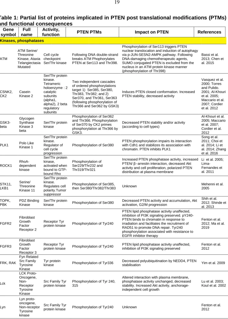
Table 1: Partial list of proteins implicated in PTEN post translational modifications (PTMs) and functional consequences
Gene symbol
Full name
Activity,
function PTEN PTMs Impact on PTEN References
Kinases, phosphatases ATM ATM Serine/ Threonine Kinase, Ataxia Telangiectasia Mutated Cell cycle checkpoint Ser/Thr kinase
Following DNA double-strand breaks ATM Phophorylates PTEN at Ser113 and Thr398.
Phosphorylation of Ser113 triggers PTEN nuclear translocation and induction of autophagy via p-JUN-SESN2-AMPK pathway. Following DNA-damaging chemotherapeutic agents, SUMO conjugated PTEN is excluded from the nucleus in an ATM protein kinase manner (phosphorylation of Thr398) Bassi et al. 2013; Chen et al. 2015 CSNK2, CK2 Casein Kinase 2 Ser/Thr protein kinase. Tetrameric holoenzyme : 2 catalytic subunits (alpha1, alpha2), 2 beta regulatory subunits
Two independent cascades of ordered phosphorylations target 1) Ser385, Ser380, Thr383, Thr382; and 2) Ser370, and Thr361, Ser363 (following phosphorylation of Thr366 and Ser362 by GSK3)
Induces PTEN closed conformation. Increased PTEN stability, decreased activity
Vasquez et al. 2000; Torres and Pulido. 2001; Al-Khouri et al. 2005; Maccario et al. 2007; Cordier et al. 2012 GSK3-beta Glycogen Synthase Kinase 3 beta Ser/Thr protein kinase Phosphorylation of Ser362 and Thr366. Phosphorylation of Ser370 by CK2 primes phosphorylation at Thr366 by GSK3.
Decreased PTEN stability and/or activity (according to cell types)
Al-Khouri et al. 2005; Maccario et al. 2007; Cordier et al. 2012 PLK1 Polo Like Kinase 1 Ser/Thr protein kinase. Regulator of cell cycle progression Phosphorylation of Ser380
PTEN phosphorylation impairs its interaction with Cdh1 and stabilizes its association with chromatin. PTEN inhibits PLK1
Song et al. 2011; Choi et al. 2014; Li et al. 2014; Zhang et al. 2016 ROCK1 RhoA- dependent kinase Ser/Thr protein kinase, activated when bound to GTP-bound Rho Phosphorylation of Ser229/Thr232 and Thr319/Thr321
Increased PTEN phosphatase activity, increased PTEN/ β-arrestin interaction, decreased Akt activity and cell proliferation, polarized PTEN distribution at plasma membrane
Li et al. 2005; Lima-Fernandes et al. 2011 STK11, LKB1 Serine/ Threonine Kinase 11 Ser/Thr protein kinase. Regulates cell polarity.Tumor suppressor Phosphorylation of Ser385,
then Ser380/Thr382/Thr383 Unknown
Mehenni et al. 2005 TOPK, PBK PDZ Binding Kinase Ser/Thr protein
kinase Phosphorylation of Ser380
Decreased PTEN activity and accumulation, Akt activation, G2/M progression Shih et al. 2012; Shinde et al. 2013 FGFR2 Fibroblast Growth Factor Receptor 2 Receptor Tyr
protein kinase Phosphorylation of Tyr240
PTEN lipid phosphatase activity unaffected, inhibition of PI3K signaling preserved. pY240-PTEN binds to chromatin in response to irradiation and facilitates the recruitment of RAD51 to promote DNA repair. Tyr240 phosphorylation associated with resistance to EGFR inhibitor therapy
Fenton et al. 2012; Ma et al. 2019 FGFR3 Fibroblast Growth Factor Receptor 3 Receptor Tyr
protein kinase Phosphorylation of Tyr240
PTEN lipid phosphatase activity unaffected, inhibition of PI3K signaling preserved
Fenton et al. 2012 FRK, RAK Fyn Related Src Family Tyrosine Kinase Tyr protein
kinase Phosphorylation of Tyr336
Decreased polyubiquitination by NEDD4, PTEN
stabilisation Yim et al. 2009
Lck LCK Proto-Oncogene, Non-Receptor Tyrosine Kinase Src Family Tyr protein kinase Phosphorylation of Tyr 240, 315
Altered interaction with plasma membrane, phosphatase activity unchanged, decreased stability. Increased Akt activity, anchorage-independent cell growth
Lu et al. 2003; Koul et al. 2002 Lyn Lyn proto-oncogene, Non-receptor Tyrosine kinase Src Family Tyr
protein kinase Phosphorylation of Tyr240 Unknown
Fenton et al. 2012
PTK2, FAK1 Protein Tyrosine Kinase 2 Cytoplasmic Tyr protein kinase concentrated at focal adhesions Phosphorylation of Tyr336
Increased phosphatase activity, protein–lipid interaction, and protein stability. FAK is positively regulated by Rock
Tzenaki et al. 2015 SRC SRC proto-oncogene, Non-receptor Tyrosine kinase Tyr protein kinase Phosphorylation of Tyr240 and Tyr315 (C2 domain), Tyr68 and Tyr155 (catalytic domain), and Tyr377 (tail)
Phosphorylation of Tyr240 and Tyr315: altered interaction with plasma membrane, phosphatase activity unaffected, decreased stability;
Phosphorylation of Tyr68 and Tyr155: PTEN destabilization. Increased Akt activity, anchorage-independent cell growth
Koul et al. 2002; Lu et al. 2003; Fenton et al. 2012 PP2A Protein Phosphatase 2A Ser/Thr-protein Phosphatase Dephosphorylation of PTEN at the Ser380, Thr382 and Thr383 cluster within the C-terminal tail
NDRG2 recruits protein phosphatase 2A (PP2A) to PTEN Nakahata et al. 2014 PTEN Phosphatase and tensin homolog deleted on chromosome 10 Protein / lipid phosphatase; Tumor suppressor Probable auto-dephosphorylation of Thr366 and Thr383
PTEN protein phosphatase activity together with lipid phosphatase activity inhibits invasion of U87 glioblastoma cells
Raftopoulou et al. 2004; Tibarewal et al. 2012 PTPN6, SHP1 Protein Tyrosine Phosphatase Non-receptor type 6 Tyr protein phosphatase Tyr dephosphorylation,
reversion of Src/Lck effects Decreased Akt activity Lu et al. 2003
Ubiquitination/Deubiquitination/SUMOylation Ndfip1 and Ndfip2 Nedd4 Family Interacting Protein 1 and protein 2 Endosomal membrane proteins that bind to and activate members of the Nedd4 family of E3 ubiquitin ligases PTEN ubiquitination
Induces polyubiquitination of PTEN by either Nedd4 or Itch. PTEN destabilization. Neuronal progenitors : Ndfip1 induces nuclear PTEN targeting and decreases cell proliferation (downregulation of cyclinD1, PLK1) Mund and Pelham 2010; Howitt et al. 2012; Howitt et al. 2015 NEDD4-1 Neural Precursor Cell Expressed, Developme ntally Down-Regulated 4, E3 Ubiquitin Protein Ligase E3 Ubiquitin Protein Ligase PTEN (mono/poly) ubiquitination
PTEN polyubiquitination by NEDD4-1, leads to PTEN degradation. NEDD4-1 can also induce PTEN monoubiquitination and nuclear translocation Wang et al. 2007; Trotman et al. 2007 SHARPIN , SIPL1 Shank-interacting protein–like 1 Component of the LUBAC complex which conjugates linear polyubiquitin chains in a head-to-tail manner to substrates Promotes PTEN polyubiquitination
Inhibits PTEN lipid phosphatase activity, does not induce PTEN degradation
He et al. 2010; De Melo et al. 2014 TRIM27, RFP Tripartite Motif Containing 27 Transcription repressor. RING-Type E3 Ubiquitin transferase Polyubiquitination
RFP-mediated ubiquitination negatively regulates PTEN phosphatase activity, resulting in increase in AKT activity and inhibition of TRAIL-mediated apoptosis. PTEN stability or localization unaffected Lee et al. 2013 WWP1 WW domain-containing ubiquitin E3 ligase 1 Ubiquitin E3 ligase Non-degradative Lys27-linked polyubiquitination of PTEN
Suppresses PTEN oligomerization, plasma membrane recruitment, and tumour suppressor functions in vitro and in vivo
Lee et al. 2019 WWP2, AIP-2 WW domain-containing E3 Ligase 2 Ubiquitin E3 ligase
PTEN polyubiqutination and degradation
Increased Akt activity, Decreased apoptosis. PTEN phosphorylation on Tyr155 prevents interaction with WWP2
Maddika et al. 2011
XIAP, BIRC4 X-Linked Inhibitor Of Apoptosis E3 Ubiquitin Protein Ligase, apoptotic suppressor protein,
PTEN mono and poly-ubiquitination
Regulates PTEN accumulation and compartmentalization van Themsche et al. 2009 STUB1, CHIP STIP1 Homology And U-Box Containing Protein 1, RING-Type E3 Ubiquitin Transferase STIP1 Homology And U-Box Containing Protein 1
PTEN polyubiqutination and degradation
HSP70 and HSP90 favor PTEN interaction with STUB1 Ahmed et al. 2012 OTUD3 OTU Deubiquitinas e 3 Deubiquitinating enzyme that hydrolyzes Lys-6- and Lys-11-linked polyubiquitin Deubiquitination: removes Lys6, Lys11, Lys27 and Lys48 types of ubiquitin chains on PTEN. Does not remove monoubiquitin on PTEN
PTEN Stabilization, inhibition of Akt signaling. Depletion of OTUD3 leads to the activation of Akt signalling, induction of cellular
transformation and cancer metastasis.
Yuan et al. 2015 USP7 (HAUSP) Ubiquitin Specific Peptidase 7 Deubiquitinates target proteins
Nuclear PTEN exclusion through decreased monoubiquitination (Lys13, Lys289)
USP7 regulates PTEN subcellular localization but not its stability. High levels of USP7 is associated with tumor aggressiveness. Nuclear/monoubiquitinated PTEN possesses greater apoptotic potential, nuclear exclusion is observed in more aggressive cancers.
Song et al. 2008 USP10 Ubiquitin Specific Peptidase 10 Cleaves ubiquitin from ubiquitin-conjugated protein substrates Deubiquitination and stabilization of PTEN
Knockdown of USP10 promotes tumor growth and invasion of Lewis carcinoma cells
Sun et al. 2018 USP11 Ubiquitin Specific Peptidase 11 Deubiquitinating enzyme (cysteine protease). X-linked tumor suppressor gene Reverses PTEN polyubiquitination in cytoplasm and nucleus
PTEN stabilization Park et al. 2019
USP-13 Ubiquitin Specific Peptidase 13 Deubiquitinates target proteins Deubiquitination : removes Lys6, Lys29 and Lys63 types of ubiquitin chains on PTEN. Does not remove
monoubiquitination
PTEN stabilization, inhibition of Akt signaling. Subcellular localization unaffected (cytoplasm). Overexpression of USP13 suppresses tumorigenesis and glycolysis in PTEN-positive but not PTEN-null breast cancer cells
Zhang et al. 2013 UBE2I SUMO-conjugating enzyme Ubc9 SUMO E2 Conjugase involved in SUMOylation of targets impacting their subcellular localization, transcriptional regulation, and protein stability Sumoylation of Lys266 targets PTEN to the plasma membrane. Sumoylation of Lys254 controls PTEN nuclear localization.
PTEN SUMOylated on Lys 266 decreases Akt activity, suppresses anchorage independent cell proliferation and tumor growth in vivo. PTEN sumoylated on Lys254 involved in DNA damage repair pathways
Huang et al. 2012; Bassi et al. 2013
PIASX SUMO E3 ligase PIAS3
SUMO E3 ligase that facilitates SUMOylation
Facilitates PTEN
SUMOylation on Lys254 and Lys266
Leads to protection from polyubiquitination, increased PTEN stability, inhibition of cell proliferation and tumour suppression
Wang et al. 2014 Acetylation/Deacetylation PCAF KAT2B Lysine Acetyltransf erase 2B Acetylates histone and non-histone proteins
Acetylation of Lys125 and Lys128 in response to growth factors. Decreased PTEN lipid Ptase activity
Decreased PTEN-induced G1 cell cycle arrest Okumura et al. 2006 HDAC6 Histone Deacetylase 6 Deacetylation of lysine residues, closely related to sirtuins Deacetylation of Lys163
Lys163 acetylation inhibited the interaction of the PTEN C-tail with the remaining part of PTEN. Deacetylation of Lys163 decreases PTEN translocation to plasma membrane
Meng et al. 2016 CREBBP, CBP CREB Binding Protein
Acetyltransferase PTEN acetylation on Lys402
Increased interaction with PDZ domain (hDLG, MAGI-2), PTEN phosphatase activity not directly affected Ikenoue et al. 2008 SIRT1 Sirtuin 1 NAD-dependent protein deacetylase
Deacetylation of Lys402 Antagonizes CBP-induced PTEN acetylation Ikenoue et al. 2008
PRDX1 Peroxiredox in 1 Antioxidant enzyme which reduces hydrogen peroxide and alkyl hydroperoxides
Prdx1 interaction with PTEN protects oxidation-induced inactivation and promotes PTEN lipid phosphatase activity under oxidative stress
Prdx1 tumour suppression of Ras- or ErbB-2-induced transformation mainly via PTEN
Cao et al. 2009 TXNIP Thioredoxin -interacting protein Negative regulator of thioredoxin- NADPH-dependent reduction of disulphide bonds in proteins Reactivation of oxidized PTEN
H2O2 produced under pathological conditions or via activation of cell surface receptors inactivates PTEN through formation of a disulfide bridge between Cys124 and Cys71. TXNIP acts to reactivate oxidized PTEN
Lee et al. 2002; Kwon et al. 2004; Hui et al. 2008 Nitrosylation eNOS Endothelial nitric oxide synthase
Synthesizes NO S-nitrosylation of PTEN S-nitrosylation of PTEN promotes its ubiquitination Gupta et al. 2017 Ribosylation TNKS1 and TNKS2 Tankyrase 1 and Tankyrase 2 Poly-ADP-ribosyltransferase (PARP) PTEN ADP-rybosylation (Glu40, Glu150, Asp326)
PTEN ribosylation promotes recognition by PAR-binding E3 ubiquitin ligase RNF146, leading to PTEN ubiquitination and degradation. Knockdown of tankyrases stabilizes PTEN, down-regulates AKT and suppresses cell proliferation and glycolysis in vitro and tumor growth in vivo Li et al. 2015 Methylation SMYD2 SET And MYND Domain Containing 2 Protein-lysine N-methyltransferase
PTEN methylation at lysine 313 favors PTEN
phosphorylation at Ser380
PTEN inactivation. Increased Akt activity Nakakido et al. 2015 NSD2 Histone-lysine N-methyltrans ferase Histone
methyltransferase PTEN methylation at Lys 349 Recruitment of PTEN to DNA damage sites
Zhang et al. 2019
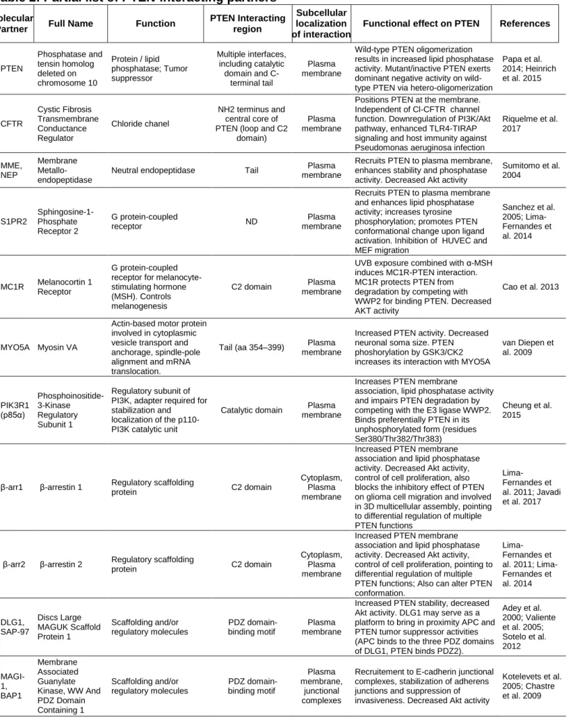
Table 2: Partial list of PTEN-interacting partners
Molecular
Partner Full Name Function
PTEN Interacting region
Subcellular localization of interaction
Functional effect on PTEN References
PTEN Phosphatase and tensin homolog deleted on chromosome 10 Protein / lipid phosphatase; Tumor suppressor Multiple interfaces, including catalytic domain and C-terminal tail Plasma membrane
Wild-type PTEN oligomerization results in increased lipid phosphatase activity. Mutant/inactive PTEN exerts dominant negative activity on wild-type PTEN via hetero-oligomerization
Papa et al. 2014; Heinrich et al. 2015 CFTR Cystic Fibrosis Transmembrane Conductance Regulator Chloride chanel NH2 terminus and central core of PTEN (loop and C2
domain)
Plasma membrane
Positions PTEN at the membrane. Independent of Cl-CFTR channel function. Downregulation of PI3K/Akt pathway, enhanced TLR4-TIRAP signaling and host immunity against Pseudomonas aeruginosa infection
Riquelme et al. 2017 MME, NEP Membrane Metallo-endopeptidase
Neutral endopeptidase Tail membrane Plasma
Recruits PTEN to plasma membrane, enhances stability and phosphatase activity. Decreased Akt activity
Sumitomo et al. 2004 S1PR2 Sphingosine-1-Phosphate Receptor 2 G protein-coupled receptor ND Plasma membrane
Recruits PTEN to plasma membrane and enhances lipid phosphatase activity; increases tyrosine phosphorylation; promotes PTEN conformational change upon ligand activation. Inhibition of HUVEC and MEF migration Sanchez et al. 2005; Lima-Fernandes et al. 2014 MC1R Melanocortin 1 Receptor G protein-coupled receptor for melanocyte-stimulating hormone (MSH). Controls melanogenesis
C2 domain Plasma membrane
UVB exposure combined with α-MSH induces MC1R-PTEN interaction. MC1R protects PTEN from degradation by competing with WWP2 for binding PTEN. Decreased AKT activity
Cao et al. 2013
MYO5A Myosin VA
Actin-based motor protein involved in cytoplasmic vesicle transport and anchorage, spindle-pole alignment and mRNA translocation.
Tail (aa 354–399) Plasma membrane
Increased PTEN activity. Decreased neuronal soma size. PTEN phoshorylation by GSK3/CK2 increases its interaction with MYO5A
van Diepen et al. 2009 PIK3R1 (p85α) Phosphoinositide-3-Kinase Regulatory Subunit 1 Regulatory subunit of PI3K, adapter required for stabilization and
localization of the p110-PI3K catalytic unit
Catalytic domain Plasma membrane
Increases PTEN membrane
association, lipid phosphatase activity and impairs PTEN degradation by competing with the E3 ligase WWP2. Binds preferentially PTEN in its unphosphorylated form (residues Ser380/Thr382/Thr383)
Cheung et al. 2015
β-arr1 β-arrestin 1 Regulatory scaffolding
protein C2 domain
Cytoplasm, Plasma membrane
Increased PTEN membrane association and lipid phosphatase activity. Decreased Akt activity, control of cell proliferation, also blocks the inhibitory effect of PTEN on glioma cell migration and involved in 3D multicellular assembly, pointing to differential regulation of multiple PTEN functions
Lima-Fernandes et al. 2011; Javadi et al. 2017
β-arr2 β-arrestin 2 Regulatory scaffolding
protein C2 domain
Cytoplasm, Plasma membrane
Increased PTEN membrane association and lipid phosphatase activity. Decreased Akt activity, control of cell proliferation, pointing to differential regulation of multiple PTEN functions; Also can alter PTEN conformation. Lima-Fernandes et al. 2011; Lima-Fernandes et al. 2014 DLG1, SAP-97 Discs Large MAGUK Scaffold Protein 1 Scaffolding and/or regulatory molecules PDZ domain-binding motif Plasma membrane
Increased PTEN stability, decreased Akt activity. DLG1 may serve as a platform to bring in proximity APC and PTEN tumor suppressor activities (APC binds to the three PDZ domains of DLG1, PTEN binds PDZ2). Adey et al. 2000; Valiente et al. 2005; Sotelo et al. 2012 MAGI-1, BAP1 Membrane Associated Guanylate Kinase, WW And PDZ Domain Containing 1 Scaffolding and/or regulatory molecules PDZ domain-binding motif Plasma membrane, junctional complexes
Recruitement to E-cadherin junctional complexes, stabilization of adherens junctions and suppression of invasiveness. Decreased Akt activity
Kotelevets et al. 2005; Chastre et al. 2009
MAGI-2, SSCAM , and MAGI-3 Membrane Associated Guanylate Kinase, WW and PDZ Domain Containing 2; and PDZ Domain Containing 3 Scaffolding and/or regulatory molecules PDZ domain-binding motif, decreased interaction with PTEN phosphorylated at Thr382 Plasma membrane, junctional complexes
Increased PTEN protein stability; favors PTEN recruitment into high molecular weight molecular complexes and enhances PTEN-mediated down-regulation of PI3K/Akt pathway Wu et al. 2000; Tolkacheva et al. 2001; Valiente et al. 2005 MAST1 , 2 and 3 Microtubule Associated Serine Threonine Kinases 1, 2 and 3 Scaffolding and/or regulatory molecules, Ser/Thr protein kinases
PDZ domain-binding motif Plasma membrane, junctional complexes
Binding of PTEN to the PDZ domain of MAST2 (MAST205) facilitates its phosphorylation by this kinase
Valiente et al. 2005.
PARD3 , PAR-3
Par-3 Family Cell Polarity Regulator
Scaffolding molecule involved in asymmetrical cell division, tight junctions and cell polarization PDZ domain-binding motif Plasma membrane, junctional complexes
The 2nd PDZ domain of Par-3 binds to phosphatidylinositol (PI) lipid membranes, the 3rd binds PTEN. Epithelial cell polarization
Wu et al. 2007 SLC9A 3R1, NHERF 1, EBP50 SLC9A3 Regulator 1 Na+/H+ exchanger regulatory cofactor, interacts with and regulates various proteins including CFTR and G-protein coupled receptors
PDZ domain-binding motif, decreased interaction with PTEN phosphorylated at Ser380/Thr382/Thr 383 Plasma membrane
Assembly of a ternary complex between PTEN, NHERFs and PDGFR. Enhances PTEN stability, restricts activation of PI3K by PDGFR; inhibits Akt pathway.
Takahashi et al. 2006 MAN2C 1 Cytosolic α-mannosidase 2C1
Catabolic enzyme for the breakdown of free oligosaccharides
ND Cytoplasm
Prevents PTEN recruitment to the cell membrane, Inhibits lipid phosphatase activity He et al. 2011 PARK7 , DJ1 Parkinsonism Associated Deglycase Peptidase ND Cytoplasm
Oxidized DJ1 Inhibits PTEN activity. High glucose increases PTEN/DJ1 interaction Kim et al. 2009; Das et al. 2011 BMI1 Proto-Oncogene, Polycomb Ring Finger Component of polycomb group complex 1 (PRC1) that modulates gene expression through epigenetic/ chromatin remodeling.
C2 domain
(186-286) Nucleus
Sequestering PTEN in the nucleus decreases PTEN's ability to inhibit the PI3K-AKT pathway
Fan et al. 2009
IPO11 Importin-11
Nucleocytoplasmic transport of protein and RNA cargoes
Monoubiquinated PTEN
Nuclear translocation
Decreased degradation: nuclear translocation protects PTEN from cytoplasmic proteins that cause its degradation Chen et al. 2017 MVP Major Vault Protein Component of a multi-subunit ribonucleoprotein structures, might be involved in nucleo-cytoplasmic transport C2 domain, Ca2+ dependent Nuclear translocation
Nuclear transport of PTEN, increased PI3K/AKT pathway activity
Chung et al. 2005; Minaguchi et al. 2006 PPP1R 10, PNUTS Protein Phosphatase 1 Regulatory Subunit 10 Protein phosphatase 1
binding protein C2-domain Nucleus
Sequesters PTEN in the nucleus in an inactive state (no induction of rad51 and p53 expression), decreases PTEN's ability to inhibit the PI3K-AKT pathway
Kavela et al. 2013 PREX2 Phosphatidylinosi tol-3,4,5-Trisphosphate Dependent Rac Exchange Factor 2 Guanine-nucleotide exchange factors for Rac family small G proteins
C-term tail (phosphorylated),
Catalytic/ C2 domains
ND
Inhibits PTEN lipid phosphatase activity. Stimulation of cell growth, decrease of glucose uptake and insulin resistance
Fine et al. 2009; Hodakoski et al. 2014
Figure legends
Fig. 1. PTEN structure and function.
(a) The PTEN protein contains five functional domains: a short N-terminal PIP2-binding
domain (PIP2-BD), the catalytic phosphatase domain, a C2 membrane-targeting/scaffold domain, a regulatory C-terminal tail containing multiple phosphorylation sites and several PEST sequences, and a PDZ-binding motif (PDZ-BM). (b) Schematic representation of the PTEN/PI3K/AKT pathway. PI3Ks are activated by a wide range of stimuli, including receptor tyrosine kinase receptors (RTKs) and G protein-coupled receptors (GPCRs). PI(3,4,5)P3 is produced from PI(4,5)P2 through the action of PI3Ks. PI(3,4,5)P3 acts a a lipid signalling intermediate to promote the recruitment and activation of a subset of pleckstrin homology (PH) domain-containing proteins, including AKT. AKT subsequently phosphorylates downstream protein substrates, leading to increased cell survival, proliferation, migration and invasion. PTEN acts as a brake on the PI3K/AKT pathway via its lipid phosphtase activity, which removes the D3 phosphate from PI(3,4,5)P3 to produce PI(4,5)P2.
Fig. 2. Molecular mechanisms controlling PTEN expression and activity.
The diagram depicts the different molecular mechanisms involved in control of PTEN expression and function. Methylation of the promoter results in PTEN silencing and hypermethylation of the promoter provokes reduced PTEN expression in different cancers. PTEN transcription is also under the control of a variety of transcription factors that bind the PTEN promoter to positively (e.g., p53, Egr-1) or negatively (e.g., NF-B, c-Jun) regulate its expression. At the post-transcriptional level microRNAs (miRNA) negatively impact PTEN expression. By acting as decoys for PTEN-targeting miRNAs, competitive endogenous RNAs (ceRNAs) that present partial sequence homology to PTEN as well as PTEN pseudogene (PTENP1) mRNAs, act to enhance PTEN expression. At the post-translational level the PTEN protein is subject to regulation by post-translational modifications (PTMs) and partner proteins, which impact PTEN conformation, localization, stability and activity. The culmination of all these regulatory events results in the "functional dose" of PTEN in the cell and corresponding functional outputs.
Fig. 3. Post-translational modifications mediating PTEN regulation.
(a) The diagram shows post-translational modifications (PTMs) implicated in PTEN
regulation. ATM: ataxia telangiectasia kinase, CBP: CREB-binding protein, CK2: casein kinase 2, FAK: Focal adhesion kinase, GSK3: glycogen synthase 3, HDAC6: histone deacetylase 6, NEDD4: neural precursor cell expressed developmentally down-regulated protein 4, PCAF: p300/CBP-associated factor, histone acetyltransferase, Rak: Fyn Related Src family kinase, ROCK: RhoA-associated, coiled-coil containing protein kinase, SMYD2: SET and MYND domain containing 2, lysine methyltransferase, Src: protein tyrosine kinase, TNKS1/2: tankyrase1/2, poly(ADP-ribose) polymerase, Ubc9: SUMO E2 Conjugase. (b) The diagram shows the PTEN “phosphorylation switch” conformational model between "closed" and "open" states. Phosphorylation of the C-terminal tail residues Ser380, Thr382, Thr383 and Ser385 promotes a PTEN intramolecular interaction, resulting in the "closed" conformation. Dephosphorylation of the C-terminal tail phosphorylation cluster results in the loss of this intramolecular interaction and PTEN switches to an "open"
conformation that is targeted to the plasma membrane where its substrate PI(3,4,5)P3 resides.
Fig. 4. A PTEN biosensor to monitor conformational change in live cells.
(a) The schematic shows the Rluc-PTEN-YFP biosensor with PTEN sandwiched
between the energy donor Rluc and the energy acceptor YFP. The diagram illustrates how PTEN conformational rearrangement may lead to changes in BRET measurements, although the actual orientations of donor and acceptor proteins are not known. (b) The BRET ratio is calculated by the ratio of light emitted by YFP at 530nm and the light emitted by Rluc at 480nm (YFP/Rluc) in the presence of the Rluc substrate coelenterazine. (c) As the biosensor is unimolecular, BRET signals remain constant over a wide range of Rluc concentrations (theoretical values are presented here based on previous experimental data). This is particularly useful in situations where, for example, a mutant of PTEN is less stable, as a change in BRET will only reflect a conformational change. Background BRET is determined using a Rluc-PTEN fusion, lacking YFP acceptor. Specific “net” BRET is then calculated by subtracting this value (background "bystander" BRET) from that obtained with the biosensor Rluc-PTEN-YFP (shaded grey area). (d) A list of potential experimental modes in which the biosensor can be used. (e) Specific BRET values multiplied by 1000 to generate mBRET (milliBRET units). Wild-type PTEN biosensor establishes the basal BRET signal (black bar and dotted line). Conformational changes are detected by changes in BRET signal that can be either negative (Condition X) or positive (Condition Y) compared to basal BRET.
References
Adey NB, Huang L, Ormonde PA, Baumgard ML, Pero R, Byreddy DV, Tavtigian SV, Bartel PL. 2000. Threonine phosphorylation of the MMAC1/PTEN PDZ binding domain both inhibits and stimulates PDZ binding. Cancer Res 60: 35-37. Ahmed SF, Deb S, Paul I, Chatterjee A, Mandal T, Chatterjee U, Ghosh MK. 2012.
The chaperone-assisted E3 ligase C terminus of Hsc70-interacting protein (CHIP) targets PTEN for proteasomal degradation. J Biol Chem 287: 15996-16006.
Al-Khouri AM, Ma Y, Togo SH, Williams S, Mustelin T. 2005. Cooperative phosphorylation of the tumor suppressor phosphatase and tensin homologue (PTEN) by casein kinases and glycogen synthase kinase 3beta. J Biol Chem
280: 35195-35202.
Alimonti A, Carracedo A, Clohessy JG, Trotman LC, Nardella C, Egia A, Salmena L, Sampieri K, Haveman WJ, Brogi E et al. 2010. Subtle variations in Pten dose determine cancer susceptibility. Nature genetics 42: 454-458.
Bassi C, Ho J, Srikumar T, Dowling RJ, Gorrini C, Miller SJ, Mak TW, Neel BG, Raught B, Stambolic V. 2013. Nuclear PTEN controls DNA repair and sensitivity to genotoxic stress. Science 341: 395-399.
Bolduc D, Rahdar M, Tu-Sekine B, Sivakumaren SC, Raben D, Amzel LM, Devreotes P, Gabelli SB, Cole P. 2013. Phosphorylation-mediated PTEN conformational closure and deactivation revealed with protein semisynthesis. Elife 2: e00691. Cao J, Schulte J, Knight A, Leslie NR, Zagozdzon A, Bronson R, Manevich Y, Beeson
C, Neumann CA. 2009. Prdx1 inhibits tumorigenesis via regulating PTEN/AKT activity. Embo J 28: 1505-1517.
Cao J, Wan L, Hacker E, Dai X, Lenna S, Jimenez-Cervantes C, Wang Y, Leslie NR, Xu GX, Widlund HR et al. 2013. MC1R is a potent regulator of PTEN after UV exposure in melanocytes. Mol Cell 51: 409-422.
Chalhoub N, Baker SJ. 2009. PTEN and the PI3-kinase pathway in cancer. Annu Rev
Pathol 4: 127-150.
Chastre E, Abdessamad M, Kruglov A, Bruyneel E, Bracke M, Di Gioia Y, Beckerle MC, van Roy F, Kotelevets L. 2009. TRIP6, a novel molecular partner of the MAGI-1 scaffolding molecule, promotes invasiveness. Faseb J 23: 916-928. Chen JH, Zhang P, Chen WD, Li DD, Wu XQ, Deng R, Jiao L, Li X, Ji J, Feng GK et
al. 2015. ATM-mediated PTEN phosphorylation promotes PTEN nuclear translocation and autophagy in response to DNA-damaging agents in cancer cells. Autophagy 11: 239-252.
Chen M, Nowak DG, Narula N, Robinson B, Watrud K, Ambrico A, Herzka TM, Zeeman ME, Minderer M, Zheng W et al. 2017. The nuclear transport receptor Importin-11 is a tumor suppressor that maintains PTEN protein. J Cell Biol 216: 641-656. Cheung LW, Walkiewicz KW, Besong TM, Guo H, Hawke DH, Arold ST, Mills GB. 2015. Regulation of the PI3K pathway through a p85alpha monomer-homodimer equilibrium. Elife 4: e06866.
Choi BH, Pagano M, Dai W. 2014. Plk1 protein phosphorylates phosphatase and tensin homolog (PTEN) and regulates its mitotic activity during the cell cycle. J
Biol Chem 289: 14066-14074.
Chon HJ, Bae KJ, Lee Y, Kim J. 2015. The casein kinase 2 inhibitor, CX-4945, as an anti-cancer drug in treatment of human hematological malignancies. Frontiers