The chromosomal complements of multipronuclear
human zygotes resulting from intracytoplasmic sperm
injection
Ervin Macas
1, Bruno Imthurn, Marinella Rosselli
and Paul J.Keller
Department of Gynaecology and Obstetrics, Division of Endocrinology, University Hospital, Frauenklinikstrasse 10, 8091, Zurich, Switzerland
'To whom correspondence should be addressed
Implementation of intracytoplasmic sperm injection (ICSI) in human in-vitro fertilization (IVF) has highlighted the need for information about the risk of nuclear spindle damage caused by this procedure. For this purpose we studied the final products of oocyte meiosis at the first cleavage division of multipronuclear zygotes arising after ICSI, and compared the results with abnormally fertilized oocytes after conventional in-vitro insemination. Of 37 successfully analysed tripronuclear zygotes, 18 had three individual metaphases. Abnormal complements of 11 zygotes in this group indicated that non-disjunction occurred predominantly at the second meiotic division of the oocytes. Nine of the 37 tripronuclear zygotes exhibited two individual metaphases. Seven were abnormal and there were some indications that non-disjunction took place during oocyte meiosis. Of the 37 tripronuclear zygotes, 10 had a single metaphase and three showed an aneuploid number of chromosomes. The overall rate of aneuploidy among tripronuclear microinjected zygotes was 56.7%. In addition, seven zygotes with more than three pronuclei arising after ICSI displayed severely depleted chromosome complements. The incidence of non-disjunction in oocytes fertilized by conventional in-vitro insemination was signi-ficantly lower (20.0%, P < 0.01), since only four zygotes had an aneuploid number of chromosomes. Our findings suggest that ICSI might interfere with regular chromosome segregation at the second meiotic division of the oocytes. Key words: cytoskeleton/human chromosomes/intracytoplasmic
sperm injection/multipronuclear zygotes
Introduction
In the last decade multipronuclear eggs have been widely used in human in-vitro fertilization (IVF) for experimental purposes (Wentz et al, 1983; Van Blerkom et al, 1984; Kola et al., 1987). Cytogenetic evidence has shown that zygotes having three pronuclei usually arise through dispermic fertilization and at the first cleavage contain one maternal and two paternal genomes (Rudak et al., 1984; Angell et al, 1986). However, the main advantage in analysing the first cleavage division of tripronuclear zygotes is that the chromosomes of gametes
remain in discrete haploid clusters. If metaphase plates are suitably spread, potential errors occurring in female or male meiosis can be analysed more accurately (Rudak et al, 1984). Implementation of the intracytoplasmic sperm injection procedure (ICSI) in human IVF has provided a source of new material for the study of triploidy (Macas et al, 1996). Multipronuclear zygotes derived from ICSI are thought to be digynic in their origin, since they consist phenotypically of only one polar body and three pronuclei, probably resulting from the incorporation of the second polar body (Van Steirteghem et al., 1993). Cytogenetic analysis of the first cleavage division of these zygotes could therefore provide useful information on the regularity of distribution of maternal chromosomes at the second meiotic division. Thus, if the microinjection procedure in some way disturbs regular chromo-some segregation at the second meiotic division through deterioration of the spindle apparatus, this error could be detected in the metaphase at the first mitotic division of tripronuclear zygotes by analysis of the maternal and second polar body chromosomes. Accordingly, the present study was designed to obtain insight into the regularity of sister chromatid exchange at the final stage of the second meiotic division of oocytes microinjected with a single spermatozoon into the cytoplasm. Furthermore, we investigated in parallel the chromosome constitution of tripronuclear zygotes arising after IVF. Although these zygotes originated predominately through dispermy, they were included as a control group, because any differences in the number of chromosomally abnormal complements found among micro- and non-microinjected oocytes would be a basic prerequisite for determining the genetic significance of ICSI.
Materials and methods
Patients
Ninety-three IVF treatment cycles implementing ICSI were performed in our centre between March 1995 and April 1996 The indications for assisted fertilization were previous failure of normal fertilization by conventional IVF (n = 3) and severe male subfertility (n = 90). The female patients were aged 20-37 years (mean 34.3) for ICSI, and 28-39 years (mean 34.8) for IVF cycles.
Stimulation and oocyte preparation
Hormonal stimulation was performed as reported earlier (Macas etaL, 1996). At 36 h after human chorionic gonadotrophin (HCG) injection, oocytes with cumulus cells were collected from antral follicles by ultrasound-guided follicular aspiration. After a maturation culture interval of 3 h, oocyte-cumulus complexes were treated with hyaluronidase (60 IU, type VHI: Sigma, St Louis, MO, USA) in Earle's medium for 30 s. The remaining cumulus cells surrounding
Chromosomal complements of human zygotes
each oocyte were then removed with a finely drawn Pasteur pipette to allow visualization of ooplasm and polar bodies. The cumulus-free oocytes were washed twice in Ham F-10 medium supplemented with 10% patient serum, transferred to 0.1 ml of the same medium covered with paraffin oil (Sigma), and incubated at 37°C under 5% of CO2 in air pnor to microinjections.
Semen treatment
The sperm assessment was performed according to the recommenda-tions of the World Health Organization (1992). Semen of each patient was assessed on several occasions pnor to admission to the treatment cycle in order to check whether enough spermatozoa were present to perform ICSI. Semen was obtained by masturbation from the husbands of the patients 3—4 h after the oocyte recovery procedure and was prepared by use of discontinuous Percoll gradients (Per Wash: FertiPro N.V., Sint-Marten-Latem, Belgium) (Van Steirteghem et al., 1993). Pnor to microinjectioa, the sperm suspension was diluted with 10% polyvinylpyrrolidone solution (Sigma) to decrease the morality of spermatozoa and facilitate their capture into the injection needle (Van Steirteghem et al., 1993).
Microinjection procedure
Sperm microinjection needles (l.d., 5 (im) and holding pipettes (i d., 15 urn) used for the ICSI procedure were made by Cook Co. (K-MPIP-1045, K-HPIP-1045. William A. Cook, Brisbane, Australia). For the microinjection procedure, the oocytes with the first polar body were transferred into drops of Hepes buffered Earle's medium, covered with sterile paraffin oil and placed on a warm plate (37°Q of an inverted Nikon Diaphot microscope using Nomarski optics (Nikon, Tokyo, Japan). The microinjection procedure was performed as desenbed by Palermo et aL (1992) and Van Steirteghem et al (1993). In brief, the oocyte was immobilized by negative pressure exerted on the holding pipette The polar body was held at 12 or 6 o'clock and the injection of the spermatozoa was achieved by firmly pushing the injection pipette through the zona pellucida and deep into the oocyte cytoplasm at 3 o'clock. Negative pressure exerted on the microinjection needle was used to penetrate through the oolemma. A small amount of cytoplasm was aspirated into the micropipette and then released back into the oocyte along with the spermatozoon. After microinjection, the oocytes were nnsed twice in preequihbrated medium and cultured at 37°C in a humidified atmosphere with 5% CO2 in air. At 14-20 h after the sperm injection,
all oocytes with two or three polar bodies and two distinct pronuclei were left to develop into embryos and then replaced into the patient's uterus Each multipronucleated oocyte was re-examined under an inverted microscope using phase or interference optics to ascertain the exact number of polar bodies, Multipronucleated zygotes were then transferred to fresh Ham F-10 medium containing 1 |ig/ml of Colcemid (Gibco, Paisley, Scotland), incubated for 14-16 h and then submitted to cytogenetic analysis.
Cytogenetic analysis
The methods used for chromosome preparations were basically those described by Dyban (1983) with slight modifications In bnef, the multipronuclear oocytes were transferred to 1 ml of the precooled hypotonic solution of sodium citrate (0.9%) and left for 5-10 min. During this time fresh fixatives were prepared: fixative A, consisting of ethanol and acetic acid (3:1) and fixative B consisting of ethanol and 75% acetic acid (1:4). After hypotonic treatment the zygotes were placed on a 'grease-free' optical slide in the minimum quantity of hypotonic solution. Fixative A was carefully released drop-wise beside the zygotes Before fixative A evaporated, a small drop of fixative B was added The zygotes were then carefully observed
under the dissecting microscope to monitor the degree of cytoplasmic spreading. After drying for 24 h the slides were stained with 2% Giemsa for 20 mm, after which each analysable metaphase was photographed.
Results
A total of 1057 oocytes was collected (average number, 11.4 per cycle), of which 835 possessed a first polar body at the time of microinjection (78.9%). Of the microinjected oocytes, 84 were damaged (10.1 %) and a total of 517 (61.9%) achieved normal fertilization (2PN).
Abnormal fertilization occurred in 52 injected oocytes (6.2%) when three pronuclei were present, in 10 (1 2%) oocytes with more than three pronuclei, and in 41 (4.9%) oocytes with only one pronucleus. Of a total of 447 inseminated oocytes (average number, 9 9 per cycle), 24 (5.4%) multipronuclear zygotes were observed after conventional IVF.
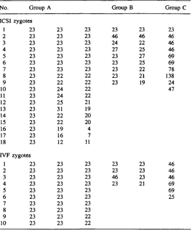
A total of 45 3PN zygotes resulting after the ICSI procedure were prepared for cytogenetic investigation, and chromosome analysis was possible on 37 of these (82.2%). The remaining eight (17.8%) were unsuitable for analysis because the chromo-somes either were inadequately spread or were lost during the preparation. The results of chromosomal investigations on 37 tnpronuclear zygotes derived after ICSI were arranged in three groups, A, B, C, depending on the number of individual metaphases found among analysed zygotes. A total of 18 zygotes in group A had three metaphases which were separated and spread in different planes of the cell. The numbers of chromosomes observed, according to the number of individual metaphases present, are summarized in Table I. Of these, 11 were aneuploid, having one normal and two abnormal haplotypes. In the majority of these abnormal zygotes (n = 7), one haploid was accompanied by two hypohaploid comple-ments. Only three zygotes have been karyotyped here:
20,X,-E.-2F; 22,X,-E; 23X 22.X.-D; 22,X,-D; 23X 22.X.-G; 22.X,-G; 23,X.
Chromosome constitution of the remaining four zygotes did not need to be karyotyped, because the presence of an aneuploid complement of second polar body chromosomes (n = 20) in one zygote, and two small metaphases with a total of 23 chromosomes (79—4; 16—7; 12—77) in another three, were sufficient grounds to suggest an error in oocyte meiosis. Approximately one-third of abnormal zygotes (n = 4) in group A displayed hypo-, hyper-, and haploid complements and three were successfully karyotyped: 21,X,-A,-B; 25,X,+A,+B; 23,X,
22.X.-E; 24.X, +E; 23X 22.X.-G; 24.X. + G, 23,X. The absolute
difference in the number of chromosomes between each of the corresponding hypo- and hyperhaploid complements was the same. This difference was due to chromosomes belonging to the same groups of the karyotype (Figure 1). The last zygote in this group exhibited an abnormal complement of second polar body chromosomes (n = 19), pointing again to an error in oocyte meiosis. Group B comprised nine zygotes with two individual metaphases and the majority of them exhibited one haploid and either hypo, or hyperhaploid complements
(n = 5). One particularity found in this group was the presence
Table I. Chromosome and No. ICSI 1 2 3 4 5 6 7 8 9 10 11 12 13 14 15 16 17 18 IVF l 2 3 4 5 6 7 8 9 10 IVF Group A zygotes 23 23 23 23 23 23 23 23 23 23 23 23 23 23 23 23 23 23 zygotes 23 23 23 23 23 23 23 23 23 23 analysis 23 23 23 23 23 23 23 22 22 24 24 25 31 22 22 19 16 12 23 23 23 23 23 23 23 23 23 23 of tnpronuclear zygotes 23 23 23 23 23 23 23 22 22 22 22 21 19 20 20 4 7 11 23 23 23 23 23 23 23 23 22 22 Group B 23 46 24 27 23 23 23 23 23 23 23 46 23 arising 23 46 22 25 27 25 22 21 19 23 23 23 21 after ICSI Group C 23 46 46 46 69 69 78 138 24 47 46 46 46 69 69 25
aneuploid complements of these zygotes, only partially suggest-ive of an error in oocyte meiosis. Ten zygotes in group C possessed a single metaphase, and their chromosome findings showed various levels of ploidy ranging from N to 6N. The total rate of aneuploidy found among microinjected zygotes was 56.7%. In addition, in 10 microinjected oocytes the evaluation of an exact number of pronuclei was difficult, because, apart from one normally shaped pronucleus, a few (three or four) small nuclei were superponated during the observation. Chromosomal analysis of seven of these zygotes revealed one haploid complement and several small metaphases with extreme hypohaploid numbers of chromosomes.
In the control group, as with the microinjected oocytes, the number of individual metaphases was not always in agreement with the number of pronuclei observed. However, in contrast to microinjected oocytes, their cytogenetical analysis revealed only four aneuploid zygotes having a single abnormal haplotype (20.0%, x2 = 7.2, P < 0.01). Table I shows the chromosome
complements of 20 tnpronuclear zygotes arising after IVF. The appearance of oocytes with more than three pronuclei was not observed in the group of in-vitro fertilized oocytes.
Discussion
Although there is much current debate about the potential risk of abnonnalities associated with the clinical application of ICSI, the possibility of oocyte cytoskeletal damage due to this procedure has to date been inadequately discussed
(Ng et al, 1993; De Jonge and Pierce, 1995; Patrizio, 1995). Our results suggest that ICSI could interfere with regular chromosome segregation at the second meiotic division, since significantly more abnormal complements were found in microinjected (56.7%) than in in-vitro fertilized oocytes (20.0%). These data also show that some aspects of ICSI might have harmful effects on the oocyte cytoskeleton, resulting in the production of chromosomally abnormal zygotes.
Effect of super-ovulation regimen
The finding of severe chromosomal defects in zygotes having more than three pronuclei {n = 7) demonstrates that the damaging effect of ICSI on the cytoskeletal system might sometimes be profound, with consequent dispersal of groups of chromosomes from the metaphase plates. To clarify this, we retrospectively evaluated all possibilities which could have had a marked influence on the organization of the meiotic spindle at the time of micromanipulation. One of the most likely possibilities was that some of the oocytes obtained after applying the super-ovulation regimen were of poor quality. These oocytes exhibited a normally expanded cumulus-corona cell complex and polar body, but the morphological characteristics of the ooplasm, which appeared partially dark and unevenly granular, signalled their poor quality. These types of oocytes were thought to represent an initial stage of the degeneration associated with follicular atresia. Spindle regression of such oocytes has therefore been strongly influenced by this degenerative process (Keefer, 1987). Moreover, it seems that the process of spindle regression was accelerated by micromanipulation itself, because none of these abnormal oocytes was found in the group of in-vitro fertilized oocytes.
Micro-manipulation procedures and their possible genetic consequences
An interesting observation in the present study was the finding of three zygotes with three pronuclei and two regular polar bodies, leading to the initial false impression that they resulted from dispermic fertilization. Cytogenetical analysis of these oocytes revealed monospermic fertilization, since one of three pronuclei contained a haploid number of chromosomes, and the sum of the other two was exactly 23 (19 + 4; 16 + 7; 12 + 11), suggesting that a small group of chromosomes split off from a maternal genome at the late anaphase or early telophase stage during the second meiotic division, forming one additional supernumerary pronucleus. Both abnormalities previously mentioned can be fairly easily recognized, if the zygotes are carefully examined following ICSI, because the presence of more than two irregularly shaped pronuclei would be highly suggestive of cytoskeletal damage and chromosomal displacement from maternal genomes. If, however, only one or two chromosomes segregated irregularly, with an insufficient amount of genetic material available for formation of the nuclear membrane, this anomaly would be practically impossible to visualize after ICSI. The findings of three 3PN zygotes with balanced hypo- and hyperhaploid complements in the present study
I j i
&
:
l t
Chromosomal complements of human zygotes
b
3 I I 2 i 4 I >
;
: j
• * • a • 4 • * G Gf
i f > \ i < >
t I.
G * YFigure 1. Karyotypc of a tnpronuclear zygote with balanced (a) hypohaploid (22,X,-E ), (b) hypcrhaploid (24.X.+E), and (c) haploid complements (23,Y).
might support this notion. The loss and gain of chromosomes in two haploid complements of these zygotes may have been associated with defects in the organization of the oocyte cytoskeleton at the time of sperm injection. However, in contrast to the previous cases, ICSI had a more subtle effect on the microtubule system, principally by disturbing the regular distribution of single chromosomes. The present results also suggest that a certain number of microinjected oocytes were chromosomally abnormal prior to microinjec-tion, and that their original chromosomal compositions were modulated after the application of ICSI. Thus, by interfering with the regular chromosome segregation of an egg with
21 chromosomes, ICSI probably caused the formation of a zygote having 20 and 22 maternal chromosomes (Figure 2); or, for example, by interfering with an egg with 26 chromosomes, resulting finally in 27 and 25 chromosomes. It is possible that the same mechanism was involved in the production of other such zygotes with unbalanced haplotypes. The further development of these zygotes would depend on the number and nature of chromosomes escaping regular division The majority, as seen in in-vivo and m-vitro conditions, would be eliminated through natural selection, since their chromosome constitution would not be compatible with survival (Plachot et al, 1988; Pellestor et al., 1994).
* 1
X 4 ft * - •.:•••&.•• . * &fi
Figure 2. Karyotype of a tripronuclear zygote with two hypohaploid complements' (a) 20,X,-E,-2F; (b) 22.X.-E, accompanied by their corresponding metaphase spreads (c).
Effect of calcium ions and hydrostatic pressure on the oocyte cytoskeleton
A number of factors can have a deleterious effect on the microtubular system. The effect of some of these factors on the oocyte meiotic spindle has been discussed in detail in one of our recent publications (Macas et ai, 1996). Rather than focus again on all of these mechanisms, we concentrated in the present study on two possibilities by which ICSI could influence the structure of the microtubule system, namely the effects of calcium ion concentration and hydrostatic pressure.
It has been known for many years that minimal concentrations of external calcium ions could disorganize the elements of the mitotic microtubular system (Inoue, 1981). For example, when microinjected in millimolar concentration into sea urchin eggs, calcium ions may rapidly depolymenze spindle microtubule(s), an effect that is limited to the region of the cell directly receiving the microinjection (Kiehart, 1979). Moreover, the presence of calcium ions at micromolar concentrations may rapidly depolymerize purified microtubules in vitro (Inoue, 1981). During ICSI, the gametes are usually contained in
Chromosomal complements of human zygotes
medium with a calcium concentration of approximately 2 mM (Edwards and Van Steirteghem, 1993). At the time of injection, a small amount of this calcium-rich medium (~5 \iM) is injected together with the spermatozoon, deep into the oocytes at some distance from the polar body.
It has been suggested, however, that the meiotic spindle may move away from the polar body after vigorous pipetting of the egg following corona radiata removal, and therefore is not necessarily associated with die polar body at the time of sperm injection (Cohen et al., 1994). This has been supported recently by the observation of Flaherty et al. (1995), who found in some unfertilized human oocytes that the sperm heads were located in close proximity to metaphase plates after insertion of the injection needle distal to the polar body region. In view of this, it is not difficult to imagine that insertion of the needle tip close to the spindle, and discharge of calcium, could sometimes lead to rapid disruption of elements of the microtubular system. On the other hand, it is well established that hydrostatic pressure is capable of altering the properties of the cytoskeletal system (Inoue, 1981). Changes in hydrostatic pressure can induce depolymerization of spindle microtubules in living cells, an effect that is also caused by low temperature (Salmon, 1975a). Furthermore, Salmon (1975b) found that brain microtubules can also be rapidly depolymerized in vitro by an increase in hydrostatic pressure. Because spindle micro-tubules of metaphase II oocytes might be exposed to changes of intracellular hydrostatic pressure during microinjection, during aspiration of the oolemma into the injection needle, we speculated that this mechanism might also sometimes con-tribute to spindle alterations causing irregular chromosome segregation during the completion of meiosis. At present, we do not know the precise degree of intracellular pressure caused by ICSI, but the fact that the microtubular system might be significantly influenced by this physical force must be taken seriously into consideration in future discussions about the negative effect of ICSI on the cytoskeletal system of the oocyte
In conclusion, the present study provides evidence that the ICSI procedure may contribute to die generation of chromosomal abnormalities at the second meiotic division. The alteration of the oocyte cytoskeleton potentially induced either through changes of intracellular hydrostatic pressure or local concentration of calcium ions during ICSI, might be responsible for abnormal segregation of chromosomes at die final stage of the second meiotic division.
Acknowledgements
The authors wish to thank Miss Manka Borsos for the excellent technical assistance and Miss Manan Widmer for photographic assistance.
Dyban, A P. (1983) An improved method for chromosome preparations from preimplantation embryos, oocytes or isolated blastomeres. Stain Technol, 58, 69-79
Edwards, R.G arid Van Steirteghem, A C . (1993) Intracytoplasmic sperm injections (ICSI) and human fertilisation does calcium hold the key to success? Hum. Reprod., 8, 988-989.
Flaherty, S P., Payne, D , Swann, N and Matthews, D (1995) Aetiology of failed and abnormal fertilisation after intracytoplasmic sperm injection
Hum. Reprod., 10, 2623-2629
Inoue, S. (1981) Cell division and the mitotic spindle J Cell BwL, 91, 131s-147s
Keefer, C.L. (1987) Morphology of unfertilised and fertilised oocytes In
Fredericks, Ch M , Paulson, JX> and DeChemey, A.H (eds), Foundations
of In Vitro Fertilisation Hemisphere Publishing Corporation, Washington,
p. 238
Kiehart, D.P (1979) Microinjection of Echinoderm eggs. I Apparatus and
procedures II Studies on the In Vivo Sensitivity of Spindle Microtubules to Calcium Ions and Evidence for a Vesicular Calcium-Sequestenng System. III. Evidence that Myosm Does Not Contribute to Force Production in Chromosome Movement Ph D Thesis, University of Pennsylvania, USA
Kola, I., Trounson, A., Dawson, G. and Rogers, P. (1987) Tnpronuclear human oocytes altered cleavage patterns and subsequent karyotypic analysis of embryos BwL Reprod., 37, 395-^01.
Macas, E., Imthurn, B , Rosselh, M and Keller, PJ. (19%) Chromosome analysis of single- and mulnpronucleated human zygotes proceeded after the intracytoplasmic sperm injection procedure / Assist Reprod. Genet,
13, 127-132
Ng, S Ch , Liow, S L., Sathananthan, H , Bongso, A and Ramam, S S (1993) Review, microinjection of human sperm directly mto human oocytes. J
Assist. Reprod. Genet, 10, 337-350
Palermo, G., Joris, H , Devroey, P. and Van Steirteghem, AC. (1992) Pregnancies after intracytoplamic injection of single spermatozoon in to an oocyte Lancet, 340, 17-18.
Patrizio, P (1995) Intracytoplasmic sperm injection (ICSI) potential genetic concerns Hum. Reprod., 10, 2520-2523
Plachot, M., Veiga, A , Montagut, J. et al (1988) Are clinical and biological IVF parameters correlated with chromosomal disorders in early life a multicentnc study Hum. Reprod., 3, 627-635.
Pellestor, F., Girardet, A , Andreo, B et al (1994) Relationship between morphology and chromosomal constitution m human preimplantation embryo. Mol Reprod. Dev, 39, 141-146
Rudak, E , Dor, J , Mashiach, Sh et al (1984) Chromosome analysis of mulrjpronuclear human oocytes fertilised in vitro Fertil Stenl, 41,538-545. Salmon, E D (1975a) Spindle microtubules thermodynamics of in vivo assembly and role in chromosome movement Ann. N ¥ Acad. Sci, 253, 383--W6
Salmon, E.D (1975b) Pressure-induced depolymensation of brain microtubules in vitro Science, 189, 884-886.
Van Blerkom, J , Henry, G and Porreco, R. (1984) Preimplantation human embryonic development from polypronuclear eggs after in vitro fertilisation
Fertil StenL, 41, 686-696.
Van Steirteghem, A C , Liu, J , Jons, H. et al (1993) Higher success rate by intracytoplasmic sperm injection than by subzonal insemination Report of a second senes of 300 consecutive treatment cycles. Hum. Reprod., 8, 1055-1060
Wentz, A.C , Repp, J E , Maxon, W S et al (1983) The problem of polyspermy in m vitro fertilisation Fertil Steal, 40, 748-754
World Health Organization (1992) WHO Laboratory Manual for the
Examination of Human Semen and Sperm—Cervical Mucus Interaction, 3rd
edn. Cambridge University Press, Cambridge, UK.
Received on May 7, 1996, accepted on August 14, 1996
References
Angell, R.R, Templeton, A A and Messinis, I.E (1986) Consequences of polyspermy in man Cytogenet Cell Genet, 42, 1-7
Cohen, J., Ahkaru, M , Munne, S and Palermo, G (1994) Micromanipulation in clinical management of fertility disorders Semin. Reprod. EndocnnoL, 12, 151-168.
De Jonge, ChJ. and Pierce, J (1995) Intracytoplasmic sperm injection - what kind of reproduction is being assisted? Hum. Reprod, 10, 2518-2528