HAL Id: hal-03064368
https://hal.archives-ouvertes.fr/hal-03064368
Submitted on 14 Dec 2020
HAL is a multi-disciplinary open access
archive for the deposit and dissemination of
sci-entific research documents, whether they are
pub-lished or not. The documents may come from
teaching and research institutions in France or
abroad, or from public or private research centers.
L’archive ouverte pluridisciplinaire HAL, est
destinée au dépôt et à la diffusion de documents
scientifiques de niveau recherche, publiés ou non,
émanant des établissements d’enseignement et de
recherche français ou étrangers, des laboratoires
publics ou privés.
Regulator of RTK Signaling during Drosophila
Development
Hélène Chanut-Delalande, Alain Jung, Magdalena Baer, Li Lin, Francois
Payre, Markus Affolter
To cite this version:
Hélène Chanut-Delalande, Alain Jung, Magdalena Baer, Li Lin, Francois Payre, et al.. The Hrs/Stam
Complex Acts as a Positive and Negative Regulator of RTK Signaling during Drosophila Development.
PLoS ONE, Public Library of Science, 2010, 5 (4), pp.e10245. �10.1371/journal.pone.0010245�.
�hal-03064368�
The Hrs/Stam Complex Acts as a Positive and Negative
Regulator of RTK Signaling during
Drosophila
Development
He´le`ne Chanut-Delalande1,2,3*., Alain C. Jung1.¤, Magdalena M. Baer1, Li Lin1, Franc¸ois Payre2,3, Markus Affolter1*
1 Biozentrum der Universita¨t Basel, Abteilung Zellbiologie, Basel, Switzerland, 2 Universite´ de Toulouse, UPS, Centre de Biologie du De´veloppement, Universite´ Paul Sabatier, Toulouse, France,3 CNRS, UMR5547, Centre de Biologie du De´veloppement, Toulouse, France
Abstract
Background:Endocytosis is a key regulatory step of diverse signalling pathways, including receptor tyrosine kinase (RTK) signalling. Hrs and Stam constitute the ESCRT-0 complex that controls the initial selection of ubiquitinated proteins, which will subsequently be degraded in lysosomes. It has been well established ex vivo and during Drosophila embryogenesis that Hrs promotes EGFR down regulation. We have recently isolated the first mutations of stam in flies and shown that Stam is required for air sac morphogenesis, a larval respiratory structure whose formation critically depends on finely tuned levels of FGFR activity. This suggest that Stam, putatively within the ESCRT-0 complex, modulates FGF signalling, a possibility that has not been examined in Drosophila yet.
Principal Findings:Here, we assessed the role of the Hrs/Stam complex in the regulation of signalling activity during Drosophila development. We show that stam and hrs are required for efficient FGFR signalling in the tracheal system, both during cell migration in the air sac primordium and during the formation of fine cytoplasmic extensions in terminal cells. We find that stam and hrs mutant cells display altered FGFR/Btl localisation, likely contributing to impaired signalling levels. Electron microscopy analyses indicate that endosome maturation is impaired at distinct steps by hrs and stam mutations. These somewhat unexpected results prompted us to further explore the function of stam and hrs in EGFR signalling. We show that while stam and hrs together downregulate EGFR signalling in the embryo, they are required for full activation of EGFR signalling during wing development.
Conclusions/Significance: Our study shows that the ESCRT-0 complex differentially regulates RTK signalling, either positively or negatively depending on tissues and developmental stages, further highlighting the importance of endocytosis in modulating signalling pathways during development.
Citation: Chanut-Delalande H, Jung AC, Baer MM, Lin L, Payre F, et al. (2010) The Hrs/Stam Complex Acts as a Positive and Negative Regulator of RTK Signaling during Drosophila Development. PLoS ONE 5(4): e10245. doi:10.1371/journal.pone.0010245
Editor: Christos Samakovlis, Stockholm University, Sweden
Received March 19, 2009; Accepted March 30, 2010; Published April 21, 2010
Copyright: ß 2010 Chanut-Delalande et al. This is an open-access article distributed under the terms of the Creative Commons Attribution License, which permits unrestricted use, distribution, and reproduction in any medium, provided the original author and source are credited.
Funding: Studies done in the laboratory of Markus Affolter are supported by research grants from the Swiss National Science Foundation, the Kantons Basel-Stadt and Basel-Land, and the European Commission via the FP6 Network of Excellence ‘‘Cells into organs’’. Experiments performed in Franc¸ois Payre laboratory are supported by Association pour la Recherche contre le Cancer nu1111 and ANR Programme Blanc Netoshape. The funders had no role in study design, data collection and analysis, decision to publish, or preparation of the manuscript.
Competing Interests: The authors have declared that no competing interests exist. * E-mail: [email protected] (HCD); [email protected] (MA)
.These authors contributed equally to this work.
¤ Current address: Laboratoire de Biologie Tumorale, Centre Re´gional de Lutte contre le Cancer, Strasbourg, France
Introduction
Receptor Tyrosine Kinase (RTK) signalling pathways regulate a wide range of biological processes and functions, including cell survival, growth, differentiation and migration [1]. Regulating levels of RTK signalling activity in time and space is an important issue since its deregulation leads to developmental defects and pathologies, including human cancers [2]. Besides ligand avail-ability, the levels of RTK signalling are strongly influenced by endocytic processes [3,4,5]. The Endosomal Sorting Complex Required for Transport (ESCRT) machinery comprises four protein complexes (ESCRT-0 to III) that sequentially act to target activated RTK to multivesicular bodies (MVB), en route to the
lysosome for degradation, thus promoting signalling downregula-tion. After endocytosis from the plasma membrane into early endosomes, ubiquitinated RTKs are bound by the ESCRT-0 complex composed of Hrs (Hepatocyte growth factor-regulated tyrosine kinase substrate) and Stam (Signal Transducing Adaptor Molecule) proteins [6,7]. The Hrs/Stam complex thus plays a key role in sorting cargo proteins either for degradation or recycling to the plasma membrane [8,9]. Indeed, Hrs selects ubiquitinated cargos for lysosomal degradation [7,10,11], recruiting components of other ESCRT complexes (I, II & III), which in turn send cargos to MVB, late endosomes and finally to lysosomes [12,13,14]. Results from numerous studies in cultured cells have shown that the Hrs/Stam complex is required for attenuation of Epidermal
Growth Factor Receptor (EFGR) signaling and suggested a general function in RTK down regulation. However, the Stam protein was reported to interact with deubiquitination enzymes [15,16], raising the possibility that it can also contribute, in interaction with or independently of Hrs, to disengagement of the cargo from the degradation pathway. Therefore, the full deciphering of the role of Hrs and Stam requires in vivo analysis of their respective function during the different processes that are regulated by signalling pathways during development.
Previous works in Drosophila have clearly show that Hrs indeed act to attenuate EGFR signalling during embryogenesis [17]. Embryonic hrs mutant cells display more EGFR signalling activity and accumulate activated receptors and ubiquitinated proteins in enlarged endosomes [17,18,19,20,21]. In addition, hrs mutant cells show a marked accumulation of other activated RTK, including PDGF/VEGF receptors, as well as other types of signalling receptors such as Patched and Smoothend (two members of the Hedgehog pathway), Notch, and Thickveins (a type-I serine-threonine kinase receptor for the TGF-b ligand Dpp) [19]. Strikingly, it has been shown that loss of hrs actually increases Dpp signalling in ovarian follicle cells and wing disc cells [19]. Furthermore, Hrs appears necessary for efficient JAK/STAT signalling during drosophila oogenesis [18]. Nevertheless, the consequences of enhanced signalling activity on developmental process due to the lack of hrs could be weak on development as shown during oogenesis where two RTK signalling pathways, EGFR and PDGF/VEGF are essential for border cell migration [19]. Despite an enhancement of these two pathways in hrs mutant border cells, the cell migration process is not affected. Jekely et al have shown that the total level of RTK activity is not critical for border cell migration but correct migration requires a restricted subcellular localisation of activity in the cell[22]. Those analyses brought new insights on mechanisms controlling signalling activity required for efficient cell migration. Altogether, these data reveal the complexity of the mechanisms relying on Hrs activity to regulate various signalling activities in vivo, suggesting that whereas required for down regulation of RTK, Hrs is also necessary for
efficient signalling of other receptors. Whether these differential activities involve the full ESCRT-0 complex, or correspond to Stam-independent function of Hrs, remains yet to be tested.
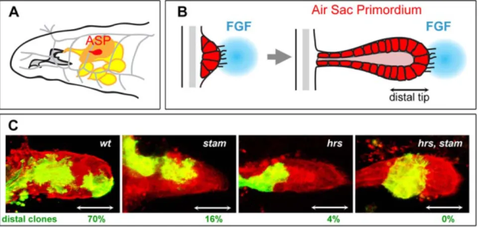
We have recently isolated the first mutations inactivating the Stam gene in Drosophila in a genetic screen aiming at identifying novel molecules involved in the formation of the larval tracheal system [23]. The Drosophila tracheal system develops during embryogenesis through the formation of branches arising from epithelial sacs containing approximately 80 cells in ten adjacent segments of the embryo. Through cell migration, rearrangement and intercalation, a complex network of interconnected epithelial tubes is formed to permit gas exchanges throughout the embryo. The development of the tracheal system has proven to be a model of choice to study branching morphogenesis and has been useful to genetically dissect the FGF pathway [24,25,26]. The FGF ligand, Branchless (Bnl), binds and activates the FGF receptor tyrosine kinase Breathless (Btl), which is expressed in all tracheal cells and triggers cell migration upon activation [27,28]. However, despite extensive genetic analyses, relatively little is known about how signalling is finely tuned during embryonic tracheal development. Indeed, genes linked to RTK signalling display pleiotropic functions during embryogenesis; the observed phenotypes, which affect several tissues as well as developmental processes, often hinder interpretations with regards to their role in trachea. Moreover, due to a strong maternal contribution to the egg during oogenesis, the function of many genes cannot be assessed during embryogenesis using zygotic loss-of-function analysis. Some of these problems can be circumvented by studying the development of tracheal structures formed at later stages, in larvae. During the third larval instar, tracheal cells of the transverse connective in the second thoracic segment bud out and form the primordium of a so-called dorsal air sac, or Air Sac Primordium (ASP) (Fig. 1A). The development of ASP relies on both cell proliferation and cell migration [29,30,31]. Genetic analyses of mosaic animals, using the MARCM (Mosaic Analysis with a Repressible Cell Marker) technique [32], have shown that during air sac morphogenesis, the FGFR pathway controls tracheal cell migration at the tip of the
Figure 1.stamandhrsare required for tracheal cell migration in the air sac primordium. A. Schematic representation of the anterior part of a Drosophila third instar larva. The air sac primordium (ASP) (red) buds from the transverse connective branch (in grey) and is attached to the wing imaginal disc (orange). The tracheal system is drawn in grey and imaginal discs other than the wing disc are colored in yellow. B. Model for the formation of the air sac primordium during larval development. Tracheal cells divide and migrate during ASP formation. Migration occurs under the control of the FGFR signalling pathway. Tracheal cells at the distal tip of the primordium are extending filapodia in the direction of the FGF ligand source (blue). Double arrow indicates the position of ASP distal tip. C. Migration behaviour of wild type, stam, hrs and stam hrs mutant cells. Confocal micrographs of the ASP of a Drosophila third instar larva are shown. All tracheal cells are labelled in red (RFP-moesin) and MARCM clones are labelled in green (mCD8-GFP). The FRT40A chromosome was used as a wild-type control. MARCM clones were induced for stam, hrs and stam, hrs. Scale bar: 15 mm. White double arrows indicate the position of ASP distal tip. Percentages of distal clones are indicated for each genotype tested. Note the strong effect of mutations in hrs and hrs, stam on cell migration. For each genotype, more than 20 clones were scored.
outgrowths (Fig. 1B), while EGFR signalling triggers cell survival and proliferation in the stalk [32,33]. Moreover, the Ras/MAPK cascade, commonly used to transduce RTK signals, is required for both tracheal cell migration and survival. However, the Pointed ETS transcription factor was found to be specifically required downstream of the Ras/MAPK cassette for cell migration [33].
To better understand the molecular mechanisms controlling air sac morphogenesis, we previously carried out a genetic mosaic screen and identified the stam gene as being essential for efficient tracheal cell migration in the ASP [23]. In the absence of Stam, mutant cells are unable to reach the distal tip of the ASP, showing that, like FGF signalling, Stam is required for efficient cell migration. These data therefore open the intriguing hypothesis that, during ASP formation, the activity of Stam, and putatively that of the ESCRT-0 complex, may be required for efficient FGF signalling. Here we explore this possibility and show that both Stam and Hrs act as positive regulators of FGF-mediated RTK activity to control cell migration in the ASP. Mutant cells lacking Stam, Hrs, or both, display abnormal accumulation of the FGF receptor Breathless (Btl) in enlarged vesicles, likely representing early endosomes. This is accompanied by a reduced expression of pointed, showing that the lack of ESCRT-0 activity specifically impairs efficient FGF signalling in ASP cells. In addition, we show that Stam and Hrs are required for the FGF-mediated formation of fine cytoplasmic extensions in terminal tracheal cells, further illustrating the importance of the ESCRT-0 complex in the positive regulation of FGF signalling. In contrast, we find that, like Hrs, Stam is required for down-regulation of EGFR signalling in the embryo. However, our data provide evidence to show that the ESCRT-0 complex is required for full activation of EGFR signalling during wing development. Taken together, our results indicate that, depending upon signalling pathways and the cellular context, the ESCRT-0 complex promotes either efficient activa-tion or down-regulaactiva-tion of RTK pathways, shedding novel light on the importance of the endocytic control of signalling activities in vivo.
Materials and Methods Drosophila stocks and genetics
The following fly strains were used: stam2L2896, stam2L3297[23] and hrsD28(gift from H. Bellen). The stam2L2896line harbours a non sense mutation in codon 6 suggesting that this stam allele represents a null allele. In addition, an Ala505to a Thr substitution was found in the stam2L2896line. The stam3297line also carried two mutations in the stam locus: a Phe428 to Ser mutation and a Tyr528-to-His substitution. This latter change may be the one affecting Stam protein function since Tyr528 is a favourable environment for
phosphorylation. The hrs, stam recombinants were generated via standard genetic procedures. As a wild type control, we used a FRT40A line. The following strains were used: UAS-stam to rescue the migration phenotype [23], UAS-Rab5-GFP (gift from M. Gonzalez Gaitan) to detect early endosomes and UAS-Btl-GFP to follow the localisation of the FGFR/Btl protein [29]. We also used the bnlP1 mutant [28] and the enhancer trap lines argosW11and pointed (l(3)7825) [34].
To obtain embryos lacking the maternal contribution of stam, progeny of adult females of the following genotype hsFLP/+; stam FRT40A/P[ovoD1], P[ovoD1]FRT40A received one heat pulse at 38uC, for 1 hr during first-instar larval development. For hrs and stam, hrs mutations, similar procedures were followed using females from genotypes hsFLP/+; hrs FRT40A/P[ovoD1],P[ovoD1]FRT40A and hsFLP/+; stam, hrs FRT40A/P[ovoD1], P[ovoD1]FRT40A, respectively.
Clonal analysis using the MARCM technique
Induction of MARCM clones in the tracheal system and in the air sac primordium. Clones in the air sacs were obtained according to procedures previously described in Cabernard and Affolter, (2005) and using the following MARCM lines: y,w,hsFlp; tubGal80, FRT40A/CyO; btlenhancer-mRFP1moe, btl-Gal4, UAS-mCD8-GFP/TM6C to visualize the clones in the ASP and y,w,hsFlp; tubGal80, FRT40A/CyO; btlenhancer-mRFP1moe, btl-Gal4/ TM6C to detect the clones in the ASP with either Rab5-GFP or Btl-GFP.
MARCM females were crossed to males carrying the mutation of interest. The progeny of these crosses was staged, and embryos that had reached 4–6 hr of development were heat-shocked for 1 h at 38uC. They were further grown at 25uC until the third instar larval stage. GFP-positive larvae were sorted using a GFP stereomicroscope (Leica MZFLIII) and wing imaginal discs were dissected [33]. For terminal cell analysis, GFP positive larvae were heat-shocked in a heat-block for 30 seconds at 70uC and mounted in glycerol.
Induction of clones in the wing imaginal discs and pupal wings. The following lines (gift from B. Bello) were used to induce MARCM clones:
N
y,w,hsFlp;FRT40A,tub-Gal80/CyoActinGFP; Tub-Gal4, UAS-mCD8-GFP/TM6,Tb,Hu to detect clones in the wing imaginal discs and pupal wingN
y,w,hsFlp;FRT40A,tub-Gal80/CyoActinGFP; Tub-Gal4/ TM6,Tb,Hu to visualise the clones in the wing imaginal discs with UAS-Rab5-GFP.MARCM females were crossed to mutant males and first instar larvae of the progeny received one pulse of heat shock at 38uC for 1 h.
Staining, electronic microscopy experiments and imaging
Immunostaining of dpERK in embryos lacking the maternal contribution of hrs and/or stam genes was performed using standard procedures (anti dpMAPK, Sigma, 1/500).
To visualise pointed and argos expression, lacZ staining was performed on wing imaginal discs or dissected pupal wings: Anti mouse Alexa 568 and 633 (1:500 Molecular Probes, Invitrogen) were used as secondary antibodies to detect the staining.
Confocal pictures were made using a Leica TCS SP2 microscope. Pictures from the entire ASP were made to detect the position of the clones. To visualise the localisation of Btl-GFP and Rab5-GFP, the pictures correspond to single section of the ASP. To count the branching points of terminal cells, pictures of all branches were done.
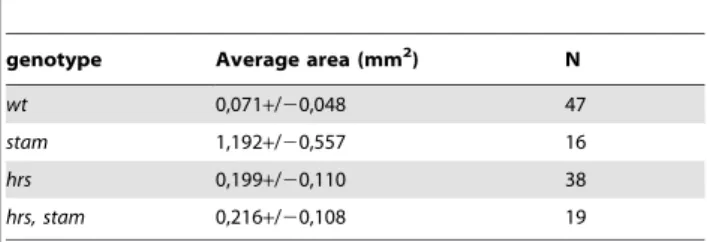
Garland cell preparation was performed according to proce-dures described previously [17,35]. Sections were photographed with a Leo 510 (Zeiss) microscope (Zentrum Mikroskopie der Universita¨t Basel). Endosome areas were measured using ImageJ software for each genotype.
Results
The Hrs/Stam complex is essential for FGF-mediated tracheal cell migration in the air sac primordium (ASP)
Using the MARCM technique, we have recently shown [23] that the loss of Stam impairs the migration of air sac primordium (ASP) cells; with only 16% of mutant clones detected at the ASP tip (Fig. 1C). Interestingly, migration of cells at the tip of the air sac primordium requires FGFR signalling activity, since mutant cells
lacking components of the pathway (like btl mutant clones, for instance) are unable to reach the distal tip of the ASP.
Since Stam is known to be a direct partner of Hrs within the ESCRT-0 complex [6], we tested a putative role of hrs during tracheal cell migration in the ASP using the hrsD28null allele. Cell migration is strongly affected in hrs mutants, as only 4% of hrs clones are able to colonize the distal tip of the ASP (Fig. 1C). Cells lacking both hrs and stam exhibited a severe migration phenotype; double mutant clones were never detected at the distal tip of the ASP (Fig. 1C). Such a strong migration phenotype was previously only observed following the inactivation of genes encoding essential components of the FGFR signalling pathway [33]. In addition to the failure to locate at the tip of outgrowing ASP, hrs, stam double mutant clones often exhibited a reduced number of cells compared to stam or hrs single mutant clones suggesting that the lack of ESCRT-0 function also affects cell proliferation to a certain extent. Taken together, these results show that hrs and stam are required for the FGF-dependent migration of tracheal cells during larval development.
Btl/FGFR accumulates in enlarged vesicles in stam and hrs mutant air sac cells
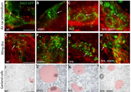
Since the ESCRT-0 complex is known to promote the endocytic degradation of many signalling receptors [17,19], we analyzed the consequences of the loss of Stam and Hrs on endosome formation in ASP cells. We induced MARCM clones carrying either stam single mutations or hrs, stam double mutations. Clones were visualised via the expression of the fusion protein Rab5-GFP, Rab5 being a marker for early endosomes [36]. In wild type cells, Rab5-GFP-positive early endosomes were detected as small dot-like structures in the cytoplasm (Fig. 2A). In stam and hrs single mutant, as well as in stam, hrs double mutant cells,
Rab5-GFP accumulates in larger dots (Fig. 2B–D), suggesting that early endosomes were enlarged in the absence of ESCRT-0 activity. Interestingly, this early endosome size defect was not restricted to tracheal cells, since we observed a similar phenotype in wing imaginal cells lacking stam, hrs or both (Fig. 2E–H). To better characterise the role of ESCRT 0 complex, we performed transmission electron microscopy (TEM) analyses of Garland cells, which are known to have a rapid rate of fluid phase endocytosis [35]. HRP (Horse Radish Peroxidase) uptake experiments allow the detection of the lumen and endosome membranes. Ultrastructural analysis of wild-type Garland cells has demonstrated that endosomes undergo maturation via a step of membrane invagination [17] (Fig. 2I). We measure the endosome area and we showed that in wild type Garland cells, the average area is 0,071+/20,048 mm2 (Table 1). Consistently with a previous report [17], we observed that the endosomes of Garland cells lacking hrs are 2,7 fold larger than wild type ones (average area 0,199+/20,110 mm2) (p,0,005) and do not exhibit HRP staining in the lumen, a phenotype likely due to defective membrane invagination (Fig. 2K and Table 1). Our analyses further revealed that stam mutant cells displayed even larger endosomes (1,192+/20,557 mm2, 16,6 fold compared to wild type) (p,0,005) than those observed in the absence of hrs (Fig. 2J and Table 1). Garland cells lacking simultaneously hrs and stam mutations exhibited endosomes of the size of those observed in hrs mutant cells (0,216+/20,108 mm2) (3 fold larger than wild type (p,0,005) (Fig. 2L and Table 1), suggesting that while hrs and stam are both involved in endosome maturation, they may have partly distinct roles during this process [7].
Having shown that the absence of the ESCRT-0 complex leads to endosomal defects in different tissues, we next examined
Figure 2.hrsandstammutants display enlarged early endosomes inDrosophila. A–D. Close-up pictures of wild type, stam, hrs and hrs, stam mutant cells induced with the MARCM system in the dorsal ASP. Scale bar: 15 mm. Mutant clones are visualised by the expression of Rab5-GFP to detect early endosomes. Note the large size of early endosomes in stam (B), hrs (C) and hrs, stam (D) mutant cells compared to FRT40A cells used as a wild type control (A). The tracheal cells were labeled with RFP-moesin (red). Arrows indicate early endosomes stained with GFP. Pictures shown are single scan sections. E–H. MARCM clones mutant for hrs, stam and hrs, stam in the wing imaginal disc of third instar larvae. Scale bar: 15 mm. Mutant cells express the Rab5-GFP fusion protein. As in the ASP, early endosomes are enlarged in stam (F), hrs (G) and hrs, stam (H) mutant cells compared to FRT40A cells used as a control (E). The cells were detected with phalloidin to follow F-actin and the shape of the cells. Arrows indicate early endosomes stained with GFP. Pictures shown are single scan sections. I–L. HRP uptake experiment in Garland cells of wild-type, stam, hrs and hrs, stam larvae. I. In wild-type Garland cells, many small endosomes are observed (highlighted in light red). Scale bar equals 460 nm. Endosomes from stam and hrs mutant larvae are dramatically enlarged (J–L), especially in stam mutants (J), compared to FRT40A larvae (I).
whether it affected the distribution of the FGF-R Btl in ASP cells. We performed mosaic analyses using a Btl-GFP fusion protein, specifically expressed in clones of control, stam or hrs mutant cells. In wild type cells, Btl-GFP mostly accumulated at the cell membrane (Fig. 3A, white arrow) and also as fine dotted structures in the cytoplasm, often lined up along the cell membrane (Fig. 3A,
yellow arrow). In hrs and stam mutant clones, Btl-GFP was no longer detected at the membrane but was found in severely enlarged cytoplasmic vesicles (Fig. 3B–C). These data show that the lack of ESCRT-0 activity in tracheal cells of the ASP leads to the accumulation of the Btl receptor in enlarged vesicles, likely corresponding to the abnormal early endosomes revealed by Rab-5 staining.
Stam and Hrs positively regulate FGFR activity in tracheal cells
Together with a reduced migration at the distal tip of the ASP, the abnormal accumulation of Btl in cells lacking Stam or Hrs suggests that ESCRT-0 regulate the FGF pathway. However, both the lack and the excess of FGF activity blocks tracheal cell migration. To determine a possible influence of the ESCRT-0 on FGF activity in ASP cells, we analysed the expression of the pointed (pnt) gene, an early target of the pathway that is specifically required for tracheal cell migration in the ASP [33]. Using a pnt-lacZ reporter line, we detected accordingly a staining in the distal Table 1. Average area of endosomes analyzed in Garland
cells from wt, stam, hrs and hrs, stam larvae.
genotype Average area (mm2) N
wt 0,071+/20,048 47
stam 1,192+/20,557 16 hrs 0,199+/20,110 38 hrs, stam 0,216+/20,108 19 doi:10.1371/journal.pone.0010245.t001
Figure 3.stamis required to properly localise FGFR/Btl and fully activate FGFR signalling. A–C. Localisation of Btl in wild type and mutant tracheal cells. High magnification pictures of wild type (A), stam (B) and hrs (C) MARCM mutant cells in the ASP. Scale bar equals 15 mm. Mutant cells were visualised via the expression of UAS-btl-GFP (green). The tracheal cells were visualised with RFP-moesin (red). White arrow indicates the presence of Btl at the cell membrane in wild type cells while yellow arrows indicate Btl as dotted structures. The dotted structures are dramatically enlarged in stam and hrs mutant cells as compared to wild type cells. D–F. pointed expression in wild type and stam mutant tracheal cells. Scale bars: 15 mm. pointed expression is restricted to the distal part, the tip, of a wild type ASP (D–D0). In a stam mutant clone located at the proximal part of the ASP, pointed expression is unchanged (E–E0). When the stam clone is positioned close to or at the distal tip of the ASP: pointed expression is lost (F–F0). Dotted lines showed the position of the stam mutant cells in the ASP (E9, F9). Arrows indicate the distal tip of the ASP (E0, F0).
doi:10.1371/journal.pone.0010245.g003
part of the ASP in wild-type cells (Fig. 3D–D0). This is in agreement with the view that tracheal cells of the ASP distal tip actively migrate under the influence of the FGFR signalling. In stam mutant clones located in the proximal part of the ASP, we did not detected modification of the pointed staining, not even in cells at the most distal part of the clones, indicating that the expression of pnt is not upregulated under conditions in which activated Btl accumulates in enlarged endosomes (Fig. 3E–E0). Furthermore, when stam mutant cells were located close to the distal tip of the ASP, they did not express pnt despite the fact that they are in the vicinity to the source of the FGF ligand (Fig. 3F–F0). Hence these results are in line with stam being required for pnt activation and thus efficient FGFR signalling in the migrating cells of the ASP. Due to their absence at the tip of the ASP, we could not use however pnt expression as readout for FGFR activity in hrs single mutant and hrs, stam double mutant backgrounds.
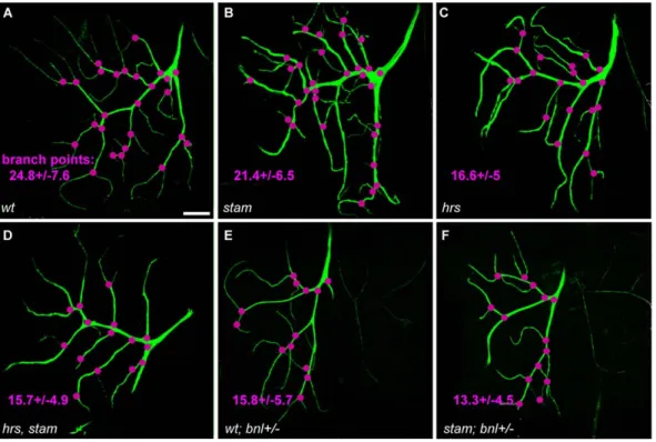
To further characterize the function of the Stam/Hrs complex in the regulation of FGFR signalling, we therefore analysed the formation of fine cytoplasmic extensions by terminal tracheal cells (TCs), a process which is also known to rely on Btl function. TC extensions are the finest branches of the tracheal system, which directly provide oxygen to individual cells. During larval development, the number of TC branches changes according to the needs of tissues for oxygen and this process is controlled, in a dose-dependent manner, by levels of FGFR signalling [37,38,39]. We thus analysed whether stam and hrs influenced the formation of TC extensions. We generated MARCM TC and counted the number of branch points formed by these mutant cells, taking into account both thick (primary) and thin (secondary and tertiary) projections [37]. To reliably compare the different genetic backgrounds, we focused on dorsal TCs. In wild type TCs, the
average number of branching points was 24.867.6 (Fig. 4A and table 2). In stam mutant cells, this number decreased to 21.466.5 (p,0.001) (Fig. 4B). hrs mutant and hrs, stam double mutant cells displayed a much reduced capacity to sprout since we observed 16.665.0 (p,0.001) and 15.764.9 (p,0.001) branching points, respectively (Fig. 4C–D and table 2). These results thus indicate that stam and hrs are necessary for efficient TC sprouting.
As previously described [28], lowering levels of FGF activity leads to a similar reduction in the number of TC extensions. For example, the strong bnlP1 allele of branchless in heterozygous condition leads a clear decrease in the number of branching points (15.865.7), when compared to wild-type controls (p,0.001) (Fig. 4E–F and table 2). In terminal cells heterozygous for bnl, the absence of stam further increased the observed defects, with 13.364.5 branching points per cell. Although we recovered very few larvae carrying MARCM clones in the tracheal system for both hrs single and hrs, stam double mutants, these mutations also enhanced the phenotype resulting from lowering levels of FGF signalling.
Taken together, our results show that the function of the ESCRT-0 complex is required for efficient FGFR signalling in different aspects of tracheal cell development, including migration at the distal tip of the ASP and the formation of cytoplasmic projections in terminal cells.
The ESCRT-0 complex displays opposite roles in the regulation of EGFR signalling throughout development
While our results show that the ESCRT-0 complex can positively regulate RTK activity during development, Hrs is required for down-regulating the EGFR activity in embryos [17]. We therefore tested whether stam acts similarly to hrs regarding
Figure 4.stamandhrsare required for the efficient formation of fine cytoplasmic extensions in tracheal terminal cells and interact with FGFR signalling. Confocal micrographs of MARCM wild type and mutant dorsal terminal cells. Scale bar: 50 mm. The clones are visualised using UAS-mCD8-GFP. FRT40A line was used as a control (A). MARCM clones were induced for stam (B), hrs (C), hrs, stam (D). (E–F). The FRT40A and stam MARCM terminal cells with altered FGFR signalling. bnl+/2 corresponds to a larvae heterozygous for bnl. Branch points (visualised in pink) were counted for mutant clonal cells for each genotype. The average number of branch points is given for each genotype.
EGFR signalling during embryogenesis. We used an antibody specific of the double phosphorylated form of ERK (dp-ERK) since EGFR signalling triggers ERK/MAPK phosphorylation in the early embryo [40]. In order to circumvent the maternal contribution of stam and hrs, we obtained embryos from germ-line clones lacking maternal and zygotic expression of stam, hrs, or both, and compared the results to wild type embryos.
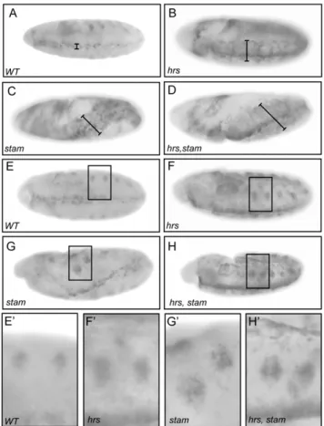
During embryogenesis, the EGFR pathway is essential to pattern the neuroectoderm. dpERK is observed in three to four rows of cells, on each side along the ventral midline in wild-type embryos, when the Spitz ligand activates the EGFR pathway (Fig. 5A). Compared to wild-type embryos, we detected an expansion of dpERK staining in the ventral ectoderm in embryos lacking the maternal contribution for hrs (Fig. 5B) in agreement with the study of Lloyd et al (2002). In embryos lacking stam and those lacking simultaneously stam and hrs (Fig. 5C–D), we observed a similar enhancement of dpERK staining, attesting for elevated levels of EGFR signalling. Moreover, mutant embryos showed morphological defects due to aberrant folding of the embryo during germ band elongation, defects that are likely due to the expanded EGFR activation. We also monitored dpERK staining in stage 10 embryos, when the EGFR signalling pathway becomes required for the invagination of tracheal cells and the formation of tracheal placodes. At this stage of embryonic development, dpERK staining is detected in tracheal pits [40]. When compared to wild type, embryos lacking hrs or stam, or both stam and hrs display a clear expansion of the dpERK staining around the tracheal pits for all mutant conditions (Fig. 5E–H9). Therefore, the data indicate that Stam and Hrs play similar role in the attenuation of EGFR signalling throughout embryonic development.
These results also suggested that the ESCRT-0 complex acts to down-regulate EGFR signalling throughout development. To further test this conclusion, we then extended our analysis to adult wing development, for which the role of EGFR regulatory cascade is well established [41]. To analyse a putative influence of hrs and stam on the regulation of EGFR signalling during wing patterning, we followed the expression of argos, a target gene of the EGFR pathway [42]. The vein differentiation process that initiates during the 3rdlarval instar proceeds into the pupal stage and, during the entire process, argos is activated in vein tissues (Fig. 6A). Cells that lack stam displayed a strong reduction of the argos expression, when clones of mutant cells were located in the vein tissue (Fig. 6B) Although stam2L2896, hrs mutant clones are tiny and do not permit to analyse argos expression, we also found a strong reduction of
argos in vein cells carrying simultaneously the stam2L3297and hrsD28 mutations (Fig. 6C). In contrast to its role in EFGR attenuation in embryonic tissue, these results thus support that the Hrs/Stam complex is required to efficiently activate EGFR signalling in the developing wing veins.
Discussion
In this study, we have analyzed roles of the endocytic ESCRT-0 complex, in vivo, through the genetic ablation of its two main components Hrs and Stam, at different steps of the Drosophila development. We show that the loss of Hrs and Stam produce similar phenotypes in all tissues examined, indicating that these proteins mainly function as a single complex throughout development. Our results indicate that Stam and Hrs participate in the efficient activation of the FGFR pathway in larval tracheal cells. In contrast, the ESCRT-0 acts to attenuate the extent of EGFR activation during embryogenesis. However, we find that Stam and Hrs are also required for the expression of EGFR targets in the adult wing. These data therefore support the view that the ESCRT-0 complex mediates directly or indirectly both positive and negative regulation of RTK signaling, depending on developmental contexts.
Table 2. Number of branching points of hrs and stam MARCM terminal cell clones in different FGFR signalling activity backgrounds.
Genotype Branching points N
wt 24.867.6 209 stam 21.466.5 207 hrs 16.665.0 200 hrs, stam 15.764.9 99 wt; bnl2/+ 15.865.7 86 stam; bnl2/+ 13.364.5 103 hrs; bnl2/+ 14.664.2 66 hrs, stam; bnl2/+ 11.264.7 23 doi:10.1371/journal.pone.0010245.t002
Figure 5.stamandhrsdownregulate EGFR signalling activity during embryogenesis. Diphospho-ERK (dpMAPK) antibody staining in wild type, hrs, stam and hrs, stam maternal-zygotic loss-of-function embryos (A–H9). At stage 10, hrs, stam and hrs, stam mutants display enhancement of the dp-ERK staining (B–D), due to an expansion of their ventral fate (see black bar length) compared to a wild type embryo (A). Mutant embryos also display an expansion of the staining in tracheal placodes (F–H) compared to wild type (E). Close up views of the dp-ERK staining in the tracheal placodes (E9–H9).
doi:10.1371/journal.pone.0010245.g005
stam and hrs are positive regulators of FGFR signaling
Tracheal cell migration in the air sac primordium (ASP) is controlled by FGFR signaling [29,33]. Indeed, tracheal cells are not able to populate the distal tip of the ASP when FGF signal reception or transmission is defective (in btl mutant cells, for instance) or when cells experience too much FGF signaling [33]. We show here that stam and hrs are required for tracheal cells to efficiently colonize the tip of the ASP. Consistent with a role of Hrs and Stam in endosomal cargo sorting, we found that Btl/FGFR receptors accumulate in enlarged cytoplasmic vesicles in stam and hrs mutant cells, whereas they accumulate at the plasma membrane in wild type cells. Rab5-positive vesicles corresponding to early endosomes were also found to be enlarged in the absence of hrs and stam, suggesting that Btl/FGFR accumulates in altered early endosomes in mutant cells.
To test whether FGFR signaling is either enhanced or diminished in the absence of stam, we monitored the expression of a pnt-lacZ reporter gene and found that stam is required for its transcriptional activation, and hence most probably for high levels of FGFR signaling. These data show that the total level of FGF activity in tracheal cells is essential for efficient cell migration, which is in contrast with border cell migration, where localized signaling activity in cell is necessary for migration and not general level [22]. In order to further investigate the role of Hrs and the Hrs/Stam complex in FGFR signaling, we analyzed the effect of these mutations in the formation of projections by tracheal TCs, another cellular process which requires FGFR signaling. It has previously been shown that the number of branches formed by terminal cells increases with increasing FGFR signaling activity [39]. We found that stam and hrs are required in terminal cells to form branches efficiently. In addition, hrs and stam genetically interact with FGFR signaling during this process, to promote the formation of terminal cell projections. Therefore, both in TC and in ASP cells, our data demonstrate that hrs and stam are positive
regulators of FGFR signaling in the Drosophila larval tracheal system.
Interestingly, it has been previously reported that awd/Nm23 (abnormal wing disc) and shibire/dynamin down-regulate FGF signaling during embryonic tracheal development through endocytosis of Btl/FGFR [43]. It is therefore possible that the ESCRT-0 complex might also act to attenuate FGFR signaling during tracheal development in embryos, albeit the early lethality of embryos lacking Stam or Hrs prevents this hypothesis to be tested experimentally. Alternatively, different machineries regulating endocytosis and eventual endosome sorting might display antagonistic activity toward FGF signaling. For example, work in mammalian cell culture has shown that Rab5, a key component of the early endocytic pathway, colocalizes with activated FGFRs and that the depletion of rab5 decreases FGFR signaling [44]. Finally, it has been shown that two different FGF ligands, KGF (Keratinocyte Growth Factor) and FGF10, can target the FGFR to the degradation pathway or the recycling pathway, respectively [45]. These results show that early endocytosis is critical for fine tuning of FGFR signaling activity; depending on the context, different endocytic mechanisms might differentially regulate FGFR signaling.
Dual function of stam and hrs in EGFR signaling
The role of the ESCRT-0 complex in regulating the degradation of EGFR receptors has been well studied in cultured cells [10,46]. Functional assays using RNA interference have shown that Hrs and Stam trigger the sorting of ubiquitinated receptors and downregulate EGFR signaling activity [10]. During Drosophila embryogenesis, hrs was also well established as a negative regulator of signaling RTK pathways such as EGFR and Torso [17]. Our recent isolation of the first mutations of the stam gene in flies [23] allowed us to show, here, that stam attenuates the EGFR activity during gastrulation and formation of tracheal placodes,
Figure 6.argosexpression is strongly reduced instam,hrsandhrs, stammutant cells in pupal wings. argos expression was analysed in wild type (A), stam2L2896(B) and hrs, stam (C) MARCM clones visualized with mCD8-GFP (green) in pupal wings (24–30 hrs APF) using a argos-LacZ
enhancer trap line. White dotted squares (in A–C) indicate the position of close-up pictures. White doted lines (in A9–C9) indicated the position of the clones in the close-up pictures. The argos staining (red) is absent in stam and hrs, stam mutant cells.
suggesting that Stam and Hrs likely function within a common ESCRT-0 complex during embryogenesis.
Having shown that EGFR signaling is strongly upregulated in the absence of hrs and stam in the embryo, we tested whether EGFR signaling was also over-activated in the pupal wing upon reduction of stam and hrs activity. Assuming that argos expression in veins represents a faithfull readout of EGFR signaling, we observed instead a loss of EGFR activity in the absence of hrs and stam at the pupal stage. Supporting our conclusions, it was recently reported that hrs is also required for EGFR signaling at earlier stages, during the patterning of larval discs [20]. Therefore, these data provide independent evidence that, contrary to what was observed during embryogenesis, hrs and stam are necessary for high level EGFR signaling during wing development, from larval to pupal stages.
Concluding remarks
We show that stam and hrs modulate RTK signalling activities either positively or negatively according to the tissue and the developmental stage. hrs and stam are essential for efficient activation of the FGFR signalling in the tracheal system, e.g., they may promote recycling of the FGFR/Btl receptor to the plasma membrane. In addition, hrs and stam contribute to either enhance or attenuate EGFR signalling activity, depending on the tissue context. Similar to what has been proposed for FGFR signalling, positive versus negative modulation of EGFR activity may depend on the ligand which activates the receptors. Whereas Spitz is the only known EGFR ligand acting during early embryogenesis, Keren could be an additional ligand with Spitz to activate EGFR during vein differentiation [47,48]. Another possible explanation for the tissue-specific antagonistic activities of the ESCRT-0 complex concerns the phosphorylation state of its main compo-nents. Depending of the amino acid residues which are phosphorylated in the Hrs and Stam proteins, their influence on
the modulation of signalling activity vary [49]. Modulation of the phosphorylation code of Stam and Hrs between tissues may turn those molecules into pathway-specific activators or repressors.
Our compared analysis of the individual roles of Hrs and Stam in vivo strongly support that these two proteins act together to modulate RTK signalling, i.e., within the ESCRT-0 complex. Regarding endosome maturation in Garland cells, our analyses show, however, that hrs and stam mutant cells do not display identical phenotype in this process; endosomes being much larger in the absence of stam than in the absence of hrs (or in the absence of both stam and hrs). Assuming that available mutations for both Stam and Hrs correspond to null alleles, which is fully compatible with their molecular nature, it remains possible that these different roles on endosome maturation may be linked to distinct protein partners of Stam and Hrs. The availability of stam loss of function mutations and rescuing transgenes, provide novel tools that will allow further dissection of the Stam protein function in vivo, and ultimately help in gaining further insights into the role of endocytosis during development.
Acknowledgments
We thank Hugo Bellen, Marcos Gonzalez-Gaitan and the Blomington stock center for sending flies. We also thank Martin Mueller, Fisun Hamaratoglu, all members of the Affolter and Payre laboratories for comments on the manuscript. We are grateful to Ursula Sauder (Zentrum fu¨r Microskopie, Biozentrum, Universita¨t Basel) for the electron micros-copy experiments and to Markus Du¨rrenberger for help with the confocal microscope.
Author Contributions
Conceived and designed the experiments: HCD ACJ MB MA. Performed the experiments: HCD ACJ MB LL. Analyzed the data: HCD ACJ MB LL. Contributed reagents/materials/analysis tools: HCD ACJ FP MA. Wrote the paper: HCD ACJ MB FP MA.
References
1. Schlessinger J (2000) Cell signaling by receptor tyrosine kinases. Cell 103: 211–225.
2. Bache KG, Slagsvold T, Stenmark H (2004) Defective downregulation of receptor tyrosine kinases in cancer. Embo J 23: 2707–2712.
3. Fischer JA, Eun SH, Doolan BT (2006) Endocytosis, endosome trafficking, and the regulation of Drosophila development. Annu Rev Cell Dev Biol 22: 181–206.
4. Katzmann DJ, Odorizzi G, Emr SD (2002) Receptor downregulation and multivesicular-body sorting. Nat Rev Mol Cell Biol 3: 893–905.
5. Marmor MD, Yarden Y (2004) Role of protein ubiquitylation in regulating endocytosis of receptor tyrosine kinases. Oncogene 23: 2057–2070. 6. Asao H, Sasaki Y, Arita T, Tanaka N, Endo K, et al. (1997) Hrs is associated
with STAM, a signal-transducing adaptor molecule. Its suppressive effect on cytokine-induced cell growth. J Biol Chem 272: 32785–32791.
7. Bache KG, Raiborg C, Mehlum A, Stenmark H (2003) STAM and Hrs are subunits of a multivalent ubiquitin-binding complex on early endosomes. J Biol Chem 278: 12513–12521.
8. Prag G, Watson H, Kim YC, Beach BM, Ghirlando R, et al. (2007) The Vps27/ Hse1 complex is a GAT domain-based scaffold for ubiquitin-dependent sorting. Dev Cell 12: 973–986.
9. Williams RL, Urbe S (2007) The emerging shape of the ESCRT machinery. Nat Rev Mol Cell Biol 8: 355–368.
10. Kanazawa C, Morita E, Yamada M, Ishii N, Miura S, et al. (2003) Effects of deficiencies of STAMs and Hrs, mammalian class E Vps proteins, on receptor downregulation. Biochem Biophys Res Commun 309: 848–856.
11. Raiborg C, Bache KG, Gillooly DJ, Madshus IH, Stang E, et al. (2002) Hrs sorts ubiquitinated proteins into clathrin-coated microdomains of early endosomes. Nat Cell Biol 4: 394–398.
12. Bache KG, Brech A, Mehlum A, Stenmark H (2003) Hrs regulates multivesicular body formation via ESCRT recruitment to endosomes. J Cell Biol 162: 435–442.
13. Katzmann DJ, Stefan CJ, Babst M, Emr SD (2003) Vps27 recruits ESCRT machinery to endosomes during MVB sorting. J Cell Biol 162: 413–423. 14. Lu Q, Hope LW, Brasch M, Reinhard C, Cohen SN (2003) TSG101 interaction
with HRS mediates endosomal trafficking and receptor down-regulation. Proc Natl Acad Sci U S A 100: 7626–7631.
15. Kim MS, Kim JA, Song HK, Jeon H (2006) STAM-AMSH interaction facilitates the deubiquitination activity in the C-terminal AMSH. Biochem Biophys Res Commun 351: 612–618.
16. McCullough J, Row PE, Lorenzo O, Doherty M, Beynon R, et al. (2006) Activation of the endosome-associated ubiquitin isopeptidase AMSH by STAM, a component of the multivesicular body-sorting machinery. Curr Biol 16: 160–165. 17. Lloyd TE, Atkinson R, Wu MN, Zhou Y, Pennetta G, et al. (2002) Hrs regulates endosome membrane invagination and tyrosine kinase receptor signaling in Drosophila. Cell 108: 261–269.
18. Devergne O, Ghiglione C, Noselli S (2007) The endocytic control of JAK/ STAT signalling in Drosophila. J Cell Sci 120: 3457–3464.
19. Jekely G, Rorth P (2003) Hrs mediates downregulation of multiple signalling receptors in Drosophila. EMBO Rep 4: 1163–1168.
20. Miura GI, Roignant JY, Wassef M, Treisman JE (2008) Myopic acts in the endocytic pathway to enhance signaling by the Drosophila EGF receptor. Development 135: 1913–1922.
21. Thompson BJ, Mathieu J, Sung HH, Loeser E, Rorth P, et al. (2005) Tumor suppressor properties of the ESCRT-II complex component Vps25 in Drosophila. Dev Cell 9: 711–720.
22. Jekely G, Sung HH, Luque CM, Rorth P (2005) Regulators of endocytosis maintain localized receptor tyrosine kinase signaling in guided migration. Dev Cell 9: 197–207.
23. Chanut-Delalande H, Jung AC, Lin L, Baer MM, Bilstein A, et al. (2007) A genetic mosaic analysis with a repressible cell marker screen to identify genes involved in tracheal cell migration during Drosophila air sac morphogenesis. Genetics 176: 2177–2187.
24. Affolter M, Bellusci S, Itoh N, Shilo B, Thiery JP, et al. (2003) Tube or not tube: remodeling epithelial tissues by branching morphogenesis. Dev Cell 4: 11–18. 25. Affolter M, Caussinus E (2008) Tracheal branching morphogenesis in
Drosophila: new insights into cell behaviour and organ architecture. Development 135: 2055–2064.
26. Lubarsky B, Krasnow MA (2003) Tube morphogenesis: making and shaping biological tubes. Cell 112: 19–28.
27. Klambt C, Glazer L, Shilo BZ (1992) breathless, a Drosophila FGF receptor homolog, is essential for migration of tracheal and specific midline glial cells. Genes Dev 6: 1668–1678.
28. Sutherland D, Samakovlis C, Krasnow MA (1996) branchless encodes a Drosophila FGF homolog that controls tracheal cell migration and the pattern of branching. Cell 87: 1091–1101.
29. Sato M, Kornberg TB (2002) FGF is an essential mitogen and chemoattractant for the air sacs of the drosophila tracheal system. Dev Cell 3: 195–207. 30. Sato M, Kitada Y, Tabata T (2008) Larval cells become imaginal cells under the
control of homothorax prior to metamorphosis in the Drosophila tracheal system. Dev Biol 318: 247–257.
31. Weaver M, Krasnow MA (2008) Dual Origin of Tissue-Specific Progenitor Cells in Drosophila Tracheal Remodeling. Science.
32. Lee T, Luo L (1999) Mosaic analysis with a repressible cell marker for studies of gene function in neuronal morphogenesis. Neuron 22: 451–461.
33. Cabernard C, Affolter M (2005) Distinct roles for two receptor tyrosine kinases in epithelial branching morphogenesis in Drosophila. Dev Cell 9: 831–842. 34. Samakovlis C, Hacohen N, Manning G, Sutherland DC, Guillemin K, et al.
(1996) Development of the Drosophila tracheal system occurs by a series of morphologically distinct but genetically coupled branching events. Development 122: 1395–1407.
35. Kosaka T, Ikeda K (1983) Reversible blockage of membrane retrieval and endocytosis in the garland cell of the temperature-sensitive mutant of Drosophila melanogaster, shibirets1. J Cell Biol 97: 499–507.
36. Wucherpfennig T, Wilsch-Brauninger M, Gonzalez-Gaitan M (2003) Role of Drosophila Rab5 during endosomal trafficking at the synapse and evoked neurotransmitter release. J Cell Biol 161: 609–624.
37. Centanin L, Dekanty A, Romero N, Irisarri M, Gorr TA, et al. (2008) Cell autonomy of HIF effects in Drosophila: tracheal cells sense hypoxia and induce terminal branch sprouting. Dev Cell 14: 547–558.
38. Ghabrial A, Luschnig S, Metzstein MM, Krasnow MA (2003) Branching morphogenesis of the Drosophila tracheal system. Annu Rev Cell Dev Biol 19: 623–647.
39. Jarecki J, Johnson E, Krasnow MA (1999) Oxygen regulation of airway branching in Drosophila is mediated by branchless FGF. Cell 99: 211–220. 40. Gabay L, Seger R, Shilo BZ (1997) MAP kinase in situ activation atlas during
Drosophila embryogenesis. Development 124: 3535–3541.
41. Blair SS (2007) Wing vein patterning in Drosophila and the analysis of intercellular signaling. Annu Rev Cell Dev Biol 23: 293–319.
42. Golembo M, Schweitzer R, Freeman M, Shilo BZ (1996) Argos transcription is induced by the Drosophila EGF receptor pathway to form an inhibitory feedback loop. Development 122: 223–230.
43. Dammai V, Adryan B, Lavenburg KR, Hsu T (2003) Drosophila awd, the homolog of human nm23, regulates FGF receptor levels and functions synergistically with shi/dynamin during tracheal development. Genes Dev 17: 2812–2824.
44. Vecchione A, Cooper HJ, Trim KJ, Akbarzadeh S, Heath JK, et al. (2007) Protein partners in the life history of activated fibroblast growth factor receptors. Proteomics 7: 4565–4578.
45. Belleudi F, Leone L, Nobili V, Raffa S, Francescangeli F, et al. (2007) Keratinocyte growth factor receptor ligands target the receptor to different intracellular pathways. Traffic 8: 1854–1872.
46. Urbe S, Sachse M, Row PE, Preisinger C, Barr FA, et al. (2003) The UIM domain of Hrs couples receptor sorting to vesicle formation. J Cell Sci 116: 4169–4179.
47. Reich A, Shilo BZ (2002) Keren, a new ligand of the Drosophila epidermal growth factor receptor, undergoes two modes of cleavage. Embo J 21: 4287–4296.
48. Shilo BZ (2003) Signaling by the Drosophila epidermal growth factor receptor pathway during development. Exp Cell Res 284: 140–149.
49. Row PE, Clague MJ, Urbe S (2005) Growth factors induce differential phosphorylation profiles of the Hrs-STAM complex: a common node in signalling networks with signal-specific properties. Biochem J 389: 629–636.