HAL Id: inserm-02380171
https://www.hal.inserm.fr/inserm-02380171
Submitted on 26 Nov 2019
HAL is a multi-disciplinary open access
archive for the deposit and dissemination of
sci-entific research documents, whether they are
pub-lished or not. The documents may come from
teaching and research institutions in France or
abroad, or from public or private research centers.
L’archive ouverte pluridisciplinaire HAL, est
destinée au dépôt et à la diffusion de documents
scientifiques de niveau recherche, publiés ou non,
émanant des établissements d’enseignement et de
recherche français ou étrangers, des laboratoires
publics ou privés.
Amine Toubal, Isabelle Nel, Sophie Lotersztajn, Agnès Lehuen
To cite this version:
Amine Toubal, Isabelle Nel, Sophie Lotersztajn, Agnès Lehuen. Mucosal-associated invariant T cells
and disease. Nature Reviews Immunology, Nature Publishing Group, 2019. �inserm-02380171�
The immune system is traditionally divided into the adaptive and innate arms that together mount efficient immune responses against invading pathogens and pre-serve body integrity. Conventional T and B cells of the adaptive immune system induce specific responses and provide long- term memory against microorganisms, whereas the innate immune system allows immediate recognition of pathogens and moulds adaptive immune responses. Mucosal- associated invariant T (MAIT) cells are innate- like T cells and thus can be considered to span both the innate and adaptive arms. They were originally identified on the basis of expression of an invariant T cell receptor (TCR) α- chain (Vα7.2–Jα33 in humans, and Vα19–Jα33 in mice) associated with a limited TCR β- chain repertoire1–3. In 2003, Lantz and
colleagues showed that MAIT cells are restricted by the non- polymorphic MHC class I- related (MR1) molecule, which is highly conserved in mammals and expressed by most cell types3. Subsequent studies revealed the
antibacterial recognition and function of MAIT cells4–7.
A critical advance in the biology of MAIT cells was the discovery by Rossjohn and McCluskey of the main bac-terial ligands presented by the MR1 molecule to MAIT cells, namely agonist ligands derived from the microbial vitamin B2 (riboflavin) biosynthesis pathway and
modi-fied by the bacterial and host cell metabolites glyoxal and methylglyoxal, as well as non- activating ligands derived from vitamin B9 (folate)8–10(Box 1). The recent
generation of a Vα7.2-specific monoclonal antibody and MR1 tetramers has allowed MAIT cell characterization in both humans and mice. Although MAIT cells were first described as being enriched in mucosal tissues such as those of the intestinal tract and lungs, they are also abundant in the blood and are present in several peri-pheral tissues in physiological conditions11–19(TaBle 1).
The highest frequency of MAIT cells is found in the liver, whereas they are less frequent in lymphoid organs. MAIT cells are more abundant in humans (comprising
1–35% of total αβ T cells) than in common laboratory mouse strains (comprising 0.01–5% of αβ T cells)4,20.
These innate- like T cells exhibit an effector- memory phenotype and are usually characterized in humans by high expression of the C- type lectin CD161. They produce a wide range of cytokines and molecules that induce cytotoxic responses, such as granzyme B and per-forin, upon activation through their TCRs and/or cyto-kine receptors, and they express integrins and chemocyto-kine receptors, allowing their migration to inflamed tis-sues21–23. Due to their localization and function, MAIT
cells can act as a first line of defence against infec-tions, but they may also contribute to autoimmune and immune- mediated diseases. Unlike existing Reviews23–32, we highlight the pathophysiological roles
of MAIT cells, emphasizing the common and distinct features of MAIT cells in various pathologies. We address the behaviour of MAIT cells in infectious and non-infectious pathologies and describe their homeo-stasis, activation, tissue migration, apoptosis and different effector functions through which they can exert protective or deleterious roles.
MAIT cells in infectious diseases
MR1-dependent MAIT cell activation in bacterial infec-tion. The first functional studies of MAIT cells showed that they are activated by certain bacteria and yeast in an MR1-dependent manner7,33,34. Subsequent analyses
characterized MAIT cell agonist ligands as metabolites derived from the bacterial vitamin B2 synthesis
path-way8–10 that can be produced by both commensal and
pathogenic strains3,35. Vitamin B
2 biosynthesis
path-ways that are unique to yeast and bacteria allow MAIT cells to sense infection by recognition of conserved microbial metabolites. In patients with Mycobacterium tuberculosis infection, MAIT cells are decreased in the blood but seem to accumulate in the lungs, suggesting that they are recruited to the infected tissues and can
Mucosal- associated invariant T cells
and disease
Amine Toubal
1,2,3, Isabelle Nel
1,2,3, Sophie Lotersztajn
2,3,4and Agnès Lehuen
1,2,3*
Abstract | Mucosal- associated invariant T (MAIT) cells are unique innate- like T cells that bridge
innate and adaptive immunity. They are activated by conserved bacterial ligands derived from
vitamin B biosynthesis and have important roles in defence against bacterial and viral infections.
However, they can also have various deleterious and protective functions in autoimmune,
inflammatory and metabolic diseases. MAIT cell involvement in a large spectrum of pathological
conditions makes them attractive targets for potential therapeutic approaches.
1Institut Cochin, Centre
National de la Recherche Scientifique (CNRS) UMR8104, INSERM U1016, Paris, France.
2Université de Paris,
Paris, France.
3Laboratoire d’Excellence
Inflamex, Paris, France.
4INSERM UMR1149, Centre
de Recherche sur l’Inflammation, Paris, France. *e- mail: agnes.lehuen@ inserm.fr
https://doi.org/10.1038/ s41577-019-0191-y
contribute to immune defence5,7,36. Functional in vitro
studies of MAIT cells from healthy individuals show that they produce interferon- γ (IFNγ) and tumour necrosis factor (TNF) upon MR1-dependent activation by lung epithelial cells infected with M. tuberculosis5,7,
thereby inducing their death. Interestingly, IFNγ production by MAIT cells could be one mechanism by which they control M. tuberculosis infection, as IFNγ is known to induce antimicrobial mechanisms such as autophagy37 and the activation of nitric oxide
synthase 2 (NOS2)38,39.
The activation of MAIT cells and their ability to kill infected cells have been confirmed in infection of epithelial cells by various bacteria, such as Escherichia coli and Shigella flexneri33. Migration of blood MAIT
cells to infected tissue is also suggested in patients who developed typhoid fever after being challenged with oral Salmonella enterica subsp. enterica serovar Typhimurium. These patients have decreased blood MAIT cell frequency compared with those who are resistant to the infection, and their remaining MAIT cells are activated and express high levels of gut- homing chemokine receptors (CCR9 and CCR6), which could allow their migration to the infected intestinal tract40.
However, the decreased blood MAIT cell frequency could also reflect their activation- induced cell death. In addition to migration, proliferation can also lead to MAIT cell expansion in infected tissue. Indeed, non- human primates vaccinated intradermally with bacil-lus Calmette–Guérin and challenged with pulmonary M. tuberculosis exhibit a higher frequency of prolifer-ating (Ki-67+) MAIT cells at the site of vaccination41.
A protective role for MAIT cells against bacterial infec-tions in humans is suggested by the better outcome
of patients with bacterial sepsis who exhibit higher frequencies of circulating MAIT cells42.
Although studies of the frequency, phenotype and acti vation status of MAIT cells in patients bring some insight into their function in bacterial infections, most studies are limited to analysis of the blood. The most con-vincing data supporting the protective role of MAIT cells in bacterial infections have been obtained in mouse models. MAIT cells in mice are exclusively identified by mouse MR1 tetramers, owing to a lack of specific antibodies recognizing the invariant TCR Vα19–Jα33 chain. Despite the low frequency of MAIT cells in mice compared with humans (TaBle 1), mouse studies showed
that MAIT cells have protective roles against infection by Mycobacterium abscessus, Mycobacterium bovis, E. coli, Klebsiella pneumoniae, Francisella tularensis and Legionella longbeachae6,7,43–47. For example,
intraperito-neal injection of E. coli or M. abscessus in Mr1−/− mice,
which are thereby devoid of MAIT cells, results in a higher bacterial load in the spleen than in MAIT cell- containing control mice. Intranasal infections of wild- type mice with F. tularensis, M. bovis, S. Typhimurium or L. longbeachae induce an accumulation of MAIT cells in the lungs, where they produce large amounts of cytokines such as IL-17A, IFNγ and TNF, depending on the type of infection43,45,47,48. MAIT cell
accumula-tion in the lungs after S. Typhimurium infecaccumula-tion is MR1 dependent and requires the presence of ligands derived from the microbial riboflavin synthesis pathway48.
MR1-independent activation of MAIT cells. TCR- independent activation of MAIT cells has also emerged as an important mechanism against both bacterial and viral infection (Fig. 1). MAIT cells express high levels
of IL-18 receptor (IL-18R) and IL-12R, and IL-18 and IL-12 have emerged as the major cytokines that acti-vate MAIT cells upon bacterial or viral infection43,45,49–68
(TaBle 2). During bacterial infection, blockade or
dele-tion of the IL-12p40 subunit impairs MAIT cell control of intracellular growth of F. tularensis and M. bovis43,45.
The effect of IL-18 on MAIT cells in bacterial infection is more uncertain. In vitro studies have shown that F. tularensis and Enterococcus faecalis, whether or not they produce riboflavin, are able to stimulate the produc-tion of IFNγ by MAIT cells in an IL-12- and/or IL-18-dependent manner49,50. Furthermore, peripheral blood
mononuclear cells or the THP1 monocytic cell line exposed to E. coli (riboflavin- synthesizing) or E. faecalis (non- riboflavin-synthesizing) produce IL-12 and IL-18 via a Toll- like receptor 8 (TLR8) pathway, and both cytokines are required for MAIT cell activation59.
However, a recent study showed that although IL-18 is required for MAIT cell activation by F. tularensis in vitro, this cytokine is not essential for MAIT cell IFNγ production in in vivo infection49. Bacterial
infec-tion in vivo might induce MAIT cell activainfec-tion through a more complex immune cell crosstalk, involving TLRs expressed by antigen- presenting cells (APCs)48,59.
Interestingly, cytokine-dependent MAIT cell activation by tissue macro phages or dendritic cells has a key role in the recruitment and differentiation of monocytes in the lungs of infected mice and thereby promotes local CD4+
Box 1 | mAIT cell ligands
the main ligands for MHC class i- related (Mr1) molecules are small organic metabolites derived from two vitamin B sources: metabolites that can activate mucosal- associated invariant t (Mait) cells are derived from riboflavin (vitamin B2), and non- stimulatory
metabolites are derived from folic acid (vitamin B9)8–10. riboflavin is synthesized by many
bacteria and yeasts, but not by mammals. the first described ligands — 7-hydroxy- 6-methyl-8-d-ribityllumazine and 6,7-dimethyl-8-d-ribityllumazine — can stimulate Mait cells when presented by Mr1, due to the presence of a ribityl tail in their structure thatinteractswiththeMAITcellT cellreceptor.Twosingle-ringpyrimidinecompounds, 5-(2-oxopropylideneamino)-6-d- ribitylaminouracil and 5-(2-oxoethylideneamino)-6- d-ribitylaminouracil, which are formed from the spontaneous reaction of the riboflavin precursor 5-amino-6-d- ribitylaminouracil with methylglyoxal or glyoxal, respectively, and are by- products of both bacteria and host cell glycolysis, were recently described as the most potent Mait cell- activating ligands. 6-Formylpterin and acetyl-6-formylpterin, the resulting products of folic acid photodegradation, can both bind to Mr1, increasing its stability and its surface expression. these two formylpterins inhibit Mait cell activation by competing with the activating ligands.
Due to its high structural plasticity, the Mr1 pocket can present a large variety of ligands, such as drugs and drug- related molecules with diverse chemical structures110.
as an example, Mr1 can present chemical agonists such as diclofenac or 5-hydroxy diclofenac, as well as chemical inhibitors such as 3-formylsalisylic acid, 5-formylsalisylic acid and 2,4-diamino-6-formylpteridine.
endogenous mammalian ligands have not yet been identified; however, their existence is likely. Mait cells are selected in the thymus in an Mr1-dependent manner, and this thymic education is maintained in germ- free mice3,35.Severalin vitrostudies
also support the presence of endogenous Mr1 ligands leading to Mait cell interaction with hepatic myofibroblasts or tumour cells in an Mr1-dependent way in the absence of exogenous ligands69,148.
T cell recruitment and activation46. These studies
high-light the ability of MAIT cells to bridge the innate and adaptive arms of an immune response.
During viral infection, MAIT cells are also activated by cytokines produced by dendritic cells or macro-phages. Of note, unlike bacteria, viruses are unable to generate riboflavin- derived ligands to activate MAIT cells. In vitro, myeloid cells exposed to hepatitis C virus (HCV), dengue virus and influenza virus activate MAIT cells to produce IFNγ, TNF and granzyme B in an IL-18-dependent manner, which might act in synergy with IL-12, IL-15 and type I interferons62,66. Through the
production of IFNγ, TNF and granzyme B, MAIT cells inhibit viral replication and may exert cytotoxic function against virus- infected cells. In patients with acute viral infection by dengue or influenza virus, MAIT cells rap-idly acquire an activated phenotype characterized by the expression of activation and exhaustion markers such as CD38, CD69, PD-1 and TIM3 (reFs62,63,66). Moreover,
there is a positive association between the frequency of activated blood MAIT cells and the severity of dengue disease66, which could be due to higher viral load and/or
inflammatory cytokine levels. Although patients with dengue infection have frequencies of circulating MAIT cells similar to those in healthy controls, circulating MAIT cells are reduced in patients with influenza virus infection. Accumulation of MAIT cells in the lungs of mice infected with influenza virus suggests their recruit-ment from the blood to infected sites, and this was associated with their increased expression of activation markers such as CD69 and CD25 and increased produc-tion of granzyme B62,66. Analysis of MR1-deficient mice
and transfer of MAIT cells demonstrated a protective role for MAIT cells in this infection67.
In chronic infections by hepatitis B virus (HBV), HCV and HIV, most studies have described a decrease in MAIT cell numbers both in the blood and in tissues such as the liver. Despite their reduction in frequency and defective in vitro response to E. coli, the ability of MAIT cells to respond to IL-12 or IL-18 is generally pre-served51–58,60,66,68. Indeed, as has been observed in dengue
and influenza virus infections, the remaining MAIT cells express higher levels of activation and/or exhaustion markers, such as CD38, HLA- DR, CD69 and PD-1, and produce IFNγ and granzyme B on activation.
However, some human studies have described a functional impairment of MAIT cells in chronic HBV68
and HIV60,61 infection, with decreased IFNγ, TNF and
IL-17A production and reduced cytotoxicity, after in vitro TCR- dependent activation, correlating with very low levels of expression of the transcription factors T- bet, EOMES, PLZF, RORγt and HELIOS61. In a small
cohort of HIV- infected patients, treatment with recom-binant human IL-7 restored the frequency of circulating MAIT cells, as was previously shown for conventional CD4+ and CD8+ T cells64, and functional analysis of
one treated patient showed that MAIT cell responsive-ness to IL-12 and/or IL-18 or E. coli stimulation was also restored65.
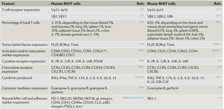
Overall, studies in vitro, in mouse models and in patients demonstrate the ability of MAIT cells to sense bacterial and viral infection. The protective role of MAIT cells in several infections, such as tuberculosis and influ-enza virus, suggests that therapeutic approaches based on the activation of MAIT cells by cytokines and/or ago-nist ligands could be developed. By contrast, the poten-tial deleterious role of MAIT cells in other infections, such as dengue, warrants further investigation. Table 1 | Features of mAIT cells in humans and mice
Feature Human mAIT cells Refs mouse mAIT cells Refs
T cell receptor expression Vα7.2–Jα33 2,20 Vα19–Jα33 2,115
Vβ2, Vβ13 2,20 Vβ8.1, Vβ8.2, Vβ6 2,115
Percentage of total T cells 1–35%, depending on the tissue (blood 5%, oral mucosa 2%, lung 3%, spleen 1%, liver 35%, adipose tissue 2%, ileum 2%, colon 2–7%, female genital tract 2–3%)
11–19 0.01–5%, depending on the tissue and
mouse strain (excluding transgenic mice) (blood 0.5%, lung 3%, spleen 0.08%, pancreatic lymph nodes 0.4%, liver 5%, adipose tissue 2%a, ileum 1%, colon 1%)
70,115
Transcription factor expression PLZF, RORγt, T- bet 4,35 PLZF, RORγt, T- bet 35,48,115
Activation and/or maturation
marker expression CD69, CD25, CD161, CD95, CD62L
low,
CD45RO, CD27
20,113 CD69, CD25, CD38, CD62L , CD44 69,70,115
Cytokine receptor expression IL-7R , IL-12R , IL-15R , IL-18R , IFNAR 28,50 IL-7R , IL-12R , IL-15R , IL-18R 115
Chemokine receptor
expression CCR2, CCR5, CCR6, CCR9, CCR10, CXCR3, CX3CR1, CXCR6
70,152 CCR2, CCR5, CCR6, CCR9, CXCR3,
CXCR6
115
Cytokines produced IFNγ, IFNα, TNF, IL-17A , IL-2, IL-4, IL-10, IL-22 5,16,50,73 IFNγ, TNF, IL-17A , IL-2, IL-4, IL-10, IL-13,
IL-22, GM- CSF
70,115
Cytotoxic mediator expression Granzyme A , granzyme B, granzyme K , perforin
34,70 Granzyme B, perforin 70
Natural killer cell and adhesion
marker expression PD-1, NKG2D, NKP80, NKP30, CD56, CD43, CD49a, CD103, CL A , αL integrin, α4β1 integrin, PSGL1, sLex
18,70,91,98,113,153–156 NK1.1 115
CL A , cutaneous lymphocyte- associated antigen; GM- CSF, granulocyte–macrophage colony- stimulating factor ; IFN, interferon; MAIT, mucosal- associated invariant T; PD-1, programmed cell death 1; PSGL1, P selectin glycoprotein ligand 1; sLex, sialyl–Lewis X motif; TNF, tumour necrosis factor. aA.T., B. Kiaf and A.L.,
MAIT cells in immune- mediated diseases
MAIT cell characteristics (TaBle 1) — including
activa-tion by inflammatory cytokines, expression of chemo-kine receptors and integrins, and the production of large amounts of cytokines and cytotoxic molecules — suggest that MAIT cells, similarly to conventional effec-tor memory T cells, could be involved in autoimmune and immune- mediated chronic diseases. However, in contrast to conventional T cells, circulating MAIT cells can function without specific antigen priming and following antigen- nonspecific activation by cytokines produced in an inflammatory context. In addition, inflammation increases the surface expression of MR1 protein69–71, suggesting that MAIT cells could be
acti-vated through their TCRs following recognition of MR1 molecules presenting bacterial, or possibly endogenous, ligands (Box 1). After activation, MAIT cells could act
directly on target cells or could participate to the recruit-ment and activation of other immune cells, as in infec-tious diseases. Numerous reports have described MAIT cell alterations in patients with various autoimmune and immune- mediated pathologies. Despite the hetero-geneity of the disease aetiologies, there are several com-mon features, such as reduced MAIT cell frequency in
the blood, their presence in the target tissue and their increased activation and dysfunction.
Decreased circulating MAIT cell frequency. As reported in chronic infectious diseases, the number of circu-lating MAIT cells decreases in most autoimmune diseases and in many inflammatory and metabolic diseases16,18,24,69,70,72–86(Fig. 2; TaBle 3). Blood MAIT cell
frequency correlates with disease severity in several pathologies87–94(TaBle 3). In multiple sclerosis (MS), the
frequency of blood MAIT cells decreases during clini-cal relapse and progression of the disease, whereas it increases during remission71,95. Similarly, after bariatric
surgery, obese patients improve in their metabolic and inflammatory parameters, simultaneously increasing in their number of blood MAIT cells16. Only a few studies
have characterized circulating MAIT cell subsets accord-ing to their CD4 or CD8 expression in autoimmune and inflammatory diseases. In humans, MAIT cells are mainly CD8 positive, approximately 10% are CD4 and CD8 double negative, and very few are CD4 positive. When MAIT cell subsets are characterized, the frequen-cies of both CD8+ and CD4−CD8− circulating MAIT cells
are similarly decreased18,70,80,87,94. The very low frequency
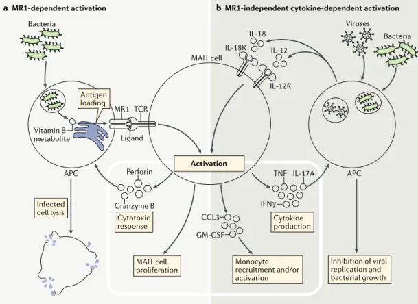
a MR1-dependent activation b MR1-independent cytokine-dependent activation
APC APC Bacteria Bacteria Viruses TCR Ligand MR1 Infected cell lysis Antigen loading MAIT cell
proliferation Monocyte recruitment and/or
activation Inhibition of viral replication and bacterial growth Cytotoxic response Activation Cytokine production Vitamin B metabolite IL-18R IL-18 IL-12 IL-12R Granzyme B TNF IL-17A IFNγ Perforin CCL3 GM-CSF MAIT cell
Fig. 1 | mAIT cell activation in bacterial and viral infection. Upon bacterial and viral infections, mucosal- associated
invariant T (MAIT) cells can be activated in a MHC class I- related molecule (MR1)-dependent manner through their T cell receptor (TCR) recognizing MR1-presenting bacterial ligands (part a), and/or in an MR1-independent manner by
inflammatory cytokines such as IL-12 and IL-18, produced by infected cells and sensed by MAIT cells through cytokine receptors (part b). MAIT cell activation induces their release of granzyme B and perforin (which kill infected cells), their
proliferation and their production of cytokines (tumour necrosis factor (TNF), interferon- γ (IFNγ) and IL-17A) that inhibit viral replication and bacterial growth. Activated MAIT cells can also promote monocyte recruitment and activation. APC, antigen- presenting cell; CCL3, CC- chemokine ligand 3; GM- CSF, granulocyte–macrophage colony- stimulating factor ; IL-12R , IL-12 receptor.
of CD4+ MAIT cells renders their specific detection
chal-lenging, due to potential contamination by conventional T cells during flow cytometry analysis. Several mecha-nisms could explain the decreased frequency of blood MAIT cells observed in most, if not all, pathologies. Presence of MAIT cells in inflamed tissues. Compared with tissues from healthy individuals or with unaf-fected tissues or blood from the same patients, the frequency of MAIT cells increases in the target tissues of patients with various immune- mediated diseases.
TaBle 3 and Fig. 2 illustrate the diversity of MAIT cell localization16,18,73,77–79,82,86,89,96–100. Mouse models allow
the analysis of MAIT cells in tissues that are hard to access in humans. In non- obese diabetic (NOD) mice, the mouse model of type 1 diabetes (T1D), MAIT cell frequency continuously increases in the pancreas until disease onset. Experiments involving transfer of MAIT cells into NOD recipient mice at different stages of dis-ease showed an incrdis-ease in the recruitment of MAIT cells to the pancreas of diabetic as compared with prediabetic mice70.
It should be noted that MAIT cell accumulation in affected tissues has not been found in all immune- mediated diseases; some diseases may instead show a redistribution of MAIT cells. For example, in the liver of patients with cirrhosis, immunohistochemical analy-sis shows preferential localization of MAIT cells in the inflammatory infiltrate around the portal tract and in the fibrotic septae69,87, and in patients with Crohn’s
dis-ease (CD), MAIT cells distribute specifically towards the injured part of ileum18. Fate- tracking studies should
help to clarify whether and how MAIT cell recruitment
to inflamed tissues contributes to their local expansion, promoting their pathogenic functions.
Several studies have suggested a key role for adhesion molecules, integrins and chemokines in the migration of MAIT cells to inflamed tissues. For example, blood MAIT cells from patients with MS express higher levels of the pro- migratory molecules P- selectin glycoprotein ligand 1 and αL integrin (also known as CD11a) than do cells from healthy individuals71. Moreover, IL-18,
which is elevated in the serum from patients with MS101,
signifi cantly upregulates the expression of α4β1 integrin (also known as VLA4) by CD8+ MAIT cells, compared
with conventional memory CD8+ T cells99, and α4β1
integrin mediates CD8+ T cell migration across the
blood–brain barrier in MS102. In rheumatoid arthritis,
high levels of TNF and IL-1β in the synovial fluid can upregulate the expression of adhesion molecules, such as E- selectin, by human endothelial cells91. E- selectin
promotes MAIT cell binding to endothelial cells via the sialyl–Lewis X (sLex) motif, which is elevated on syno-vial fluid MAIT cells compared with blood MAIT cells. In patients with rheumatoid arthritis, fewer circulat ing MAIT cells express the sLex motif than do circulat-ing MAIT cells from healthy controls91, suggesting that
sLex+ MAIT cells have migrated to the synovial fluid.
TNF also upregulates the expression of CC- chemokine ligand 20 (CCL20), which might attract MAIT cells expressing CCR6 in synovial fluid91. Similarly, in
chil-dren with recent- onset T1D, the frequency of blood MAIT cells is positively correlated with their expression of CCR6 (reF.70), suggesting the involvement of CCR6
in the migration of MAIT cells to inflamed tissues. CCR6+ cells recognize CCL20 and β-defensins, which
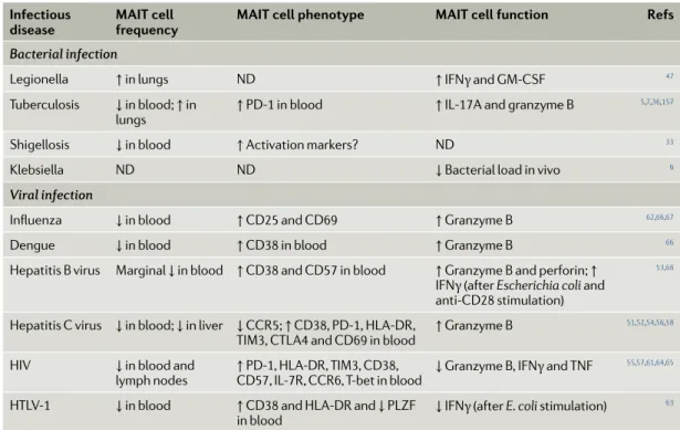
Table 2 | mAIT cells in infectious diseases
Infectious
disease mAIT cell frequency mAIT cell phenotype mAIT cell function Refs
Bacterial infection
Legionella ↑ in lungs ND ↑ IFNγ and GM- CSF 47
Tuberculosis ↓ in blood; ↑ in
lungs ↑ PD-1 in blood ↑ IL-17A and granzyme B
5,7,36,157
Shigellosis ↓ in blood ↑ Activation markers? ND 33
Klebsiella ND ND ↓ Bacterial load in vivo 6 Viral infection
Influenza ↓ in blood ↑ CD25 and CD69 ↑ Granzyme B 62,66,67
Dengue ↓ in blood ↑ CD38 in blood ↑ Granzyme B 66
Hepatitis B virus Marginal ↓ in blood ↑ CD38 and CD57 in blood ↑ Granzyme B and perforin; ↑ IFNγ (after Escherichia coli and anti- CD28 stimulation)
53,68
Hepatitis C virus ↓ in blood; ↓ in liver ↓ CCR5; ↑ CD38, PD-1, HL A- DR ,
TIM3, CTL A4 and CD69 in blood ↑ Granzyme B
51,52,54,56,58
HIV ↓ in blood and
lymph nodes ↑ PD-1, HL A- DR , TIM3, CD38, CD57 , IL-7R , CCR6, T- bet in blood ↓ Granzyme B, IFNγ and TNF
55,57,61,64,65
HTLV-1 ↓ in blood ↑ CD38 and HL A- DR and ↓ PLZF
in blood ↓ IFNγ (after E. coli stimulation)
63
CCR , CC- chemokine receptor ; CTL A4, cytotoxic T lymphocyte antigen 4; GM- CSF, granulocyte–macrophage colony- stimulating factor ; HTLV-1, human T cell lymphotropic virus type 1; IFNγ, interferon- γ; MAIT, mucosal- associated invariant T; ND, not determined; PD-1, programmed cell death 1; TIM3, T cell immunoglobulin and mucin domain- containing 3; TNF, tumour necrosis factor.
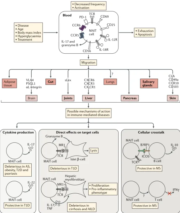
• Disease
• Age
• Body mass index
• Hyperglycaemia • Treatment • Exhaustion • Apoptosis • Decreased frequency • Activation Blood Migration Brain Adipose tissue
Joints Liver Pancreas
Salivary glands
Skin
Gut Lungs
Possible mechanisms of action in immune-mediated diseases VLA4 PSGL1 αL integrin sLex ? CXCR6 CXCR3 CX3CR1 CLA CD49a CCR10 CD103 PD-1 CCR5 CCR6 TCR CD69 CD25 CD56 IL-17 and granzyme B MAIT cell
MAIT cell MAIT cell
MAIT cell
MAIT cell MAIT cell T cell
MAIT cell
B cell B7RP1
ICOS
Cytokine production Direct effects on target cells Cellular crosstalk
Protective in T1D Protective in MS Protective in MS Deleterious in AS, obesity, T2D and psoriasis Deleterious in T1D Deleterious in cirrhosis and AILD
• Proliferation • Pro-inflammatory phenotype IL-17 IL-17, TNF Lysis Islet β-cell TCR TCR ? Granzyme B MR1 IL-22 TCR Hepatic myofibroblast MR1 IL-10 IFNγ IL-18R IL-12R
Fig. 2 | mAIT cell alterations and function in immune- mediated diseases. In most autoimmune and immune- mediated
diseases, mucosal- associated invariant T (MAIT) cell numbers in the blood are decreased. This may reflect two possible mechanisms: migration of MAIT cells to inflamed tissues, or increased MAIT cell death. MAIT cells express numerous chemokine receptors (such as CCR5, CCR6 and CXCR6), integrins (such as α4β1 integrin, CD49a and CD103) and other adhesion molecules (such as P- selectin glycoprotein ligand 1 (PSGL1), αL integrin and sialyl–Lewis X motif (sLex)). In some disease settings, MAIT cells are chronically activated (showing expression of CD25, CD69 or CD40L) by MHC class I-related molecule (MR1) and/or cytokines (IL-12, IL-18, IL-6 and interferon- α (IFNα)), and this can lead to a dysfunctional or exhaustion state (characterized by the expression of PD-1 and impaired IFNγ and tumour necrosis factor (TNF) secretion) and apoptosis (marked by increased levels of FAS and intracellular active caspase 3, as well as decreased levels of BCL-2). Disease, age, body mass index, hyperglycaemia or treatment could also impact MAIT cell frequency and functions. MAIT cells have been found to have both protective and deleterious roles in various immune- mediated diseases, through multiple mechanisms involving cytokine production, direct cell contact and cellular crosstalk. MAIT cells may act directly on target cells — for example, granzyme B- mediated lysis of islet β- cells in type 1 diabetes (T1D) and MR1-dependent or cytokine- dependent activation of profibrogenic functions of liver myofibroblasts in cirrhosis and autoimmune liver disease (AILD). MAIT cells may also act through immunoregulatory pathways involving more complex cellular crosstalk such as interactions with B and T cells and with myeloid cells. Bold text is used in the middle part of the figure for tissues to which MAIT cells have been shown to migrate or for which migration is strongly supported. AS, ankylosing spondylitis; B7RP1, B7-related protein 1; CL A , cutaneous lymphocyte- associated antigen; ICOS, inducible T cell co- stimulator ; IL-12R , IL-12 receptor ; MS, multiple sclerosis; PD-1, programmed cell death 1; T2D, type 2 diabetes; TCR , T cell receptor.
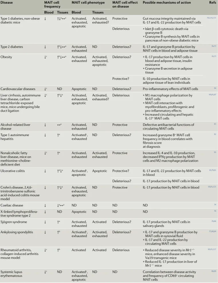
Table 3 | mAIT cells in immune- mediated diseases
Disease mAIT cell
frequency mAIT cell phenotype mAIT cell effect on disease Possible mechanisms of action Refs Blood Tissues Blood Tissues
Type 1 diabetes, non- obese
diabetic mice ↓ ↑↓
a↔a Activated,
exhausted, apoptotic
Activated,
exhausted? Protective Gut mucosa integrity maintained via IL-17 and IL-22 production by MAIT cells
70,124,131
Deleterious • Islet β- cell cytotoxic death via granzyme B
• Granzyme B synthesis by MAIT cells in pancreas of non- obese diabetic mice Type 2 diabetes ↓ ↑b↓↔a Activated,
exhausted ND Deleterious? IL-17 and granzyme B production by MAIT cells in blood and adipose tissue
16,73
Obesity ↓c ↑b↓↔a Activated,
exhausted Activated, exhausted, apoptotic
Deleterious? • IL-17 production by MAIT cells in blood and adipose tissue, insulin resistance
• Granzyme B secretion in adipose tissue
16,73
Protective? IL-10 production by MAIT cells in adipose tissue of lean individuals
Cardiovascular diseases ↓c ND Apoptotic ND Deleterious? Pro- inflammatory effects of MAIT cells 94
Liver cirrhosis, autoimmune liver disease, carbon tetrachloride- exposed mice, mice undergoing bile duct ligation
↓c ↑b↓a Activated,
exhausted, apoptotic
Activated,
exhausted? Deleterious • M1 macrophage polarization by MAIT cells
• MAIT cell interaction with myofibroblasts, profibrogenic and pro- inflammatory effects
• Increased circulating and hepatic IL-17+ MAIT cells
69,85,87
Alcohol- related liver
disease ↓ ↔
a Activated,
exhausted ND Protective Defective antibacterial functions of circulating MAIT cells
83
Type 1 autoimmune
hepatitis ↓ ↑
a Activatedc,
exhausted ND Deleterious? Increased granzyme B
+ MAIT cell
frequency in blood correlates with fibrosis score
at diagnosis
82
Nonalcoholic fatty liver disease, mice on methionine–choline- deficient diet
↓c ↑a Activated,
exhausted Activated, exhausted Protective Increased IL-4 and IL-10 production, decreased IFNγ production by MAIT cells and M2 macrophage polarization
93
Ulcerative colitis ↓ ↑d↓a Activatedc,
apoptotic Apoptotic Protective? IL-17 and IL-22 production by MAIT cells in blood
18,79,81
Deleterious? IL-17 production by MAIT cells in blood Crohn’s disease,
2,4,6-trinitrobenzene sulfonic acid- induced colitis mouse model
↓ ↑a↓a Activated,
exhausted, apoptotic
ND Protective IL-17 production by MAIT cells in blood 18,81,125
Coeliac disease ↓ ↓a↔a ND ND ND ND 74
X- linked
lymphoprolifera-tive syndrome type 2 ↓ ND Apoptotic ND ND ND
76
Sjögren syndrome ↓ ↑a Activated,
exhausted Activated Deleterious? IL-17 production by MAIT cells in salivary glands
78,86
Ankylosing spondylitis ↓ ↑b Activatedc,
exhausted Activated, exhausted Deleterious? • IL-17 and granzyme B production by MAIT cells in synovial fluid
• IL-17 and IL-22 production by circulating MAIT cells
77,80,84
Rheumatoid arthritis, collagen- induced arthritis mouse model
↓c ↑b Activated Activated Deleterious • Reduced disease severity in Mr1−/−
mice, enhanced disease severity in Vα19 transgenic mice
• Reduced IL-17 production in liver of
Mr1−/− mice 89,91,117 Systemic lupus erythematosus ↓ c ND Activatedc, exhausted, apoptotic
ND ND Correlation between disease activity and frequency of CD69+ circulating
MAIT cells
are chemoattractants that are increased in the pancreas and gut of diabetic patients and mouse models103–105.
In addition to CCR6, other skin- homing receptors, such as CCR10, CD49a, CD103 and cutaneous lymphocyte- associated antigen, are expressed by some blood MAIT cells19,97,98 and could participate in their migration to
dermatitis herpetiformis lesions, in which MAIT cells are abundant97. Increased expression of the chemokine
receptors CXCR3 and CX3CR1 on hepatic MAIT cells in
non- infectious chronic liver diseases might reflect their activation rather than their recruitment, as MAIT cells are already abundant in the liver at the steady state15.
Of note, even though MAIT cells express various chemo-kine receptors (TaBle 1), until now, no specific subset of
MAIT cells has been identified as preferentially trafficking to distinct tissues.
Blood MAIT cells are activated and exhausted. The decreased frequency of blood MAIT cells in immune- mediated diseases could also result from their death by apoptosis subsequent to sustained activation. Accordingly, in many autoimmune and inflammatory diseases, circulating MAIT cells express higher lev-els of activation and/or exhaustion markers, such as CD38, CD25, CD69 and PD-1 (reFs16,18,69,70,73,83,85,87,89,93).
Furthermore, in some diseases MAIT cell activation correlates with disease activity score (TaBle 3). As has
been observed in infectious diseases, MAIT cells could
be activated via MR1-dependent or MR1-independent pathways. In systemic lupus erythematosus (SLE), the increased activation of MAIT cells has partly been explained by the increased ability of monocytes to acti-vate MAIT cells in a TCR- dependent manner88. The
activation of MAIT cells by MR1 in autoimmune and immune- mediated diseases is also suggested by the observation of increased MR1 expression in inflamed tissues71. In the context of T1D, in vitro exposure of
a human pancreatic β- cell line to pro- inflammatory cytokines increases both MR1 transcript levels and MR1 surface expression70. Interestingly, gut homeostasis is
altered in many autoimmune and immune- mediated diseases (Box 2), which could lead to increased levels of
bacteria-derived ligands for MR1. The observations of thymic selection of MAIT cells in germ- free mice106
and TCR-dependent activation of myofibroblasts by MAIT cells in the absence of exogenous ligands69
support the existence of endogenous ligands (Box 1).
Moreover, correlations between circulating MAIT cell frequency, CD69+ MAIT cell frequency and cytokine
(IL-18, IL-6 and IFNα) plasma levels in patients with SLE88, MS99 and ulcerative colitis (UC)79 suggest that
MAIT cells can also be activated by cytokines in these diseases. The exact mechanism of MAIT cell activation is not yet well defined. As in infectious diseases, the combined triggers for MAIT cell activation of inflamma-tory cytokines and TCR- mediated signals might lead to
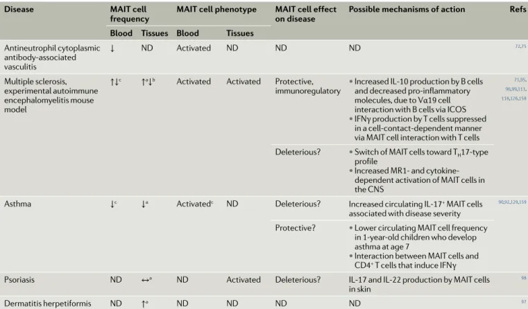
Disease mAIT cell
frequency mAIT cell phenotype mAIT cell effect on disease Possible mechanisms of action Refs Blood Tissues Blood Tissues
Antineutrophil cytoplasmic antibody- associated vasculitis ↓ ND Activated ND ND ND 72,75 Multiple sclerosis, experimental autoimmune encephalomyelitis mouse model
↑↓c ↑a↓b Activated Activated Protective,
immunoregulatory • Increased IL-10 production by B cells and decreased pro- inflammatory molecules, due to Vα19 cell interaction with B cells via ICOS
• IFNγ production by T cells suppressed in a cell- contact-dependent manner via MAIT cell interaction with T cells
71,95,
96,99,111,
118,126,158
Deleterious? • Switch of MAIT cells toward TH17-type
profile
• Increased MR1- and cytokine- dependent activation of MAIT cells in the CNS
Asthma ↓c ↓a Activatedc ND Deleterious? Increased circulating IL-17+ MAIT cells
associated with disease severity
90,92,129,159
Protective? • Lower circulating MAIT cell frequency in 1-year- old children who develop asthma at age 7
• Interaction between MAIT cells and CD4+ T cells that induce IFNγ
Psoriasis ND ↔a ND Activated Deleterious? IL-17 and IL-22 production by MAIT cells
in skin
98
Dermatitis herpetiformis ND ↑a ND ND ND ND 97
Exhausted? denotes a cell status based only on phenotype and not confirmed by functional analysis. Exhausted denotes cells with impaired cytokine production and function. Deleterious? denotes a deleterious effect is suggested or hypothetical and not yet demonstrated. Protective? denotes a protective effect is
suggested or hypothetical and not yet demonstrated. ↑, increased frequency ; ↓, decreased frequency ; ↑↓, both increased and decreased frequency reported;
↔, no change; CNS, central nervous system; ICOS, inducible T cell co- stimulator ; IFNγ, interferon- γ; MAIT, mucosal- associated invariant T; MR1, MHC class I- related molecule; ND, not determined; TH17 , T helper 17. aCompared with tissues from healthy individuals or with unaffected tissues. bCompared with blood.
cCorrelation with disease severity. dComparison between active and inactive forms of the disease.
MAIT cell exhaustion or dysfunction. Indeed, alterations of MAIT cell responses in vitro suggest an exhausted status. After phorbol myristate acetate–ionomycin stim-ulation or upon MR1-dependent activation, MAIT cells from patients with autoimmune and immune- mediated diseases are usually less activated and produce fewer T helper 1 (TH1)-type cytokines than MAIT cells from
controls16,18,73,77,78,82,83,85–87,89,93. In patients with SLE, MAIT
cell exhaustion has been associated with dysfunction of the Ca2+–calcineurin–NFAT1 signalling pathway
down-stream of TCR stimulation89. Although MAIT cells from
most immune- mediated diseases show defective activ-ation and/or IFNγ and TNF production in vitro, several studies have described an increased frequency of MAIT cells producing IL-17 in the same pathologies16,18,70,73,77.
These observations suggest there is heterogeneity among MAIT cells, a hypothesis that is worthy of more detailed analysis.
Increased apoptosis of blood MAIT cells. MAIT cells in many immune- mediated diseases show increased apoptosis, possibly as a result of chronic activation. In children with recent- onset T1D, fewer circulating MAIT cells express the anti- apoptotic factor BCL-2, and this is positively correlated to the overall frequency of MAIT cells70. A similar correlation is also observed in
patients with liver cirrhosis69. In patients with UC, CD
and SLE, more circulating MAIT cells express activated caspases (which mediate apoptotic cell death) than in control individuals81,88. X- linked lymphoproliferative
syndrome type 2 (XLP2) is a rare human genetic immuno deficiency with some clinical signs similar to CD. XLP2 results from mutations in the gene encoding X-linked inhibitor of apoptosis (XIAP) and leads to a pro- apoptotic phenotype in invariant natural killer T (iNKT) cells and MAIT cells, which could explain the decreased frequency of such cells in the blood of these patients76.
It would be interesting to analyse whether chronic inflammation, often associated with autoimmune diseases, dampens XIAP expression by MAIT cells. Increased apoptosis of MAIT cells in target tissues. As we mentioned above, MAIT cells have the propensity to migrate to inflamed tissues, and in many autoimmune and immune- mediated diseases MAIT cell frequency is increased in target tissues. However, in some tissues, such as the liver, colon and adipose tissue, where MAIT cells are abundant in the steady state, their frequency tends to decrease upon inflammation. These results sug-gest that increased apoptosis of MAIT cells could over-ride potential MAIT cell recruitment in these inflamed tissues. For example, MAIT cells are less frequent in inflamed intestinal mucosa of patients with UC and CD and of children with coeliac disease, compared with healthy tissue74,81. Decreased frequency of MAIT cells is
also observed in adipose tissue from obese adults com-pared with the adipose tissue from lean adults, and in co- cultures of MAIT cells and adipose tissue from obese individuals BCL-2 expression is decreased compared with adipose tissue from lean individuals16,73. Similarly,
MAIT cells are less frequent in the livers of patients with cirrhosis and autoimmune liver disease (AILD) and have an activated/exhausted phenotype (CTLA4+ and PD-1+),
as compared with control livers69,87. Intrahepatic MAIT
cell frequency is negatively correlated with the PD-1 expression of the cell. Increased MAIT cell proliferation is observed in the blood of patients with CD18 and with
liver cirrhosis69,83, as well as in vitro in the presence of
inflamed adipose tissue from obese patients16; however,
this proliferation is not sufficient to compensate for the loss of MAIT cells.
Other factors impacting MAIT cell frequency. The marked heterogeneity among patients with regard to factors — such as age107,108, body mass index16,73,
physi-cal activity109 and ongoing treatment69,90,110 — that could
impact MAIT cell frequency and function might explain the conflicting results in human studies71,111. For
exam-ple, decreased blood MAIT cell frequency is observed in adults but not in children with coeliac disease74,
and in obesity MAIT cell frequency is increased in chil-dren but decreased in adults16,73. Whether the strong
decrease of MAIT cell frequency reported in patients with severe asthma results from the pathophysiology or from the inhaled corticoid treatment is unclear90.
Indeed, the lower MAIT cell frequencies in the blood, sputum and endobronchial biopsies of asthmatic patients are positively associated with disease severity and negatively associated with inhaled corticosteroids. No change in MAIT cell frequency is observed in the broncho- alveolar lavage fluid from asthmatic patients, which could reflect the weak diffusion of the inhaled corticosteroids in more distal airways and the alveolar compartment90. However, in another study, after
exclu-sion of patients on corticoid treatment, the re- evaluated circulating MAIT cell frequency was still decreased in patients with inflammatory bowel diseases112.
Moreover, decreased MAIT cell frequency has been observed in other autoimmune or immune- mediated Box 2 | mAIT cells and gut alteration in immune- mediated diseases
studying mucosal- associated invariant t (Mait) cells in autoimmune, inflammatory and metabolic diseases can shed new light on the pathophysiology of these cells. Despite their different features, many of the pathologies listed are associated with alterations in gut homeostasis149–151, including microbiota composition, intestinal permeability and
gut immunity. Given the key characteristics of Mait cells (activation by bacterial ligands, homing capacity and gut localization), they might represent sensors and/or enactors of such gut modifications. But what is the first step during autoimmune, inflammatory and metabolic diseases? are Mait cells first impaired by the inflammation occurring in inflamed tissues, or is microbiota dysbiosis the initial impact modifying Mait cell function? inflammation outside of the gut can activate and alter the function of the Mait cells that subsequently migrate to the gut mucosa, where they are no longer able to maintain gut homeostasis, as suggested by the decreased production of iL-17 and iL-22 by intestinal Mait cells in the non- obese diabetic (NOD) mouse model of type 1 diabetes24,70. impaired Mait cells might also lose their ability to provide
efficient antibacterial function. together, these Mait cell alterations could promote gut mucosa and microbiota alterations.
in a second hypothesis, initial modifications of gut homeostasis, such as increased bacterial translocation and modification of the intestinal microbiota, could alter the equilibrium between different bacteria and the level of activating and/or inhibiting ligands, thereby impacting Mait cell functions. identifying the kinetics and nature of Mait cell interactions with the gut mucosa and microbiota represents a critical step for exploring Mait cell- based treatments for autoimmune, inflammatory, metabolic and immune- mediated diseases.
diseases in patients without immune-modulating treatments16,69,70,73,74,83,93.
An impact of hyperglycaemia on MAIT cell fre-quency has been proposed. In cardiovascular disease, circulating MAIT cell frequency negatively correlated to glycated haemoglobin (HbA1c) levels94, and in obese
patients, decreased circulating MAIT cell frequency was greater in adults with uncontrolled HbA1c levels73.
Moreover, exposure of MAIT cells to glucose increases their apoptosis in a dose- dependent manner94. While no
correlation was observed between HbA1c level and blood
MAIT cell frequency in children with T1D, granzyme B+ MAIT cell frequency was inversely correlated with
HbA1c levels at the onset of disease, but not in children
with established disease70. The potential role of
gran-zyme B+ MAIT cells in T1D is further discussed below.
Together, these observations suggest that decreased MAIT cell frequency in various diseases might have multifactorial origins.
Pathogenic roles for MAIT cells
Pathogenic role of MAIT cells in the liver. During chronic liver injury, signals originating from immune cells, as well as bacterial by- products, play central roles in the progression of liver fibrosis by activating the fibrogenic functions of hepatic myofibroblasts and their inflamma-tory properties. In the liver of patients with cirrhosis and AILD, MAIT cells accumulate in the fibrotic septa69,87,
making contact with hepatic myofibroblasts. Their profi-brogenic functions were demonstrated in mouse models, in which deficiency of Mr1 led to resistance to fibrosis induced by chronic carbon tetrachloride administration or bile- duct ligation, whereas expression of a Vα19TCR transgene, leading to an excess of MAIT cells, resulted in mice that were more prone to fibrosis69. In vitro,
MAIT cells enhanced the fibrogenic functions of hepatic myofibroblasts, characterized by increased pro-liferation and collagen secretion, and exacerbated their pro- inflammatory properties69,87. Interestingly, hepatic
myofibroblasts express MR1 at the cell surface and are activated by MAIT cells via an MR1-dependent pathway. By contrast, the myofibroblast pro- inflammatory prop-erties can be induced in a contact- independent manner by TNF or IL-17A released by MAIT cells69,87. In
addi-tion to directly interacting with fibrogenic cells, MAIT cells may also favour fibrosis progression by fostering a local inflammatory reaction, in that they promote polar-ization of macrophages towards a pro- inflammatory phenotype69.
Pathogenic role of IL-17-producing MAIT cells. All human and most mouse MAIT cells express RORγt, a master transcription factor controlling IL-17 pro-duction22,113–115. IL-17 has pathogenic roles in many
autoimmune, metabolic and liver diseases116,
suggest-ing that MAIT cells could be deleterious via IL-17 production. In the collagen- induced arthritis murine model, MAIT cells are pro- inflammatory117, in that
the severity of the disease is reduced in Mr1−/− mice
and enhanced after T cell transfer from Vα19TCR- transgenic mice. Accordingly, total splenocytes from Mr1−/− mice produce lower levels of IL-17 after anti- CD3
and anti- CD28 stimulation or IL-23 treatment. Human studies highlight a switch towards a TH17-type profile
of blood MAIT cells, with an increase in secretion of IL-17 in patients with CD18, UC18,79, ankylosing
spon-dylitis (AS)77, obesity16,73, type 2 diabetes (T2D)16 and
cirrhosis69, and of IL-22 in patients with UC18 and AS84.
Analysis of MAIT cells in patients with MS has fur-ther shown increased expression of RORγt, CCR6 and IL-7R compared with healthy controls118. Similarly,
IL-17 production is increased in MAIT cells from the salivary glands of patients with Sjögren syndrome (SS) compared with controls78,86, as well as after stimulation
with IL-23 in vitro78. In AS, the altered cytokine profile
of blood MAIT cells is also observed in the synovial fluid in favour of IL-17 production77,84, and IL-7
prim-ing of MAIT cells increases IL-17 production in patients compared with healthy controls77. Of note, IL-7 levels
are elevated in the joints and gut of AS patients119,120,
and there is a positive correlation between IL-7 and IL-17 levels in these patients121. Interestingly,
circu-lating MAIT cells from patients with AS have higher IL-7R expression levels than in controls77. There is a
report of IL-17 and IL-22 production by a MAIT cell clone derived from psoriatic skin lesions98. Adipose
tissue from obese individuals had an elevated fre-quency of MAIT cells that produce IL-17 compared with controls16,73, which might induce insulin resistance in
adipocytes122,123. Interestingly, the pathogenic role for
IL-17-producing MAIT cells is suggested in asthmatic children, in whom there is a correlation between cell frequency and disease severity (defined as the number of exacerbations)92. Most of these analyses in humans
have shown correlations between MAIT cells, IL-17 production and disease status, but further studies are warranted to demonstrate causality.
Pathogenic role of cytotoxic MAIT cells. MAIT cells could have a pathogenic role in autoimmune and immune- mediated diseases via their cytotoxic proper-ties. T1D is the most obvious example of this activity70.
Analysis of NOD mice has revealed an increasing level of granzyme B expression in pancreatic islets from the prediabetic stage to diabetes onset. In humans, more circulating MAIT cells produce granzyme B in chil-dren with recent onset T1D than do control MAIT cells. Interestingly, granzyme B+ MAIT cell frequency
corre-lated negatively with the frequency of MAIT cells and with the age of the child at diagnosis, in agreement with the current view that T1D is more aggressive in the youngest children. These data in NOD mice and T1D patients sug-gest that MAIT cells could kill pancreatic β- cells, even though to date MAIT cells have not been detected in diseased islets124. As we mentioned earlier, MR1
expres-sion is upregulated on the human β- cell line EndoC- βH1 in the presence of pro- inflammatory cytokines, and in co- cultures MAIT cells from healthy controls can kill EndoC- βH1 cells in an MR1-dependent manner70.
A similar pathogenic local effect of granzyme B+
MAIT cells is also suggested in patients with AS77 and in
obesity16. Of note, in NOD mice the presence of
gran-zyme B+ MAIT cells in the intestinal mucosa increases
pathogenic role in this tissue before and at T1D onset (L. Beaudoin and A.L., unpublished observations). Together, these studies show that MAIT cells can mediate various pathogenic mechanisms.
A protective role for MAIT cells
Immunoregulatory role of MAIT cells. In contrast to their potentially pathogenic activities, MAIT cells also have immunoregulatory activities that may be protec-tive in disease. MAIT cells have a protecprotec-tive role in the 2,4,6-trinitrobenzene sulfonic acid (TNBS)-induced colitis mouse model125,126. MAIT cells are decreased in
the colonic mucosa after TNBS treatment, and trans-fer of Jα33TCR+ MAIT cells reduces the severity of the
disease, weight loss, intestinal bleeding and colonic inflammation. In experimental autoimmune encepha-lomyelitis (EAE)126, the mouse model of MS, transgenic
expression of the invariant Vα19TCR in T cells protects mice from EAE induction, and adoptive transfer of liver invariant Vα19TCR+ T cells into wild- type mice
with EAE reduces the disease severity. Conversely, EAE in Mr1−/− mice is exacerbated126. The suppression of
EAE by invariant Vα19TCR+ T cells is associated with
reduced expression of pro- inflammatory molecules and increased production of IL-10 by B cells, through an inducible T cell co- stimulator-dependent interaction with invariant Vα19TCR+ T cells126. IL-10 can inhibit
excessive gut inflammation127 and is implicated in IgA
production128. A regulatory role of IL-10-secreting
MAIT cells is also suggested by their presence in the adipose tissue of lean individuals but their reduced fre-quency in obese individuals73. In vitro experiments also
support the immunoregulatory function of MAIT cells from both patients with MS and healthy adults95, as they
suppress IFNγ production by conventional T cells in a cell contact-dependent manner.
In a longitudinal study, the circulating MAIT cell frequency was shown to be lower in 1-year- old children who later develop asthma than in 1-year- olds who do not. A higher MAIT cell frequency is correlated with stronger IFNγ secretion by CD4+ T cells, suggesting a
protective role of MAIT cells associated with a potent TH1-type immune response129. It would be interesting to
determine whether there is a link between these obser-vations in children and the hygiene hypothesis, implying that activation of the immune system at a young age can promote immunoregulatory mechanisms130 and that
MAIT cells could represent an ideal immune cell type to maintain regulatory function in the whole body. Prevention of T1D by MAIT cells. Concerning T1D, an initial study suggested a protective role for MAIT cells, as diabetes is delayed in Vα19TCR- transgenic NOD mice131. More recently, analysis of Mr1−/− NOD mice
con-firmed the protective role of MAIT cells, as these mice develop accelerated diabetes, despite the potential path-ogenic role of MAIT cells in the pancreas70. Indeed, this
study suggests that MAIT cells could have a protective role in the gut mucosa through their production of IL-17 and IL-22, two cytokines required for the maintenance of gut barrier integrity. In Mr1−/− NOD mice,
exacerba-tion of diabetes development is associated with a loss of
gut barrier integrity, allowing the translocation of bacte-rial compounds and subsequent dendritic cell activation in pancreatic lymph nodes70. Of note, a protective role of
IL-17 has been established in several intestinal inflam-mation models116, and IL-22+ MAIT cells have been
observed in the blood of patients with UC18 and AS84.
Protective role of MAIT cells in the liver. In patients with nonalcoholic fatty liver disease (NAFLD), MAIT cells accumulate around fatty hepatocytes. Although mouse and human studies have demonstrated the profibrogenic properties of MAIT cells, Mr1−/− mice
fed a methionine–choline- deficient diet show enhanced steato sis, are more prone to liver injury and have more CD11c+ M1-like inflammatory macrophages but fewer
CD206+ M2-like macrophages93. Future studies will be
needed to confirm this protective role of MAIT cells in a more relevant model of nonalcoholic steatohepa-titis, and the contribution of IL-17 needs to be evalu-ated, as it is a major driver of NAFLD progression132,133.
In addition, it remains unclear whether gut- derived bacterial components impact MAIT cell activation during NAFLD.
Patients with alcoholic liver disease, either severe alcoholic hepatitis or end- stage cirrhosis, are at high risk of developing bacterial infections, because of a leaky gut, gut dysbiosis, increased bacterial translocation and dys-regulation of immune cell functions. In in vitro exper-iments, stool extracts from patients with alcoholic liver disease induced MAIT cell depletion and suppressed their antibacterial cytokine (IL-17, TNF and IFNγ) responses and the expression of PLZF and RORγt, which are involved in MAIT cell maturation and effector functions83. These data suggest that MAIT cell functional
defects in patients with alcohol- related liver disease could contribute to this population’s high suscepti bility to bacterial infection, which is also described during autoimmune hepatitis82. Interestingly, prophylactic
administration of antibiotics to patients with severe alco-holic cirrhosis, a treatment usually administered so as to avoid bacterial infections, partially prevents blood MAIT cell reduction and activation69.
Together, MAIT cells can have multiple functions, and their pathogenic or protective role may rely on their activation or exhaustion status, tissue localization and cytokine profile, and on the chronicity of the disease. Even though the characterization of blood MAIT cells might not be as informative as tissue analysis, blood MAIT cells do seem to reflect the status of MAIT cells in inflamed tissues.
MAIT cells in human cancers
Recent studies have investigated the role of MAIT cells in cancer, and the observations reported are similar to those made in autoimmune and inflammatory dis-eases (TaBles 3,4). Circulating MAIT cell frequency is
decreased in patients with colorectal cancer (CRC)134–136,
hepatocellular carcinoma (HCC)137 and lung cancer136.
MAIT cells accumulate in tumour tissues from patients with colon adenocarcinomas138 and CRC, particularly
in those with advanced CRC134–136, and in hepatic
patients with HCC, the frequency of MAIT cells was decreased in the liver, and in the tumour, when com-pared with healthy controls. As we observed in immune- mediated diseases, this could reflect increased apoptosis of MAIT cells, possibly as a result of their chronic activa-tion. Indeed, in patients with HCC, tumour- infiltrating MAIT cells had an exhausted phenotype, with high PD-1, CTLA4 and TIM3 expression, and produced less IFNγ and IL-17 than MAIT cells from healthy donors137.
Similarly, MAIT cells from colonic adenocarcinomas produced less IFNγ than MAIT cells from healthy colons138, suggesting that tumour- infiltrating MAIT
cells might be altered due to tumour microenviron-mental factors. Furthermore, tumour- infiltrating MAIT cells produced less IFNγ than MAIT cells from healthy livers in response to recombinant IL-12 and IL-18
(reF.139). As was observed in other diseases, circulating
MAIT cells in patients with CRC produce more IL-17 and less IFNγ and TNF134. Of note, chemotherapy does
not alter the ability of MAIT cells to produce IFNγ, nor their frequency139, in agreement with their expression
of the multidrug resistance transporter ABCB1 (reF.11).
High frequencies of tumour- infiltrating MAIT cells are associated with poor clinical outcomes for patients with HCC. In contrast, in the context of haematological malignancies, a higher frequency of MAIT cells after allogeneic haematopoietic cell transplantation is asso-ciated with less graft- versus-host disease (GVHD)140,141.
Mouse studies have also shown a beneficial role of MAIT cells against GVHD and highlighted their role in maintaining gut integrity142. All these studies suggest
that MAIT cells could represent attractive therapeu-tic targets; however, their precise role in cancer needs further investigation.
Conclusion and perspectives
MAIT cells provide efficient protection against acute bacterial and viral infections. However, in chronic pathological situations, including chronic viral infec-tions and autoimmune, inflammatory and metabolic diseases, MAIT cells can play a pathogenic role by sus-taining inflammation and cytotoxicity. This ambivalent function is reminiscent of NKT cells, which share many features with MAIT cells22,143,144, although the ligands
recognized by the two innate- like T cell populations are quite distinct. Several endogenous ligands of NKT cells have been described145,146; however, the MAIT cell
endogenous ligands remain to be identified. Although both MAIT and NKT cells are conserved in mammals, MAIT cells are much more abundant than NKT cells in humans144. Moreover, MAIT cell localization in the
gut mucosa and their recognition of bacterial ligands might play a key role in sensing any modification of the microbiota composition147, either commensal or
pathogenic, which might reflect the infectious, meta-bolic and general health status of the individual (Box 2).
Further studies should be performed to provide a bet-ter understanding of the functional plasticity of MAIT cells, the epigenetic regulation of MAIT cell function, the impact of their TCR repertoire, and their inter-actions with other immune and non- immune cells in physiological and pathological situations. Many of the data presented in this Review highlight MAIT cells as biomarkers in various diseases; however, new insights into their biological functions are still awaited, to inform the design of therapeutic approaches based on MAIT cell manipulation.
Published online xx xx xxxx Table 4 | mAIT cells in cancer
Cancer mAIT cell
frequency mAIT cell phenotype mAIT cell effect on disease
Possible mechanisms of action Refs Blood Tissues Blood Tissues
Kidney ↓ ↑ ND ND ND Increased MAIT cells in tumour tissue 160
Brain ↓ ↑ ND ND ND Increased MAIT cells in tumour tissue 160
Lung ↓ ND ND ND ND ND 136
Breast ↔ ND ND ND ND Increased IL-17 produced by breast duct MAIT cells
136,161
Colorectal ↓ ↑ ND ND Protective? • Increased MAIT cells in tumour tissue
• Increased CD69 and PD-1 expression
• Decreased IFNγ production
134–136,138
Hepatic metastasis ND ↑ ND ND Protective and/
or deleterious? •• Increased MAIT cells in tumour tissue Decreased IFNγ production and increased IL-17 production
139
Hepatocellular carcinoma ↓ ↓ Exhausted Exhausted Deleterious? • Decreased MAIT cells in tumour tissue, more IL-8 produced (pro-tumour cytokine)
• High MAIT cell frequency associated with poor clinical outcome
137
Graft- versus-host disease (in individuals treated for cancer with bone marrow transplant)
↓ ND ND ND Protective Low MAIT cell frequency associated with poor clinical outcome
140–142
Exhausted denotes cells with impaired cytokine production and function. Deleterious? denotes a deleterious effect is suggested or hypothetical and not yet demonstrated. Protective? denotes a protective effect is suggested or hypothetical and not yet demonstrated. ↑, increased frequency ; ↓, decreased frequency ; ↔, no change; IFNγ, interferon- γ; MAIT, mucosal- associated invariant T; ND, not determined; PD-1, programmed cell death 1.