practices of cell-safe device design and operation
The MIT Faculty has made this article openly available.
Please share
how this access benefits you. Your story matters.
Citation
Varma, Sarvesh and Joel Voldman. “Caring for Cells in
Microsystems: Principles and Practices of Cell-Safe Device Design
and Operation.” Lab on a Chip 18, 22 (2018): 3333–3352 © 2018 The
Royal Society of Chemistry
As Published
http://dx.doi.org/10.1039/c8lc00746b
Publisher
Royal Society of Chemistry (RSC)
Version
Final published version
Citable link
http://hdl.handle.net/1721.1/119013
Terms of Use
Creative Commons Attribution-NonCommercial 3.0 Unported
CRITICAL REVIEW
Cite this:Lab Chip, 2018, 18, 3333
Received 18th July 2018, Accepted 8th October 2018 DOI: 10.1039/c8lc00746b rsc.li/loc
Caring for cells in microsystems: principles and
practices of cell-safe device design and operation
Sarvesh Varma
and Joel Voldman
*
Microfluidic device designers and users continually question whether cells are ‘happy’ in a given micro-system or whether they are perturbed by micro-scale technologies. This issue is normally brought up by engineers building platforms, or by external reviewers (academic or commercial) comparing multiple tech-nological approaches to a problem. Microsystems can apply combinations of biophysical and biochemical stimuli that, although essential to device operation, may damage cells in complex ways. However, assays to assess the impact of microsystems upon cells have been challenging to conduct and have led to subjective interpretation and evaluation of cell stressors, hampering development and adoption of microsystems. To this end, we introduce a framework that defines cell health, describes how device stimuli may stress cells, and contrasts approaches to measure cell stress. Importantly, we provide practical guidelines regarding de-vice design and operation to minimize cell stress, and recommend a minimal set of quantitative assays that will enable standardization in the assessment of cell health in diverse devices. We anticipate that as micro-system designers, reviewers, and end-users enforce such guidelines, we as a community can create a set of essential principles that will further the adoption of such technologies in clinical, translational and com-mercial applications.
Introduction
A cell's health and biological function are closely regulated by its microenvironment. Adverse perturbations caused by exter-nal stimuli (e.g., injury, molecular sigexter-nals) can steer cellular homeostasis towards a dysfunctional state. To this end, countless platforms and technologies (including micro-fluidics) have been developed to gain insight into disease biology, as well as for engineering diagnostics and therapeu-tics. In their application, these devices typically leverage a va-riety of physical forces and biochemical factors to study and manipulate a broad range of cell types.1,2 Paradoxically, the device microenvironment itself may inevitably impose unde-sirable changes upon cell health, thereby biasing or invalidating the device's utility. However, how does one avoid unintended device-imposed biological artifacts that can bias results from the intended biological study and application? In other words, how does one design and maintain the device microenvironment in a way that does not stress or harm cells? In this review, our objective is to address these ques-tions and provide practical guidelines for designing and using devices in ways that lower cell damage; thus, negating device-imposed biological artefacts and extending the utility of the device to the broader community.
First, we will define cell health and how various perturba-tions can steer cells to a stressed state or towards cell death. Next, we will outline common considerations for device de-sign and operating conditions that should help in maintaining viable cells. These considerations should assist the device designer in keeping‘device-treated’ cells alive and appearing similar to the appropriate reference cells (e.g. cells cultured by standard protocols). With such precautions such cells may appear healthy, however they may still be stressed in ways not always obvious to the end-user. For those inter-ested in maintaining healthy (and not just viable) cells, in the latter part of this review, we recommend quantitative as-says that can be used to measure and minimize cell stress within the context of microsystems.
Cell health and stress responses
For a microsystem engineer or user, the first concern is to en-sure that their particular platform does not kill cells. Cell death is commonly observed as a decrease in cell numbers due to the exposure to a device environment, while cell damage is com-monly inferred through changes in cell morphology or func-tion. However, cell death can be masked by proliferation of sur-viving cells and ‘washing off’ or disintegration of dead cells. Similarly, activation of cell death mechanisms (e.g. apoptosis) or stress mechanisms may not be reflected in cell morphology or proliferation. In this way, common inferences made by the device designers may be both inaccurate and misrepresentative
Department of Electrical Engineering and Computer Science, Massachusetts Institute of Technology, 77 Massachusetts Avenue, Room 36-824, Cambridge, USA. E-mail: voldman@mit.edu; Fax: +617 258 5846; Tel: +617 253 1583
Open Access Article. Published on 16 October 2018. Downloaded on 11/14/2018 1:54:17 PM.
This article is licensed under a
Creative Commons Attribution-NonCommercial 3.0 Unported Licence.
View Article Online
of cell health. Hence, before describing what can help maintain live and healthy cells, we will first describe to the device de-signers what it means for a cell to be healthy.
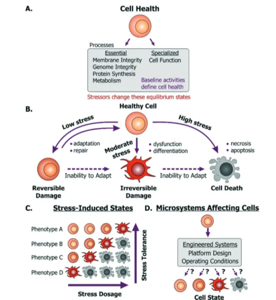
A dysfunctional or damaged cell is an undesirable conse-quence that needs to be prevented to maintain a healthy physiological state. However, what this healthy state refers to is dependent on a few important aspects. Cells in vivo provide unique functionality through specialized processes, which are tied to their phenotype and microenvironment. While the in vivo purpose and functions may differ among cells, they are all susceptible to stress damage in similar ways. Specifi-cally, stressors can inflict injury to cell membranes, ATP gen-eration processes, protein synthesis and genome integrity and replication processes- all of which can influence the other. We define these as essential processes that are relevant to all cells. The baseline ‘activity’ of each of these essential and specialized processes collectively defines what we will
term the“healthy state” of a particular cell (Fig. 1A). Impor-tantly, these states will differ between the natural in vivo microenvironment and typical in vitro culture conditions and depending on the cellular phenotype and origin, the relevant reference must be considered as the healthy state. Addition-ally, within in vitro culture, primary cells and transformed cell lines (including cancer cell lines) will maintain distinct homeostatic equilibria for each of the essential cellular pro-cesses. For cancer or diseased cells this equilibrium is de-fined as the healthy state, even though it may in fact be asso-ciated with disease in vivo. With this context, a stressor is defined as a stimulus that steers the cell away from its preexisting equilibrium healthy state. For example, any per-turbation which causes ATP depletion, loss of ionic homeo-stasis (Ca++, Na+, K+, etc.), generation of reactive free radicals, mitochondrial damage, pH imbalance, defects in membrane permeability, genetic mutations, is a stressor.
Fig. 1 Cell health and its responses to stressors. A. Cell health is defined as the collective equilibrium activities of essential and specialized cellular processes; while a cell stressor is defined as a stimulus that causes excursion from its equilibrium state. B. Emergent cell states following exposure to low to high stress dosages. C. A cell's base phenotype can influence its response to stress. D. Cell health may be perturbed within microsystems based on platform design or operating conditions.
Open Access Article. Published on 16 October 2018. Downloaded on 11/14/2018 1:54:17 PM.
This article is licensed under a
Any external stimulus can potentially harm otherwise healthy cells based on the stimulus' intrinsic characteristics, quantifiable‘amount’ of stimulus and its duration of expo-sure. For example, brief exposure to high-energy radiation (e.g. gamma radiation) could damage cells, as could prolonged exposure to comparatively lower-energy radiation (e.g. UV radiation) at the same intensity. Here, we refer to this overall“amount of stress” as a ‘stress dosage’. Typically, a cell would be able to adapt or repair itself following low stress dosage (Fig. 1B). Moderate dosages can stress a cell beyond its tolerance, where it may be forced to change irreversibly to a dysfunctional phenotype. Likewise, high stress dosages or inability to adapt to stress dosage can lead to cell death. Im-portantly, irreversible damage can activate cell death mecha-nisms before the effects manifest themselves visually.
Given the broad diversity of cell types, there is wide varia-tion in cells' sensitivity to environmental stress. A particular dose can either stress, irreversibly damage, or even kill a par-ticular cell depending on its genotype and phenotype (Fig. 1C). Examples of observable sub-lethal and reversible changes include cell swelling and fatty changes, while irre-versible changes include lysosomal rupture, membrane breakdown and nuclear fragmentation. The central goal is to engineer platforms and their operating conditions to mini-mize perturbations from the healthy phenotype (Fig. 1D) by leveraging known biological stress responses. Within mam-malian cells there are several stress pathways: pro-survival mechanisms as well as apoptosis pathways that are conserved among species and various tissue types. Probing such con-served pathways provides a means to discover conditions that are safe for a variety of cell types.
Several cell-stressing stimuli are found broadly in vitro. These stressors exist both in macro-scale and in microscale systems. For instance, all aspects of cell culture, handling, manipulation or analysis affect phenotype. Studies have re-vealed how different aspects of the cellular microenvironment regulate cell state,3 such as the culture substrate chemical composition4,5 its mechanical properties,6–8 the culture me-dium composition9and culture architecture (2D vs. 3D).10,11 In this context, cell state regulation by these factors is of equal significance to both macroscale and microscale cell culture system design. This similarity of how cells can get stressed in macroscale systems and microscale systems is important. In particular, the microsystems designer can apply knowledge learned at the macroscale to the microscale. Given the lack of relevant reviews directed to the microsystems community, we will discuss how these stressors emerge within microsystems through design and instrumentation choices. With this con-text, we will first discuss mechanisms of cell injury and then explain how they are activated in microsystems.
Exposure to stressors within microsystems can impact cells through direct and indirect ways. For instance, fluid shear stress (FSS) can directly damage or rupture the cell membrane; high-energy light exposure can directly cause DNA strand breaks; and electric fields can induce joule heating that denatures intracellular proteins (Fig. 2).
In addition to such direct damage, microsystem stressors can harm cells indirectly. While indirect damage can occur by stress-induced ionic imbalance, pH changes, etc., the predomi-nant cell damage occurs from excessive generation of reactive oxygen species (ROS). These species attack nucleotides (causing DNA strand breaks or crosslinking), cellular proteins (by induc-ing protein aggregation), or membrane lipids (through peroxi-dation), as well as lead to mitochondrial damage. In this way essential cellular processes (Fig. 1A) such as membrane inte-grity, genome inteinte-grity, protein synthesis, metabolism, are all susceptible to damage in microsystems through direct damage as well as to indirect damage orchestrated via ROS.
These stressors can further impinge upon conserved stress pathways that regulate cell state and function. ROS generated by phototoxicity can damage DNA and thus activate p53-regulated DNA repair and cell-cycle arrest mechanisms. More generally, excessive intracellular ROS as well as cellular heating can damage proteins that are then recognized and degraded by protein folding and trafficking chaperones via the heat-shock pathway. Mechanical injury, including that by FSS, can activate multiple mechanisms such as ROS, calcium signaling, mitogen-activated kinase pathway (MAPK), inflam-matory nuclear factor kappa-B (NF-κB) pathway and others. Calcium imbalance and oxidative stress can both stress the endoplasmic reticulum,12 which initiates the unfolded pro-tein response pathway.13 Glucose-oxygen deprivation (or Fig. 2 Examples of direct and indirect cell damage by microenvironment stressors. FSS, light and heat are the prominent initiators of cell stress. Each of these can cause direct and indirect harm to cells. FSS can directly damage cell membranes and cytoskeleton; high-dosage light exposures can damage DNA; and cell heating can directly denature proteins. Each of these stressors also in-duces intracellular ROS. ROS imparts indirect harm to cells by attacking cellular lipids, nucleotides and proteins, thereby impairing a number of the essential cell health processes.
Open Access Article. Published on 16 October 2018. Downloaded on 11/14/2018 1:54:17 PM.
This article is licensed under a
metabolic stress) also upregulate this pathway through its regulator BiP,14and can also drive KRAS and BRAF gene mu-tations, leading to tumorigenesis.15 On the other hand, the MAPK/ERK pathway regulates cell cycle and proliferation and its dysregulation can steer cells towards a cancerous state.16
Additionally, the NF-κB pathway closely regulates the ho-meostasis between survival and apoptotic pathways.17In sep-tic infections, toll-like receptors and other receptors recog-nize unique non-self-molecules (termed pathogen-associated molecular patterns) to sense microbes and for consequent ac-tivation of innate inflammatory pathways (regulated by sev-eral transcription factors such as NF-κB, AP-1, ATF-6, etc.).18 On the other hand, sterile stimuli like dead cell debris, toxins or cytotoxic irritants (e.g. silica dioxide, iron oxide, and other crystalline particulates) can also activate innate inflamma-tion, mediated by the IL-1 pathway and by the secretion of a cohort of cytokines and chemokines.18,19
Altogether then, when introducing cells to device microenvi-ronments it becomes important to identify cell-stressing stim-uli and use that information to design appropriate devices and operating conditions that minimize adverse effects.20,21
Device design and operation
With this framework of cell health and cell stress responses, we will now cover recommendations for the microsystem de-signer to improve cell health in devices.
One important consideration is to define which cells are intended to be used within a particular device. There are two broad categories of devices that study or manipulate cells. First, there are platforms that are designed for specific models, for instance devices to study stem cell fate choice,22 hepatocyte toxicity,23,24cell mechanobiology,25etc. Addition-ally, in this category are‘organ-on-a-chip’ devices, which may utilize distinct cell types to mimic complex physiological be-havior.2,26,27 A key aspect in the validation of such in vitro models is to recapitulate specific phenotypes. Hence, device operating conditions are established to demonstrate such functions26,27 in order to convince others (e.g., biologists) of the relevance of such systems.
The second category consists of devices that are not spe-cific to a particular cell type. Such devices include cell sorters that utilize optical,28–30 electrical,31–33 magnetic,34–36 acous-tic,37,38or hydrodynamic inertial forces,39,40etc. Other exam-ples include sample preparation devices,41–44 droplet-based platforms,45–48 or platforms meant for automated cell culture49–51or perfusion culture.52–55In such devices, assess-ment of the device microenvironassess-ment can arguably be harder because there may not necessarily be a ‘standard’ or well-defined cellular function that could be measured for valida-tion, as for the cell-type specific devices.
Although this review, will focus on the second class of de-vices, there are several device design and usage conditions that are relevant to both these categories. To cover these, we classify microfluidic experimentation into three generic stages: 1) introducing cells into devices 2) keeping cells in
de-vices 3) retrieving cells or information from the device. For each of these three stages, we will cover predominant stressors and provide recommendations to designers for min-imizing their impact on cells. We have incorporated essential messages from this section into Table 1, which should better aid the readers in distilling our specific recommendations.
STAGE 1: cell preparation, device treatments, and cell introduction
Cells constantly respond to environmental stimuli and hence can become stressed even before they are introduced into a device. Specifically, cell states can be influenced by how they are harvested and prepared before device loading. Relevant stressors in this context include:
Cell harvesting and suspension
Relevance. The first step of interfacing cells with micro-fluidic devices involves harvesting or sourcing cells, as well as immobilization that can allow for the user to introduce cells into the device. Although the complexity and nature of these processes may vary between devices, it is important to realize common aspects of cell harvesting and suspension that may damage cells prior to device exposure. Importantly, as this step interfaces macroscale cell handling to microscale device environments, users need to consider nutrient and ionic imbalances at both scales and how they may impact cells. For instance, cells maintained in culture typically have access to nutrients in the medium, which are maintained in excess. Furthermore, any macroscale mixing or convective transport can assist in transport of nutrients to cells, as well as the removal of waste products from the cellular boundary layer. However, as cells enter the microscale, the designers need to consider specific channel length scales, as well as transport (diffusive, reactive and convective) timescales in their device that would be different from the macroscale cul-ture or from cell suspensions.
Recommendations. Barring mechanical stresses that may emerge from dissociation (e.g. for adherent cells), centrifuga-tion (e.g. during purificacentrifuga-tion), or cell isolacentrifuga-tion methods, an important factor in regulating cell viability and health is the suspension liquid that is used for introducing cells in de-vices. Particularly, maintaining cells in an iso-osmotic liquid is critical. Cells can also get stressed by ionic imbalances (e.g. calcium) as well as pH imbalances, and hence liquids with chemical composition and properties similar to culture me-dia should be used. In regards to maintaining cellular ho-meostasis with the soluble microenvironment, we recom-mend designers to calculate relevant transport numbers such as the Peclet number, Damkohler number and the Sherwood number to estimate which transport mechanism dominates in their microenvironment, and subsequently design for de-vice functionalities that allow for tuning that transport mo-dality to avoid undesirable molecular imbalances. We point the reader to relevant analytical considerations from specific reviews elsewhere.56
Open Access Article. Published on 16 October 2018. Downloaded on 11/14/2018 1:54:17 PM.
This article is licensed under a
Table 1 Esse ntial c onside rations for de vice des ign and recommendatio n s for o p erating c on di tions tha t should a ss ist in lowering c ell d amag e during d is tinc ts ta g e so fd e v ic eo p e ra ti o n s Issu e Releva nce to cell stress Recom menda tions Ref. Design Op erating co ndition s STAGE 1 : cell prepa ratio n and introd uct ion Cell harve sting and su spens ion • Mecha nical stress es (e.g. by cell isolati on, pu rification, trypsini zation ) • Minim ize cell su spens ion and cell loa ding tim e • Mai ntain cells in iso-osmoti c soluti ons 56, 58, 80, 75 • Ionic and nutri ent im balanc es in suspe nsio n soluti ons • S uspend cells in soluti ons comp osition ally simi lar to culture me dium • Mai ntain liquid sterilit y De vice treatm ent • Cytoto xicity (complement activ ation, prote in adso rption or fouli ng) • Use bioc ompat ible mater ials for fabri cation • Th orough wa shing if alcoh ol, deterge nts or disi nfecta nts are used fo r device primin g 18, 19, 57 • Anti-fou lin g coatings , UV tre atment, autoc laving • Ad sorbing or cros slinkin g ECM protein s pr ior to cult ure STAGE 2 : cell maint enance in device s Shea r stress • Memb rane and cytoskeletal damage • Design high er and wide r cha nnels to low er FSS • Low er FSS by flow rate. 58, 59, 70 • Activati on of mecha no-stre ss pathw ays • Designs that “shield ” cells from FSS (e.g. mi crowells ) • Low er tran sient FSS g radients for cells flowing through dev ices • ROS-induced stress • Avo id recirc ulating cells in peris taltic pu mps Bu bbles • Memb rane damage and necros is • Avoid abru pt geo metri es that caus e dead volumes • Pr ime wi th low surfa ce-tens ion liquids (e.g. ethano l) 73 • Use inte grated on-chip, or off-chip debu bblers • De ad-e nd flow into gas-perm ea ble mat erials • Avoid elec trol ysis genera ted bu bbles • Op erate at pressures above atm ospher ic pr essure and avoid liqui d su ction op erations Patho gen cont aminati on • Activati on of immune pathw ays and cell death mechani sms • Avoid re using dev ices and tubin g and interfa ces • Ass emble dev ices and interfac es with standa rd as eptic tec hniques 17, 59 • Use fabri cation mat erials co mpati ble wit h deterge nts and disi nfecta nts • Pr efilte r liquids and use in-li ne 0.2 μ M filte rs in fluidi c system • S terilize dev ices with bleach and ethano l and wa sh thoroughly Nutr ient stress and im bala nce • Met abolic stres s b y w a ste accu mulati on and ina dequ ate nutrie nt perfusion • Avoid cell enca psulati on in env ironm ents with limite d nutri ent transpo rt • Avo id long term nutrie nt depr ivati on by me dia perfu sion 56, 59, 74, 75, 80, 87 • Alterat ion of juxta crine and parac rine sig naling • Avoid elas tomeri c mat erials wi th pr opensity of so lvent or cr oss-linker leac hing to cells • Us e solve nt extracti on and surfa ce pa ssivation • Ensu re O2 and CO 2 availab ility and equili bration to cells • Pr ovide pre-e quilib rated medium by conve ctive or diff usive tran spo rt • Minim ize medium evap orati on • Ca n co nsider using conditio ned me dium, increa sed seru m o r g rowth factor conte nt for cells with lim ited nutri ent tran sport STAGE 3 : cell or inform ation retrieva l Lig ht • DNA damage , p h ototoxicity • Minim ize light expo sure s • Us e genet ically-e ncode d prob es inste ad of fluores cent orga nic dyes 90, 99, 100 • ROS-induced stress • Us e radica l-scavenging co mpone nts • Low er ligh t so urce inten sity, use sho rter exposures and longe r w a velength s Heat • Heat shoc k pathw ay activ ation • Integra te on-c hip or off-ch ip pr obes to mon i-tor and re gula te devic e tempera tures • Avo id heati ng cells (> 2 °C) from their ph ysiological setp oint 109, 119, 120 • ROS-induced stress • Avoid ther mosen sitive hyd rogels needi ng large therma l g radients for gelati on • Avoid fabri cation mat erials and interfac es that are poor therma l co nducto rs • Interfa ce dev ices with heat sink s
Open Access Article. Published on 16 October 2018. Downloaded on 11/14/2018 1:54:17 PM.
This article is licensed under a
If the liquid contains dissociation enzymes (e.g. trypsin) then these should be inactivated to prevent membrane dam-age that can cause necrosis. Furthermore, it is important to ensure that liquids are sterile, which can be achieved with autoclaving or alternatively, filtering with a 0.2μm filter, and by maintaining aseptic techniques. These considerations are also applicable to liquids in which cells may be recovered into after they are exposed to devices.
Device treatment
Relevance. Material bulk and surface properties impact cell viability and function. While the material and surface proper-ties are often determined by the device application, it is com-mon practice to‘treat’ devices prior to introducing cells.
Recommendations. This can be achieved by using anti-fouling (e.g. PEG-based, polyzwitterion-based or paralene-based) coatings.57These coatings lower non-specific protein adsorption and can shield biological liquids and cells from charged surfaces. Omitting these precautions increases the risk of activating the complement-system proteins (present in serum-containing liquids), which can lead to activation of in-nate inflammatory pathways in mammalian cells. For device sterilization, it is common to autoclave devices, use UV-treat-ments, or perfuse devices with 70–80% ethanol. While UV-treatments can kill bacteria, they also generate short-term radical species, and leave endotoxins on device surfaces that can be detrimental to cells. On the other hand, autoclaving or ethanol perfusion (particularly in PDMS devices) poses the risk of water or ethanol absorption into liquid-permeable ma-terial. We recommend rigorous and prolonged flushing of the device with appropriate buffers or media to mitigate these risks. When cells are kept in devices for long durations, it is important to also consider potential transport of molecu-lar species to and from the bulk device material, which we will address in the subsequent sections. For adherent cell cul-ture devices, users should consider covalently attaching or adsorbing appropriate extracellular matrix proteins to device surfaces to improve cell health.
STAGE 2: cell maintenance in devices
Considerations for cell maintenance in devices depends on the device application as well as the time cells spend in devices. However, there are certain considerations that are generally im-portant for maintaining viable and healthy cells in devices. These include minimizing mechanical stresses (particularly by fluid shear stress) and nutritional imbalances. While it is pref-erable to maintain cells at physiologic temperatures at all times, the considerations for thermal stress minimization are broadly related to lowering thermal gradients induced by physi-cal forces and will be discussed in the following sections.
Shear stress
Relevance. Since cells are cultured, sorted and manipu-lated in liquid environments, cell-based operations involve fluid flows with, or around, cells. Such flows consequently
impart FSS upon cells. FSS is thus the ubiquitous mechanical stressor in microfluidic systems. As a microsystem designer, one needs to decide how to sustain flows within a device en-vironment (i.e., through pumps or pressure sources). These choices will impact how cells are introduced into the device, maintained in the device, and how they may eventually be re-trieved from such environments. Hence flow systems and op-erating conditions play a critical role in regulating cell physi-ology in microsystems.
Depending on the application, the intensity and duration of applied FSS can vary significantly across platforms. For in-stance, flow-based microfluidic sorters can impart short-but-intense FSS (100–1000s dynes per cm2 for ms-sec durations) while cell culture devices can subject cells to ‘chronic-but-gentle’ FSS (0.001–1 dyne per cm2for hrs-days duration).58–60 Other microfluidic devices provide moderate FSS for ∼min durations.
FSS can have beneficial properties towards cells, such as maintenance of endothelial cell function.61 Nevertheless, in most microsystem applications it is viewed as a stressor.62–65 Consequently, to lower FSS-induced damage, microsystem de-signers may lower FSS by decreasing fluid flow rates, design-ing high-aspect-ratio chambers, and by other geometric de-signs (e.g. microwells) that shield cells from applied FSS.59 However, the‘dosage’ at which FSS becomes a stressor is not always obvious. Despite lowered magnitudes, reported ‘safe FSS’ setpoints vary drastically among devices, even among those working with the same cell types. For instance, Villa-Diaz et al. cultured human embryonic stem cells (hESCs) in their microfluidic platform and reported that long-term expo-sure to the device FSS of 0.6 dynes per cm2did not affect cell adhesion, and did not impact hESC differentiation.66In con-trast, Titmarsh et al. reported an optimal hESC culture FSS of 0.005 dynes per cm2, beyond which cells would detach and show signs of differentiation in their platform.67On the other hand, Yoshimitsu et al. reported that device FSS of 0.01 dyne per cm2 did not affect the self-renewal marker Oct3/4 in hu-man induced pluripotent stem cells after 3 days of culture.68 These examples provide evidence for a ∼100× variance in what was concluded as non-harmful FSS, making it challeng-ing to identify absolute deleterious effects within a low FSS regime. This is important because many devices are designed to deliver FSS values to be below a certain setpoint (hence ‘safe’ for cells), however such notions are not generally appli-cable across cells or platforms.
Dose-dependent FSS activates complex biological cascades and mechanisms, such as activation of mechanosensitive pathways and calcium signaling in mammalian cells (reviewed elsewhere69), and as noted previously, can induce ROS and lead to compromised viability.64In our lab, we spe-cifically investigated sub-lethal stresses that can be generated by flow regimes relevant to various microfluidic devices by developing a cell-based FSS sensor.58 In this work, we ob-served sensor activation both when cells were cultured in per-fusion as well as when they were introduced into a ‘flow-through’ microfluidic cell sorter at high FSS, learning that
Open Access Article. Published on 16 October 2018. Downloaded on 11/14/2018 1:54:17 PM.
This article is licensed under a
FSS can activate stress pathways which would not be appar-ent by assessing metrics such as viability or cell morphology alone. In this way our results and those of others collectively support FSS as a cell stressor.
Recommendations to lower FSS-induced stress. The most straightforward method of lowering FSS is to lower flow rates (regardless of how FSS is generated). This is beneficial for de-vices whose dimensions are fixed for other reasons. In other cases, designing higher or wider channels prior can lower FSS. It is also important to note that inferring and lowering FSS-induced stress on non-adherent cells or cells that flow through a device environment requires a few additional con-siderations to those suggested above.70 Specifically, a free-floating cell that can move with the flow may be able to reach a force equilibrium to balance the velocity gradient of the ap-plied flow (e.g. a FSS gradient force generated in Poiseuille may be balanced by a wall-induced lift force). While at such an equilibrium the cell may not experience WSS, it will be-come important to consider the timescale required to achieve this equilibrium and whether cells are perturbed in that tran-sient state. We and others have found that such trantran-sient stresses could indeed be sensed by circulating cells in micro-fluidic devices,58,70and such an impact could be lowered by reducing the shear stress gradients (e.g. by the flow rate). Fur-thermore, in cases where the channel characteristic dimen-sion is similar to the free-floating cell's diameter (e.g. in con-striction channel devices), one would also need to consider additional mechanical stresses could arise and cause cell deformation-induced cell damage. In regards to flow sources, from our experience and others,71,72we have found that peri-staltic pumps can activate stresses and alter cellular function when recirculating media72 or cells71 and hence should be used with caution in such applications. These considerations are also applicable to Stage 3, where fluid flows are utilized for retrieval of biological information or cells.
Bubbles
Relevance. Most microfluidic engineers and users would acknowledge bubbles as a source for unpredictable device fail-ure. Although bubbles can be avoided by careful introduction of bubble-free liquids into devices, typically the source for bubble generation relates to how devices are used or maintained over time. For instance, electric field-induced electrolysis can induce bubbles in the short term as well as change the dissolved gas composition in the perfused liquids. Recommendations to prevent bubbles. Although undesir-able bubble generation impedes every fluidic device applica-tion, precautions can lower occurrence of such failure modes. Device designs that minimize the abrupt geometries that cause dead volumes make it easier to remove bubbles during flow filling. Devices can also be ‘primed’ to remove pre-existing gas and bubbles in the device, by washing with a low surface-tension liquid like ethanol, or by dead-end flow into gas-permeable materials. For PDMS- or gas-permeable culture devices, we advise users to pre-equilibrate devices in the
rele-vant CO2environment prior to cell culture. Care should also
be exercised for using gas-permeable tubing through which dissolved gasses can be transported and hence impact bubble generation. Additionally, to avoid externally generated bub-bles from being introduced into devices, one can use inte-grated micro-debubblers73or discrete macroscale debubblers at the device inlet. Finally, operating at pressures above at-mospheric pressure (pushing liquids) avoids bubble introduc-tion through permeable devices that can occur when operat-ing below atmospheric pressure (pulloperat-ing liquids).
Pathogen contamination
Relevance. Pathogen exposure, endotoxin- or device material-induced toxicity are important regulators of cell health. With our recommendations mentioned earlier, one can sterilize and prime devices before introducing cells with aseptic techniques. While a large majority of microfluidic de-vices are single-use, some users may wish to reuse dede-vices and fluidic components. In some other cases, one may need to disconnect and reconnect tubing during experimentation. In such cases, it is important to ensure that these operations do not expose cells to pathogen contamination, which can lead to cell inflammatory response and eventual death.
Recommendations to prevent contamination. We recom-mend users to sterilize all fluidic components prior to use. If users wish to reuse tubing and connectors, it is critical to wash them immediately after experimentation. One protocol that works in our hands is as follows: wash with water or sa-line buffer (to flush systemic fluids), bleach or ethanol (for system decontamination), water (to remove residual solvent), a strong enzymatic detergent (to remove protein residues), and a final wash with water again. For device recycling, users should ensure that device materials are compatible with chemicals used for cleaning. Finally, to minimize pathogen contamination during experimentation, it is beneficial to form fluidic connections in a sterile environment (e.g. in a biosafety cabinet) and use inline 0.2μm filters in the fluidic network as appropriate.
Nutrient stress and imbalance
Relevance. Long-term cell culture devices need to provide cells with sufficient nutrients and oxygen, while removing waste products to maintain cellular homeostasis. Nutrient deprivation may happen when lowering device culture FSS by restricting media flow rate, and consequently lowering the nu-trient delivery and waste perfusion rates. Nunu-trient deprivation is also be relevant to devices where cells are encapsulated in isolated volumes such as droplets, gels, or microchambers. Encapsulated cells may also be prevented from receiving solu-ble signaling (e.g. paracrine growth factors) that they would otherwise receive from neighboring cells in culture. Such iso-lation will also disable juxtacrine signaling. In the presence of convective perfusion, autocrine soluble can also be removed, further altering the cellular microenvironment.
Open Access Article. Published on 16 October 2018. Downloaded on 11/14/2018 1:54:17 PM.
This article is licensed under a
Another route towards nutrient depletion in such culture devices is by the adsorption and losses of nutrients to the de-vice materials, such as PDMS. Specifically, PDMS is known to adsorb proteins and absorb hydrophobic molecules, imparting nutritional imbalance upon cells.74Furthermore, media evapo-ration in PDMS microfluidic devices can lead to osmotic changes that can hinder cell growth and development.65,75
While depriving cells of carbon sources or failing to remove waste products such as lactate is universally harmful, oxygen has a more varied effect on cell physiology. Metabolically active cells such as hepatocytes require high amounts of oxygen,76,77 while other cells types (e.g., stem cells78,79) have enhanced phe-notypes at the low oxygen tensions found in most in vivo micro-environments. While prolonged changes in oxygen tension can manifest into functional adaptations, short-term exposures are less commonly found in microsystems but remain to be ex-plored in the context of cell health.
Recommendations to lower nutrient stress. While some cells can adapt and remain viable after short-term nutrient deprivation (e.g. suspending in PBS during FACS sorting), longer-term deprivation should be avoided by perfusion or provision of‘fresh’ medium with dissolved gasses.
It is important to provide‘fresh’ equilibrated medium ei-ther by convective flow or diffusive transport. Since different cells have different metabolic requirements, the exact amount of medium required must be determined empirically using cell health assays described later on. Nutrient stress is affected by the cell density, and a helpful guideline is to use densities similar to those used in traditional culture dishes. An important caveat is that both the areal density and volu-metric cell density matter, an aspect that has been reviewed in detail elsewhere,80so a direct translation from macro to micro is not possible. At one extreme, it is important to avoid creating a confluent-like soluble environment that can cause reduction in cell growth.81In the other extreme, excess me-dium availability that will desirably dilute waste products will also undesirably dilute autocrine factors important for cell growth, as often observed with macroscale single-cell cloning. One workaround is to create conditioned medium (CM) that contains secreted factors (but may also contain waste prod-ucts) and use it for perfusion or medium replacement.82 Other strategies include increasing serum content (hence growth factor availability) in the medium, or using chemically defined medium that has been optimized for single-cell cul-ture and maintenance.83In all cases, use of cell health assays as described later in this review are critical to determine that the operating conditions are suitable.
For devices with restricted gas-permeability (e.g. 3D-printed plastic devices or droplet-cell culture platforms) it is important to maintain cells in liquids containing relevant amounts of dissolved O2 and CO2 to prevent stress induced
by hypoxia and pH imbalance in addition to that by nutrition imbalance.84For mitigating leaching of uncured crosslinkers from PDMS devices that can impart cell stress, we recom-mend solvent extraction as well as surface passivation tech-niques,85or use of newer elastomers that have been
specifi-cally designed for long-term cell culture.86 Other detailed strategies for lowering PDMS adsorption, evaporation in de-vices, and related nutrient deprivation in microsystems have been reviewed elsewhere.59,87
Stage 3: cell or information retrieval
Depending on the particular application, there can be several modalities (optical, electrical, mechanical, thermal forces, etc.) in which a device may be used to monitor cells or re-trieve cells. We will recommend strategies to minimize cell stress induced by some of such approaches.
Light
Relevance. Light is broadly used within microsystems, par-ticularly for monitoring and manipulating cells. Visible light has been used to image live cells, with and without fluores-cent molecules present as proteins or staining dyes, at tissue-scale resolution to near-molecular resolution.88,89 However most cells in vivo are not exposed to light, and microscopy-induced photoxicity90,91 can become a stressor in micro-systems. Ultraviolet light, which is known to cause cell stress via DNA damage and ROS,92 has been utilized in micro-systems for cell encapsulation via photopolymerization.93–95 UVA and violet light (340–380 nm) is typically used for imag-ing blue fluorophores (such as Hoechst 33342), blue light (460–500 nm) is used for imaging green fluorophores (such as GFP, or Calcein-AM); and green light (528–553 nm) is used to image red fluorophores (such as ethidium homodimer-1). In other applications, optical forces have been applied in microsystems to sort cells with the use of lasers.28–30,96–98 Op-tical tweezers typically employ near infrared (NIR)-wavelength lasers, which stress by indirect photothermal heating.
Recommendations to lower light-induced damage. Photo-toxicity has been studied extensively and several precaution-ary measures have been identified to minimize detrimental effects, comprehensively reviewed elsewhere.90,99,100 Briefly, we recommend using genetically-encoded probes instead of fluorescent organic dyes, which will lower ROS generation by imaging.101Otherwise, the use of radical-scavenging compo-nents can be added the imaging medium.91To lower photo-xicity in long-term imaging, users should lower the light source intensity, use optimized optical filters, and use shorter exposure times.102 Longer excitation wavelengths have also been shown to reduce stress.92,103Users should also consider increasing binning for increasing signal and thus allowing re-duced exposures that lower phototoxicity. It should be also realized that many imaging modalities (widefield, confocal) expose cells not being imaged to light, increasing photo-damage. Thus, we recommend assessing cell phenotype both in regions of interests and in regions of the device which have not been imaged when optimizing imaging parameters.
Heat
Relevance. As mentioned earlier, heat stress in micro-systems generally occurs as an indirect consequence of
Open Access Article. Published on 16 October 2018. Downloaded on 11/14/2018 1:54:17 PM.
This article is licensed under a
physical forces used for cell manipulation, since all dissipa-tion eventually couples into the thermal domain. For in-stance, laser light exposures can raise culture medium tem-peratures, typically on the order of 1 K/100 mW.104,105 Heat can also be generated by electric fields and forces used to manipulate, sort or retrieve cells. For instance, electric fields that have been used to manipulate cells, such as via dielectrophoresis, can lead to Joule heating in conductive liq-uids (∼16 K for 4 V for 50 μm spaced electrodes in culture media, scaling quadratically with voltage106). Depending on the ambient temperature, such elevated temperatures can heat cells significantly greater than the in vivo mammalian temperature of 37°C. Integrated electrodes in microsystems have been used to maintain physiologically heated cell cul-ture,107 however applying higher temperatures with such electrodes can induce substantial heat stress. This aspect was explored in microdevices to intentionally apply elevated tem-peratures to study cellular heat shock,108,109 to heat cells for mimicking cell injury110and even to lyse cells.111
In addition, cells can be exposed to thermal stresses in de-vices where they are encapsulated within, or retrieved from thermosensitive hydrogels. Hydrogels with sol–gel transition temperature close to physiological temperature have been also been used in cell printing applications. Certain biomate-rials (e.g. collagen and elastin) form gels when heated above their transition temperature, while others (e.g. agarose and gelatin) form gels when cooled (reviewed elsewhere112). How-ever in such processes, cells may be exposed to large thermal gradients (e.g. 40°C to 4 °C, and 70 °C to room temperature) for seconds-minutes duration.113,114
Cells may also experience thermal stimuli by other physical forces such as from radiation forces in acoustophoresis. Spe-cifically, losses in the piezoelectric transducers used in these devices generate voltage-dependent heat which can couple into the fluid.115,116Such heating can cause drifts in the reso-nance frequencies and hinder device performance,115 hence many devices have incorporated heat sinks or active tempera-ture controllers to maintain operational temperatempera-tures (typi-cally pinned around room temperature117,118). While subjecting cells to ambient temperature may not activate heat shock pathways, prolonged exposures can activate unique cold shock stress pathways in mammalian cells.119,120Overall, cell exposure to significant temperature changes above or be-low their physiologic range can occur in a variety of platforms, and, in return, pose risks of cell damage in microsystems.
Recommendations for lowering heat-stress. Most micro-fluidic materials (polymers, glass) are poor thermal conduc-tors, and thus cannot quickly transfer heat out of or into the device; silicon is a notable exception, though it is not trans-parent and expensive and thus not commonly used. Even with thermal insulators, keeping thermal path lengths short (thin substrates, narrow sidewalls) can increase heat transfer and help regulate temperatures.
When using thermosensitive hydrogels, designers can model the heat transfer within the biomaterial, and also mea-sure gelation properties of microscale thermosensitive
hydro-gels for cell encapsulation.121In order to minimize thermal gradients applied to cells during gelation (e.g. encapsulation) or during release, designers may modify concentrations of cross-linking agents and may add chemical modifications to the biomaterial.122
In terms of operating conditions, we recommend users to ensure that the cells generally do not experience elevated temperatures (>2 °C) from their physiological setpoint to avoid heat shock pathway activation. Temperature regulators, or electrical or molecular thermal probes should be used to characterize the temperature in the device. In many cases, users may need to keep devices at room temperature and use heat sinks to ensure that the temperatures in their device do not exceed the heat shock activation thresholds. The impact of keeping cells on ice or introducing them to ice-cooled de-vices is poorly characterized in comparison to what is known about cell cryopreservation or heat shock activation and hence should be characterized in the particular device and cellular context as appropriate.
Methods for assessing cell health in microsystems
With the stresses delineated, it is clear that empirical investi-gation is important to develop operating conditions that en-sure cell health in devices. Cell health assays can be divided into those provide a gross/global view of cell state (generic as-says) and those that provide specific information.
Generic assays. Cell health has been reported both by assaying cells directly, and by indirect assessment of the cellular microenvironment. While there exist examples of the latter, (e.g. monitoring medium pH123,124or dissolved oxygen levels124,125), it is generally more common to assay cells directly.
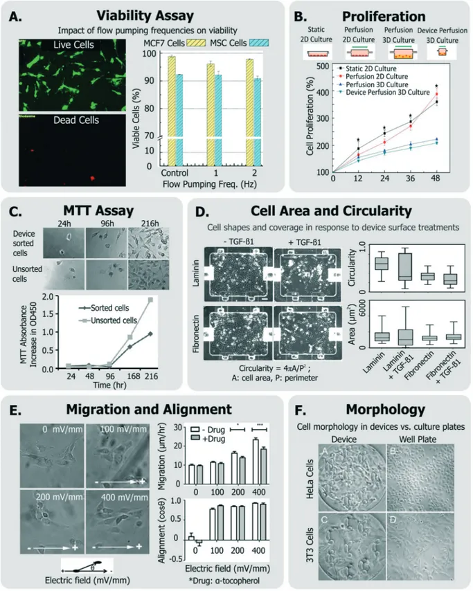
To investigate if a device and its operating conditions are not detrimental to cells, the most obvious (hence most popu-lar) measurement reported is that of cell viability30,39,126–130 (Fig. 3A), followed by assessment of cell morphology and proliferation49,50,54,129–132(Fig. 3B).
Viability can be quantified by a variety of assays,133though it is most conveniently done so by using colorimetric or fluores-cent probes which can imaged within microsystems. In many cases,132,134 viability has been assessed by exclusively labeling live cells using cell-permeant Calcein-AM stain, which becomes fluorescent and cell membrane-impermeant by intracellular es-terases in viable cells. On the other hand, others135–138 have measured fractions of cells with compromised membranes (la-beled dead), with Trypan blue stain.
While simple, the drawback of using these assays is that they do not label the “other” cells. A better approach is to have two stains, so that both live and dead cells are positively labeled. These ‘live/dead’ stains30,63,129,139,140 typically use Calcein-AM (which makes live cells fluoresce green) and ethidium homodimer-1 (which makes dead cells fluoresce red). This method is particularly useful as it quantifies rela-tive impacts of proliferation and cytotoxicity.
Viability-only assays fail to identify early apoptotic (and thus likely stressed) cells, as these cells do not have a
Open Access Article. Published on 16 October 2018. Downloaded on 11/14/2018 1:54:17 PM.
This article is licensed under a
Fig. 3 Exemplary generic assays for measuring cell health. A. Viability of MCF7 and MSC cells assessed in response to device flow pumping rates using a live-dead assay (adapted from ref. 129). B. Comparison of cell proliferation rates within device perfusion platforms and macroscale ana-logues (adapted from ref. 139, with permission from the Royal Society of Chemistry). C. Dynamics of cell metabolism assessed of device-sorted cells compared to unsorted cells using the MTT assay (adapted from ref. 131). D. Changes in cell circularity and area quantified in response to de-vice surface with laminin or fibronectin in order to assess ability of cells to undergo EMT within dede-vice environment (adapted from ref. 63, with permission from the Royal Society of Chemistry). E. Impact of applied electric fields upon migration rate and cell alignment to electric fields (adapted from ref. 150). F. Morphology and adhesion of HeLa and BALB/3T3 cells in a microfluidic cytotoxicity analysis device qualitatively com-pared to culture plates (adapted from ref. 51, with permission from the Royal Society of Chemistry).
Open Access Article. Published on 16 October 2018. Downloaded on 11/14/2018 1:54:17 PM.
This article is licensed under a
compromised membrane and are undetectable by Trypan blue, propidium iodide (PI), ethidium homodimer-1, and 7-aminoactinomycin D (7-AAD) stains. Phosphatidylserines, which flip from the cytoplasmic to extracellular leaflet during early apoptosis, can be stained with annexin V. Though not previously applied for device design or operating condition optimization, annexin V staining combined with PI141or with 7-AAD142has been conducted within microfluidics to identify viable (double negative), early apoptotic (annexin V positive, PI negative), or late apoptotic and dead (double positive) cells. Viability and apoptosis assays within microfluidic de-vices have been reviewed elsewhere.143
Viability can also be discerned by measuring cellular metab-olism, such as by monitoring culture glucose consumption rates,144or by monitoring the activity of cytochrome p450 intra-cellular enzymes (in the case of hepatocytes124,145). The meta-bolic dye 3-(4,5-dimethylthiazol-2-yl)-2,5-diphenyltetrazolium bromide (MTT) is applicable to a broad variety of cells, where the cell-soluble tetrazolium is metabolized to cell-insoluble blue formazan crystals that can be measured by colorimetric methods.133The MTT assay has been also been utilized to re-port on cell proliferation as a means of representing cell heath (Fig. 3C).131,146Although the readout of the MTT assay is sensi-tive to cell metabolism, it is also sensisensi-tive to the number of cells in the assay, the incubation time, requires colorimetric measurement, does not report on how many dead cells are in the system, and the short path length of microsystems makes colorimetric assay difficult. Given these challenges, one would choose MTT over live/dead assay when the system is not ame-nable to detailed microscopic imaging, or where metabolism it-self is of primary interest.
Cell proliferation has been also measured qualitatively, and used to infer that cells did not incur damage within different microsystems.51,54,147,148Other generic assays include measure-ments of cell shape and elongation63 spreading,149 migra-tion,150or other aspects of cell morphology.54,66,107,146 For in-stance, among a panel of assays, Park et al. quantified cellular circularity, elongation and area in response to various device surface coatings and treatments to discern the epithelial-mesenchymal transition (EMT) potential of device cultured cells63(Fig. 3D). Wu et al. investigated the impact of applied electric fields (EFs) upon fibroblast cell alignment migration rate, where they noted both increased migration rate and ROS production with increased field strengths150 (Fig. 3E). In an-other example, Wang et al. validated their microfluidic cytotox-icity analysis platform by qualitatively comparing cell morphol-ogy and adhesion of two cell lines (BALB/3T3 and HeLa cells) within their device to that within standard culture well plates (Fig. 3F), noting similar appearance.51
Specific assays. A viable cell does not ensure that the cell is stress-free or unperturbed. With this rationale, many groups have investigated cell health in their platforms be-yond the generic assays. One broad way to group these assays is based on whether they affect upstream, short-term, and rel-atively broad aspects of cell phenotype, or downstream, long-term, and more specific aspects.
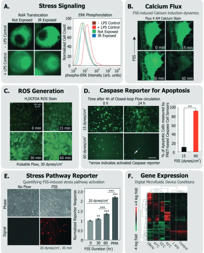
Some groups have investigated short-term changes (sec-onds-min range) in response to the device conditions (Fig. 4). Such assays typically focus on intracellular signaling. For in-stance, Perroud et al. measured NF-κB (a transcription factor that regulates inflammatory programs) translocation via engineered reporters, and extracellular signal-regulated nase (ERK) phosphorylation (a mitogen activated protein ki-nase) via staining, within cells that were sorted through their microfluidic device30 (Fig. 4A). El-Ali et al. focused more broadly on the stress-inducible mitogen-activated protein ki-nase (MAPK) pathway,151 through the phosphorylation of ERK, JNK and p38 kinases in cells passed through their de-vice.128Importantly, NF-κB, ERK, and MAPK are involved in transducing many of the stressors described earlier.
Calcium is an intracellular messenger important in regu-lating several essential cellular functions, and disturbances in its homeostasis can lead to many diseases.152FSS-induced calcium signaling has been measured by many groups137,149 (Fig. 4B). For instance, Yin et al. measured calcium flux in CHO cells in response to a large range of FSS (∼0.01–10 dynes per cm2) in their devices and noted FSS-dose depen-dent induction of Ca++signals, which matched signals from chemical agonists, even at moderate FSS.137
Cellular ROS and its direct effects can also be assessed. A common example is the probe 2′-7′-dichlorodihydrofluoresce diacetate (DCFDA), which is taken up by a variety of cells, where it fluoresces in response to intracellular ROS.153 Wu et al. used DCFDA to measure cell stress induced by electric fields in their device,150Lo et al. used the same measurement against oxygen gradients in their device,154 and Chin et al. used it to measure ROS induction in response to shear stress155(Fig. 4C). An immediate effect of ROS is DNA dam-age, which can be characterized by measuring histone γ-H2AX phosphorylation,156which is known to occur
follow-ing DNA double strand breaks.157
Other examples of short-term cellular responses to stress include changes in membrane receptor display, as well as ex-pression of immediate-response stress genes. Adams et al. in-vestigated platelet activation in their acoustophoresis device in response to the applied acoustic fields.117Specifically, they reported that expression of CD62 did not immediately vary in platelets with or without device acoustic fields, compared to off-chip prothrombin-treated positive controls. Wang et al. developed a microfluidic platform which utilized optical forces in sorting cells.28They measured the viability of sorted HeLa cells, and furthermore measured the expression of heat shock sensitive HSPA6 gene and multifactorial cell stress sen-sitive Fos gene immediately after sorting to assess if the de-vice activated stress pathways.
Short-term molecular assays have the advantage that they can be run quickly after exposure, rather than requiring ex-tended culture. This feature is appealing for the device user or designer. They also have a higher level of specificity than generic assays, as they focus on individual molecules and pathways. The drawback is their assay complexity; since they may require genetically modified cell lines (in the case of
NF-Open Access Article. Published on 16 October 2018. Downloaded on 11/14/2018 1:54:17 PM.
This article is licensed under a
Fig. 4 Exemplary specific assays for measuring cell health. A. Translocation of transcription factor RelA and flow cytometry measurement of ERK-phosphorylation in cells exposed to IR laser within a microfluidic sorter (adapted from ref. 30). B. Flow-induced changes in calcium flux and cell area measured using fluorescent Fluo-4 AM dye (adapted from ref. 149, with permission from the Royal Society of Chemistry). C. Accumulation of ROS in cells exposed to pulsatile flow measured using the fluorescent probe H2DCFDA (adapted from ref. 155, with permission from the Royal
So-ciety of Chemistry). D. Quantification of apoptotic cells among cells continually circulated within a closed-loop flow system using a FRET-based caspase reporter (adapted from ref. 71, under CC BY 4.0 license). E. Quantification of FSS-induced stress pathway activation and RFP induction using a cell-based FSS sensor (adapted from ref. 58, with permission from the Royal Society of Chemistry). F. Microarray analysis of cells exposed to various stimuli experienced in a digital microfluidic device in contrast to cells given heat shock (adapted from ref. 21, with permission from the Royal Society of Chemistry).
Open Access Article. Published on 16 October 2018. Downloaded on 11/14/2018 1:54:17 PM.
This article is licensed under a
κB translocation) or immunofluorescence with phospho-specific antibodies (in the case of kinase assays).
For longer term changes (∼hours), an assay will focus on gene expression or cell function. Assays focusing on expres-sion or function have the advantage of specificity; upregulation of a gene involved in the heat shock pathway is strong evidence that the cell underwent stress. Additionally, expression assays do not require many cells (a few thousand is routine, and single cell is possible), and the results can be quantitative (e.g., fold upregulation). Following this approach to quantify changes in gene expression, Hur et al. performed microarray analysis of 100 s of genes in MCF7 cells sorted through their inertial microfluidic device against unsorted controls to assess whether there were any global transcrip-tional changes as a result of sorting.140Similarly, Sharei et al. developed a microfluidic platform for intracellular delivery, and measured expression profiles of the POUF5 and ALP genes within mouse embryonic stem cells that passed through the device, to assess if certain conditions could initi-ate cell differentiation programs.127The drawbacks of these approaches are that 1) it takes∼hours for expression to oc-cur, which can increase optimization time, and 2) the exact time from stress to readout will affect the assay results, since the amount of mRNA or protein present will depend on both synthesis and degradation. Additionally, most of these assays are endpoint, and so require cell recovery and isolation, though live-cell reporters do exist.
As an alternative, some groups have utilized live cell-based sensors that can provide an integrated response to environ-mental stressors. Specifically, Davidsson et al. utilized a luciferase-based sensor in HeLa cells (activated by MAPK through G-coupled protein receptors158) to monitor long-term cell health their microfluidic platform.159Regmi et al. used a caspase-activation FRET reporter to monitor apoptosis activa-tion in continually circulating tumor cells within a micro-fluidic flow system71 (Fig. 4D). In this way, their approach allowed for classifying live, dying and dead cells in response to the flow conditions. Our own group has generated a suite of ‘open-source’ cell-based sensors that report on stress-induced pathways via fluorescence.58,109,160,161 Specifically, we engineered distinct sensors in NIH3T3 cells to express red fluorescence protein (RFP) when they were exposed to FSS,58,161 genotoxic stress,160,161 or to heat shock that acti-vated the respective stress pathways.161 For instance, short term FSS exposures activated our FSS sensors, and their in-duced RFP could be quantified by both microscopy and flow cytometry58(Fig. 4E). Our sensors have further been adapted and used by other groups to investigate stress in distinct de-vice microenvironments. For instance, Au et al. utilized our heat shock sensor109and further performed microarray analy-sis (Fig. 4F) and comet assay to investigate if their digital microfluidic platform biased cell health.21
Late-term changes in cell health (∼days) have also been reported. Lopacinska et al. measured neuronal-like PC12 cell viability with Calcein staining, cell cycle with PI staining, me-tabolism with MTT, and gene expression changes with
micro-arrays in response to common culture substrates used in microsystems such as polydimethylsiloxane (PDMS), poly-methylmethacrylate (PMMA) and polystyrene.146 As another example, Villa-Diaz et al. measured expression of OCT3/4 and NANOG genes in cultured hESCs to assess if their device FSS altered pluripotency after days of culture.66
A common approach to investigate the cellular inflamma-tory response is to quantify the release of relevant cytokines, chemokines and interleukins.162,163These serve as important markers for assessing acute and chronic septic and aseptic inflammation. Despite the prevalence of such assays in bio-logical sciences, they are infrequently performed in the con-text of microsystem design or assessment. This may in part be a result of technical challenges in assaying secreted pro-teins in microsystems (e.g. low cell numbers or analyte dilu-tion). These challenges have motivated the development of intricate microsystems for inline detection of inflammatory biomarkers,162,163 or surface patterning of protein detection regions within microfluidic channels.164–166While these plat-forms may allow for sensitive and multiplexed detection of secreted analytes, they require specific expertise in device integration and operation and hence it can be challenging to integrate such platforms within typical device workflow and optimization. Notably, bead-based protein sensors that can be introduced and incubated with cells within devices can be relatively convenient in this regard, and can enable both im-aging-based167 and off-chip168 non-destructive readouts. Al-ternative strategies exist (e.g. those applied for immunology studies169) may also provide similar benefits. Broadly, if these aforementioned challenges can be solved with other specific and conveniently-translatable methodologies, then sensitive and specific quantification of secreted cytokines from cells within device environment will serve as a powerful, non-destructive assay for cell health.
Though extensive device optimization or characterization using a variety of cell health assays is not commonly found in literature, the examples from various groups discussed above do demonstrate a few important points. First, the microsystems community does care about cell health in their platforms, and further, it does employ a variety of techniques and assays to investigate the phenomenon. Second, there are several examples of groups using similar assays (e.g. viability, proliferation), but there lacks a consensus when it comes to applying assays specific (and likely more informative) to cell health. We believe that the latter is related to certain inher-ent challenges in adapting established cell health specific as-says within the device design and workflow.
Current challenges in assessing cell
health within microsystems
To systematically assess cell state in a device microenviron-ment, one has to consider the variety of assays (generic and specific) used for assessing cell health. In this regard, it is common to adapt standard assays within engineered micro-systems, or apply them to cells that have experienced the
Open Access Article. Published on 16 October 2018. Downloaded on 11/14/2018 1:54:17 PM.
This article is licensed under a
device microenvironment. While some assays, such as imaging-based assays, are easily translatable to microscale technologies, there exist challenges in finding assays that are specific to cell health, quantitative, and conveniently adapt-able. Some of these challenges are elaborated below.
Challenges and tradeoffs
Challenge of low cell numbers. In microscale platforms where cells are retained in small volumes (for culture, analy-sis, manipulation, etc.), there are inherent limitations on the number of cells available for running any assay. ‘Flow-through’ devices such as sorters that do not work with rare cells, or instead work with cell lines are an exception to this limitation, since the cell availability is directly related to the starting amount of cells. In most other cases, with limited cell numbers it becomes challenging to run biochemical as-says such as western blots, that typically require >100 000 cells. Even qRT-PCR, which can be run at the single-cell level, is much simpler when one uses>1000 cells.
Complexity of specific assays. Many specific assays are de-structive, costly and complex, and hence technically prohibi-tive. Assays of intracellular protein levels or gene expression have been performed by our group82and others49,63while de-veloping various platforms. However, within the microfluidic community the usage of such methods to interrogate device bias is not routinely performed for certain reasons.
First, it is usually not obvious as to which cell health bio-markers to measure, and hence which assay to run. Second, many relevant assays (such as qRT-PCR, western blotting, flow cytometry) require may steps such as washing or mixing biological materials, isolation, purification, etc. These addi-tional steps need to be incorporated into the device workflow based on practicality and convenience. Obtaining starting materials (cells or cellular constituents) can be done by either taking the cells off-chip for processing, or bringing the assay reagents on to the chip. Retrieving immobilized cells from microfluidic devices is often a non-trivial task. While at-tached cells can be harvested by flushing enzymatic dissocia-tion buffers,132,170 one has to be careful about cell losses in the fluidic network. Alternatively, cells can be retrieved with optical methods,171 or mechanically (e.g. with micromanipu-lators147,172or micropipettes50), each of which require sophis-ticated technical expertise. On the other hand, while bringing assay reagents to cells is possible through specific fluidic ar-chitectures173 or device designs,49,174 these approaches may not be easy to integrate within one's own device workflow. Fi-nally, many specific assays are end-point and thus destroy the cell, making it laborious to investigate longitudinal as-pects of cell health.
Lack of standardization. Perhaps the most noteworthy chal-lenge in assessing cell health in microsystems is the lack of standard assays and associated quantitative metrics. Given the broad diversity of both specific and generic assays to study cell health, microsystems engineers need to pick specific assays which are convenient to adapt given a certain amount of cells
in their particular workflow. Such standardization would in turn allow inter-platform and inter-laboratory comparisons.
Guidelines for best practices
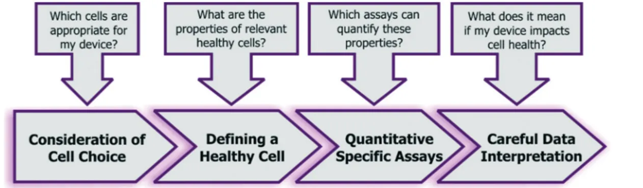
Given the combined complexity of cell stress biology, vast number of assay choices, and large parameter space for microsystem design and operation, it's likely that there may be not be an exclusive ‘universal design or operating condi-tion’ for optimizing all devices for healthy cells. However, it is possible to outline as set of best practices for the micro-systems engineer and end-user, which they can adapt in their own device context (Fig. 5):
Consideration of cell choice. When designing, it's impor-tant to determine if the platform will be used for specific cell types or a variety of cells. For specific phenotypes, one must determine if the microenvironment will mimic physiologic (e.g., bioreactor platforms) or pathologic (disease models) conditions. This is important because it guides the pheno-type selection process and defines desirable biological states. For instance, in a disease-mimic model cells can be inten-tionally stressed, whereas in a physiologic model cells should remain in their in vivo healthy state. For instance, culturing (diseased) cancer cells in a disease-relevant microenviron-ment makes sense, and by using them one can investigate if the microenvironment is enabling the cancerous cells to re-main in their equilibrium biological state. However, using cancer cells to check whether a generic cell culture platform, sorting device, etc., stresses cells is not informative. Such cells were not normal to begin with (i.e., they may have dys-functional or dysregulated stress-response pathways), and it is impractical to judge and learn from perturbations that make such cells more or less cancerous. For instance, Barnes J. et al. compared primary blood cells, primary epithelial cells, non-transformed cell lines and cancer cell lines in their ability to withstand identical FSS conditions.175 Here they noted that cancer cell lines demonstrated high resistance to FSS-induced damage, in contrast to non-transformed cell lines and primary cells that took a relatively bigger hit on their viability. Results such as these demonstrate the impor-tance of carefully choosing relevant cells for validating the impact of engineered systems on cell health.
Defining the reference cell. Prior to any optimization, it is important for device designers and users to establish the ref-erence‘healthy cell’. In some cases it may make sense to use cells cultured in incubators as a reference (such as when working with cell lines). Cells exposed to the device environ-ment can then be compared against cells from a culture incu-bator to determine device-induced effects. When working with primary cells, rare cells, or cells that require laborious processing prior to usage it may not always be convenient to obtain a reference population. Furthermore, in some of these cases, the cells may already have become stressed or dysfunc-tional in-part due to the ‘pre-processing’ and hence may be inappropriate for device optimization. Here, one should use a multi-step optimization, first using a conveniently available
Open Access Article. Published on 16 October 2018. Downloaded on 11/14/2018 1:54:17 PM.
This article is licensed under a