Research Article
Screening of Bioactive Peptides Using an Embryonic Stem Cell-Based
Neurodifferentiation Assay
Ruodan Xu,1,2Maxime Feyeux,2Stéphanie Julien,2Csilla Nemes,3Morten Albrechtsen,1 Andras Dinnyés,3,4,5and Karl-Heinz Krause2,6
Received 31 October 2013; accepted 27 January 2014; published online 21 February 2014
Abstract. Differentiation of pluripotent stem cells, PSCs, towards neural lineages has attracted significant attention, given the potential use of such cells for in vitro studies and for regenerative medicine. The present experiments were designed to identify bioactive peptides which direct PSC differentiation towards neural cells. Fifteen peptides were designed based on NCAM, FGFR, and growth factors sequences. The effect of peptides was screened using a mouse embryonic stem cell line expressing luciferase dual reporter construct driven by promoters for neural tubulin and for elongation factor 1. Cell
number was estimated by measuring total cellular DNA. We identified five peptides which enhanced
activities of both promoters without relevant changes in cell number. We selected the two most potent
peptides for further analysis: the NCAM-derived mimetic FGLL and the synthetic NCAM ligand,
Plannexin. Both compounds induced phenotypic neuronal differentiation, as evidenced by increased neurite outgrowth. In summary, we used a simple, but sensitive screening approach to identify the neurogenic peptides. These peptides will not only provide new clues concerning pathways of neurogenesis, but they may also be interesting biotechnology tools for in vitro generation of neurons. KEY WORDS: bioactive peptides; embryonic stem cells; neural differentiation.
INTRODUCTION
Neural differentiation of pluripotent stem cells (PSCs) has become a topic of major interest in contemporary biomedical research (1,2). From a basic science point of view, PSC neural differentiation recapitulates key steps of embryo-genesis and fetal development, giving unprecedented access to the underlying mechanisms (3). Therefore, PSC-derived neurons have a variety of biotechnology applications. Indeed, they have an important potential for in vitro use, including disease modeling (4,5), development of central nervous system (CNS) drugs (6), and neurotoxicity testing (7). Much discussed is also the potential of PSC-derived neurons for in vivo use, in particular cell replacement therapy (8–12).
However, in order to allow PSC-derived neurons to live up to their biotechnology potential, there is a need for better tools to direct and control neural differentiation.
Traditionally, growth factors (13), extracellular matrix proteins (14), and other endogenous neurogenic proteins (15) have been used to direct neurogenesis of PSCs in vitro (16,17). This approach is logical because it allows mimicking physiological steps of neural differentiation; however, there are limits to the use of whole lengths proteins: they are costly, tend to be relatively unstable. Also, if proteins are supposed to be further developed for an in vivo use, they have major limitations, in particular cross of the blood brain barrier, and potential of immunogenicity (indeed, the adult organism may recognize as non-self proteins which are only expressed during embryonic development (18)). For these reasons, there have been increasing efforts to develop small molecules that mimic growth factor actions, but would not have the limitations of the latter (19–21). There are good examples for a successful application of this approach, for example the so-called dual Smad inhibition (22) or Notch inhibition through gamma-secretase inhibitors (23). However, the specificity of
small molecule inhibitors is often limited and they have a potential for unpredictable toxicity (24). The use of peptides as biological drugs potentially combines the advantages of both approaches described above. Similar to physiological proteins, small peptides can be designed to act within developmentally relevant protein networks. Yet because of their much smaller size, they can be synthesized at lower cost with higher stability and high chemical and biological
1ENKAM Pharmaceuticals A/S, Copenhagen, Denmark.
2Department of Pathology and Immunology, University of Geneva
Medical Center, Geneva, Switzerland.
3Biotalentum Ltd, Gödöllő, Hungary.
4Molecular Animal Biotechnology Laboratory, Szent Istvan
University, Godollo, 2100, Hungary.
5Department of Farm Animal Health, Faculty of Veterinary
Medicine, Utrecht University, Utrecht, 3584 CL, The Netherlands.
6To whom correspondence should be addressed. (e-mail:
ABBREVIATIONS: PSC, Pluripotent stem cells; CNS, Central nervous system; NCAM, Neural cell adhesion molecule; FGFR, Fibroblast growth factor receptor; NSCs, Neural stem cells; MEFs,
Murine embryonicfibroblasts; NPCs, Neural progenitor cells.
400 1550-7416/14/0300-0400/0 # 2014 American Association of Pharmaceutical Scientists
diversity can be achieved. Also they are not immunogenic. Peptides often act through physiological receptor pathways and therefore induced rapid activatory or inhibitory re-sponses. Basically, there are two approaches to the discovery of bioactive peptides: screening of randomly generated large peptide libraries or generation of targeted peptide libraries based on known bioactive sequences within proteins.
During the embryonic development, the neural cell adhesion molecule (NCAM) plays an important role in neurogenesis. It is critically involved in proliferation, migra-tion, survival, and differentiation of neural progenitors (25). NCAM is able to interact with itself through both homophilic cis- and trans-interactions forming zipper-like complexes. NCAM binds to several heterophilic ligands as well, like: thefibroblast growth factor receptors (FGFRs), the glial cell line-derived neurotrophic factor (GDNF), and its cognate receptor, GDNF family receptor α (GFRα) among others (26). Besides brain-derived neurotrophic factor (BDNF), nerve growth factor (NGF), the last decade of research has dramatically widened the picture of the functional roles of NCAM showing that NCAM is a multifunctional regulator of cell adhesion, intracellular signaling, and cytoskeletal dynam-ics, all phenomena of key importance in stem cell differenti-ation (27). Based on the structure–function relationship of
NCAM, small peptides mimicking NCAM activities mediated via its different binding sites were discovered (27–29). For example, peptides derived from the structure of NCAM and growth factors induced the neural differentiation in primary cerebellar granule neurons or hippocampus neurons (29–37). It was the purpose of this study to investigate neurogenic properties of small peptides derived from bioactive sequences within neurotrophic proteins. The initial screen was performed with a dual promoter/reporter line (derived from mouse embryonic PSCs). Key results were confirmed by immunofluo-rescence and neurite outgrowth assays. Among 15 peptides investigated in this study, at least two were found to have potent neurogenic activity during PSCs differentiation. Our results indicate that a peptide design based on bioactive sequences of relevant proteins, such as NCAM or related growth factors, represents a promising approach for the understanding and control of neural differentiation.
MATERIALS AND METHODS Peptides
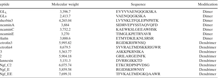
All peptides were synthesized by Peptides&Elephants GmbH (Potsdam, Germany). Peptides were synthesized using the Fmoc-protection strategy on TentaGel resin using Fmoc-protected amino acids. The peptides were synthesized in two forms: (a) dimeric peptides composed of two linear monomers linked together by a C-terminal lysine residue (FGLL, FGLs, Inherbin3, EncaminA, EncaminC, EncaminE, and Gliafin), and (b) tetrameric dendrimers composed of four monomers coupled to a lysine backbone (Betrofin3, Betrofin4, C3, P2, Plannexin, hNgf_C2, hNgf_E, and hNgf_EE;29–38; see overview of peptides in Table I). Peptides were at least 85% pure as estimated by mass spectrometry and analytical high-performance liquid chromatography (HPLC). The peptides were dissolved in MilliQWater, and the concentrations of peptides were determined spectrophotometric by measuring the absorption at 205 nm (39).
Reagents
Murine CGR8 ES cell line was purchased from European Collection of Cell Culture. The human embryonic stem (hES) H1 was obtained from WiCell Research Institute, Inc., WA01. The bone marrow stromal MS5 cell line was kindly provided by Katsuhiko Itoch (40). Cell culture medium, fetal bovine serum, knockout serum replacement, penicillin/streptomycin, N2 supple-ment, non-essential amino acid, sodium pyruvate, FGF2, and FGF8 were purchased from Gibco, Invitrogen Corporation (Paisley, Scotland). Dual-luciferase® Reporter Assay System was from Promega (Madison, WI, USA); Poly-L-ornithine was from Sigma-Aldrich. Antibody and dilution were as follows: rabbit anti-βIII-tubulin (1:2,000; Covance, Princeton, NJ, USA). The fluorochrome-coupled secondary antibody was used, AlexaFluor® 555 Goat Anti-rabbit (1:1,000; Invitrogen-Molecular Probes).
Mouse ES Cell Culture
Dual luciferase expressing CGR8-2luc cells were obtained by transduction of mouse ES CGR8 cells with the 2 k7 EF1-αS Rluc/Tα1α Fluc vector as previously described (41,42). EF1-αS
corresponds to the short promoter of the eukaryotic translation elongation factor 1 alpha 1 (EEF1A1) and Tα1α to the Tubulin alpha1 (TUBA1A) promoter. Cells were cultured on 0.1% gelatin-coated dishes in CGR8-2luc maintenance medium: BHK21 medium, 10% fetal bovine serum (FBS), 2% sodium pyruvate, 1% non-essential amino acids, 2 mM L-glutamine, beta-mercaptoethanol (0.1 μM), 1% penicillin/streptomycin, and leukemia inhibitory factor LIF (43). CGR8-2luc cells grow in a feeder-independent manner and therefore no murine embryonicfibroblasts (MEFs) or other feeder cells were used. CGR8-2luc Differentiation and Exposure to Peptides
The overview schemes of differentiation protocols (proto-col 1 and 2) were shown in Fig.1. For the primary screening assay, cells were cultured as described previously (42). Briefly,
103CGR8-2luc cells per well were plated on gelatin-coated 96-well plates in differentiation medium: BHK21 medium, 20% FBS, 2% sodium pyruvate, 1% non-essential amino acids, 2 mM L-glutamine, beta-mercaptoethanol (0.1 μM), 1% penicillin– streptomycin; 48 h later, the medium was removed and replaced by 300 μl fresh differentiation medium with various concentrations of growth factors, peptides, or solvent control. Seventy-two hours later, cells were assayed for Firefly and Renilla luciferase activity.
To perform the neurite elongation assay, neuronal differen-tiation was carried out as described (42,44). Briefly, irradiated
MS5 cells (1.75×10⁵per well) were seeded in 6-well plates. The next day, CGR8-2luc cells (0.6×10³to 3×10³cells per well) were plated on the MS5 layer in SR medium (DMEM high glucose supplemented with 15% knockout serum, non-essential amino acids, 2-mercaptoethanol, Pen/Strep) for 5 days with growth factors, peptides, or solvent control found in the primary screen. Five days later, cells were then trypsinized and seeded 1,000 cells/ well onto polyornithine-coated 24-well plates in N2 medium (DMEM high glucose, N2 supplement with 10 ng/ml human basic fibroblast growth factor, Pen/Strep) and cultured for four additional days without compounds addition.
Dual-Luciferase Assay
Luciferase activity was measured with Dual-luciferase® Reporter Assay System kit. CGR8-2luc ES cells were lysed in 96-well plates according to the manufacturer’s instructions. Luminescence measurements were performed in triplicates on Fluostar Optima reader (BMG Labetch GmbH, Germany). Luminescence counts were normalized by com-parison to the control wells without treatment after subtrac-tion of the background luminescence.
Propidium Iodide and Resazurin (AlamarBlue®) Assay DNA quantity was determined by Propidium iodide (PI). Propidium iodide was added to cell homogenates after lucifer-ase test, at afinal concentration of 50 μg/ml and incubated for an additional 2 h. After incubation, thefluorescence intensity was measured with a Fluostar Optima microplate reader (excitation 544 nm±15 nm, emission 620 nm±15 nm).
Cell viability was determined using alamarBlue® (Invitrogen, Carlsbad, CA92008, USA). In brief, CGR8-2luc cells were seeded in 96 wells at a non-confluent cell density and incubated for 24 h under standard cell culture conditions. After 4 h of exposure to Methylmercury (MeHg), 20 μl of alamarBlue® solution (diluted 1:10 from stock solution) was added to each well. After a 2-h incubation at 37°C,fluorescence was measured (excitation 544 nm, emission 590 nm) and corrected to background control (no cells). Results are given as fractional survival as compared to untreated cells.
Human ES Cell Culture
The human embryonic stem (hES) cells H1 (WiCell Research Institute, Inc., WA01) cell line was maintained on irradiated MEFs freshly isolated from mouse 13.5 dpc embryos (C57BL/6 strain). The hES cells cultures were fed daily DMEM/ F12 glutamax supplemented with 20% knockout serum replace-ment, 1 mM nonessential amino acids, 1% penicillin/streptomycin, 0.55 mM 2-mercaptoethanol, and 5 ng/ml recombinant human FGF2. hES cells were enzymatically treated with collagenase for passaging every 5–7 days.
Neural Differentiation of H1 hESC Line
Neural induction of H1 hES cell line was based on dual SMAD inhibition, slightly modified from (45). The overview scheme of differentiation protocol 3 was shown in Fig.1. Briefly,
undifferentiated hESC colonies were starved in N2B27 medium supplemented with FGF2 (5 ng/ml, Invitrogen). The next day H1 hES cells were manually detached from the feeder-layer, collected in differentiation medium composed of N2B27 medi-um and transferred for 6 h to low-attachment plate. Cells were Table I. Overview of the Peptides
Peptide Molecular weight Sequence Modification
FGLL 3,396.7 EVYVVAENQQGKSKA Dimer
FGLs 2,413.7 VAENQQGKSKA Dimer
Inherbin3 4,265.04 LVYNKLTFQLEPNPHTK Dimer
EncaminA 3,884 SIDRVEPYSSTAQVQFD Dimer
EncaminC 3,752.2 KAEWKSLGEEAWHSK Dimer
EncaminE 3,270 TIMGLKPETRYAVR Dimer
Gliafin 3,888.6 ETMYDKILKNLSRSR Dimer
Betrofin3 5,995.82 RGIDKRHWNSQ Dendrimer
Betrofin4 8,679.5 SYVRALTMDSKKRIGWR Dendrimer
C3 5,363.77 ASKKPKRNIKA Dendrimer
P2 5,904.18 GRILARGEINFK Dendrimer
Plannexin 5,151.3 DVRRGIKKTD Dendrimer
hNgf_C2 6,075.74 ETKCRDPNPVDSG Dendrimer
hNgf_E 5,859.58 RGIDSKHWNSY Dendrimer
hNgf_EE 7,699.31 TFVKALTMDGKQAAWR Dendrimer
Fig. 1. Overview scheme of the differentiation protocols. Protocol 1 is the primary screening assay. CGR8-2luc cells per well were plated on gelatin-coated 96-well plates in differentiation medium. Forty-eight hours later, the medium was removed and replaced by peptides.
Seventy-two hours later, cells were assayed for Firefly and Renilla
luciferase activity. Protocol 2 is the neurite outgrowth assay. CGR8-2luc cells were cocultured on MS5 cells with peptides exposure for 5 days. Five days later, cells were then seeded on polyornithine-coated plates and were cultured for four additional days without compounds addition. Protocol 3 is the human ESC neural differen-tiation morphology assay. The obtained neural stem cells from dual SMAD protocol, cells were cultured on laminin-polyornithine-coated plates with peptides exposure from day 12 to day 24
then seeded on 300 ng/cm2poly-ornithine (Sigma) and 500μg/ cm2laminin (Trevigen) sequentially coated tissue culture plates. Differentiation medium was changed after 24 h then every other day. LDN193189 (1 μM, Axonmedchem) and SB431542 (20μM, Tocris Biosciences) were added from day 0 and on for every medium change until rosette neural stem cell (R-NSC) arose at day 8 to 12. R-NSC were manually collected at day 10– 14, enzymatically detached using 0.05% trypsin (Invitrogen), and seeded at 105cells/cm2on polyornithine and laminin-coated tissue culture plates in N2B27 medium supplemented with FGF2 (10 ng/ml, Invitrogen), EGF (10 ng/ml, R&D systems). Cells were maintained in the same medium and passaged every 2–3 days for no more than 25 passages.
Immunofluorescence Analysis
Immunostaining with rabbit anti-βIII-tubulin and Alexa Fluor 555-conjugated anti-rabbit IgG secondary antibody was performed as described previously (42). In brief, cells plated on polyornithine-coated glass cover slips werefixed with 2% PFA, permeabilized with 0.5% (v/v) Triton X-100 and stained for Tubulin and counterstained with DAPI to visualize the
nucleus. Negative control immunostaining was performed withoutfirst antibody. After wash with PBS, coverslips were mounted on glass slides using FluorSaveTM Reagent (Calbiochem, San Diego, CA, USA).
Determination of Neurite Elongation
Images were acquired on a Mirax Micro digital slide scanner (Carl Zeiss) or a Zeiss axioplan microscope equipped for epifluorescence. Immunostaining and nuclear staining quantifications were performed using the Mirax Viewer (Carl Zeiss) and MetaXpress (Molecular Devices) software with the following settings of cell body recognition: min width 4μm, max width 28 μm, 40 gray levels above background. Total neurite outgrowth was quantified using the neurite outgrowth analysis module, and total cell numbers were quantified with the count nuclei analysis module.
Peptide Stability Assay
Peptides were dissolved in FBS (270μl) to a concentra-tion of 0.2 mM (30 μl of 2 mM) and incubated at 37°C.
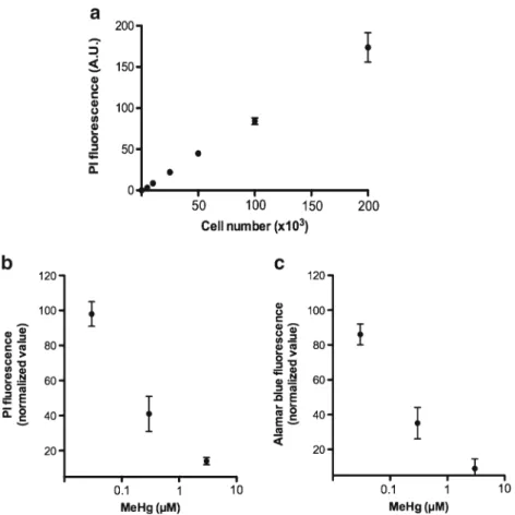
Fig. 2. Correlation betweenfluorescence and cell count. Cells were grown for 24 h under
undifferentiation and differentiation conditions at 37°C and were subsequently tested in a
96-well plate (200μl final volume). a Numbers of CGR8 cells were cultured in maintenance
medium for 24 h, PI was added to each well (final concentration in well 50 μg/ml), and
fluorescence was determined after 2 h incubation. b 200×103CGR8 cells in differentiation
medium were cultured with Methylmercury for 48 h, PI was added to each well (final
concentration in well 50μg/ml), in which the cells were incubated for 2 h at 37°C, and after
whichfluorescence was determined. c 200×103CGR8 cells in differentiation medium were
cultured with Methylmercury for 48 h. Each well was added 20μl of 10× resazurin in PBS.
Aliquots (30μl) were removed at various time intervals and quenched with aq. 60 μL trichloroacetic acid (5%). The aliquots were vortexed and incubated for 15 min at 4°C prior
to centrifugation at 18,000×g for 2 min. The supernatants were analyzed by RP-HPLC to quantity peptide relative to time zero.
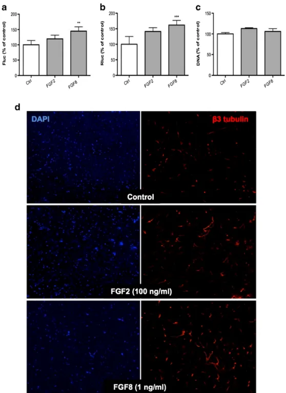
Fig. 3. Effect of cognate ligands of FGF receptor on CGR8-2luc neural differentiation. a, b Dual luciferase activity of FGF2 and FGF8 in CGR8-2luc cells. CGR8-2luc were grown on 0.1% gelatin for 48 h, afterwards continued to culture in the presence of the indicated concentrations of FGF2 or FGF8 for 72 h. Cells were
subsequently tested a Firefly, b Renilla luciferase activity, and c DNA quantity by PI assay. Results are
expressed as percentages±SEM with untreated controls set at 100%. Error bars indicate SEM based on three independent experiments. d Effect of FGF2 and FGF8 on neurite extension. CGR8-2luc were grown on MS5 for 5 days with FGF2 (100 ng/ml) or FGF8 (1 ng/ml) in SR medium. Cells were subsequently dissociated and replated in N2 medium for additional 4 days without compounds addition. Vehicle, FGF2,
and FGF8-treated ES cells were examined forβ-III tubulin (red), and nuclei were stained with DAPI (blue)
on day 9 of differentiation. **P<0.01, ***P<0.005, compared with untreated cells (control; Student’s paired
Statistical Analysis
The data were plotted as mean (SEM from three independent experiments. Statistical significance was ac-cepted at p <0.05. The results were analyzed with one-way analysis of variance (ANOVA) or t test using Prism 5 software (GraphPad Software, San Diego, CA).
RESULTS
Propidium Iodide Fluorescence as an Approximation of Cell Number
The dual reporter line detects the activity of a Tα1 promoter fragment and an EF1-α promoter fragment. The initial concept of this promoter construct was to use the Tα1 promoter as a reporter for neural differentiation and the EF1-α promoter as a house keeping promoter that would reflect the cell number. However, as published previously (46), the EF1-α promoter is responsive to cell differentiation
(down-regulation) and to neuroactive and neurotoxic com-pounds (upregulation or downregulation). Thus, while the EF1-α promoter activity contributes to fingerprinting of the effect of a compound on neural differentiation, it does not provide information about the number of cells. We have therefore added a novel parameter to our neural differen-tiation assay, namely propidium iodide (PI) fluorescence. This assay is performed by adding PI to cell homogenates after the luciferase assays is performed. To validate the assay, we first investigated the correlation between cell number (undifferentiated CGR8 cells) and the PI fluores-cence. As shown in Fig. 2a there is a good correlation. To study the behaviour of the assay in a more complex situation, we investigate neurotoxicity of Methylmercury comparing the classical cytotoxicity assay Alamar blue with our PI assay. As shown in Fig. 2 (panel b and c) the PI assay correlated well with the Alamar blue assay. Thus, we conclude that the PI assay provides an approximation of the cell number in our assay system. Of importance, the assay is readily combined with the dual luciferase assay and PI measurement can be performed in the same homogenates used for measuring luciferases activity.
Effect of Growth Factors in the Assay System
To validate our assay system, we analyzed the effects of two growth factors FGF2 and FGF8. We first investi-gated their impact in a spontaneous differentiation assay, based on culture of CGR8-2luc cells in a basal neural differentiation medium (see Methods). A 3-day exposure of cells to FGF8 led to increased Tα1 and EF1-α promoter activities without a significant effect on the PI signal (Fig. 3a–c). A similar tendency, however without statistical significance was observed for FGF2. We next investigated the impact of the growth factors in a stroma cell-enhanced differentiation assay. Figure 3d shows im-munofluorescence analysis of FGF2- and FGF8-treated differentiated CGR8-2luc cells. These results show that both growth factors, in particularly FGF8, enhance neurite outgrowth in differentiating CGR8-2luc cells (for quanti-fication see below).
Primary Screen of Bioactive Peptides
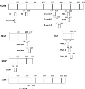
Peptides derived from growth factors could mimic the effect of growth factors or even be more efficacious. We have therefore used peptides designed based on well-defined regions of NCAM, BDNF (brain-derived neurotrophic factor), GDNF (glial cell derived neurotrophic factor), NGF (nerve growth factor), FGFR (fibroblast growth factor receptor), and EGFR (epidermal growth factor receptor). Schematic representations of the origins of the peptide sequences are shown in Fig.4.
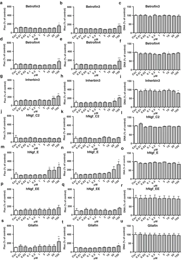
These putatively bioactive peptides were investigated using the neural differentiation assays described above. Dose– response analysis ranging from 10 nM to 100μM was performed for all peptides, and Tα1 and EF1-α promoter activities, as well as DNA content were measured (Figs.5and6). The obtained results allowed grouping the peptides into three categories, based on their responses in the dual luciferase assay: group 1 had no effects or even showed mild inhibitory effect; for group 2, effects were only observed at 100μM; for group 3, a dose– response was observed starting from either 3 or 10μM. Group 1 comprises 4 NCAM-derived peptides (FGLs, EncaminA, Encamin C, P2) and 2 NGF-derived peptides (hNgf_C2, hNgf_EE). Obviously group 1 is not of further interest for our Fig. 4. Schematic showing positions in NCAM, BDNF, NGF, GDNF, and EGFR of the various bioactive peptides described in this study
Fig. 5. Effect of peptides derived from NCAM on CGR8-2luc neural différentiation. CGR8-2luc were grown on 0.1% gelatin for 48 h and
continued to culture in the presence of the indicated concentrations (0.01–
100μM) of FGLs, FGLL, EncaminA, EncaminC, EncaminE, C3, P2, and
Plannexin for 72 h. Cells were subsequently tested a, d, g, j, m, p, s, v Firefly, b, e, h, k, n, q, t, w Renilla luciferase activity, and c, f, i, l, o, r, u, x DNA quantity. Results are expressed as percentage±SEM with untreated controls set at 100%. Error bars indicate SEM based on three independent experiments. *P<0.05, **P<0.01, ***P<0.005, compared with controls
(one-way, repeated-measures ANOVA, Bonferroni’s post hoc test)
P2 Ctr l 0 .0 10 .0 3 0. 1 0 .3 1 3 1 0 3 0 100 0 200 400 600 µM Fluc (% of control) Plannexin Ctr l 0 .0 10 .0 3 0.1 0 .3 1 3 1 0 3 0 100 0 200 400 600 *** µM Fluc (% of control) Plannexin Ctr l 0 .0 10 .0 3 0. 1 0 .3 1 3 1 0 3 0 100 0 200 400 600 ** *** µM Rluc (% of control) Plannexin Ctr l 0 .0 10 .0 3 0.1 0.3 1 3 1 0 3 0 100 0 50 100 150 ** ** µM DNA (% of control) P2 Ctr l 0 .0 10 .0 3 0. 1 0 .3 1 3 1 0 3 0 100 0 200 400 600 ** µM Rluc (% of control) C3 Ctrl0 .0 10 .0 3 0. 1 0 .3 1 3 1 0 3 0 100 0 50 100 150 ** * µM DNA (% of control) EncaminE Ctr l 0 .0 10 .0 3 0.1 0 .3 1 3 1 0 3 0 100 0 200 400 600 ** µM Rluc (% of control) EncaminE Ctrl0 .0 10 .0 3 0. 1 0.3 1 3 1 0 3 0 100 0 50 100 150 µM DNA (% of control) C3 Ctr l 0 .0 10 .0 3 0.1 0 .3 1 3 1 0 3 0 100 0 200 400 600 * ** *** µM Rluc (% of control) C3 Ctr l 0 .0 10 .0 3 0. 1 0 .3 1 3 1 0 3 0 100 0 200 400 600 ** *** µM Fluc (% of control) FGL L Ctr l 0 .0 10 .0 3 0 . 1 0 .3 1 3 1 0 3 0 100 0 200 400 600 * ** *** *** µM Fluc (% of control) FGL L Ctr l 0 .0 10 .0 3 0. 1 0 .3 1 3 1 0 3 0 100 0 200 400 600 * *** *** *** µM Rluc (% of control) EncaminA Ctr l 0 .0 10 .0 3 0.1 0.3 1 3 10 30 100 0 200 400 600 * µM Rluc (% of control) EncaminA Ctr l 0 .0 10 .0 3 0. 1 0.3 1 3 1 0 3 0 100 0 50 100 150 µM DNA (% of control) EncaminC Ctr l 0 .0 10 .0 3 0.1 0 .3 1 3 1 0 3 0 100 0 50 100 150 µM DNA (% of control) EncaminC Ctr l 0 .0 10 .0 3 0.1 0.3 1 3 1 0 3 0 100 0 200 400 600 * * µM Rluc (% of control) EncaminC Ctrl0.010.03 0.1 0.3 1 3 10 30 100 0 200 400 600 * * ** µM Fluc (% of control) EncaminE Ctr l 0 .010 .03 0.1 0.3 1 3 1 0 30 100 0 200 400 600 *** µM Fluc (% of control) EncaminA Ctrl0 .0 10 .0 3 0.1 0 .3 1 3 1 0 3 0 100 0 200 400 600 µM Fluc (% of control) FGLs Ctr l 0.010.03 0.1 0.3 1 3 10 30 100 0 200 400 600 µM Rluc (% of control) FGLs Ctr l 0 .0 10.03 0.1 0 .3 1 3 1 0 30 100 0 200 400 600 µM Fluc (% of control) FGLs Ctrl0 .0 10 .0 3 0.1 0.3 1 3 1 0 3 0 100 0 50 100 150 µM DNA (% of control) FGL L Ctr l 0 .0 10 .0 3 0 . 1 0 .3 1 3 1 0 3 0 100 0 50 100 150 ** *** µM DNA (% of control) P2 Ctr l 0 .0 10 .0 3 0.1 0 .3 1 3 1 0 3 0 100 0 50 100 150 µM DNA (% of control) f h g i c b d a e o q p r l k m j n x u t s w v
Inherbin3 Ctr l 0 .0 10 .0 3 0. 1 0 .3 1 3 1 0 3 0 100 0 200 400 600 *** * µM Rluc (% of control) Betrofin4 Ctr l 0 .0 10 .0 3 0. 1 0.3 1 3 1 0 30 100 0 200 400 600 *** µM Rluc (% of control) Gliafin Ctr l 0 .0 10 .0 3 0. 1 0.3 1 3 1 0 3 0 100 0 200 400 600 ** µM Fluc (% of control) hNgf_EE Ctr l 0 .0 10 .0 3 0.1 0 .3 1 3 10 3 0 100 0 200 400 600 µM Fluc (% of control) hNgf_E Ctr l 0 .0 10 .0 3 0. 1 0 .3 1 3 1 0 3 0 100 0 50 100 150 ** µM DNA (% of control) hNgf_E Ctr l 0 .0 10 .0 3 0. 1 0 .3 1 3 10 3 0 100 0 200 400 600 * µM Rluc (% of control) hNgf_EE Ctr l 0 .010 .0 3 0. 1 0.3 1 3 10 3 0 100 0 200 400 600 µM Rluc (% of control) hNgf_EE Ctr l 0 .0 10.0 3 0. 1 0.3 1 3 1 0 3 0 100 0 50 100 150 µM DNA (% of control) hNgf_C2 Ctr l 0.0 10 .0 3 0. 1 0 .3 1 3 1 0 30 100 0 50 100 150 µM DNA (% of control) hNgf_C2 Ctr l 0 .010 .0 3 0. 1 0.3 1 3 10 3 0 100 0 200 400 600 µM Rluc (% of control) hNgf_C2 Ctr l 0 .0 10 .0 3 0. 1 0.3 1 3 1 0 3 0 100 0 200 400 600 µM Fluc (% of control) hNgf_E Ctr l 0 .0 10 .0 3 0. 1 0 .3 1 3 1 0 3 0 100 0 200 400 600 ** * µM Fluc (% of control) Betrofin4 Ctr l 0 .0 10 .0 3 0. 1 0 .3 1 3 1 0 3 0 100 0 200 400 600 *** µM Fluc (% of control) Inherbin3 Ctr l 0.010.03 0 . 1 0 .3 1 3 1 0 3 0 100 0 200 400 600 *** ** µM Fluc (% of control) Betrofin4 Ctr l 0.010.03 0. 1 0 .3 1 3 1 0 3 0 100 0 50 100 150 * µM DNA (% of control) Betrofin3 Ctr l 0 .0 10 .0 3 0. 1 0.3 1 3 1 0 3 0 100 0 200 400 600 * µM Fluc (% of control) f h g i c d b e a o p r l k m j n u t s q Betrofin3 Ctr l 0 .0 10 .0 3 0. 1 0 .3 1 3 1 0 3 0 100 0 200 400 600 *** µM Rluc (% of control) Betrofin3 Ctr l 0 .0 10 .0 3 0. 1 0 .3 1 3 1 0 3 0 100 0 50 100 150 µM DNA (% of control) f Inherbin3 Ctr l 0 .0 10 .0 3 0. 1 0.3 1 3 10 30 100 0 50 100 150 ** µM DNA (% of control) Gliafin Ctr l 0 .0 10 .0 3 0. 1 0.3 1 3 1 0 3 0 100 0 50 100 150 µM DNA (% of control) Gliafin Ctr l 0 .0 10 .0 3 0. 1 0 .3 1 3 10 3 0 100 0 200 400 600 * µM Rluc (% of control)
Fig. 6. Effect of peptides derived from BDNF, EGFR, NGF, and GDNF on CGR8-2luc neural differentiation. CGR8-2luc were grown
on 0.1% gelatin for 48 h, and continued to culture in the presence of the indicated concentrations (0.01–100 μM) of Betrofin3, Betrofin4,
Inherbin3, hNgf_C2, hNgf_E, hNgf_EE, and Gliafin for 72 h. Cells were subsequently tested a, d, g, j, m, p, s Firefly, b, e, h, k, n, q, t
Renilla luciferase activity, and c, f, i, l, o, r, u DNA quantity. Results are expressed as percentage±SEM with untreated controls set at 100%. Error bars indicate SEM based on three independent experiments. *P<0.05, **P<0.01, ***P<0.005, compared with controls
studies, however it provides an important control, as it excludes that peptide addition has non-specific effects in our experimen-tal system. Group 2 comprises one NCAM-derived peptide (EncaminE) and two BNDF-derived peptides (Betrofin 3, Betrofin 4) and one NGF-derived peptide (Glafin). This group might have some effect on neural differentiation; however, as these effects are observed only at very high concentrations, it is difficult to exclude that the effects are non-specific and we have therefore not further investigated this group. Group 3 comprises three NCAM-derived peptides (FGLL, C3, and Plannexin), one EGF-derived peptide (Inherbin 3) and one NGF-derived peptide (hNgf_E). This group is obviously of most interest for future work on neural differentiation. For all these peptides, there was (a) a parallel increase in Tα1 and EF1-α promoter activities and (b) a decrease in total DNA content (i.e., cell number). FGLLwas the most potent compound and elicited effects on Tα1 and EF1-α promoter activities already at 3 μM. Also, while most of the active peptides enhanced Tα1 and EF1-α promoter activities to a similar extent, FGLLhad a preferen-tial impact on the neuronal Tα1 promoter. The C3 peptide also was relatively potent, showing activity in our assay starting from 10 μM; however as opposed to FGLL, it did not have a preferential activity on neuronal Tα1 promoter. Indeed, its effects appeared slightly more pronounced on the ubiquitous EF1-α promoter. Also, the effect on DNA content was relatively modest, as compared to the one seen with FGLL. Inherbin3 and hNgf_E have effects resembling these of the C3 peptides, however slightly less potent. The responses to Plannexin show unique features. Its activity, similar for Tα1 and EF1-α promoter activities, starts only at 30μM, however decreases at 100 μM, and even the DNA content appears decreased to a lesser extend at 100μM as compared to 30 μM.
Peptide Stability
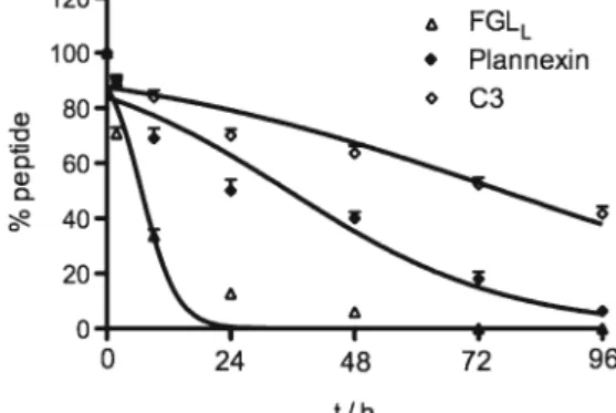
For the three most active peptides, FGLL, Plannexin, and C3, we measured peptide stability. For this purpose, peptides in aqueous solution were kept at 37°C, and samples for HPLC analysis were taken after 2, 8, 24, 48, and 96 h. As shown in Fig.7, C3 was most stable and approximately 50% of the peptide was still detected after 96 h. Plannexin showed an intermediate stability, with a half life of approximately 28 h. Finally, and unexpectedly, the most active compound, FGLL, had only a relative short half life of approximately 6 h.
Impact of Peptides on Neurite Outgrowth
Next we investigated the effect of two active peptides, FGLL and Planexin, on neural maturation by investigating the capacity of neural progenitor cells (NPCs) to extend neurites. As neurite extension requires a relatively high degree of NPC differentiation, cells were differentiated for 5 days in coculture with MS5 cells (44), and compounds of interest were added during this differentiation phase. Neurite extension was investigated during a 4 day culture on polyornithin-coated plates, after the 5 day differentiation protocol. Thus, we have investigated the effect of the peptides on NPC maturation, and not a direct effect on neurite extension. As a positive control, we chose phenazopyridine, a neurogenic small molecule that we have previously shown to enhance neurite outgrowth (42). We chose the P2 peptide as negative control; as in the primary
screen, it was found not to enhance neural differentiation and even had a moderate inhibitory effect (Fig.5). As can be seen in Fig. 8, FGLL and Plannexin, at concentrations of 30 μM (number of surviving cells after exposure to FGLL and Plannexin: 4.38±0.4×10⁵, 4.42±0.3×10⁵) and 100μM (number of surviving cells after exposure to FGLLand Plannexin: 4.27± 0.3 × 10⁵, 4.31 ± 0.4 × 10⁵) caused an increase of neurite outgrowth, as compared to control (number of surviving cells, 4.2±0.4×10⁵) and P2 peptide (number of surviving cells, 4±0.2× 10⁵). Automated imaging of neurite outgrowth (MIRAX) confirmed the visual impression: both FGLL and Plannexin enhanced neurite outgrowth. For comparison, the effect of the full length growth factors FGF2 and FGF8 are included in the quantitative analysis (for pictures see Fig.3). Interestingly, the peptides were more powerful than the growth factors in this system, which corroborates data obtained with the dual luciferase assay (compare Figs.3to5).
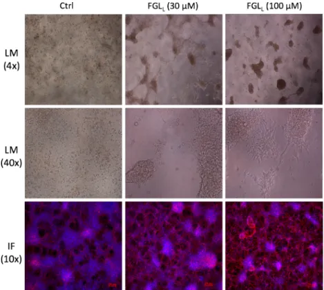
Effect of FGLLon Neural Differentiation of Human ES Cells We next determined the effect of the most powerful peptide, FGLL, on neural differentation of human ES cells. H1 human ESCs were differentiated according to standard protocols (Methods) and FGLL(30 and 100μM) was added daily from day 1 to day 10 of the differentiation protocol. As can be seen in Fig.9, light microscopic analysis performed at day 18, showed homogenously appearing, evenly spread cells under control conditions. In contrast, cultures in the presence of FGLL were more mature, with polarized individual cells migrating outwards from spherical clusters. Individual cells displayed multiple neurite outgrowths, in particular with the higher concentrations of FGLL. Immunostaining with β III tubulin antibodies and counterstaining with DAPI (day 24) revealed more abundant nuclei under control conditions, but more abundantβ III tubulin-positive neurites in cells treated with FGLL. The density of the neurite network appeared to be increased with the higher FGLLconcentration.
DISCUSSION
In this study, we have investigated the effect of peptides, derived from proteins within neural growth factor pathways, on early neural differentiation. We have performed an initial screen in a mouse ES cell dual reporter gene expression
Fig. 7. In vitro stability in serum at 37°C for dimeric peptide FGLL
and dendrimeric peptides Plannexin and C3. Half-lives (T1/2[h]) are shown in parentheses (±SEM)
assay. The peptide with the most potent efficacy on neural differentiation was FGLL, a peptide derived from the neural adhesion molecule NCAM. FGLLenhanced neural promoter activity at low micromolar concentrations, enhanced neurite outgrowth and enhanced neural differentiation.
Stem cell-based neuronal systems for in vitro testing are becoming increasingly important for the detection of neurotoxic and neuroactive compounds (47–49). We have recently de-scribed and validated a stem cell based dual luciferase reporter assay for neuronal in vitro testing (42,46). To overcome certain
Fig. 8. Effect of Phenazopyridine, P2, FGLL, and Plannexin in neuronal differentiation on CGR8-2luc. CGR8-2luc were
grown on MS5 for 5 days with compounds of indicated concentrations in SR medium. Cells were subsequently dissociated
and replated in N2 medium for additional 4 days without compounds addition. Vehicle and Phenazopyridine, P2, FGLL, and
Plannexin-treated ES cells that examined forβ-III tubulin (red) and nuclei were stained with DAPI (blue) at day 9 of
differentiation. Results are expressed as ratio of total neurite outgrowth and total cell numbers±SEM with untreated controls. Error bars indicate SEM based on three independent experiments. *P<0.05, **P<0.01, compared with controls
limitations of our assay, we have now developed a novel parameter to our neural differentiation assay, which can be readily performed after the dual luciferase assay. It quantifies the total amount of DNA and therefore serves as a convenient approximation of the cell number. We have compared the PI test to other test investigating cell number and cytotoxicity (Fig.3) and found it to be robust and reliable.
To understand the impact of the peptides on the dual reporter assay, wefirst studied the impact of the two growth factors FGF2 and FGF8. These results show that FGF8 enhances neural differentiation as demonstrated by the in-creased Tα1 promoter activity. However, while neural differen-tiation is generally accompanied by a decrease in EF1-α promoter activity (presumable because of a decreased need of protein synthesis in post-mitotic differentiated neurons, (46)), FGF8 increased EF1-α promoter activity. This concomitant activation of the neural promoter and the house-keeping promoter is, in our hands, a signature profile for growth-factor like activity. This is in line with the results observed with PI analysis. Indeed, none of the two growth factors FGF2 and FGF8 enhanced cell number when added during neural differentiation. This is most likely due to the fact that the selected growth factors induced at the same time NPC proliferation and neural differentiation (which leads to cell cycle arrest as mature neurons are postmitotic), which would account for the absence of an effect on cell number.
Out of the 15 peptides analyzed in the study, five were considered to be neuroactive. Interestingly all of thosefive peptides induced an increase in Ta1 and in EF1a promoter activity. In this respect, they resemble the effect of the growth factor FGF8.
Interestingly, the activity pattern observed with thefive neuroactive peptides also resembles the pattern observed with several antide-pressants in the dual luciferase assay (46). And indeed, recent data suggest that antidepressants might exert their therapeutic benefit through activation of neurogenesis from NSCs (50–53). Further research will be necessary to understand whether the peptides described in this study might have antidepressant activity in vivo.
FGLL and Plannexin had the strongest neurogenic effects in our study. The peptide structure of FGFL and Plannexin provides possible hints about their targets. First, both peptides are derived from NCAM. However, FGFL, a 15-amino-acid-long peptide, synthesized to correspond to the secondfibronectin type III module of NCAM (which binds to FGFR1), mimics the heterophilic binding of NCAM to FGFR1. Plannexin, a synthetic 10-amino-acid-long peptide, mimics a homophilic trans-binding site in the NCAM Ig2 module which binds to the other NCAM Ig3 module, subsequently leading to activation of the FGFR. The NCAM-derived FGLL peptide is an agonist of the FGFR, has the direct interaction with FGFR, without prior NCAM binding (in contrast to Plannexin). Second, the peptides are capable of binding to specific extracellular regions of FGFR or NCAM, with different binding affinity. FGLL binds to FGFR1 isotype c with an apparent dissociation constant (Kd) of 2.58±2.06μM (54), whereas Plannexin binds to the Ig1–2–
3 fragment of NCAM with a dissociation constant (Kd) of 5.07±1.8×10⁻⁷M (31). That might explain distinct effects in the primary screen: while Plannexin showed an FGF8-like pattern with an approximately equal activation of the Tα1 and the EF1-α promoter, FGLLpreferentially activates Tα1.
Fig. 9. The effect of FGLLon human ES cells neural differentiation. Neural differentiation
after 18 days, cells exhibit increasingly neuritic extensions in the presence of FGLLfrom 30
to 100μM under 4 and 40 times magnification of light microscope (LM). Neural positive
cells were marked withβ-III tubulin (red), and nuclei were stained with DAPI (blue) at
We evaluated the stability of the three most effective peptides FGLL,C3 and Plannexin in serum and found out the degradation of dimeric peptide is faster than dendrimeric peptide. The dimeric peptide FGLLwas degraded relatively fast with half-lives (T1/2) of less than 6 h, but tetrameric dendrimer Plannexin and C3 composed of four monomers with the sequence coupled to a lysine backbone led to a five-to 13 fold increase in T1/2 in serum. This suggests that dendrimeric peptides, such as Plannexin and C3, showed superior stability. Thus, the dendrimerization contributes more than dimerization per se to stability. A puzzling result of these experiments is the fact that the least stable peptide is the one with the highest bioactivity. The most simple explanation would be that only a short term stimulation is needed for the effects on neural differentiation and therefore degradation is not relevant. However, we cannot exclude two alternative explanations: (a) short-term stimulation is more powerful than long stimulation and (b) the dimeric peptide is a more efficient receptor agonist than the tetrameric peptide. In summary, we have demonstrated that pluripotent stem cells with a dual reporter system are a powerful tool to investigate neurogenic effects of peptides. Indeed, peptides are interesting and potentially powerful tools for pharmacological interventions in vitro and in vivo. First, peptides are easy to synthesize at relatively low costs and peptide libraries that display high levels of chemical and biological diversity can therefore be generated. Also, as opposed to proteins, small peptides are unlikely to invoke an immune response since they fall below the immunogenic thresh-old. However, the biological impact of a peptide cannot be predicted based on its structure and pharmacological preparation. Thus, biologically relevant assay systems are required. The approach described in this study provides such a biologically relevant, embryonic stem cell based testing system. The system can be used even for higher throughput screening and is suitable to analyze larger peptide libraries in the future. Yet, FGLL, identified in this study as a potent inducer of neural differentiation is already a peptide that should merit further attention. Because of its relatively low stability in serum, it might not be the ideal peptide for systemic administration in vivo, however it might be an interesting addition to in vitro neural differentiation protocols. Also, it might be interesting to think about its potential to be cotransplanted with ESC-derived neurons for cell therapy applications.
ACKNOWLEDGMENTS
Research was financed by EU FP7 grant (STEMCAM, PIAP-GA-2009-251186)
REFERENCES
1. Chiu AY, Rao MS. Cell-based therapy for neural disorders–
anticipating challenges. Neurotherapeutics. 2011;8(4):744–52.
2. Nikoletopoulou V, Tavernarakis N. Embryonic and induced pluripotent stem cell differentiation as a tool in neurobiology.
Biotechnol J. 2012;7(9):1156–68.
3. Li G et al. The ventral hippocampus is the embryonic origin for adult
neural stem cells in the dentate gyrus. Neuron. 2013;78(4):658–72.
4. Yuan SH, Shaner M. Bioengineered stem cells in neural development and neurodegeneration research. Ageing Res
Rev. 2013;12(3):739–48.
5. Han SS, Williams LA, Eggan KC. Constructing and deconstructing stem cell models of neurological disease.
Neuron. 2011;70(4):626–44.
6. Mackay-Sim A. Patient-derived stem cells: pathways to drug discovery for brain diseases. Front Cell Neurosci. 2013;7:29. 7. Hoelting L et al. A 3-dimensional human embryonic stem cell
(hESC)-derived model to detect developmental neurotoxicity of
nanoparticles. Arch Toxicol. 2013;87(4):721–33.
8. Alsanie WF, Niclis JC, Petratos S. Human embryonic stem cell-derived oligodendrocytes: protocols and perspectives. Stem Cells
Dev. 2013;22:2459–76.
9. Moon J et al. Stem cell grafting improves both motor and
cognitive impairments in a genetic model of Parkinson’s disease,
the aphakia (ak) mouse. Cell Transplant. 2013;22(7):1263–79.
10. Kriks S et al. Dopamine neurons derived from human ES cells
efficiently engraft in animal models of Parkinson’s disease.
Nature. 2011;480(7378):547–51.
11. Relano-Gines A et al. Prion replication occurs in endogenous adult neural stem cells and alters their neuronal fate: involve-ment of endogenous neural stem cells in prion diseases. PLoS Pathog. 2013;9(8):e1003485.
12. Benraiss A, Goldman SA. Cellular therapy and induced neuronal
replacement for Huntington’s disease. Neurotherapeutics.
2011;8(4):577–90.
13. Jaeger I et al. Temporally controlled modulation of FGF/ERK signaling directs midbrain dopaminergic neural progenitor fate in mouse and human pluripotent stem cells. Development.
2011;138(20):4363–74.
14. Akama K et al. Proteomic identification of differentially
expressed genes in mouse neural stem cells and neurons differentiated from embryonic stem cells in vitro. Biochim
Biophys Acta. 2008;1784(5):773–82.
15. Kuo YC, Huang MJ. Material-driven differentiation of induced pluripotent stem cells in neuron growth factor-grafted poly(epsilon-caprolactone)-poly(beta-hydroxybutyrate)
scaf-folds. Biomaterials. 2012;33(23):5672–82.
16. Solozobova V, Wyvekens N, Pruszak J. Lessons from the embryonic neural stem cell niche for neural lineage
differentia-tion of pluripotent stem cells. Stem Cell Rev. 2012;8(3):813–29.
17. Li YC, Lin YC, Young TH. Combination of media, biomaterials and extracellular matrix proteins to enhance the differentiation of neural stem/precursor cells into neurons. Acta Biomater.
2012;8(8):3035–48.
18. Berg JM, Tymoczko J, Stryer L. Biochemistry. 2002.
19. Kaushansky K. Small molecule mimics of hematopoietic growth
factors: improving on Mother Nature? Leukemia. 2001;15(4):673–4.
20. Berrera M, Cattaneo A, Carloni P. Molecular simulation of the binding of nerve growth factor peptide mimics to the receptor
tyrosine kinase A. Biophys J. 2006;91(6):2063–71.
21. Zhang L et al. Small-molecule blocks malignant astrocyte prolifer-ation and induces neuronal gene expression. Differentiprolifer-ation.
2011;81(4):233–42.
22. Chambers SM et al. Combined small-molecule inhibition accel-erates developmental timing and converts human pluripotent
stem cells into nociceptors. Nat Biotechnol. 2012;30(7):715–20.
23. Chi Z et al. Botch promotes neurogenesis by antagonizing notch.
Dev Cell. 2012;22(4):707–20.
24. Hefti FF. Requirements for a lead compound to become a clinical candidate. BMC Neurosci. 2008;9 Suppl 3:S7.
25. Ditlevsen DK et al. NCAM-induced intracellular signaling
revisited. J Neurosci Res. 2008;86(4):727–43.
26. Walmod PS et al. Zippers make signals: NCAM-mediated molecular interactions and signal transduction. Neurochem Res.
2004;29(11):2015–35.
27. Yang HJ et al. A novel role for neural cell adhesion molecule in modulating insulin signaling and adipocyte differentiation of mouse
mesenchymal stem cells. J Cell Sci. 2011;124(Pt 15):2552–60.
28. Berezin V, Bock E. NCAM mimetic peptides: pharmacological
and therapeutic potential. J Mol Neurosci. 2004;22(1–2):33–9.
29. Cambon K et al. A synthetic neural cell adhesion molecule mimetic peptide promotes synaptogenesis, enhances presynaptic function, and facilitates memory consolidation. J Neurosci.
2004;24(17):4197–204.
30. Chen Y et al. The fibroblast growth factor receptor (FGFR)
peptide FGL activate FGFR substrate 2alpha differently. J
Neurosci Res. 2010;88(9):1882–9.
31. Kohler LB et al. A peptide derived from a trans-homophilic binding site in neural cell adhesion molecule induces neurite outgrowth and
neuronal survival. J Neurosci Res. 2010;88(10):2165–76.
32. Klementiev B et al. A peptide agonist of the neural cell adhesion molecule (NCAM), C3, protects against developmental defects induced by a teratogen pyrimethamine. Int J Dev Neurosci.
2002;20(7):527–36.
33. Kiryushko D et al. Neural cell adhesion molecule induces intracellular signaling via multiple mechanisms of Ca2+
homeo-stasis. Mol Biol Cell. 2006;17(5):2278–86.
34. Fobian K et al. Peptides derived from the solvent-exposed loops 3 and 4 of BDNF bind TrkB and p75(NTR) receptors and stimulate neurite outgrowth and survival. J Neurosci Res.
2010;88(6):1170–81.
35. Pedersen MV et al. Neuritogenic and survival-promoting effects of the P2 peptide derived from a homophilic binding site in the neural cell adhesion molecule. J Neurosci Res.
2004;75(1):55–65.
36. Hansen SM et al. NCAM-derived peptides function as agonists
for the fibroblast growth factor receptor. J Neurochem.
2008;106(5):2030–41.
37. Li S et al. Fibroblast growth factor-derived peptides: functional
agonists of thefibroblast growth factor receptor. J Neurochem.
2008;104(3):667–82.
38. Xu R et al. A peptide antagonist of the ErbB1 receptor inhibits receptor activation, tumor cell growth and migration in vitro and xenograft tumor growth in vivo. Cell Oncol.
2010;32(4):259–74.
39. Scopes RK. Measurement of protein by spectrophotometry at
205 nm. Anal Biochem. 1974;59(1):277–82.
40. Itoh K et al. Reproducible establishment of hemopoietic supportive stromal cell lines from murine bone marrow. Exp
Hematol. 1989;17(2):145–53.
41. Fujii DK et al. Neurite outgrowth and protein synthesis by PC12 cells as a function of substratum and nerve growth factor. J
Neurosci. 1982;2(8):1157–75.
42. Suter DM et al. Phenazopyridine induces and synchronizes neuronal differentiation of embryonic stem cells. J Cell Mol
Med. 2009;13(9B):3517–27.
43. Dinsmore J et al. Embryonic stem cells differentiated in vitro as a novel source of cells for transplantation. Cell Transplant.
1996;5(2):131–43.
44. Barberi T et al. Neural subtype specification of fertilization and
nuclear transfer embryonic stem cells and application in
parkin-sonian mice. Nat Biotechnol. 2003;21(10):1200–7.
45. Feyeux M et al. Early transcriptional changes linked to naturally
occurring Huntington’s disease mutations in neural derivatives of
human embryonic stem cells. Hum Mol Genet. 2012;21(17):3883–95.
46. Kern I et al. Embryonic stem cell-based screen for small molecules: cluster analysis reveals four response patterns in
developing neural cells. Curr Med Chem. 2013;20(5):710–23.
47. Krug AK et al. Human embryonic stem cell-derived test systems for developmental neurotoxicity: a transcriptomics approach.
Arch Toxicol. 2013;87(1):123–43.
48. Krug AK et al. Evaluation of a human neurite growth assay as
specific screen for developmental neurotoxicants. Arch Toxicol.
2013;87(12):2215–31.
49. Krause KH et al. Monocrotophos in Gandaman village: India school lunch deaths and need for improved toxicity testing. Arch
Toxicol. 2013;87(10):1877–81.
50. Han X et al. Imipramine treatment improves cognitive outcome associated with enhanced hippocampal neurogenesis after
trau-matic brain injury in mice. J Neurotrauma. 2011;28(6):995–1007.
51. Mowla A et al. Megadose clonazepam dependence: a case report.
J Clin Psychopharmacol. 2007;27(5):542–3.
52. Finkel SI et al. A randomized, placebo-controlled study of the
efficacy and safety of sertraline in the treatment of the
behavioral manifestations of Alzheimer’s disease in outpatients
treated with donepezil. Int J Geriatr Psychiatry. 2004;19(1):9–18.
53. Santarelli L et al. Requirement of hippocampal neurogenesis for the
behavioral effects of antidepressants. Science. 2003;301(5634):805–9.
54. Kiselyov VV et al. Structural basis for a direct interaction between FGFR1 and NCAM and evidence for a regulatory role