Development of Biodegradable Microfluidic Networks
for Tissue Engineering
by
Kevin R. King
B.S. Electrical Engineering
University of Illinois Urbana Champaign, 1999
Submitted to the Department of Electrical Engineering and Computer
Science in partial fulfillment of the requirements for the degree of
Master of Science in Electrical Engineering
MASSACHUSETTS INSTITUTE OF TECHNOLOGY
September 2002
© Massachusetts Institute of Technology 2002. All rights reserved.
Author
Department of Electricatii iieering and Computer Science
September 1, 2002
Certified by
Joel Voldman
AAssistant Professor, Electrical Engineering
Certified by_
Jeffrey T. Borenstein
Principa lember of\Technical Staff, Draper Laboratory
Certified by_
Joseph P. Vacanti
John Homans Professor
ofSur9pryfarvaro Nedica!,
School
Accepted by___
"Arthur
Smith
Chairman, Department Committee on Graduate Students
B aAKZR79ASSAUSS INOTUTE
Development of Biodegradable Microfluidic Networks for Tissue
Engineering
by
Kevin Robert King
Submitted to the Department of Electrical Engineering and Computer Science on September 1, 2002 in partial fulfillment of the requirements for the degree of
Master of Science in Electrical Engineering
ABSTRACT
Biodegradable tissue engineering scaffolds currently suffer from poorly controlled geometries, lack of reproducibility, and severe mass transport limitations.
Microfabrication is an ideal tool for attacking problems in tissue engineering due to its control of diverse size scales, from microns to centimeters, all with micron resolution and submicron precision. By enabling precision geometries and flexible designs,
microfabrication has the potential to offer creative solutions to some of the major problems facing the field of tissue engineering; those of achieving rapid vascularization and recapitulating normal complex tissue microarchitecture.
In this thesis, silicon micromachining, soft lithography, and traditional polymer processing are combined to develop a fully biodegradable microfabrication platform using poly(DL-lactic-co-glycolic) acid (PLGA 85:15). First, micron scale structures fabricated on silicon substrates are transferred into the surface of biodegradable films using polymer melt replica molding. Next, microchannel networks are sealed and made three-dimensional by stacking and irreversibly bonding the biodegradable films using a newly developed thermal fusion bonding process. The process is modeled and
optimized to guide bonding of a wide range of microchannels while avoiding parasitic microstructure deformation. An extruded inlet/outlet scheme is implemented and combined with micromolding and fusion bonding to build fully patent, leak-free biodegradable microfluidic networks with predictable fluidic resistances. Finally, this thesis concludes with the first demonstration of long-term continuous-flow cell culture in prototype microfluidic networks, demonstrating the feasibility of using the newly
developed biodegradable microdevices as cell-seeded tissue engineering scaffolds. In comparison to conventional scaffold fabrication techniques, these processes offer two orders of magnitude improvement in fabrication time and spatial resolution while offering flexible designs and unmatched reproducibility. Through these features, they have enabled construction of the first fully biodegradable microfluidic networks. Thesis Supervisor: Joel Voldman Ph.D.
Title: Assistant Professor of Electrical Engineering Thesis Supervisor: Jeffery T. Borenstein Ph.D.
Title: Principle Member of Technical Staff, Draper Laboratory Thesis Supervisor: Joseph P. Vacanti M.D.
Acknowledgements
This thesis was performed as part of a long collaboration between Dr. Vacanti at MGH and Dr. Jeff Borenstein at Draper Laboratory in the general area of
"microfabrication approaches to tissue engineering". Because the work was performed at both locations, I have had the benefit of interacting with many people with diverse backgrounds, experiences, and perspectives.
As I sat in the first Vacanti Lab group meeting two years ago, I vividly recall his referring to the microfabrication tissue engineering effort and saying, "As a surgeon whose focus is the patient, my goal is clear." Referring to the therapeutic objective of the research, these four words will remain with me as a constant reminder of why biomedical engineering research is important. I would like to thank Dr. Vacanti for initially taking me into his lab despite my lack of prior cell culture experience and for seeing to it that I was surrounded by helpful and enthusiastic investigators in a dynamic research environment. His vision and commitment to the end goal of helping patients is not only inspiring, but also provided constant motivation for this work.
I initially joined Jeff and the MEMS group at Draper Laboratory to contribute to the microfabrication and device development side of the project, however it quickly
became my research "home." Jeff has provided endless support - technical,
administrative, and personal - throughout the course of my research. Quick to help in any way possible, he made sure there were no obstacles to this exploration. I
appreciate his confidence and trust as well as the flexibility he gave me to shape and define the direction of this work. I have had the chance to explore a wide range of topics from the theoretical to the experimental and from the basic science to the applied. By
giving me the space to mold the project and make my own mistakes, he made the successes that much more rewarding.
This thesis would not have been possible without the help of Joanne Wang, an outstanding MIT undergraduate who helped with nearly every aspect of the project. Joanne's curiosity was infectious and I'm extremely grateful for her endless commitment to the project. She offered a critical sounding board for ideas and provided a
companionship that made the journey an enjoyable one.
Thank you to Joel for reviewing this thesis and helping me put together the written document. In just a few helpful discussions he was able to give me the
necessary guidance to assemble my work into an organized and coherent thesis. I wish him the best of luck in his new position as a professor at MIT.
I'd like to thank the Whitaker Foundation and Draper Laboratory for their financial support through graduate fellowships; the many people at Draper Laboratory, MGH and MIT who provided helpful discussions, technical assistance, and/or friendship over the course of the past two years; and finally my family and friends for all of their support.
This thesis was prepared at The Charles Stark Draper Laboratory, Inc. under No. DAMDIT-99-2-9001. Publication of this thesis does not constitute approval by Draper or the sponsoring agency of the findings or conclusions contained herein. It is published for the exchange and stimulation of ideas. This project was sponsored by the
Department of the Army, Cooperative Agreement DAMD-99-2-9001. The content of this thesis does not necessarily reflect the position or the policy of the government and no official endorsement should be inferred.
Contents
1. INTRODUCTION... 13 1.1 Biodegradable Polymer Tissue Engineering
1.1.1 Tissue Engineering Scaffolds 1.1.2 Biodegradable Scaffold Approach 1.1.3 Polylactic Glycolides
1.2 Organ Fabrication Concept
1.3 Existing Scaffold Fabrication Techniques 1.3.1 Scaffold Design Criteria
1.3.2 Scaffold Fabrication and Polymer Processing 1.4 Microtechnology for Medicine and Biology
1.4.1 Microfabrication and Micromachining 1.4.2 Soft Lithography
1.4.3 Cell Patterning 1.4.4 Microfluidics
1.4.5 Cells in Microfluidic Devices 1.5 Thesis Objectives and Overview 1.6 References
2. MICROSTRUCTURING PLGA SURFACES...39 2.1 Master Mold and Tooling Fabrication
2.1.1 Photomask Fabrication
2.1.2 Silicon Master Mold Fabrication 2.1.3 Polymeric Tooling
2.2 Biodegradable Polymer Micromolding 2.2.1 Solvent Casting
2.2.2 Melt Processing and Compression Molding 2.3 Microstructure Characterization
2.3.1 Complex Channel Networks 2.3.2 High Resolution Structures 2.4 Discussion
2.5 Conclusion
3. PLGA FUSION BONDING...61 3.1 Thermal Fusion Bonding of Microstructures
3.2 Thermoplastic Material Properties 3.2.1 Glass Transition Temperature 3.2.2 Determination of Glass Transition
3.2.3 Fusion Bonding Near the Glass Transition Temperature 3.3 Fusion Bonding Model Overview
3.4 Intimate Contact - 'Wetting" 3.4.1 Qualitative Observations
3.4.2 Previous Models for Intimate Contact
3.4.3 Experimental Determination of Intimate Contact Time 3.4.4 Results
3.5 Interdiffusion - "Healing" 3.5.1 Qualitative Observations
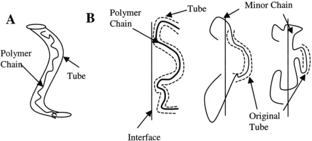
3.5.2 Reptation and Interdiffusion
3.5.3 Interdiffusion Time
3.6 Bonding Time 3.7 Plastic Deformation
3.7.1 Elastic Deformation
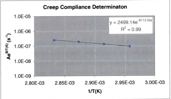
3.7.2 Viscoelastic Deformation 3.8 Creep Compliance Estimation
3.8.1 Macroscopic Beam Approach
3.8.2 Experimental Method 3.8.3 Results
3.8.4 Empirical Estimation of Creep Compliance
3.9 Microchannel Deformation
3.9.1 Prediction of Microchannel Deformation Time
3.9.2 Microchannel Bonding Experiments 3.9.3 Results
3.9.4 Model Limitations and Uncertainty
3.10 Process Window Determination 3.11 Bond Morphological Characterization 3.12 Discussion 3.13 Conclusion 3.14 References 4. BIODEGRADABLE MICROFLUIDICS...98 4.1 Design 4.1.1 Layout Specification
4.1.2 Mask and Layout Fabrication 4.2 Device Fabrication
4.2.1 Silicon and PDMS Mold Fabrication 4.2.2 Inlets and Outlets - Modification of PDMS 4.2.3 Compression Molding of PLGA with Connectors 4.2.4 Thermal Fusion Bonding
4.2.5 Device Connectors 4.3 Morphological Characterization
4.3.1 PLGA Molded Films
4.3.2 Microchannel Patterns 4.3.3 Bonding
4.3.4 Completed Microfluidic Devices 4.4 Functional Microfluidics Characterization
4.4.1 Patency, Perfusion, and Leak Testing 4.4.2 Pressure Burst Tests
4.4.3 Microfluidic Resistance Characterization 4.5 Three-dimensional Microfluidics
4.5.1 Multi-layer Fabrication Approach 4.5.2 Multi-layer Device Characterization
-U 4.6 Microfluidics-enabled Scaffolds 4.6.1 Fabrication Approach 4.6.2 Device Characterization 4.7 Conclusion 4.8 References
5. MICROFLUIDIC CELL CULTURE...119 5.1 Cell Culture
5.1.1 Cell Culture 5.1.2 Imaging
5.2 PDMS Prototype Microfluidic Device 5.2.1 Design 5.2.2 Fabrication 5.3 Microfluidic Bioreactor 5.3.1 Design 5.3.2 Dynamics 5.4 Seeding
5.4.1 Sterilization and Assembly 5.4.2 Cell Seeding
5.5 Continuous-Flow Culture
5.5.1 Characterization of Culture in NET2 5.5.2 Characterization of Culture in NET1 5.5.3 Growth to Confluency
5.6 PLGA Microfluidic Culture 5.7 Discussion
5.8 Conclusion 5.9 References
6. FUTURE WORK AND CONCLUSIONS...139 6.1 Summary of Contributions
6.2 Future Work 6.3 Final Comments
Appendix A Detailed Microfabrication Process Flow...-147 Appendix B PLG A Material Data...151
List of Figures
Figure 1-1: Tissue Engineering with Biodegradable Polymers Figure 1-2: PLA, PGA, and PLGA Chemistry and Degradation Figure 1-3: Generic Bulk Organ Fabrication Concept
Figure 1-4: Conventional Porous and Fibrous Scaffolding
Figure 1-5: Oxygen Transport and Hypoxia-induced Angiogenesis Figure 1-6: Complex Liver Microarchitecture
Figure 1-7: Microfabrication Process Flow
Figure 1-8: Soft Lithography Fabrication Process Flow Figure 1-9: Biodegradable Microfabrication Process Flow Figure 2-1: Silicon Master Mold Fabrication
Figure 2-2: PDMS Elastomeric Tooling Fabrication Figure 2-3: Solvent Casting Process
Figure 2-4: Melt Molding Process Figure 2-5: Melt Molding Apparatus
Figure 2-6: Thermal Gravimetric Analysis - Temperature Ramp Figure 2-7: Degradation Rate vs. Temperature
Figure 2-8: PLGA 85:15 Melt Rheology
Figure 2-9: Film Thickness vs. Time (Experiment and Theory) Figure 2-10: Light Microscope Images - Silicon, PDMS, and PLGA
Figure 2-11: SEM - Solvent Cast PLGA Figure 2-12: SEM - Melt Processed PLGA
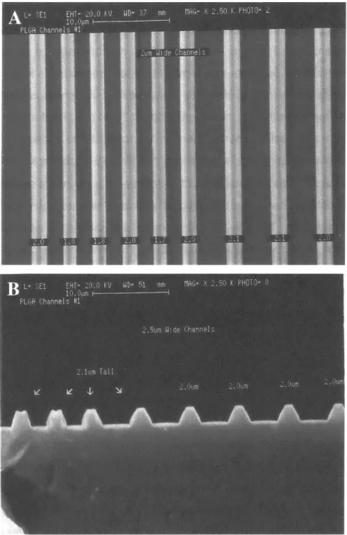
Figure 2-13: SEM - High Resolution Structures in PLGA
Figure 2-14: SEM - High Resolution Structures - 2gm LINES AND SPACES
Figure 3-1: Thermoplastic Modulus Temperature Dependence Figure 3-2: Differential Scanning Calorimetry Schematic
Figure 3-3: PLGA 85:15 Glass Transition Temperature Determination Figure 3-4: Thermal Fusion Bonding Model Overview
Figure 3-5: Process Optimization Overview
Figure 3-6: Surface 'Wetting" or Intimate Contact Schematic Figure 3-7: Images of Intimate Contact and Microchannel Collapse Figure 3-8: Temperature Dependence of Intimate Contact Time Figure 3-9: Reptation of Polymer Chains
Figure 3-10: Interdiffusion Bonding of Surface Microstructures Figure 3-11: Experiment for Estimation of Interdiffusion Time Figure 3-12: Plastic Deformation and Collapse of Microchannels Figure 3-13: Beam Model of Microchannel
Figure 3-14: Simple Loaded Beam Models of Polymer Structures Figure 3-15: Macroscopic Cantilever Beam Experimental Setup Figure 3-16: Cantilever Creep Time vs. Dimensions
Figure 3-18: Microchannel Deformation Time vs. Dimensions Figure 3-19: Predicted/Experimental Deformation Times Figure 3-20: Sample Process Window vs. Temperature
Figure 3-21: SEM Morphology of Bonded Microfluidic Channels Figure 3-22: SEM Morphology of Bonded High Resolution Channels Figure 3-23: SEM Morphology of Bonded High Aspect Ratio Channels Figure 4-1: Microfluidic Network Design for NET-1 and NET-2
Figure 4-2: Mask Layouts for NET-1 and NET-2
Figure 4-3: Modification of PDMS Tooling for Inlet/Outlet Ports Figure 4-4: Melt Molding of Extruded Inlet and Outlet Connectors Figure 4-5: Thermal Fusion Bonding Model Results for NET-2 Figure 4-6: Images of Biodegradable Inlet and Outlet Fabrication Figure 4-7: SEM of Microfluidic Channel Networks - Unbonded Films Figure 4-8: Optical Monitoring of Microfluidics Bonding Process Figure 4-9: Images of Fully Bonded PLGA Networks
Figure 4-10: SEM of Microchannel Cross-sections
Figure 4-11: Completed Microfluidic Networks NET-1 AND NET-2 Figure 4-12: Perfusion of Microfluidic Networks
Figure 4-13: Schematic of Hydraulic Resistance Measurement Setup Figure 4-14: Pressure-Flow Relationships for NET-1 and NET-2 Figure 4-15: Schematic of Multi-layer Fluidics Fabrication Process Figure 4-16: SEM of Multi-layer Network Cross-section
Figure 4-17: Perfusion of Three-dimensional Microfluidics
Figure 4-18: Interconnected Porous Scaffold Fabrication Approach Figure 4-19: Characterization of Interconnected Porous Scaffold Figure 5-1: Microfluidic Cell Culture Network Design
Figure 5-2: Prototype PDMS Microfluidic Networks
Figure 5-3: Continuous-flow Microfluidic Bioreactor Schematic Figure 5-4: Rationale for Culture Flow Rate Determination Figure 5-5: Determination of Continuous-flow Culture Flow Rate Figure 5-6: Image of Cell Seeding in Microfluidic Channels Figure 5-7: Early Stages of NET-2 Microchannel Cell Culture Figure 5-8: Late Stages of NET-1 Microchannel Cell Culture Figure 5-9: Time Course of NET-1 Microchannel Cell Culture Figure 5-10: Microchannel Percent Confluency vs. Time Figure C.1: Schematic of Rectangular Microchannel
Figure C.2: PSPICE Simulation of Microfluidic Resistive Network NET2
-I
List of Tables
Table 1-1: Tissue Engineering Scaffold Design Criteria Table 1-2: Conventional Scaffold Processing Techniques Table 1-3: Advanced Scaffold Processing Techniques Table 2-1: Silicon Master Mold Microfabrication Processes Table 2-2: Compression Molding Parameters
Table 3-1: Prediction of Fusion Bonding Dynamics
Table 3-2: Boundary Conditions for Solving Beam Deformation Table 3-3: Estimated Creep Compliance Constants
Chapter 1
Introduction
This thesis describes the development of a biodegradable polymer microfabrication platform for large-scale tissue engineering. In contrast to conventional biodegradable scaffold fabrication techniques, microtechnology is
used to improve spatial resolution by two orders of magnitude while offering exceptional design flexibility and unmatched reproducibility. A new
biodegradable polymer bonding technique is introduced, and a predictive model is developed and tested to enable rapid optimization of the bonding process. This powerful platform is then used to build the first ever fully biodegradable microfluidic devices. The dimensional control and design flexibility afforded by microfabrication are leveraged to construct microfluidic networks fashioned after the massively parallel highly branched microvasculature of a large organ. Then the same design is used to demonstrate the first long-term sterile cell culture in microfluidic devices of any kind, an achievement that has broad applications in the areas of tissue engineering, lab-on-a-chip devices, and biomedical
microdevices in general.
Chapter 1 begins with a brief introduction to the field of tissue engineering and biodegradable polymers followed by a description of the high level goal of this thesis - to develop a biodegradable scaffold fabrication platform that will enable engineering of large vital organs. This is followed by background on conventional biodegradable polymer scaffold fabrication and it's shortcomings, as well as an introduction to the relevant areas of microtechnology. Finally, the chapter concludes with a detailed overview of this thesis.
1.1 Biodegradable Polymer Tissue Engineering
1.1.1 Tissue Engineering Scaffolds
Organ transplantation is the only permanent therapy for patients with end-stage organ failure [1]. The most significant challenge facing organ
transplantation is the need for available tissue. There are more than 80,000 patients currently on the organ donor waiting list, with more than 70,000 awaiting livers and kidneys alone [2]. These are complex tissues with intricate
arrangements of multiple cell types. It is this well-preserved microarchitecture that determines the physiologic organ-specific function. The shortage of
available organs as well as the need for other replacement tissues has inspired rapid growth in the field of "tissue engineering".
Tissue engineering is an emerging field aimed at addressing the growing disparity between needed and available tissues by developing alternative therapies to tissue and organ transplantation based on implantation of living cells. Several excellent reviews of the field can be found in the literature [1, 3-6].
In these approaches, cells are not simply implanted alone, but rather in
association with a support matrix. The nature and complexity of the supports is
highly variable and has increased considerably over the past several decades
owing to new understandings of the relationships between cell growth and cell support.
As early as 1933, investigators were implanting living cells. At that time, the living elements were encased in a simple polymer membrane [7]. Today, methods for implanting live cells are diverse, ranging from encasement in chemically specific hydrogels [8], to seeded on porous polymer surfaces [9], to containment in silicon microwells [10].
1.1.2 Biodegradable Scaffold Approach
In 1993, the success of support matrices inspired a novel approach involving the implantation of cells on synthetic bioresorbable scaffolding [11]. This now common tissue engineering paradigm (Figure 1.1) consists of three
principle steps: 1) Cell Acquisition - cells can be obtained by tissue biopsy or differentiated from progenitors or stem cells and expanded in culture. 2) Cell Seeding - cells are expanded in culture, seeded on an engineered polymer scaffold, and cultured in vitro to increase cell number and tissue integrity. 3) Implantation - the cell/polymer construct is implanted in vivo to integrate and serve its organ-specific function while the polymer is resorbed in a biologically compatible fashion. This approach has been extensively explored for a wide
range of tissues over the past decade [12-15], and its feasibility has been
thoroughly demonstrated. The focus is now shifting toward one of understanding mechanisms, optimizing scaffolds, selecting appropriate cells, and perfecting
implantation methods in order to make the approach therapeutically viable. This thesis focuses on development of a fabrication platform for building high
performance scaffolding that can be used to address many of the major challenges currently facing tissue engineering.
1.1.3 Polylactic Glycolides
The most commonly used synthetic bioresorbable polymers are the (o-hydroxy) aliphatic polyesters, polyglycolic acid (PGA), polylactic acid (PLA), and their copolymers (PLGA) (Figure 1.2) [16]. These materials have a long history
of Food and Drug Administration (FDA) approval for use as surgical sutures, pins, and microsphere drug delivery vehicles [17, 18]. The linear chain polymers are degraded by hydrolysis to yield lactic acid and glycolic acid, natural
biomolecules that are readily metabolized in vivo (Figure 1.2) [19]. Due to the presence of an additional methyl moiety, the polylactic acids are sterically hindered from hydrolytic attack and therefore degrade slower than the glycolic acids. By varying the monomer ratio in PLGA copolymers, the degradation kinetics can be systematically varied from days to months. In this thesis, the slowly degrading 85L:15G copolymer is used (details in Appendix B).
There is a long history of growing cells on PLGA scaffolds. PLGA is an amorphous thermoplastic linear chain polymer with a glass transition temperature between 450C -550C. It is readily dissolved in many organic solvents, and is amenable to a wide range of processing techniques. The ability to control physicochemical properties of the resorbable material has made it particularly attractive for scaffolding and has lead to the development of many different scaffold fabrication methods, each designed to optimize specific design criterion.
C+H CH3 H-a-C-C-OH Poly(lactic acid) H CH3 H20 Lactic acid H O-- O HHH H ~ O
Poly(glycolic acid) H-O-C-C-OH
H O H O Glycolic acid
H O--- O--- OH
CH3 H
Poly(lactic-co-glycolic acid)
Figure 1.2 Chemical structure ot the (cx-hydroxy) aliphatic polyesters polyglycolic acid (PGA),
1.2 Organ Fabrication Concept
It is clear that physiologically, functional units of tissue microarchitecture are organized on the size scale of microns to millimeters [3]. If vital organ fabrication is to be possible, these functional units must be recapitulated in a
grossly parallel fashion [20]. In addition, the scaffold must be fashioned with a microvascular equivalent to oxygenate and nourish cells in all areas of the construct [21]. At this stage, it is not yet clear to what extent the scaffold should explicitly depict microarchitecture or microvasculature. However, the processes developed in this thesis fortunately allow design of engineered tissue architecture at any level that it might be required, from small 5-10p m capillaries and hepatic sinusoids to larger millimeter-scale organized branchings of vascular-like conduits. Such a capability enables a wide range of configurations of varied complexity. Organ-specific parenchymal cells could be placed in generic pore-like wells, patterned in stacked planar cellular sheets, or even encouraged to develop into self-organized spheroids. Alternatively, more explicit topologies can be imagined, such as vascular networks designed to mimic physiologic
parameters; either function-determining flow rates and shear stresses or morphological metrics describing vessel branching patterns and distributions.
As an example of a non-explicit design at the microscale, consider the engineering of a generic bulk organ such as the liver. In the case of the liver, the desired organ-specific function is metabolic modification and detoxification of the blood. By building a large microfluidic scaffold that could be placed in-line with the vascular circulation, blood would have access to the cell-seeded interior of the device. Stacking alternating layers of microfabricated wells (-200ptm cubes) and overlaying microfluidic networks allows one to design microwell
interconnectivity, enabling efficient and systematic seeding as well as continuous flow culturing of small units of tissue in vitro and even perfusion with whole blood
in vivo. Eventually, more complexity could be built in by incorporating
nanoporous membranes or creating other selective barriers to cells while
remaining permeable to gas and macromolecules. The generic organ fabrication concept is shown schematically in Figure 1.3.
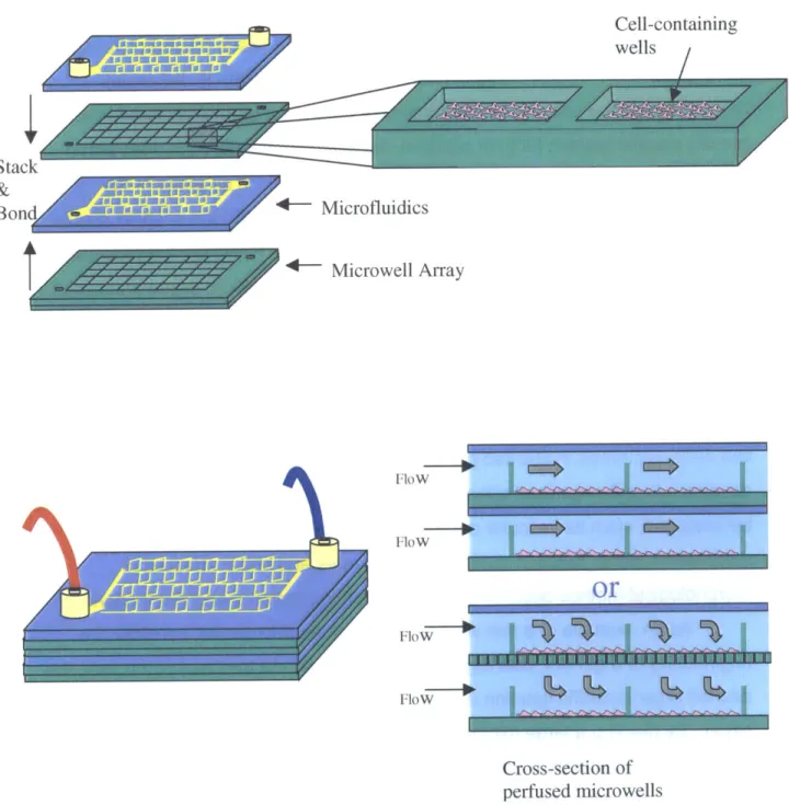
uWj,; iamAu -A
Cell-containing wells
Stack
&
Bon d W.E EEw-s 4 - Microfluidics
*- Microwell Array Flow Flow
or
FloW FloW Cross-section of perfused microwellsFigure 1.3 Example of a generic organ fabrication design using three-dimensional
microfabricated biodegradable scaffolding. Combinations of cell-containing microwells and microfluidic interconnections allow scaffolds to be seeded and perfused efficiently and methodically.
1.3 Existing Scaffold Fabrication Technologies
1.3.1 Scaffold Design Criteria
Several design criteria have been identified to guide scaffold fabrication, each motivated by a functional requirement. Initial emphasis was placed on the need for large surface-area-to-volume ratios to seed attachment-dependent cells, and large void volumes to allow three-dimensional growth of tissue within the matrix. These criteria inspired the development of three-dimensional porous scaffolds with pore sizes ranging from 500gm, and porosities ranging from 50-99% [9, 22, 23]. Pore interconnectivity was emphasized as a critical determinant of mass transport, host tissue in-growth, and tissue engineered cellular
outgrowth. It was specified that diffusive transport of oxygen and metabolites be sufficient to maintain cell viability until the in-growth of vascularized tissue or the development of de novo blood vessels could be achieved. The scaffold was also
required to provide sufficient mechanical support to prevent collapse under the stress of a fibrous capsule [24]. At the same time, it was desired that the scaffold
not significantly restrain growth of new tissue from inside. These final criteria are typically met been by selecting resorbable polymers that degrade on the same time scale as tissue growth.
A wide variety of porous and fibrous scaffolds have been developed over the last decade and today their fabrication is fairly routine. Investigators are still
in search however, of the optimal scaffold that is sufficiently flexible to
simultaneously meet diverse design criteria. At these early stages, the criteria are still somewhat speculative and dynamic. Therefore, a versatile approach is needed to both aid in systematic identification of important scaffold features, and adapt to new design criteria as relationships between tissue development and scaffold design are revealed. The microfabricated scaffolding platform described in this thesis addresses these issues by providing 1) unmatched geometrical resolution and reproducibility, 2) explicit control of mass transport and pore interconnectivity, 3) dramatic improvements in design flexibility to allow
manufacturability by making the fabrication process rapid, efficient, and accessible to investigators without specialized equipment.
Geometrical Precision and Reproducibility
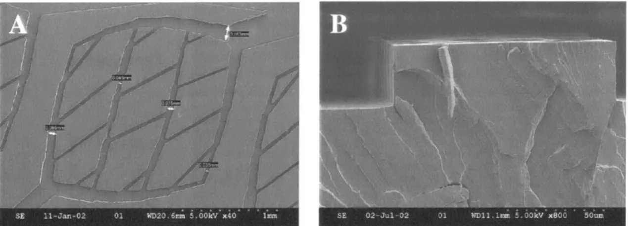
Scaffold pore size has been demonstrated to influence implanted cell growth and vascularization [25]. Evidence from examination of scaffolds with pores and conduits of different sizes and interconnectivity suggest that optimum pore sizes are tissue specific. Meeting strict requirements on pore size and interconnectivity will require improved control and precision because current methods only control bulk properties such as average pore size, shape, and distribution (Figure 1.4) [23]. The reproducibility inherent in high precision methods such as microfabrication will allow experiments to be more effectively compared by enabling systematic variation of scaffold properties.
.44-Figure 1.4 Conventional A) porous and B) fibrous scaffolds with control limited to bulk properties
of average pore size size, shape, distribution, and interconnectivity. Nominal pore sizes and interfiber distances are several hundred microns.
Mass Transport Limitations
Metabolically active cells require a continuous supply of nutrients and removal of waste. In porous scaffolds, cells rely solely on diffusive transport to deliver oxygen and metabolites. The thickness of cell-seeded scaffolds is
therefore limited to millimeters in size, the diffusion distance of oxygen in tissue, before cells at the scaffold center become hypoxic [26]. In order to maintain the
viability of larger cell-seeded scaffolds, methods for rapid vascularization must be developed [27].
The formation of blood vessels follows one of two paths - vasculogenesis or angiogenesis. Vasculogenesis is the development of blood vessels de novo from groups of endothelial cells, as occurs during early development.
Angiogenesis, in contrast, is branching of preexisting vessels to form new blood vessel networks (often hypoxia induced). This occurs physiologically in
pregnancy and wound healing, and pathologically in large vascularized tumors. Several promising methods have been demonstrated to improve tissue engineering scaffold vascularization, however this work is still in its infancy.
Early attempts involved prevascularization [28]. More recently, naked DNA has been delivered via the scaffold to transfect host cells [29] and cause host
synthesis of Vascular Endothelial Growth Factor (VEGF), a potent angiogenic factor. Angiogenic factors implanted directly into scaffolds have also been shown to accelerate vascularization, but the resulting vessels remained immature,
unstable, and eventually regressed [30]. However, when more complex scaffolds were designed to deliver two factors VEGF, and Platelet Derived Growth Factor
(PDGF) with different release kinetics, more mature and sustainable vessels
developed [31]. Another promising approach involves preseeding scaffolds with endothelial cells. This avoids the requirement that host cells migrate and invade the scaffold, as such a process can require days or weeks depending on scaffold size [32]. Despite these encouraging results, there is still no clear solution to the problem of rapidly vascularizing large cell-seeded scaffolds.
Hypoxic cells >100p&m from nearest blood vessel
Con entration
Tissue Depth
Tissue Parenchymal
capillary cells
Figure 1.5 Cells more than hundreds of microns from blood vessels become hypoxic and recruit
Tissue Microarchitecture
Structure-function relationships have long been emphasized in physiology, even at the microscale, the level of cells and molecules. The microscopic
arrangement of multiple cell types and the patterns of molecular ligands present on extracellular matrix components are critical for the development of a natural tissue microarchitecture, and this microarchitecture is a key determinant of organ-specific function. For example, the zonation of liver lobules and the
tortuous renal tubule system are evidence for the importance of microarchitecture in physiology [34]. Another microstructural component, the distributed capillary system and hierarchical branching blood vessel networks are fundamental to the
maintenance of the tissues they perfuse. If the field of tissue engineering is to attempt growth of complex bulk organs such as desperately needed livers and kidneys, guidance and recapitulation of the complex microarchitecture will be important [20, 21]. Many of the complex organized arrangements, such as liver sinusoids, renal tubules, and capillary beds are actually massively parallel systems of conduits only tens of microns in diameter, and can be viewed as examples of biological microfluidic networks. Therefore, the primary goal of this thesis is to develop a microfluidics-enabled biodegradable scaffold.
Hepatic lobule
v V
v iLlv
Figure 1.6 Example of biological microfluidics (10-20um): A) Schematic of interconnected sinusoids making up a hepatic lobule, B) Histology of well-preserved hexagonal hepatic lobule with interconnected sinusoids. [34]
Manufacturability and Scalability
In addition to mass transport limitations, there are additional challenges related to scalability. Scaffold size is currently limited by transport-dependent fabrication processes such as leaching of pore-forming particulates or porogens, solvent evaporation, and scaffold drying. In addition, as more cells are implanted and scaffold sizes increase, inadequate pore interconnectivity is becoming a major barrier to well-distributed cell seeding, and microvascular in-growth [23].
Design Variable Rationale Design Criteria
Void Volume Volume for cell growth 50-90% porosity
Volume for mass transfer Maximize Surface Area Area for cell attachment Maximize
Interconnectivity Efficient transport Designable interconnections Cell and tissue in- out growth ideally fully interconnected High Resolution Design microarchitecture Requires <10m resolution and Precision Organize cells and molecules and -1 gm precision
Design Flexibility Mass transport in vitro Heterogeneous feature sizes, Vascularization approaches shapes and distributions Scalablility Deliver therapeutic # of cells ~100cm3 sized scaffolds Manufacturabiltiy Rapid and simple fabrication Minutes rather than days
& Accessibility Reproducibility Micron reproducibility
Table 1.1 Summary of commonly used tissue engineering support scaffold design criteria.
1.3.2 Scaffold Fabrication and Polymer Processing
Initial design criteria requiring high porosity and sub-millimeter pore sizes inspired the development of several now conventional synthetic scaffold
fabrication techniques. Solvent casting and particulate leaching involves casting a polymer solution around a bed of salt particles and evaporating the organic solvent to solidify the polymer [35]. Once dry, the salt is "leached" or dissolved in an aqueous solution, leaving behind pores. In this approach, pore size is
controlled by mechanically filtering the porogen before casting, and pore geometry is simply the arbitrary shape of the porogen. Over the past decade, this method and its many variations [36-38] have been adapted to a wide range of porogens and have become one of the most commonly used techniques for scaffold fabrication. However, while the method has been optimized to yield good control over bulk scaffold properties such as total porosity and average
pore size, individual pore properties are not well controlled. This method has additional drawbacks related to its reliance on transport-dependent processes such as leaching and vacuum drying, causing fabrication time to scale poorly with scaffold size.
Textile materials, such as woven and non-woven fiber meshes have also been used to create porous structures with controllable average interfiber
distances and density, but once again, micron scale control of fiber arrangement is not possible [39]. Several methods have been developed to incorporate druc 3 in the scaffold. Phase inversion, in which two phases - a polymer rich phase and a polymer lean phase are formed by inducing phase separation with subsequent sublimation of the solidified solvent [9]. Alternatively, high pressure processing can build scaffolds without using harmful chemicals or high temperatures [40]. The method involves the low temperature pressurization of a polymer solution using CO2 and the creation of a thermodynamic instability to nucleate pores.
Other methods include melt molding [41], hydrocarbon templating for rapid porogen removal [42], and membrane lamination in which thin films of
degradable polymer are cast and laminated by locally dissolving the surfaces with applied solvent [1].
Processing Advantages Disadvantages
Technique
Solvent Casting Simple process and low Slow porogen leaching and viscosity for filling small gaps drying. Potential for cytotoxic and interstices residual solvents
Fiber Bonding Can shape and modulate Poor control of fiber arrangement strength of fibrous scaffolds & local interfiber distances
Phase Inversion Can incorporate active agents. Requires sublimation apparatus. Limited pore sizes and shapes. High Pressure Can incorporate active agents Requires specialized equipment Processing such as angiogenic factors
Melt Molding Easily shaped in the melt form High temperatures required for non-amorphous polymers Hydrocarbon No thickness limitation Solvent residue still requires long Templating because of rapid leaching drying times
Membrane Lamination Can create large composite Residual solvents and slow structures processing
More recently, advanced processing techniques have emerged to address some of the shortcomings of conventional scaffolding methods. In response to new design criteria requesting higher resolution and design flexibility, Solid Free-form Fabrication (SFF) [39, 43], Three-Dimensional Printing (3DP) [44, 45], ink jetting polymer solution, direct write laser etching [46], electrospinning, solvent
casting on microstructured surfaces [47], stamping microstructured surfaces into solvent cast films [48], and several other novel processes have been developed. Although each addresses one or many of the criterion discussed here, methods are not available that meet a large number of the stated design criteria. Nearly all approaches suffer resolution limitations that limit their reproducibility to the size scale of several hundred microns. Most techniques still rely on solvents and therefore necessitate long drying steps that leave processing times at days.
Finally, while many of the advanced processes allow arbitrary designs to be realized, they require specialized equipment and are therefore not readily adapted by a wide range of investigators.
Techniques Resolution Precision & Design Process Time and Demonstrated Reproducibility Flexibility Equipment
Solid Free 200-500pm Limited by trapped Very Requires specialized Form porogens and Flexible equipment
Fabrication particle sizes Days to dry
3D Printing 200-500gm Trapped porogens Very Specialized equipment Flexible Hours to construct,
Days to dry
Laser Etching 50-1 00lam Spot size limited Planar Requires laser source structures Serial process
Electrospinning 200-500gm Poor Arbitrary Specialized equipment fiber required. Limited to placement fibrous structures MIMIC Casting 50pm Shrinkage limited Planar Microfabricated mold
structures required. Minutes to cast. Days to dry Stamping 50pm Shrinkage limited Planar Microfabricated mold
structures required. Minutes to cast. Days to dry
Despite the many fabrication alternatives, the limitations of geometrical precision, manufacturability, rapid vascularization, and recapitulation of tissue microarchitecture remain. In this thesis, high-precision techniques collectively known as microfabrication are employed to process biodegradable PLGA with a wide range of geometries, from microns to centimeters. Microfabrication, an ideal tool for addressing many of the scaffold fabrication limitations, offers improved precision and reproducibility, manufacturability and scalability, and design flexibility. Micromachined devices are also particularly appropriate tools for addressing problems in medicine and biology because of their ability to
organize, manipulate, and analyze on the size scale of individual cells and
macromolecules. As it is the technology of choice in this thesis, the next section reviews microfabrication and microtechnology, providing background for the PLGA microfabrication platform development, the main topic of this thesis.
1.4 Microtechnology in Medicine and Biology
1.4.1 Microfabrication and Micromachining
Microfabrication, originally developed for the microelectronics industry, leverages photolithography to achieve micron scale resolution. By exposing photosensitive polymer thin films through lithographic masks, high-resolution patterns are faithfully translated onto surfaces of substrates such as silicon or glass. Etching and deposition compliment photolithographic patterning by selectively modifying the substrate in a subtractive or additive fashion. This planar processing technology is straightforward, and has been commonly used to build microelectronic integrated circuits and microelectromechanical systems
(MEMS) for several decades [49-52]. In addition to dimensional precision,
microfabrication offers advantages of micron resolution, massively parallel processing, exceptional reproducibility, and well-established manufacturability.
1. Polished blank 2. Spin coat 3. Expose photoresist
silicon wafer photoresist through photomask
-
- - E --- H4. Develop (remove) 5. Selectively etch 6. Remove
exposed photoresist bulk silicon photoresist
Figure 1.7 Typical silicon microfabrication process: Thin film deposition (Step 2),
photolithography (Steps 3-4), and etch (Step 5).
There are several examples of silicon-based tissue engineering devices. Nanoporous silicon microwells have been fabricated for immunoisolation of pancreatic islet cells [53]. A controlled release microchip was developed to release very small doses of drug from an addressable array [54]. In addition, silicon has been used as a microstructured substrate for demonstration of
several two-dimensional in vitro tissue models by building designable networks of neurons [55], topology-guided growth of cells, and removable cellular sheets [56]. Despite these successful demonstrations, silicon has only had a modest impact
as a biomaterial, due in part to its rigidity and opacity. However, recent reports on porous silicon are renewing interest in the material [57]. Silicon,
fundamentally has additional disadvantages of expense, long fabrication times, and the need for cleanroom fabrication facilities. These drawbacks have lead to the development of rapid prototyping polymeric solutions that combine
high-resolution photolithography and silicon microfabrication with a wide range of biocompatible polymeric materials.
1.4.2 Soft Lithography
One of the most commonly micropatterned polymers is
polydimethylsiloxane (PDMS). This silicone elastomer is a network polymer prepared as a liquid prepolymer, cast on a microfabricated silicon surface, cured, and peeled to yield precise patterns in the flexible material [58]. This
high-resolution replica molding process, limited only by the high-resolution of the microfabricated mold, represents an alternative paradigm to silicon-based microfabrication. Using PDMS, photolithographically defined features can be translated into a wide range of materials without developing new material-specific etchants or compromising already optimized processes [59, 60]. The advent of microstructured PDMS technology, dubbed "soft lithography," and its many closely related processes [61-63] has enabled a host of new applications in the areas of medicine and biology.
-
-m
t
=t
1. Etched silicon 2. Cast, cure and peel 3. Plasma treat surfaces 4. Resulting bonded
master mold PDMS from silicon mold and bring into contact PDMS microchannels
1.4.3 Cell Patterning
Initially, micropatterned PDMS stamps were used to print or "ink" biomolecules on surfaces for the purpose of controlling surface properties and promoting specific attachment of cells - "cell patterning". This approach was exploited by several investigators to demonstrate the importance of microscale
phenomena in governing the differentiated function of living cells. Investigators used patterning of cell attachment moieties to limit the area of cell attachment and thereby modulate the shape of individual cells [64]. With the aid of
microfabrication, they demonstrated that constraining cell shape can influence cell cycle and tube formation in endothelial cells [65], and they continue to employ these techniques to probe the importance of mechanical signal transduction and its connection with gene expression. In other studies, investigators utilized designable microscale through-holes in PDMS layers to mechanically mask cell seeding [66] and systematically vary the amount of heterotypic interaction between arrangements of parenchymal and
nonparenchymal cells [67, 68]. They used this control to demonstrate
preservation of hepatocyte-specific functions such as albumin and urea synthesis that are historically difficult to maintain in culture. It is important to note that all of these studies were performed on a single two-dimensional surface because microfabrication is fundamentally a planar processing technology. The
demonstrations were not intended to be implanted or used in vivo. However, through their work, these and other investigators have demonstrated the power of using micropatterning to exert microscale control and potentially maintain and even augment differentiated function of implantable cells.
1.4.4 Microfluidics
The development of an oxygen plasma surface treatment that enables bonding of patterned PDMS films to silicon, glass, and other layers of PDMS has recently elevated replica molding soft lithography to a three-dimensional
immediately exploited to inexpensively build microfluidic devices and complex multi-layer networks [70, 71]. PDMS microfluidics ultimately lead to an explosion of new biomedical applications. The fact that precision silicon master molds could be fabricated once and used repeatedly to inexpensively build microfluidic devices made this approach immediately accessible to chemistry and biology labs not equipped with dedicated cleanrooms, and it significantly broadened the utilization of microtechnology. In a short time, soft lithography found applications in analytical chemistry, biochemistry, molecular biology, and medical diagnostics, and today remains a prominent fabrication technique for exploration in medicine and biology, including the study of living cells.
1.4.5 Cells in Microfluidic devices
Initial microfluidic applications focused on biomolecules, developing systems for separation and analysis. However, more recently, living cells have been introduced into microfluidic channels in cell sorting chips [72], microchannel embryo manipulation for in vitro fertilization [73], and a variety of approaches to microscale flow cytometry [74, 75]. Adjacent laminar flows have been creatively employed to assemble dynamic chemical gradients for the study of cellular chemotaxis [76-78], to dynamically effect cell attachment [79, 80], and to introduce molecules into cells from solution with subcellular resolution [81]. However, while cells have been introduced into microfluidic systems for analysis and manipulation, they are not typically cultured long term in microfluidic
channels. Instead, a common approach involves using a three-sided PDMS microfluidic network on a standard polystyrene culture surface to seed cells in known locations with subsequent removal of the PDMS before medium changes or flow is required to maintain oxygen tension and metabolite balance [70]. In this thesis, long-term continuous-flow cell culture in biodegradable microdevices and microfluidics is proposed. Therefore, the thesis concludes with a description of a viable two-week mammalian cell culture in PDMS prototype microfluidic networks, demonstrating the feasibility of seeding and culturing cells under flow conditions in the confined geometries of biodegradable microfluidic networks.
1.5 Thesis Objectives and Overview
This thesis describes the development of a high resolution, high precision, designable, reproducible, and manufacturable biodegradable microfabrication platform. Microstructured films are fabricated, and a bonding technique is
developed to enable scaling into three-dimensions. The power of the processes
is demonstrated by building a fully biodegradable microfluidic network with
channels on the same size scale as physiologic capillaries. Finally, to
demonstrate the feasibility of seeding and culturing cells in the networks, prototype devices are populated with endothelial cells and continuous-flow cultured to near confluence. It is hoped that the techniques developed in this thesis will augment the tissue engineering toolset and enable creative solutions to the challenging problems of vascularizing cell-seeded constructs, and guiding the organization of complex tissue microarchitecture. The specific objectives of this thesis are:
1) To develop methods for reliably creating high resolution, high precision microstructures in PLGA biodegradable polymer films.
2) To develop a low temperature, solventless, adhesiveless bonding process capable of sealing PLGA microchannels without causing significant plastic deformation of microstructures.
3) To develop an inlet and outlet scheme that, combined with the microstructuring and bonding processes, can enable fabrication and characterization of fully biodegradable microfluidic networks.
4) To characterize and test the microfluidic networks for occlusions, leaks, and fluidic resistance, and explore the feasibility of using the networks to support cell culture.
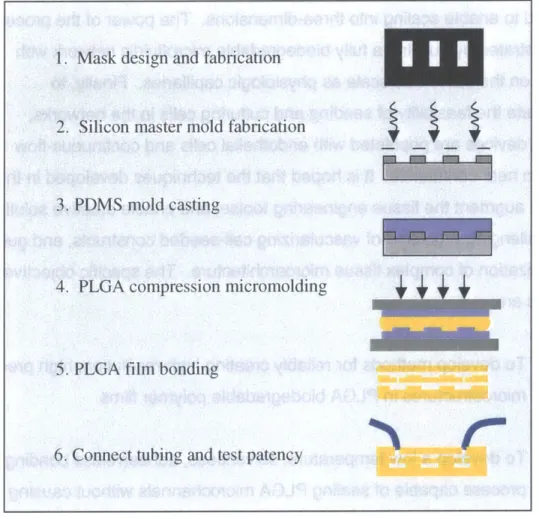
The primary accomplishment described in this thesis is the reproducible fabrication of a three-dimensional biodegradable microfluidic network. An overview of the fabrication process is shown pictorially in Figure 1.9.
Figure 1.9 Biodegradable microfluidics fabrication process overview. 1) Photomasks are
designed, fabricated, and 2) used to define microstructures in silicon. 3) This pattern is
transferred to a secondary PDMS mold and 4) used to compression micromold the biodegradable polymer, polylactic-co-glycolic acid (PLGA 85:15) by controlling time, temperature, and applied force. 5) The resulting films are bonded using an optimized thermal fusion process, and 6) fitted with connectors and tubing for perfusion and seeding with cells.
1. Mask design and fabrication
2. Silicon master mold fabrication
3. PDMS mold casting
4. PLGA compression micromolding
mt
5. PLGA film bonding
Chapter 1 provided background for the work, an overview of the thesis, and a forward-looking proposal for a generic biodegradable microfabricated scaffold to aid organ fabrication.
Chapter 2 describes the creation of micron scale features in silicon and the high fidelity transfer of the surface microstructures from silicon masters to flexible elastomeric PDMS films and then to PLGA biodegradable polymer films. Specific attention is focused on comparing solvent polymer processing with melt
polymer processing. Melt processing is ultimately chosen as the preferable process, and the remainder of the chapter describes process parameter selection and explores the range of structures that can be fabricated using the approach.
Chapter 3 describes the development of a thermal fusion bonding process that enables stacking of PLGA microstructured films to form three-dimensional biodegradable constructs. A theoretical model is developed to guide the selection of process times and temperatures in order to avoid temperature-induced plastic deformation and microchannel collapse, the dominant mode of failure during bonding. Using the model, a wide range of geometries are bonded in a robust and reproducible fashion.
Chapter 4 describes the development of extruded biodegradable fluidic connector ports that are integrated with the patterned PLGA surfaces to create monolithic microfluidic networks. The chapter describes fabrication of two complex branching network designs, demonstrates their patency, and characterizes their resistance to pressure-driven flow.
Chapter 5 describes a feasibility study in which endothelial cells are seeded and cultured in prototype PDMS microfluidic networks similar to the designs fabricated in PLGA throughout Chapter 4. Continuous flow of medium is used to support the cultures, and they are maintained sterile and unoccluded for two weeks, achieving 80-90% confluency.
Chapter 6 provides a summary of the major accomplishments in the work, and discusses future work and conclusions.
1.6 References
[1] Lanza, R.P., R. Langer, and W.L. Chick, eds. Principles of Tissue Engineering. Academic Press: Austin, 1997.
[2] UNOS, U.N.f.O.S., Report of the U.S. Organ Transplant Waiting List. UNOS: Richmond, VA, 2002.
[3] Griffith, L.G. and G. Naughton, "Tissue Engineering -Current Challenges and Expanding Opportunities". Science. 295: p. 1009-1014, 2002.
[4] Vacanti, J.P. and R. Langer, "Tissue Engineering: the Design and Fabrication of Living Replacement Devices for Surgical Reconstruction and Transplantation". The Lancet. 354(Suppl I): p. 32SI-34SI, 1999.
[5] Marler, J.J., J. Upton, R. Langer, and J.P. Vacanti, "Transplantation of Cells in Matrices for Tissue Engineering". Advanced Drug Delivery Reviews. 33: p. 165-182, 1998.
[6] Niklason, L.E. and R. Langer, "Prospects for Organ and Tissue Replacement". Journal of American Medical Association. 285(5): p. 573-576, 2001.
[7] Fuchs, J.R., B.A. Nasseri, and J.P. Vacanti, "Tissue Engineering: A 21st Century Solution to Surgical Reconstruction". Annals of Thoracic Surgery. 72: p. 577-591, 2001.
[8] Lee, K.Y. and D.J. Mooney, "Hydrogels for Tissue Engineering". Journal of Chemical Reviews, 2001.
[9] Mikos, A.G. and J.S. Temenoff, "Formation of Highly Porous Biodegradable Scaffolds for Tissue Engineering". Electronic Journal of Biotechnology. 3(2), 2000.
[10] Desai, T.A. and M. Ferrari, Microfabricated Biocapsules for the Immunoisolation
of Pancreatic Islets of Langerhans, in Bioengineering. University of California
San Francisco and University of California Berkeley: Berkley, CA, 1998. [11] Langer, R. and J.P. Vacanti, "Tissue Engineering". Science. 260(5110): p.
920-926, 1993.
[12] Shinoka, T., D. Shumtin, and P.X. Ma, "Creation of Viable Pulmonary Artery Autografts Through Tissue Engineering". Plastic Reconstructive Surgery. 88: p. 733-759, 1998.
[13] Niklason, L.E., J. Gao, and W.M. Abbott, "Functional Arteries Grown In Vivo". Science. 284: p. 489-493, 1999.
[14] Organ, G.M., D.J. Mooney, L.K. Hansen, B. Schloo, and J.P. Vacanti,
"Transplantation of Enterocytes Ultilizing Polymer-Cell Constructs to Produce Neointestine". Transplant Proc. 24: p. 3009-3011, 1992.
[15] Shinoka, T., P.X. Ma, and D. Shumtin, "Tissue engineered Hear Valves". Circulation. 94(Suppl II): p. 164-168, 1996.
[16] Peter, S.J., M.J. Miller, A.W. Yasko, M.J. Yaszemski, and A.G. Mikos, "Polymer Concepts in Tissue Engineering". Journal of Biomedical Materials Research
(Applied Biomaterials). 43: p. 422-427, 1998.
[17] Wang, N., X.S. Wu, C. Li, and M.F. Feng, "Synthesis, Characterization,
Biodegradation, and Drug Delivery Application of Biodegradable Lactic/Glycolic Acid Polymers. Part I: Synthesis and Characterization". Journal of Biomaterial Science Polymer Edn. 11(3): p. 301-318, 2000.
[18] Wu, X.S. and N. Wang, "Synthesis, Characterization, Biodegradation, and Drug Delivery Application of Biodegradable Lactic/Glycolic Acid Polymers. Part II: Biodegradation". Journal of Biomaterial Science Polymer Edn. 12(1): p. 21-34, 2001.
[19] Baldwin, S.P. and M.W. Saltzman, "Materials for Protein Delivery in Tissue Engineering". Advanced Drug Delivery Reviews. 33: p. 71-86, 1998.
[20] Desai, T.A., "Micro and nanoscale structures for tissue engineering constructs". Medical Engineering & Physics. 22: p. 595-606, 2000.
[21] Bhatia, S.N. and C.S. Chen, "Tissue Engineering at the Micro-Scale". Biomedical Microdevices. 2(2): p. 131-144, 1999.
[22] Chen, G., T. Ushida, and T. Tateishi, "Scaffold Design for Tissue Engineering". Macromol. Biosci. 2: p. 67-77, 2002.
[23] Yang, S., K.-f. Leong, Z. Du, and C.-k. Chua, "The Design of Scaffolds for Use in Tissue Engineering. Part I. Traditional Factors". Tissue Engineering. 7(6): p. 679-689, 2001.
[24] Babensee, J.E., J.M. Anderson, L.V. McIntire, and A.G. Mikos, "Host Response to Tissue Engineered Devices". Advanced Drug Delivery Reviews. 33: p.
111-139, 1998.
[25] Zeltinger, J., J.K. Sherwood, D.A. Graham, R. Mueller, and L.G. Griffith, "Effect of Pore Size and Void Fraction on Cellular Adhesion, Proliferation, and Matrix Deposition". Tissue Engineering. 7(5): p. 557-572, 2001.
[26] Mooney, D.J. and A.G. Mikos, "Growing New Organs". Scientific American. 280: p. 60-65, 1999.
[27] Soker, S., M. Machado, and A. Atala, "System for Therapeutic Angiogenesis in Tissue Engineering". World Journal Urology. 18: p. 10-18, 2000.
[28] Mikos, A.G., S.M. Leite, J.P. Vacanti, and R. Langer, "Prevascularization of Porous Biodegradable Polymers". Biotechnology and Bioengineering. 42: p. 716-723, 1993.
[29] Shea, L.D., E. Smiley, J. Bonadio, and D.J. Mooney, "DNA Delivery from
Polymer Matrices for Tissue Engineering". Nature Biotechnology. 17: p. 551-554, 1999.
[30] Murphy, W.L., M.C. Peters, D.H. Kohn, and D.J. Mooney, "Sustained Release of Vascular Endothelial Growth Factor from Mineralizaed Poly(lactic-co-glycolide) Scaffolds for Tissue Engineering". Biomaterials. 21: p. 2521-2527, 2000.
[31] Richardson, T.P., M.C. Peters, A.B. Ennett, and D.J. Mooney, "Polymeric System for Dual Growth Factor Delivery". Nature Biotechnology. 19: p. 1029-1034, 2001.
[32] Nomi, M., A. Atala, P. De Coppi, and S. Soker, "Principals of Neovascularization for Tissue Engineering". Molecular Aspects of Medicine, 2002.
[33] World Technology Evaluation Center, I., WTEC Panel Report on Tissue
Engineering Research. National Institute of Standards and Technology:
Gaithersburg, MD, 2002.
[34] Young, B. and J.W. Heath, Wheater's Functional Histology. New York: Churchill Livingstone, 2000.
[35] Murphy, W.L., R.G. Dennis, J.L. Kilney, and D.J. Mooney, "Salt Fusion: An Approach to Improve Pore Interconnectivity within Tissue Engineering Scaffolds". Tissue Engineering. 8(1): p. 43-52, 2002.
[36] Liao, C.-J., C.-F. Chen, J.-H. Chen, S.-F. Chiang, Y.-J. Lin, and K.-Y. Chang, "Fabrication of Porous Biodegradable Polymer Scaffolds Using a Solvent Merging/Particulate Leaching Method". Journal of Biomedical Materials Research. 59: p. 676-681, 2001.
[37] Chen, G., T. Ushida, and T. Tateishi, "Preparation of poly(L-lactic acid) and poly(DL-lactic-co-glycolic acid) foams by use of ice microparticulates".
Biomaterials. 22: p. 2563-2567, 2001.
[38] Zein, I., D.W. Hutmacher, K.C. Tan, and S.H. Teoh, "Fused Deposition Modeling of Novel Scaffold Architectures for Tissue Engineering Applications".
Biomaterials. 23: p. 1169-1185, 2002.
[39] Yang, S., K.-f. Leong, Z. Du, and C.-k. Chua, "The Design of Scaffolds for Use in Tissue Engineering. Part H. Rapid Prototyping Techniques". Tissue Engineering.
8(1): p. 1-11, 2002.
[40] Harris, L.D., B.-S. Kim, and D.J. Mooney, "Open Pore Biodegradable Matrices Formed with Gas Foaming". Journal of Biomedical Materials Research. 42: p. 396-402, 1998.
[41] Widmer, M.S., P.K. Gupta, L. Lu, R.K. Meszlenyi, G.R.D. Evans, K. Brandt, T. Savel, A. Gurlek, C.W.J. Patrick, and A.G. Mikos, "Manufacture of Porous Biodegradable Polymer Conduits by an Extrusion Process for Guided Tissue Regeneration". Biomaterials. 19: p. 1945-1955, 1998.
[42] Shastri, V.P., I. Martin, and R. Langer, "Macroporous Polymer Foams by Hydrocarbon Templating". Proceedings of the National Academy of Sciences. 97(5): p. 1970-1975, 2000.
[43] Wu, B.M., S.W. Borland, R.A. Giordano, L.G. Cima, E.M. Sachs, and M.J. Cima, "Solid Free-form Fabrication of Drug Delivery Devices". Journal of Controlled Release. 40: p. 77-87, 1996.
[44] Griffith, L.G., B.M. Wu, M.J. Cima, M.J. Powers, B. Chiagnaud, and J.P. Vacanti, "In Vitro Organogenesis of Liver Tissue". Annals of New York Academy of Sciences: p. 382-397, 1997.
[45] Kim, S.S., H. Utsunomiya, J.A. Koski, B.M. Wu, M.J. Cima, J. Sohn, K. Mukai, L.G. Griffith, and J.P. Vacanti, "Survival and Function of Hepatocytes on a Novel Three-Dimensional Synthetic Biodegradable Polymer Scaffold with an Intrinsic Network of Channels". Annals of Surgery. 228(1): p. 8-13, 1998.
[46] Kancharla, V.V. and S. Chen, "Fabrication of Biodegradable Polymeric
Microdevices Using Laser Micromachining". Biomedical Microdevices. 4(2): p. 105-109, 2002.
[47] Vozzi, G., C.J. Flaim, F. Bianchi, A. Ahluwalia, and S.N. Bhatia,
"Microfabricated PLGA Scaffolds: a comparative study for application to tissue engineering". Materials Science and Engineering C. 20: p. 43-47, 2002.
[48] Lu, L. and A.G. Mikos, Modulation of Cell Morphology and Function Using
Synthetic Biodegradable Polymers, in Bioengineering. Rice University: Houston,
[49] Voldman, J., M.L. Gray, and M.A. Schmidt, "Microfabrication in Biology and Medicine". Annual Reviews in Biomedical Engineering. 1: p. 401-425, 1999. [50] Trimmer, W.S., Micromechanics and MEMS Classic and Seminal Papers to
1990. New York: IEEE Press, 1996.
[51] Madou, M., Fundamentals of Microfabrication. New York: CRC Press, 1997. [52] Wolf, S. and R.N. Tauber, Silicon Processing for the VLSI Era Volume 1: Process
Technology. Sunset Beach, CA: Lattice Press, 1986.
[53] Desai, T.A., D.J. Hansford, L. Kulinsky, A.H. Nashat, G. Rasi, J. Tu, Y. Wang, M. Zhang, and M. Ferrari, "Nanopore Technology for Biomedical Applications". Biomedical Microdevices. 2(1): p. 11-40, 1999.
[54] Santini, J.T.J., M. Cima, and R. Langer, "A Controlled-Release Microchip". Nature. 397(6717): p. 335-338, 1999.
[55] Curtis, A. and C. Wilkinson, "Review: Topographical Control of Cells". Biomaterials. 18: p. 1573-1583, 1997.
[56] Kaihara, S., J.T. Borenstein, R. Koka, and J.P. Vacanti, "Silicon Micromachining to Tissue Engineer Branched Vascular Channels for Liver Fabrication". Tissue Engineering. 6: p. 105-117, 2000.
[57] Chin, V., B.E. Collins, M.J. Sailor, and S.N. Bhatia, "Compatibility of Primary Hepatocytes with Oxidized Nanoporous Silicon". Advanced Materials. 13(24): p.
1877-1880, 2001.
[58] Jackman, R.J., J.L. Wilbur, and G.M. Whitesides, "Fabrication of Submicrometer Features on Curved Substrates by microcontact Printing". Science. 269: p. 664-666, 1995.
[59] Yang, H., P. Deschatelets, S.T. Brittain, and G.M. Whitesides, "Fabrication of High Performance Ceramic Microstructures from a Polymeric Precursor Using Soft Lithography". Advanced Materials. 13(1): p. 54-58, 2001.
[60] Xu, B., F. Arias, and G.M. Whitesides, "Making Honeycomb Microcomposites by Soft Lithography". Advanced Materials. 11(6): p. 492-495, 1999.
[61] Whitesides, G.M., E. Ostuni, S. Takayama, X. Jiang, and D.E. Ingber, "Soft Lithography in Biology and Biochemistry". Annual Reviews in Biomedical Engineering. 3: p. 335-373, 2001.
[62] Xia, Y. and G.M. Whitesides, "Soft Lithography". Angew. Chem. Int. Ed. 37: p. 550-575, 1998.
[63] Xia, Y. and G.M. Whitesides, "Soft Lithography". Annual Reviews in Material Science. 28: p. 153-184, 1998.
[64] Singhvi, R., A. Kumar, G.P. Lopez, G.N. Stephanopoulos, D.I.C. Wang, G.M. Whitesides, and D.E. Ingber, "Engineering Cell Shape and Function". Science. 264: p. 696-698, 1994.
[65] Chen, C.S., M. Mrksich, S. Huang, G.M. Whitesides, and D.E. Ingber,
"Geometric Control of Cell Life and Death". Science. 276: p. 1425-1428, 1997. [66] Folch, A., B.-H. Jo, 0. Hurtado, D.J. Beebe, and M. Toner, "Microfabricated
Elastomeric Stencils for Micropatterning Cell Cultures". Journal of Biomedical Materials Research. 52(2): p. 346-353, 2000.
[67] Bhatia, S.N., U.J. Balis, M.L. Yarmush, and M. Toner, "Probing Heterotypic Cell Interactions: Hepatocyte Function in Microfabricated Co-cultures". Journal of Biomaterial Science Polymer Edn. 9(11): p. 1137-1160, 1998.