HAL Id: hal-01494065
https://hal.sorbonne-universite.fr/hal-01494065
Submitted on 22 Mar 2017HAL is a multi-disciplinary open access archive for the deposit and dissemination of sci-entific research documents, whether they are pub-lished or not. The documents may come from teaching and research institutions in France or abroad, or from public or private research centers.
L’archive ouverte pluridisciplinaire HAL, est destinée au dépôt et à la diffusion de documents scientifiques de niveau recherche, publiés ou non, émanant des établissements d’enseignement et de recherche français ou étrangers, des laboratoires publics ou privés.
Tolerance to Patients With Hepatitis C Virus-induced
Cryoglobulinemia Vasculitis
Cloé Comarmond, Marlène Garrido, Stanislas Pol, Anne-Claire Desbois,
Myrto Costopoulos, Magali Le Garff-Tavernier, Si Nafa Si Ahmed, Laurent
Alric, Hélène Fontaine, Bertrand Bellier, et al.
To cite this version:
Cloé Comarmond, Marlène Garrido, Stanislas Pol, Anne-Claire Desbois, Myrto Costopoulos, et al.. Direct-acting Antiviral Therapy Restores Immune Tolerance to Patients With Hepatitis C Virus-induced Cryoglobulinemia Vasculitis. Gastroenterology, WB Saunders, 2017, 152 (8), pp.2052-2062. �10.1053/j.gastro.2017.02.037�. �hal-01494065�
M
AN
US
CR
IP
T
AC
CE
PT
ED
1 Manuscript Number: GASTRO 16-01825Title: Direct-acting Antiviral Therapy Restores Immune Tolerance to Patients With Hepatitis C Virus-induced Cryoglobulinemia Vasculitis
Cloé Comarmond1,2,3,4, Marlène Garrido1,2,3, Stanislas Pol5, Anne-Claire Desbois1,2,3,4, Myrto Costopoulos6, Magali Le Garff-Tavernier6, Si Nafa Si Ahmed7, Laurent Alric8, Hélène Fontaine5, Bertrand Bellier1,2,3, Anna Maciejewski1,2,3, Michelle Rosenzwajg1,2,3, David Klatzmann1,2,3, Lucile Musset9, Thierry Poynard10, Patrice Cacoub1,2,3,4*, David Saadoun1,2,3,4*
1: Sorbonne Universités, UPMC Université Paris 06, UMR 7211, Département Hospitalo-Universitaire Inflammation-Immunopathologie-Biotherapie (DHU i2B), F-75005, Paris, France
2: INSERM, UMR_S 959, F-75013, Paris, France 3: CNRS, FRE3632, F-75005, Paris, France
4: AP-HP, Groupe Hospitalier Pitié-Salpêtrière, Département de Médecine Interne et Immunologie Clinique, F-75013, Paris, France
5: Department of Hepatology, APHP, Hôpital Cochin, Paris, France
6: Biological Hematology, APHP, Groupe Hospitalier Pitié-Salpétrière, Paris, France 7: Department of Hepatology, Hôpital d'Orléans, Orléans, France
8: Department of Internal Medicine and Digestive Diseases, Centre hospitalier universitaire Purpan, UMR 152, IRD Toulouse 3 University, Toulouse, France
9: Department of Immunology, UF d’Immunochimie et d’autoimmunité, APHP, Groupe Hospitalier Pitié-Salpêtrière, Paris, France
10: Department of Hepatology, UMR_S 938 & Institute of Cardiometabolism and Nutrition (ICAN), AP-HP, Groupe Hospitalier Pitié-Salpétrière, Paris, France
Address correspondence to: David Saadoun, MD, PhD, Service de Médecine Interne et d'Immunologie
Clinique, Hôpital Pitié-Salpêtrière, 83 boulevard de l’hôpital, 75013 Paris, France. Phone: +33(0)142178009, Fax: +33(0)142178033. E-mail: david.saadoun@aphp.fr
Word count: 3643, 2 Tables, 5 Figures, 50 references. Disclosures: None.
Author Contributions. CC, PC, DS: study concept and design; CC, MG, SP, ACD, MC, MLGT, SNSA,
LA, HS, BB, AM, MR, DK, LM, TP, PC, DS: acquisition of data; CC, PC, DS: analysis and
interpretation of data; CC, PC, DS: drafting of the manuscript.
Permission Information: All illustrations and figures in the manuscript are entirely original and do not
require reprint permission.
* Equal contribution
M
AN
US
CR
IP
T
AC
CE
PT
ED
2 ABSTRACTBackground & Aims: Interferon-free direct-acting antiviral (DAA) therapies are effective in patients with hepatitis C virus-induced cryoglobulinemia vasculitis (HCV-CV). We analyzed blood samples from patients with HCV-CV before and after DAA therapy to determine mechanisms of these drugs and their effects on cellular immunity. Methods: We performed a prospective study of 27 consecutive patients with HCV-CV (median age 59 years) treated with DAA therapy (21 patients received sofosbuvir plus ribavirin for 24 weeks, 4 patients received sofosbuvir plus daclatasvir for 12 weeks, and 2 patients received sofosbuvir plus simeprevir for 12 weeks) in Paris, France. Blood
samples were collected from these patients before and after DAA therapy, and also from 12 healthy donors and 12 individuals with HCV infection without CV. HCV load, cryoglobulins, and cytokines were quantified by flow cytometry, cytokine multiplex assays, and ELISAs.
Results: Twenty-four patients (88.9%) had a complete clinical response of CV to DAA therapy at week 24, defined by improvement of all the affected organs and the absence of relapse. Compared to healthy donors and patients with HCV infection without CV, patients with HCV-CV, before DAA therapy, had a lower percentage of
CD4+CD25hiFoxP3+ regulatory T cells (P<.01) but higher proportions of IgM+CD21– /low memory B cells (P<.05), CD4+IFNγ+ cells (P<.01), CD4+IL17A+ cells (P<.01), and CD4+CXCR5+IL21+ follicular T-helper (Tfh) cells (P<.01). In patients with HCV-CV, there was a negative correlation between numbers of IgM+CD21–/low memory B cells and T-regulatory cells (P=.03), and positive correlations with numbers of Tfh cells (P=.03) and serum levels of cryoglobulin (P=.01). DAA therapy increased patients’ numbers of T-regulatory cells (1.5%±0.18% before therapy vs 2.1%±0.18% after
therapy), decreased percentages of IgM+CD21–/low memory B cells (35.7%±6.1% before therapy vs 14.9% ± 3.8% after therapy), and decreased numbers of Tfh cells (12%±1.3% before therapy vs 8%±0.9% after therapy). Expression levels of B lymphocyte stimulator receptor 3 and programmed cell death 1 on B cells increased in patients with HCV-CV after DAA-based therapy (mean fluorescence units, 37 ± 2.4 before therapy vs 47 ± 2.6 after therapy; P<.01 and 29 ± 7.3 before therapy vs 48 ± 9.3 after therapy; P<.05, respectively).
Conclusions: In a prospective clinical trial of patients with HCV-CV, DAA-based therapy restored disturbances in peripheral B- and T-cell homeostasis.
M
AN
US
CR
IP
T
AC
CE
PT
ED
3 IntroductionCirculating mixed cryoglobulins (MC) are detected in 40%–60% of patients with chronic hepatitis C virus (HCV) infection, whereas overt cryoglobulinemia vasculitis (CV) is observed in only 5%-10% of cases 1–4. It involves the activation of B cells, which generates pathogenic immunoglobulin (Ig) M and IgG with rheumatoid factor activity 5,6.
CV leads to clinical manifestations ranging from purpura, arthralgia and fatigue to more serious lesions with neurologic and renal involvement2. Despite success with combination
antiviral treatment with or without immunosuppressive drugs, HCV-CV remains a serious disease with an estimated 5-year survival rate of 75% 4,7.
Treatment of HCV-CV remains difficult and requires targeting the downstream B cell arm of autoimmunity and the viral trigger to obtain clinical resolution of symptoms8,9. Two prospective randomized controlled trials have demonstrated the superiority of rituximab monotherapy compared with conventional immunosuppressive therapy in patients with HCV-CV in whom prior antiviral therapy failed to induce disease remission 10,11
. Rituximab was effective in 71.4% to 83% of patients with CV 10,11
. However, in the absence of HCV clearance, frequent relapses occurred when B cells reemerged in the peripheral blood. Direct-acting antivirals (DAA) have proven to be very effective in patients with HCV-CV. A complete clinical response was achieved in 87.5% of HCV-CV patients treated with sofosbuvir plus ribavirin, and was closely correlated with virological response12–14. We previously reported a quantitative defect in regulatory T cells (Tregs) and clonal expansion of CD27+IgM+CD21-/low memory B cells in patients with HCV-CV15–17. It has been demonstrated that antiviral treatment can lead to the disappearance of symptoms and can induce an immunologic response, i.e., a significant decrease in plasma cryoglobulin levels and increase in Tregs 15,18. In addition, a decrease in B lymphocyte stimulator receptor 3 (BR3) staining on B cells was associated with an increase in serum
M
AN
US
CR
IP
T
AC
CE
PT
ED
4 B cell activating factor of the tumor necrosis factor family (BAFF) after rituximab in HCV-CV 19.However, little evidence is available about immunologic dynamics after IFN free DAA-based therapy. As a first examination of the immunologic effects of DAAs in HCV-CV, we evaluated both B and T cell subsets in patients at baseline (W0) and after DAA therapy at end of treatment (EOT) and post treatment (PT). Our results indicate that in addition to their virological effect, DAA-based therapy effectively normalize many of the disturbances in peripheral B- and T-lymphocytes homeostasis.
METHODS
Study population
Twenty-seven consecutive patients with HCV-CV (median age 59 years [range 53–66]) were included in the study and received interferon-free DAA therapy. All were positive for HCV RNA. The HCV viral load was quantified using the Abbott HCV RealTime assay (Abbott, Rungis, France), with a lower limit of detection of 12 IU/mL. To be eligible, the patient must have been at least 18 years of age or older (without any upper age limit), been informed of the study, and presented with active HCV vasculitis defined by clinically active vasculitis with skin, joint, renal, peripheral nerve, central neurological, digestive, pulmonary and/or cardiac involvement (no histological evidence needed if patient had purpura), and chronic active HCV infection (positive HCV RNA). Exclusion criteria included non-active cryoglobulinemia vasculitis, HIV or active HBV infection, and current decompensated cirrhosis.
Cryoglobulins were measured and classified as previously described 20,21. Twelve healthy donors (HD) and 12 HCV patients, age and sex-matched controls, with positive
M
AN
US
CR
IP
T
AC
CE
PT
ED
5 viremia and without circulating cryoglobulins were also included. The study was approved by the institutional ethics committee of Pitié-Salpêtrière Hospital and was performed in accordance with the Declaration of Helsinki. All participants provided informed consent.Treatment
Twenty-seven consecutive patients with HCV-CV were treated with DAAs according to current guidelines. Twenty-one patients received antiviral therapy with sofosbuvir 400 mg daily plus ribavirin (200–1400 mg/day orally) for 24 weeks. Four patients received sofosbuvir plus daclatasvir for 12 weeks. Two patients received sofosbuvir plus simeprevir for 12 weeks. None of the patients received corticosteroids, immunosuppressants or rituximab in the 6 months before inclusion. Twenty-four patients (88.9%) had a complete clinical response of their CV at week 24. The complete clinical response of CV was defined by improvement of all the affected organs involved at baseline and the absence of clinical relapse. The skin and articular improvement were evaluated clinically (i.e. disappearance of purpura and/or ulcers and/or skin necrosis, disappearance of arthralgia and/or arthritis). Renal improvement was evaluated biologically (i.e. proteinuria <0.3g/24h, disappearance of hematuria and improvement of GFR > 20% at week 24 if GFR < 60 ml/min/1.73 m² at diagnosis). Peripheral neurological improvement was evaluated clinically (i.e. improvement of pains and paraesthesia by visual analogue scales, improvement of muscular testing in case of motor impairment at baseline) and/or electrophysiologically (i.e. improvement of electromyogram abnormalities at week 24 compared to baseline). Twenty-two patients (81.5%) had a sustained virological response (HCV RNA negative) at week 12 post-treatment.
M
AN
US
CR
IP
T
AC
CE
PT
ED
6 Flow cytometryPeripheral blood mononuclear cells (PBMCs) were obtained by density-gradient centrifugation. PBMCs were stained with the following monoclonal antibodies (at predetermined optimal dilutions) for 30 minutes at 4°C: Fluorescein isothiocyanate (FITC)-conjugated CD21, PE-conjugated CD27 and B lymphocyte stimulator receptor 3 (BR3) and CXCR5, Alexa Fluor 450-conjugated CD3, allophycocyanin (APC)-conjugated IgM, energy-coupled dye (ECD)-(APC)-conjugated CD4 and CD19, PerCP-Cy7-conjugated CD25 and PD-L1, VioBlue-PerCP-Cy7-conjugated IgD, and Brilliant Violet 510-conjugated CD127 (BioLegend). Memory B cells IgM+ CD21-/low were identified as IgM+CD21-/low cells in CD19+ B lymphocytes. Detection of intracellular FoxP3 was accomplished using fixed and permeabilized cells according to the manufacturer’s instructions (eBioscience), and then incubated with APC-conjugated FoxP3. Tregs were identified as Foxp3+CD25+ cells in CD4+ T lymphocytes. FACS analyses were performed on a Navios flow cytometer using Kaluza analysis software (Beckman Coulter).
Analysis of cytokine production
PBMCs from HD, HCV without cryoglobulinemia and HCV-CV patients were stimulated for 4 hours with 50 ng/mL phorbol myristate acetate (PMA) and 1 mM ionomycin (Sigma-Aldrich) in the presence of brefeldin A (BD PharMingen). Cells cultured in the presence of Brefeldin A were stained for cell surface markers and then permeabilized with Cytofix/Cytoperm buffer (BD PharMingen) and stained with fluorescein isothiocyanate (FITC)-conjugated IFNγ (BD PharMingen), Alexa Fluor 647-conjugated IL-17A (eBioscience), and Alexa Fluor 647-647-conjugated IL-21 (Bio-Legend). Th1 were identified as IFNγ+CD4+ cells in CD3+ T lymphocytes. Th17 were identified
M
AN
US
CR
IP
T
AC
CE
PT
ED
7 as IL17A+CD4+ cells in CD3+ T lymphocytes. Tfh were identified as CD4+CXCR5+IL21+ cells in CD3+ T lymphocytes.The levels of the following serum cytokines were measured using Human Cytokine MILLIPLEX according to the recommendations of the manufacturer (Merk Millipore): IFNγ, TNFα, IL12p70, IL-4, IL-5, IL-10, IL-13, IL-17A, and IL-21. The levels of B cell activating factor of the tumor necrosis factor family (BAFF) (R&D Systems) were determined by enzyme-linked immunosorbent assay.
Antigen-specific stimulation with virus-like particles (VLPs)
PBMCs from HCV-CV patients were stimulated during 48 hours with peptide pools (15-mers overlapping by 10 amino acids; Eurogentec) spanning the whole sequence of the H77 strains E1 and E2, as previously described 22. During the last 4 hours, brefeldin A was added. Tfh, Th1 and Th17 were identified by flow cytometry as previously described.
Data analysis and statistics
Analyses were performed using GraphPad Prism 6.0 software (Graph-Pad Software, San Diego, CA). Except when otherwise indicated, values are expressed as medians [IQR]. Categorical variables were compared using the Fisher’s exact or chi-squared tests, and continuous variables were compared using the t-test or Mann-Whitney U test when appropriate. Correlation significance was calculated with the Pearson equation. All tests were two-tailed with a significance level of 0.05.
M
AN
US
CR
IP
T
AC
CE
PT
ED
8 RESULTSMain characteristics of HCV-CV patients.
Patient characteristics are detailed in Table 1. Twenty-seven patients with HCV-CV, with a median age of 59 years [range 53–66] were included. The main clinical features of cryoglobulinemia vasculitis included purpura (89%), peripheral neuropathy (48%), glomerulonephritis (26%), and skin ulcers (15%). The median cryoglobulin level was 0.35 [0.17–0.81] g/L. Twenty-four patients (88.9%) had a complete clinical response of their CV. Twenty two patients (81.5%) had a sustained virological response (HCV RNA negative) at week 12 post treatment. Ten (27%) patients presented negativation of cryoglobulinemia and 17 (63%) patients had persistant cryoglobulinemia after DAA-based therapy.
Abnormalities in peripheral cell homeostasis of HCV-CV.
Compared with healthy donors (HD) and HCV controls, pre-treatment abnormalities in HCV-CV patients included a decreased percentage of CD4+CD25hiFoxP3+ regulatory T cells (1.5% ± 0.2% versus 3.7% ± 0.2% for HD, P < .01, and versus 2% ± 0.4% for HCV controls, P = .6) (Figure 1A, 1B). We also found increases in: IgM+CD21-/low memory B cells (35.7% ± 6.2% versus 7% ± 1.2% for HD, P = .0004, and versus 23.1% ± 4.6% for HCV controls, P = .23) (Figure 1C, 1D); CD4+IFNγ+ (21.6% ± 1.9% versus 10.8% ± 0.8% for HD, P < .0001, and versus 16.2% ± 1.7% for HCV controls, P = .15); CD4+IL17A+ (2.4% ± 0.4% versus 0.85% ± 0.12% for HD, P = .007, and versus 0.88% ± 0.16% for HCV controls, P = .002); and CD4+CXCR5+IL21+ follicularhelper T cells (Tfh) (12.2% ± 1.4% versus 4.5% ± 0.75% for HD, P = .0001, and versus 6% ± 1.6% for HCV controls, P = .003) (Figure 2).
M
AN
US
CR
IP
T
AC
CE
PT
ED
9 We have evaluated the effect of antiviral therapy in PBMCs of HCV-CV patients after antigen-dependent stimulation by VLPs. We observed an expansion in Tfh, Th1 and Th17 at baseline (W0) that decreased after antiviral therapy (EOT and PT) (Suppl Figure 1). Proportions of CD4+CXCR5+IL21+ after VLPs stimulation were 3.9% ± 0.1% at W0, 2.7% ± 0.4% at EOT and 2.5% ± 0.3% at PT. Proportions of CD4+IL21+ after VLPs stimulation were 33.5% ± 0.1% at W0, 8.3% ± 0.9% at EOT and 5.8% ± 0.4% at PT. Proportions of CD4+IFNγ+ after VLPs stimulation were 16.3% ± 2.9% at W0, 2.8% ± 1.4% at EOT and 3.6% ± 0.9% at PT. Proportions of CD4+IL17A+ after VLPs stimulation were 4.2% ± 0.8% at W0, 0.9% ± 0.4% at EOT and 1.% ± 0.3% at PT.We also examined the expression of CD27, IgD, and CD38 in B cells to differentiate naive (IgD+CD27-), switched memory (IgD-CD27+), unswitched memory (IgD+CD27+) cells and plasmablasts (IgD-CD27highCD38high). We did not observe statistically significant differences in naive B cells (41.2% ± 7.3% in HCV-CV versus 52% ± 5.8% for HD, P = .47, and 50.3% ± 7.3% for HCV controls, P = .63), switched memory B cells (37% ± 6.3% in HCV-CV versus 24.3% ± 3.3% for HD, P = .52, and 21.6% ± 4.3% for HCV controls, P = .23), unswitched memory B cells (16.6% ± 4.7% in HCV-CV versus 14% ± 2.5% for HD, P = .37, and 15.2% ± 3.4% for HCV controls, P = .75), and plasmablasts (2.3% ± 0.39% in HCV-CV versus 1% ± 0.19% for HD, P = .09, and 1.7% ± 0.44% for HCV controls, P = .59) (data not shown).
Correlation between IgM+ CD27+ CD21-/low memory B cells with regulatory T cells
(Tregs), T follicular helper (Tfh) and cryoglobulin levels.
We next asked whether the expansion of IgM+CD21-/low memory B cells was correlated with Tregs and Tfh frequency, cryoglobulin levels and HCV viral load. IgM+CD21-/low memory B cells were negatively correlated with Tregs (r = ─ 0.46,
M
AN
US
CR
IP
T
AC
CE
PT
ED
10 P=0.03), and positively correlated with Tfh (r = 0.44, P = .03) and cryoglobulin levels (r = 0.59, P=0.01). There was no correlation with the baseline HCV viral load (P = .88) (Figure 3).IFN free DAA-based therapy improve peripheral cell homeostasis in HCV-CV.
Regulatory T cell deficiency, IgM+CD21-/low memory B cell expansion and Tfh (CD4+CXCR5+IL21+) expansion did significantly reverse after DAA therapy (1.5% ± 0.18% at W0 versus 2.1% ± 0.18% at EOT and versus 2.7% ± 0.3% at PT, 35.7% ± 6.1% at W0 versus 14.9% ± 3.8% at EOT and versus 10.3% ± 2.9% PT, 12% ± 1.3% at W0 versus 8% ± 0.9% at EOT and versus 5.6% ± 1.2% PT, respectively) (Figure 4). The cryoglobulin level decreased from 0.35 (0.17–0.81) at baseline (W0) to 0.22 (0.06–0.52) g/L at EOT (P = .009; Figure 4G) to 0.13 (0-0.33) g/L at PT (P < .001; Figure 4G). We did not observe a significant difference in frequency of CD4+IFNγ+ (21.6% ± 1.9% versus 21.2% ± 2.1%, P = .85) or CD4+IL17A+ (2.4% ± 0.4% versus 1.6% ± 0.3%, P = .1) before and after DAA. Comparison between HCV-CV virological responders (n=22) and non responders (n=5) to DAA-based therapy did not show significant difference in terms of proportion of Tregs, IgM+CD21-/low memory B cell, Tfh, Th1, and Th17 cells before and after DAA.
DAA-based therapy did not impact T- and B cells homeostasis in HCV patients without cryglobulinemia (Suppl Figure 2).
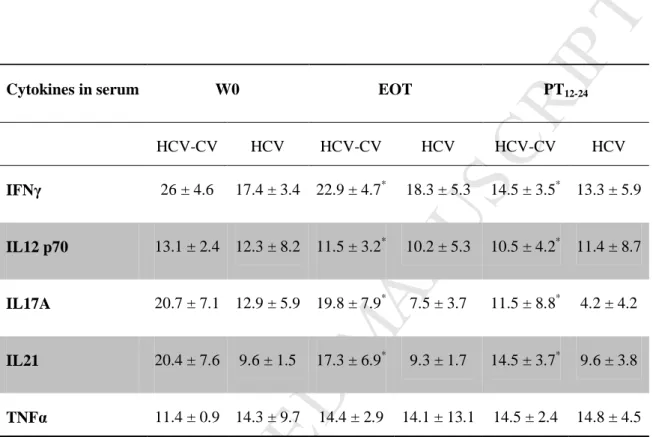
Serum levels of IFNγ, IL12 p70, IL17A and IL21 decreased significantly in HCV-CV patients after DAA therapy (26 ± 4.6 versus 22.9 ± 4.7 pg/ml; 13.1 ± 2.4 versus 11.5 ± 3.2 pg/ml; 20.7 ± 7.1 versus 19.8 ± 7.9 pg/ml; and 20.4 ± 7.6 versus 17.3 ± 6.9 pg/ml, respectively, P < .05) (Table 2). Serum levels of TNFα, IL-4, IL-5, IL-10 and IL-13 did not differ significantly before and after DAA therapy. We did not observed significant
M
AN
US
CR
IP
T
AC
CE
PT
ED
11 changes in serum levels of IFNγ, IL12 p70, IL17A, IL21 and TNFα in HCV patients without cryoglobulinemia between W0, EOT and PT (Table 2).Serum levels of BAFF tended to decrease after DAA therapy but did not reach statistical significance (1898 ± 371 versus 1491 ± 194 pg/ml, P = .58).
BR3 and PD-L1 expression on B cells are modulated by DAA therapy, and
expression of BR3 was negatively correlated with BAFF in serum.
BR3 staining levels on B cells were increased in HCV-CV patients when compared before and after DAA therapy (MFI 37 ± 2.4 versus 47 ± 2.6, P < .01) (Figure 5A, 5B). Similarly, PD-L1 staining levels on B cells were increased in HCV-CV patients
when compared before and after DAA therapy (MFI 29 ± 7.3 versus 48 ± 9.3, P < .05) (Figure 5C, 5D). MFI of BR3 was negatively correlated with BAFF in serum (r = ─ 0.46,
P = .039) (Figure 5E).
We analyzed PD-1 and CTLA-4 expression on Tregs before and after DAA-based therapy. We did not observed difference relative to PD-1 expression (MFI 1.9 ± 0.7 at W0 versus 1.7 ± 0.5 at EOT), but an increase in CTLA-4 expression (MFI 9.3 ± 2.6 at W0 versus 11.7 ± 3.8 at EOT) after DAA-based therapy.
DISCUSSION
Mixed cryoglobulinemia vasculitis is a serious autoimmune condition, which is predominantly induced by chronic HCV infection. It is an example of infection-associated tolerance failure, which leads to an immune response directed against host tissues. As such, it affords a particularly interesting insight into the delicate interrelation between invading infection, host tissue and the host immune system. The pathogenesis of mixed
M
AN
US
CR
IP
T
AC
CE
PT
ED
12 cryoglobulinemia vasculitis has traditionally been considered to be immune complex mediated. However, a number of T cell abnormalities have been described, including polarization toward Th1 subsets23 and quantitative deficiency of circulating regulatory T cells. In this study we identified significant T and B cell abnormalities in patients with HCV-CV compared with healthy donors and HCV controls. We observed for the first time Tfh expansion associated with Th1 and Th17 polarization in HCV-CV. As previously reported16,17, HCV-CV patients had Tregs deficiency and marked expansion of autoreactive IgM+CD21-/low memory B cells. We also observed a negative correlation between regulatory T cell deficiency and IgM+CD21-/low memory B cell expansion, as well as a positive correlation between Tfh expansion and IgM+CD21-/low memory B cell expansion in HCV-CV. HCV has been shown to lower B and T cell activation thresholds24,25
, which may promote immune injury to self. Therefore, the finding that enhanced frequency and function of CD4+CD25+ cells was associated with a lower degree of HCV-associated liver inflammation26,27
, as well as the finding that activated Tregs in patients with chronic HCV infection suppress effector cell activity in a non antigen-specific manner28
, may both suggest a beneficial role for Tregs in reducing immune-mediated bystander injury in chronic HCV infection. However, the exact role and function of Tregs in HCV infected patients without cryoglubulinemia is still debated 29.
IL-21 is known to drive Th1 and Th17 differentiation, and to decrease Treg cell frequency 30. IL-21 also drives autoreactive B cell responses, promoting Tfh differentiation and germinal center reaction by skewing the follicular regulatory T cell to follicular helper T cell balance31
. Interestingly, in chronic HCV infection without cryoglobulinemia vasculitis, IL-21 production by Tfh is impaired when compared with healthy controls32
. The decreased frequency of IL-21 producing CXCR5+CD4+ T cells and the lower serum IL-21 levels in chronic HCV patients without CV did not lead to an
M
AN
US
CR
IP
T
AC
CE
PT
ED
13 altered Tfh-B cell interaction (21). Conversely, we observed Tfh (i.e. CXCR5+CD4+ IL-21+) expansion in HCV-CV compared with healthy donors and HCV controls, which could contribute to aberrant B cell activation, generating pathogenic IgM and IgG with rheumatoid factor activity.In the present study we have further demonstrated that following DAA use, complete remission in autoimmune manifestations as well as viral clearance are associated with normalization of the significant disturbances in peripheral T and B lymphocyte homeostasis. This is very relevant as in the first place, immunological changes are expected to be different according to the achievement of viral clearance or not. In addition, several studies have shown that despite HCV eradication, some patients still present immune activation (positive cryocrit) or clinical symptoms (active vasculitis)
14,33
. The immune changes associated to these relevant clinical outcomes still remain to be understood. DAAs have been developed to target non-structural viral proteins involved in HCV viral replication (NS3, NS4A, NS4B, NS5A and NS5B), thereby resulting in disruption of the viral life cycle. In addition to this direct action, they may also help in the recovery of innate immune processes via interferon production34
. However, in our study only 2 patients received protease inhibitors. Although we did not observe any correlation between HCV viremia and IgM+CD21-/low memory B cell expansion before antiviral therapy, HCV clearance is closely associated with clinical and immunological response12,15,33. After DAA-based therapy, a significant decrease in Th1 (IFNγ, IL-12 p70) and Th17 (IL-17A) cytokine serum levels was observed.
We previously showed that the use of low-dose interleukin-2 leads to regulatory T cell recovery and concomitant clinical improvement in patients with HCV-CV35
M
AN
US
CR
IP
T
AC
CE
PT
ED
14 are central players in a number of autoimmune diseases, as they facilitate both the aberrant generation of autoantibodies and the formation or maintenance of ectopic follicles36. Taken together, our results suggest critical cross-talk between Tregs, Tfh and autoreactive IgM memory B cells for maintaining tolerance in HCV-CV patients. Of particular interest in this context is the two-way interaction between B cells and T cells. B cells provide signals to T cells through antigen presentation, and T cells provide “help” to B cells through the delivery of cytokines and cell-surface ligands. These interactions create the potential for a positive feedback loop or “vicious cycle.”It has recently emerged that the PD-1/PD-L1 pathway has a role in regulating lymphocyte activation, promoting regulatory T cell development and function, the breakdown of tolerance and the development of autoimmunity37
. This pathway exerts critical inhibitory functions in the setting of persistent antigenic stimulation such as during encounters with self-antigens, chronic viral infections, and tumors 38. Ligation of PD-1 by either PD-L1 or PD-L2 attenuates effector T cell proliferation, cytokine secretion and survival. In the presence of anti-CD3 and TGF-β, PD-L1 can induce a profound increase in the de novo generation of CD4+Foxp3+ Tregs from naive CD4+ T cells 39
. Elevated PD-L1 expression on B cells is an important regulator of Tfh cell activity40
. PD-L1 directly modulates TH1 cell differentiation by promoting a tolerogenic regulatory T cell phenotype 41
. The absence of PD-L1 on B cells leads to an increase in Tfh cells with abundant IgG production42. In our study, we found increased PD-L1 expression on B cells after DAA therapy. Together, these results suggest that the normalization of PD-L1 expression on B cells in HCV-CV after DAA therapy contributes to restoring immune tolerance, and controlling B cell responses and B-regulatory T cell interaction.
M
AN
US
CR
IP
T
AC
CE
PT
ED
15 BAFF is a critical factor for B cell survival and maturation. It has been well demonstrated that BAFF is involved in the pathogenesis of many autoimmune and B cell lymphoproliferative disorders 43–46. B lymphocyte stimulator receptor 3 (BR3) is the most abundant B lymphocyte stimulator receptor. We previously demonstrated a decrease in BR3 staining on B cells in patients with HCV-CV, especially in clonal autoreactive IgM memory B cells 19. Studies have shown that BAFF/BAFF-R signaling plays an essential role in the survival and maintenance of both follicular and marginal zone B cells, but not germinal center, normal B cells 47,48. BAFF is also involved in the regulation of Tfh cells. BAFF directly stimulates T-cell proliferation and cytokine production49
.
In the present study, BR3 and PD-L1 expressions on B cells increased after DAA therapy. This is in sharp contrast with the results found after rituximab treatment, i.e. a marked increase in serum BAFF concentration and decrease in BR3 staining, a factor which may indicate an increase in BAFF receptor-ligand activity 19. It may be hypothesized that repopulation of the B cell compartment in a BAFF-rich environment favors autoreactive clones, which may be of special importance in HCV-induced B cell proliferative disorders. Along this line, it has recently been shown that rituximab does not reset defective early B cell tolerance checkpoints50
. Taken together, these results indicate that BAFF receptor-ligand system normalization is a characteristic of remission induced by IFN free DAA-based therapy.
In conclusion, our study identified Tfh expansion associated with Th1 and Th17 polarization, and marked expansion of IgM+CD21-/low memory B cells and Treg deficiency in HCV-CV. We also demonstrated that IFN free DAA-based therapy
M
AN
US
CR
IP
T
AC
CE
PT
ED
16 dramatically improves abnormalities in B cell homeostasis, with a decreased proportion of autoreactive memory B cells and decreased cryoglobulin levels after treatment. In addition, the results reported herein indicate that DAA may be an efficient therapy in HCV-CV patients not only because it reduces or abolishes the production of cryoglobulin, but also because it improves T cell homeostasis by restoring regulation/activation and Th1/Th17 imbalances.M
AN
US
CR
IP
T
AC
CE
PT
ED
17 REFERENCES1. Ramos-Casals M, Stone JH, Cid MC, Bosch X. The cryoglobulinaemias. Lancet. 2012;379(9813):348–360.
2. Cacoub P, Comarmond C, Domont F, Savey L, Saadoun D. Cryoglobulinemia Vasculitis. Am. J.
Med. 2015;
3. Cacoub P, Comarmond C, Domont F, et al. Extrahepatic manifestations of chronic hepatitis C virus infection. Ther. Adv. Infect. Dis. 2016;3(1):3–14.
4. Dammacco F, Sansonno D. Therapy for hepatitis C virus-related cryoglobulinemic vasculitis. N.
Engl. J. Med. 2013;369(11):1035–1045.
5. Charles ED, Orloff MIM, Nishiuchi E, et al. Somatic hypermutations confer rheumatoid factor activity in hepatitis C virus-associated mixed cryoglobulinemia. Arthritis Rheum.
2013;65(9):2430–2440.
6. Meltzer M, Franklin EC, Elias K, McCluskey RT, Cooper N. Cryoglobulinemia--a clinical and laboratory study. II. Cryoglobulins with rheumatoid factor activity. Am. J. Med. 1966;40(6):837– 856.
7. Cacoub P, Terrier B, Saadoun D. Hepatitis C virus-induced vasculitis: therapeutic options. Ann.
Rheum. Dis. 2014;73(1):24–30.
8. Saadoun D, Resche-Rigon M, Sene D, et al. Rituximab combined with Peg-interferon-ribavirin in refractory hepatitis C virus-associated cryoglobulinaemia vasculitis. Ann. Rheum. Dis.
2008;67(10):1431–1436.
9. Saadoun D, Resche Rigon M, Sene D, et al. Rituximab plus Peg-interferon-alpha/ribavirin compared with Peg-interferon-alpha/ribavirin in hepatitis C-related mixed cryoglobulinemia.
Blood. 2010;116(3):326-334-505.
10. De Vita S, Quartuccio L, Isola M, et al. A randomized controlled trial of rituximab for the treatment of severe cryoglobulinemic vasculitis. Arthritis Rheum. 2012;64(3):843–853.
11. Sneller MC, Hu Z, Langford CA. A randomized controlled trial of rituximab following failure of antiviral therapy for hepatitis C virus-associated cryoglobulinemic vasculitis. Arthritis Rheum. 2012;64(3):835–842.
12. Saadoun D, Thibault V, Si Ahmed SN, et al. Sofosbuvir plus ribavirin for hepatitis C virus-associated cryoglobulinaemia vasculitis: VASCUVALDIC study. Ann. Rheum. Dis. 2015; 13. Sise ME, Bloom AK, Wisocky J, et al. Treatment of hepatitis C virus-associated mixed
cryoglobulinemia with direct-acting antiviral agents. Hepatol. Baltim. Md. 2016;63(2):408–417. 14. Gragnani L, Visentini M, Fognani E, et al. Prospective Study of Guideline-Tailored Therapy with
Direct-Acting Antivirals for Hepatitis C Virus-Associated Mixed Cryoglobulinemia. Hepatol.
Baltim. Md. 2016;
15. Landau D-A, Rosenzwajg M, Saadoun D, et al. Correlation of clinical and virologic responses to antiviral treatment and regulatory T cell evolution in patients with hepatitis C virus-induced mixed cryoglobulinemia vasculitis. Arthritis Rheum. 2008;58(9):2897–2907.
16. Boyer O, Saadoun D, Abriol J, et al. CD4+CD25+ regulatory T-cell deficiency in patients with hepatitis C-mixed cryoglobulinemia vasculitis. Blood. 2004;103(9):3428–3430.
17. Terrier B, Joly F, Vazquez T, et al. Expansion of functionally anergic CD21-/low marginal zone-like B cell clones in hepatitis C virus infection-related autoimmunity. J. Immunol. Baltim. Md
1950. 2011;187(12):6550–6563.
18. Saadoun D, Resche-Rigon M, Thibault V, Piette J-C, Cacoub P. Antiviral therapy for hepatitis C virus--associated mixed cryoglobulinemia vasculitis: a long-term followup study. Arthritis Rheum. 2006;54(11):3696–3706.
19. Landau D-A, Rosenzwajg M, Saadoun D, Klatzmann D, Cacoub P. The B lymphocyte stimulator receptor-ligand system in hepatitis C virus-induced B cell clonal disorders. Ann. Rheum. Dis. 2009;68(3):337–344.
20. Musset L, Diemert MC, Taibi F, et al. Characterization of cryoglobulins by immunoblotting. Clin.
Chem. 1992;38(6):798–802.
21. Brouet JC, Clauvel JP, Danon F, Klein M, Seligmann M. Biologic and clinical significance of cryoglobulins. A report of 86 cases. Am. J. Med. 1974;57(5):775–788.
M
AN
US
CR
IP
T
AC
CE
PT
ED
18 22. Garrone P, Fluckiger A-C, Mangeot PE, et al. A prime-boost strategy using virus-like particlespseudotyped for HCV proteins triggers broadly neutralizing antibodies in macaques. Sci. Transl.
Med. 2011;3(94):94ra71.
23. Loffreda S, Muratori P, Muratori L, et al. Enhanced monocyte Th1 cytokine production in HCV-infected cryoglobulinemic patients. J. Hepatol. 2003;38(2):230–236.
24. Wack A, Soldaini E, Tseng C, et al. Binding of the hepatitis C virus envelope protein E2 to CD81 provides a co-stimulatory signal for human T cells. Eur. J. Immunol. 2001;31(1):166–175. 25. Zhao L-J, Zhang X-L, Zhao P, et al. Up-regulation of ERK and p38 MAPK signaling pathways by
hepatitis C virus E2 envelope protein in human T lymphoma cell line. J. Leukoc. Biol. 2006;80(2):424–432.
26. Cabrera R, Tu Z, Xu Y, et al. An immunomodulatory role for CD4(+)CD25(+) regulatory T lymphocytes in hepatitis C virus infection. Hepatol. Baltim. Md. 2004;40(5):1062–1071.
27. Bolacchi F, Sinistro A, Ciaprini C, et al. Increased hepatitis C virus (HCV)-specific CD4+CD25+ regulatory T lymphocytes and reduced HCV-specific CD4+ T cell response in HCV-infected patients with normal versus abnormal alanine aminotransferase levels. Clin. Exp. Immunol. 2006;144(2):188–196.
28. Boettler T, Spangenberg HC, Neumann-Haefelin C, et al. T cells with a CD4+CD25+ regulatory phenotype suppress in vitro proliferation of virus-specific CD8+ T cells during chronic hepatitis C virus infection. J. Virol. 2005;79(12):7860–7867.
29. Zhai N, Chi X, Li T, et al. Hepatitis C virus core protein triggers expansion and activation of CD4(+)CD25(+) regulatory T cells in chronic hepatitis C patients. Cell. Mol. Immunol. 2015;12(6):743–749.
30. Terrier B, Geri G, Chaara W, et al. Interleukin-21 modulates Th1 and Th17 responses in giant cell arteritis. Arthritis Rheum. 2012;64(6):2001–2011.
31. Ding Y, Li J, Yang P, et al. Interleukin-21 Promotes Germinal Center Reaction by Skewing the Follicular Regulatory T Cell to Follicular Helper T Cell Balance in Autoimmune BXD2 Mice.
Arthritis Rheumatol. Hoboken NJ. 2014;66(9):2601–2612.
32. Spaan M, Kreefft K, de Graav GN, et al. CD4+ CXCR5+ T cells in chronic HCV infection produce less IL-21, yet are efficient at supporting B cell responses. J. Hepatol. 2015;62(2):303– 310.
33. Foy E, Li K, Wang C, et al. Regulation of interferon regulatory factor-3 by the hepatitis C virus serine protease. Science. 2003;300(5622):1145–1148.
34. Gragnani L, Fognani E, Piluso A, et al. Long-term effect of HCV eradication in patients with mixed cryoglobulinemia: a prospective, controlled, open-label, cohort study. Hepatol. Baltim. Md. 2015;61(4):1145–1153.
35. Saadoun D, Rosenzwajg M, Joly F, et al. Regulatory T-cell responses to low-dose interleukin-2 in HCV-induced vasculitis. N. Engl. J. Med. 2011;365(22):2067–2077.
36. Crotty S. T follicular helper cell differentiation, function, and roles in disease. Immunity. 2014;41(4):529–542.
37. Dai S, Jia R, Zhang X, Fang Q, Huang L. The PD-1/PD-Ls pathway and autoimmune diseases.
Cell. Immunol. 2014;290(1):72–79.
38. Sharpe AH, Wherry EJ, Ahmed R, Freeman GJ. The function of programmed cell death 1 and its ligands in regulating autoimmunity and infection. Nat. Immunol. 2007;8(3):239–245.
39. Francisco LM, Salinas VH, Brown KE, et al. PD-L1 regulates the development, maintenance, and function of induced regulatory T cells. J. Exp. Med. 2009;206(13):3015–3029.
40. Khan AR, Hams E, Floudas A, et al. PD-L1(hi) B cells are critical regulators of humoral immunity. Nat. Commun. 2015;6:5997.
41. Amarnath S, Mangus CW, Wang JCM, et al. The PDL1-PD1 axis converts human TH1 cells into regulatory T cells. Sci. Transl. Med. 2011;3(111):111ra120.
42. Hams E, McCarron MJ, Amu S, et al. Blockade of B7-H1 (programmed death ligand 1) enhances humoral immunity by positively regulating the generation of T follicular helper cells. J. Immunol.
Baltim. Md 1950. 2011;186(10):5648–5655.
43. Sellam J, Miceli-Richard C, Gottenberg J-E, et al. Decreased B cell activating factor receptor expression on peripheral lymphocytes associated with increased disease activity in primary Sjögren’s syndrome and systemic lupus erythematosus. Ann. Rheum. Dis. 2007;66(6):790–797.
M
AN
US
CR
IP
T
AC
CE
PT
ED
19 44. Bolkun L, Lemancewicz D, Jablonska E, et al. BAFF and APRIL as TNF superfamily moleculesand angiogenesis parallel progression of human multiple myeloma. Ann. Hematol. 2014;93(4):635–644.
45. Krumbholz M, Specks U, Wick M, et al. BAFF is elevated in serum of patients with Wegener’s granulomatosis. J. Autoimmun. 2005;25(4):298–302.
46. Jiang C, Loo WM, Greenley EJ, Tung KS, Erickson LD. B cell maturation antigen deficiency exacerbates lymphoproliferation and autoimmunity in murine lupus. J. Immunol. Baltim. Md
1950. 2011;186(11):6136–6147.
47. Sasaki Y, Casola S, Kutok JL, Rajewsky K, Schmidt-Supprian M. TNF family member B cell-activating factor (BAFF) receptor-dependent and -independent roles for BAFF in B cell physiology. J. Immunol. Baltim. Md 1950. 2004;173(4):2245–2252.
48. Rauch M, Tussiwand R, Bosco N, Rolink AG. Crucial role for BAFF-BAFF-R signaling in the survival and maintenance of mature B cells. PloS One. 2009;4(5):e5456.
49. Ng LG, Sutherland APR, Newton R, et al. B cell-activating factor belonging to the TNF family (BAFF)-R is the principal BAFF receptor facilitating BAFF costimulation of circulating T and B cells. J. Immunol. Baltim. Md 1950. 2004;173(2):807–817.
50. Chamberlain N, Massad C, Oe T, et al. Rituximab does not reset defective early B cell tolerance checkpoints. J. Clin. Invest. 2016;126(1):282–287.
M
AN
US
CR
IP
T
AC
CE
PT
ED
Table 1. Baseline characteristics of the 27 patients with HCV-CV.
Characteristics N = 27 Age, years 59 [53–66] Male gender (n, %) 14 (52) HCV genotype (n, %) 1a 1b 2 3 4 5 9 (33) 8 (30) 2 (7.5) 2 (7.5) 4 (15) 2 (7.5)
Metavir liver fibrosis score (n, %) Stage 0 Stage 1 Stage 2 Stage 3 Stage 4 2 (7.5) 2 (7.5) 6 (22) 6 (22) 11 (41)
Baseline HCV RNA (log10 IU/mL) 5.7 [4.9–6.4]
ALT level (IU/L) 59 [37–77]
Serum cryoglobulin level (g/L) 0.35 [0.17–0.81]
Serum C4 level (g/L) 0.09 [0.04–0.16]
Serum rheumatoid factor level (IU/mL) 27 [7–78]
Purpura (n, %) 24 (89)
Skin ulcer (n, %) 4 (15)
Polyneuropathy (n, %) 13 (48)
Kidney involvement (n, %) 7 (26)
Values are expressed as medians [IQR].
M
AN
US
CR
IP
T
AC
CE
PT
ED
Table 2. DAA-based therapy decreases Th1, Th17 polarization and IL21 production in HCV-CV patients.
Cytokines in serum W0 EOT PT12-24
HCV-CV HCV HCV-CV HCV HCV-CV HCV IFNγ 26 ± 4.6 17.4 ± 3.4 22.9 ± 4.7* 18.3 ± 5.3 14.5 ± 3.5* 13.3 ± 5.9 IL12 p70 13.1 ± 2.4 12.3 ± 8.2 11.5 ± 3.2* 10.2 ± 5.3 10.5 ± 4.2* 11.4 ± 8.7 IL17A 20.7 ± 7.1 12.9 ± 5.9 19.8 ± 7.9* 7.5 ± 3.7 11.5 ± 8.8* 4.2 ± 4.2 IL21 20.4 ± 7.6 9.6 ± 1.5 17.3 ± 6.9* 9.3 ± 1.7 14.5 ± 3.7* 9.6 ± 3.8 TNFα 11.4 ± 0.9 14.3 ± 9.7 14.4 ± 2.9 14.1 ± 13.1 14.5 ± 2.4 14.8 ± 4.5
Data are expressed as means plus or minus SEM *
Statistically significant (P < .05) difference for the comparison between HCV-CV patients at W0 (before DAA), end of DAA therapy (EOT) and between 12 and 24 weeks post treatment (PT).