HAL Id: hal-03003056
https://hal.archives-ouvertes.fr/hal-03003056
Submitted on 20 Nov 2020
HAL is a multi-disciplinary open access
archive for the deposit and dissemination of
sci-entific research documents, whether they are
pub-lished or not. The documents may come from
teaching and research institutions in France or
abroad, or from public or private research centers.
L’archive ouverte pluridisciplinaire HAL, est
destinée au dépôt et à la diffusion de documents
scientifiques de niveau recherche, publiés ou non,
émanant des établissements d’enseignement et de
recherche français ou étrangers, des laboratoires
publics ou privés.
Mycolic Acid Cyclopropanation
Rosa Corrales, Virginie Molle, Jade Leiba, Lionel Mourey, Chantal de
Chastellier, Laurent Kremer
To cite this version:
Rosa Corrales, Virginie Molle, Jade Leiba, Lionel Mourey, Chantal de Chastellier, et al..
Phos-phorylation of Mycobacterial PcaA Inhibits Mycolic Acid Cyclopropanation. Journal of Biological
Chemistry, American Society for Biochemistry and Molecular Biology, 2012, 287 (31), pp.26187
-26199. �10.1074/jbc.m112.373209�. �hal-03003056�
Phosphorylation of Mycobacterial PcaA Inhibits Mycolic Acid
Cyclopropanation
CONSEQUENCES FOR INTRACELLULAR SURVIVAL AND FOR PHAGOSOME MATURATION
BLOCK
*□SReceived for publication, April 17, 2012, and in revised form, May 22, 2012Published, JBC Papers in Press, May 23, 2012, DOI 10.1074/jbc.M112.373209 Rosa Milagros Corrales‡, Virginie Molle‡, Jade Leiba‡, Lionel Mourey§¶, Chantal de Chastellier储**‡‡1, and Laurent Kremer‡§§2
From the‡Laboratoire de Dynamique des Interactions Membranaires Normales et Pathologiques, Universités de Montpellier II et I, CNRS, UMR 5235, case 107, Place Eugène Bataillon, 34095 Montpellier Cedex 05, France, the§Institut de Pharmacologie et de Biologie Structurale, CNRS, 205 route de Narbonne, BP 64182, 31077 Toulouse, France, the¶Université de Toulouse, Université Paul Sabatier, IPBS, Toulouse, F-31077, France, the储Centre d’Immunologie de Marseille-Luminy, Aix-Marseille Univ, UM 2, Marseille, F-13288, France, the **Inserm, U 1104 and‡‡CNRS, UMR7280, Marseille, F-13288, France, and the§§Inserm, DIMNP, Place Eugène Bataillon, 34095 Montpellier Cedex 05, France
Background:Mycolic acid cyclopropanation plays an important role in mycobacterial virulence but molecular mechanisms controlling cyclopropanation remain unknown.
Results:Phosphorylation of the cyclopropane synthase PcaA decreases mycolic acid cyclopropanation and intracellular survival and abrogates the phagosome maturation block (PMB).
Conclusion:PMB modulation is dependent on mycobacterial protein phosphorylation.
Significance:This study highlights Ser/Thr-dependent phosphorylation of a mycolic acid biosynthetic enzyme in mycobacte-rial virulence.
Pathogenic mycobacteria survive within macrophages by residing in phagosomes, which they prevent from maturing and fusing with lysosomes. Although several bacterial components were seen to modulate phagosome processing, the molecular regulatory mechanisms taking part in this process remain elu-sive. We investigated whether the phagosome maturation block (PMB) could be modulated by signaling through Ser/Thr phos-phorylation. Here, we demonstrated that mycolic acid cyclopro-pane synthase PcaA, but not MmaA2, was phosphorylated by mycobacterial Ser/Thr kinases at Thr-168 and Thr-183 both in
vitro and in mycobacteria. Phosphorylation of PcaA was
associ-ated with a significant decrease in the methyltransferase activ-ity, in agreement with the strategic structural localization of these two phosphoacceptors. Using a BCG⌬pcaA mutant, we showed that PcaA was required for intracellular survival and prevention of phagosome maturation in human monocyte-de-rived macrophages. The physiological relevance of PcaA phos-phorylation was further assessed by generating PcaA phospho-ablative (T168A/T183A) or phosphomimetic (T168D/T183D) mutants. In contrast to the wild-type and phosphoablative pcaA alleles, introduction of the phosphomimetic pcaA allele in the ⌬pcaA mutant failed to restore the parental mycolic acid profile and cording morphotype. Importantly, the PcaA
phosphomi-metic strain, as the⌬pcaA mutant, exhibited reduced survival in human macrophages and was unable to prevent phagosome maturation. Our results add new insight into the importance of mycolic acid cyclopropane rings in the PMB and provide the first evidence of a Ser/Thr kinase-dependent mechanism for modulating mycolic acid composition and PMB.
Tuberculosis (TB)3caused by Mycobacterium tuberculosis (Mtb), remains a major threat to global health, claiming the life of two million individuals annually (1). Mtb is an obligate human pathogen predominantly growing within host phago-cytes. In these cells, bacilli reside in phagosomes, which they prevent from undergoing maturation and fusion with lyso-somes (2, 3). In this manner, the bacilli remain in a weakly acidic and non-cytolytic environment. An early requirement for the phagosome maturation block (PMB) is the establishment of an all-around close apposition between the phagosome membrane and the mycobacterial surface that will occur only when pha-gosomes contain a single mycobacterium (loner phapha-gosomes) (reviewed in Ref. 4). Whenever a phagosome contains more than one mycobacterium or mycobacterial clumps (social pha-gosome), the close apposition is not maintained in regions spanning adjacent bacteria. Such phagosomes systematically mature and fuse with lysosomes (reviewed in Ref. 4). Likewise, physical abrogation of the close apposition, by binding of
par-*This work was supported by grants from the French National Research Agency (ANR-09-MIEN-004) (to V. M. and L. K.) and funds from the Institut National de la Santé et de la Recherche Médicale (INSERM) (to C. dC.). This work was also supported by the InfectioPôle Sud (to R. M. C.) and Région Languedoc-Roussillon and the University of Montpellier 1 (to J. L.).
□S
This article containssupplemental Tables S1 and S2 and Figs. S1–S3. 1To whom correspondence may be addressed. Tel.: (⫹33)-4-91-26-91-22;
Fax: (⫹33)-4-91-26-94-30; E-mail: dechastellier@ciml.univ-mrs.fr. 2To whom correspondence may be addressed. Tel: (⫹33)-4-67-14-33-81; Fax:
(⫹33)-4-67-14-42-86; E-mail: laurent.kremer@univ-montp2.fr.
3The abbreviations used are: TB, tuberculosis; CFU, colony-forming unit; EM, electron microscopy; FAS-II, type II fatty acid synthase; HMDM, human monocyte-derived macrophage; Mtb, Mycobacterium tuberculosis; PMB, phagosome maturation block; SAH, S-adenosyl-homocysteine; SAM, S-ad-enosyl-methionine; STPK, Ser/Thr protein kinase; TDM, trehalose dimyco-late; TLC, thin layer chromatography.
THE JOURNAL OF BIOLOGICAL CHEMISTRY VOL. 287, NO. 31, pp. 26187–26199, July 27, 2012
© 2012 by The American Society for Biochemistry and Molecular Biology, Inc. Published in the U.S.A.
at CNRS, on August 13, 2012
www.jbc.org
ticles to the mycobacterial surface (3), leads to phagosome mat-uration and fusion with lysosomes. Mycobacteria are, however, not destroyed in phagolysosomes. Instead, they can rescue themselves from this cytolytic environment to again reside in non-maturing phagosomes that no longer fuse with lysosomes (Ref. 3, reviewed in Ref. 4). This, however, does not make the PMB redundant. The preferred site of residence for a mycobac-terium in host macrophages remains a non-matured phago-some, regardless of whether this is achieved directly by the PMB or by re-establishing it after rescue from phagolysosomes (3). In addition, the PMB could be a strategy for sequestering patho-genic mycobacteria away from antigen-presenting compart-ments, thereby altering the host immune response (5).
The molecular basis for the PMB is the focus of intense research. Several mycobacterial determinants, including cell surface-associated lipids such as lipoarabinomannan (LAM) and cord factor, as well as proteins, were reported to take part in this process (6 –9). Among these, the mycobacterial Ser/Thr Protein kinase (STPK), PknG, secreted into macrophage pha-gosomes, is one of the candidates mediating both the PMB and intracellular survival of BCG and Mtb (10). It has, therefore, been proposed that modulation of phagosome-lysosome fusion by PknG could be mediated through phosphorylation of host proteins following secretion within the macrophage (10).
The family of STPK, expressed by both BCG and Mtb, is composed of 11 members (11). Whether membrane-associated STPKs play a role in regulating phagosome maturation through phosphorylation of mycobacterial proteins remains to be estab-lished. Recent studies have demonstrated the participation of STPKs in the regulation of many physiological pathways, such as those connected to cell shape/division and cell envelope bio-synthesis (12). In particular, it was shown that all major type II fatty acid synthase (FAS-II) components are phosphorylated by STPKs and that, in general, phosphorylation inhibits the activ-ity of the FAS-II enzymes (12–16). These studies culminated with the recent demonstration that InhA, the primary target of isoniazid, is controlled via phosphorylation and that phosphor-ylation of InhA severely impeded enoyl reductase activity, mycolic acid biosynthesis, and mycobacterial growth in a man-ner similar to isoniazid (17). Although these studies suggest that Mtb controls in a very subtle manner its FAS-II system by regulating each step of the elongation cycle, no information is available regarding whether STPK-dependent phosphorylation may also regulate the activity of enzymes involved in functional modification of mycolic acids, such as cyclopropane synthases. Previous studies identified the pcaA gene as required for mycolic acid cyclopropanation in both BCG and Mtb (18). The corresponding pcaA mutants failed to produce serpentine cords and, importantly, were unable to persist within and kill infected mice, indicating that PcaA is necessary for the estab-lishment of a lethal and chronic infection (18). An attractive hypothesis is that regulation of PcaA activity by phosphoryla-tion represents a strategy employed by pathogenic mycobacte-ria to regulate mycolic acid cyclopropanation and, as a conse-quence, mycobacterial pathogenesis.
Herein, we explored whether PcaA represents an endoge-nous substrate for mycobacterial STPKs and whether it partic-ipates in the PMB and intracellular survival of bacilli in human
monocyte-derived macrophages. From our data, we propose that STPK-dependent phosphorylation of PcaA plays a critical role in regulating mycolic acid cyclopropanation, which in turn affects phagosome processing and intramacrophage survival. EXPERIMENTAL PROCEDURES
Bacterial Strains, Growth Conditions—Escherichia coli strains used for cloning and expression of recombinant pro-teins are listed insupplemental Table S1. Bacteria were grown in LB medium at 37 °C. Media were supplemented with ampicillin (100 g/ml) or spectinomycin (100 g/ml) as required. BCG Pasteur 1173P2 and mc22801 (BCG Pasteur pcaA::Tn5370) (18) strains were grown on Middlebrook 7H10 agar plates with OADC enrichment (Difco) or in Sauton’s medium containing 0.05% tyloxapol (Sigma) with kanamycin (25g/ml) or hygromycin (50 g/ml) when required.
Cloning, Expression, and Purification of PcaA, MmaA2, and Mutant Proteins—pcaA and mmaA2 were amplified by PCR using Mtb H37Rv chromosomal DNA as a template and prim-ers listed insupplemental Table S2. Amplified products were cloned into the pETPhos vector (19), generating pETPhos_ PcaA and pETPhos_MmaA2. Site-directed mutagenesis was directly performed on pETPhos_PcaA using inverse PCR amplification with self-complementary primers (supplemental Table S2). The duet strategy (20) was used to generate phos-phorylated PcaA in the pCDFDuet-1 vector harboring the PknF kinase domain. All constructs were verified by DNA sequenc-ing. The different His-tagged recombinant proteins were over-expressed in E. coli C41(DE3) and purified using TALON Metal affinity resin (Clontech) as described previously (14). Cloning, expression, and purification of GST-tagged STPKs were described earlier (16).
Overexpression and Purification of PcaA from BCG—pcaA was cloned into the shuttle vector pVV16 (21) using the primers listed in supplemental Table S2. The resulting construct pVV16_PcaA_WT was used for directed mutagenesis to gen-erate the pVV16_PcaA_T168A/T183A mutant. The con-structs were electroporated into mc22801. Transformants were grown and used for purification of the His-tagged PcaA protein as described above. The recombinant proteins were used for immunoblotting using phosphothreonine anti-body according to manufacturer’s instructions (Santa Cruz Biotechnology).
In Vitro Kinase Assay—In vitro phosphorylation was per-formed as described (22) with 4g of PcaA or MmaA2 in 20 l of buffer P (25 mMTris-HCl, pH 7.0; 1 mMDTT; 5 mMMgCl2;
1 mM EDTA; 50 M cold ATP) with 200 Ci/ml (65 nM)
[␥-33P]ATP (PerkinElmer, 3000 Ci/mmol) and 0.6 – 4.2g of kinase to define the optimal autophosphorylation activity of each kinase for 30 min at 37 °C.
Enzymatic Assays—Methyltransferase activity of PcaA and MmaA2 was measured by using the couple assay for mycolic acids as described (23) with slight modifications. Briefly, reac-tions were performed in a total volume of 600l and followed spectrophotometrically at 412 nm to detect the conversion of SAH (methyltransferase product) to homocysteine by the Mtb SAH hydrolase. Assays were performed in presence of 100 mM
(pH 7.5) phosphate buffer containing 1.5MSahH, 250 M
at CNRS, on August 13, 2012
www.jbc.org
NAD, and 40Mof unsaturated fatty acids (cis,
cis-11,14-ei-cosadienoic acid or linoelaidic acid, Sigma) pre-equilibrated for 5 min at 37°. After addition of 400M5,5
⬘-dithio-bis(2-nitro-benzoic acid) (DTNB), the solution was blanked and added to pre-incubated 100MSAM and 2Mof either PcaA or MmaA2
to start the reaction.
Complementation Studies and Immunoblotting—mc22801
was transformed with pMV261, pMV261_PcaA_WT,
pMV261_PcaA_T168A/T183A or pMV261_PcaA_T168D/
T183D(supplemental Table S1) constructed as follows. The pcaA gene (lacking the stop codon) and a 500-bp promoter sequence was cloned in fusion with an HA epitope in the C-ter-minal position by using the primers listed in supplemental Table S2. Site-directed mutagenesis was directly performed on pMV261_PcaA_WT using inverse PCR amplification with self-complementary primers (supplemental Table S2). Western blotting was performed on whole-cell extracts from cultures harvested at early stationary phase according to standard pro-tocols with an anti HA-11 (12CA5) monoclonal antibody (Roche).
Analysis of the Mycolic Acid Profile—Mycobacterial cultures were metabolically radiolabeled with 1Ci/ml [2-14C]acetate (56 mCi/mmol, Amersham Biosciences) and added to mid log-arithmic phase cultures for 5 h. Labeled mycolic acids were then extracted as described previously (24) and loaded onto thin layer chromatography (TLC) silica-coated plates. Mycolic acids were then separated using petroleum ether/acetone (95/5, v/v) and exposed overnight to a film.
Cording Assay—Approximately 50 colony-forming units (CFU) were plated on cord reading agar, incubated at 37 °C for 3 weeks and observed under a light microscope MVX10 (Olympus).
Purification of Blood Monocytes, Differentiation into Macro-phages (HMDM), and Infection—Human blood samples, pur-chased from the French National Blood provider of
Montpel-lier, were collected from fully anonymized non-tuberculous control donors. This study was conducted according to the principles expressed in the Helsinki Declaration. Purified monocytes, isolated as described (25), were seeded onto 24-well plates at a density of 7⫻ 105/ml in complete culture medium (RPMI containing 10% FCS) and differentiated into macro-phages (HMDM) with rh-M-CSF (10 ng/ml) for 7 days. HMDM were infected with exponentially growing BCG cultures (DO⫽ 0.8) at an MOI of 1:1 or 5:1. Single-cell bacterial suspensions were generated by 2⫻ 10s pulses in a water-bath sonicator, followed by passage through a 26-gauge needle to disrupt remaining bacterial clumps. Before infection, residual bacterial aggregates were removed by low-speed centrifugation (120⫻ g) for 2 min. Infected cells were washed with PBS and reincubated in bacteria-free medium. For CFU scoring, cells were lysed with 0.1% Triton X-100 in PBS at selected times post-infection. Serial dilutions of the lysate were plated onto Middlebrook 7H10 agar medium supplemented with OADC. Colonies were counted after incubation at 37 °C for about 3 weeks.
Labeling of Endomembrane Compartments with Endocytic Tracers—Two methods were used as follows: (i) horseradish peroxidase (HRP) uptake. At 1 or 5 days post-infection, infected cells were incubated for 90 min at 37 °C in complete medium containing 25 g/ml HRP (26). Endocytosis was stopped by fixing the cells overnight with 2.5% glutaraldehyde prepared in 0.1Mcacodylate buffer, pH 7.2, containing 0.1Msucrose, 5 mM
CaCl2and 5 mMMgCl2. Cells were then processed for HRP cytochemistry as described below (26). (ii) Latex bead uptake and transfer to lysosomes. Uninfected cells were washed twice with medium and exposed for 30 min at 37 °C to hydrazide-modified latex beads of 0.1m in diameter diluted in medium. After three washes in medium, cells were incubated for 2 h at 37 °C in bead-free medium to chase the tracer to lysosomes (27). Cells were then infected with BCG at a MOI of 5:1 for 4 h, FIGURE 1. Mtb PcaA is phosphorylated by multiple STPKs. A, structure of Mtb␣-mycolic acids. Sites of cyclopropanation by PcaA and MmaA2 are indicated.
B, in vitro phosphorylation of PcaA (left panels) and MmaA2 (right panels) by multiple kinases. Mtb STPKs purified as GST fusions were incubated with His-tagged
PcaA or MmaA2 and [␥-33]ATP. Samples were separated by SDS-PAGE, stained with Coomassie Blue or visualized by autoradiography. Upper bands reflect the autophosphorylation activity of each kinase (Pkn); the lower bands correspond to phosphorylated PcaA.
at CNRS, on August 13, 2012
www.jbc.org
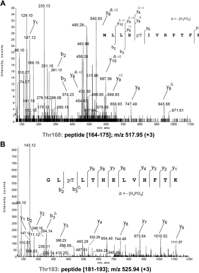
FIGURE 2. Identification of the PcaA phosphorylation sites. A, MS/MS spectra at m/z 517.95 (⫹3) of peptide (164–175) of PcaA. Unambiguous location of the phosphate group on Thr-168 was shown by observation of the “y” C-terminal daughter ion series. Starting from the C-terminal residue, all “y” ions lose phosphoric acid (⫺98 Da) after the phosphorylated residues. B, MS/MS spectra at m/z 525.94 (⫹3) of peptide (181–193) of PcaA. Unambiguous location of the phosphate group on Thr-183 was shown by observation of the “y” C-terminal daughter ion series. Starting from the C-terminal residue, all “y” ions lose phosphoric acid (⫺98 Da) after the phosphorylated residues.
at CNRS, on August 13, 2012
www.jbc.org
washed, and re-incubated in BCG-free medium. One day later, cells were fixed with glutaraldehyde and processed for EM.
Horseradish Peroxidase Cytochemistry—Cells exposed to HRP and fixed with glutaraldehyde were washed overnight at 4 °C with sucrose-containing buffer. After three washes with sucrose-free buffer, cells were incubated with 3,3 ⬘-diaminoben-zidine tetrahydrochlorate (DAB)-H2O2as before (26) and then processed for EM.
Processing for Transmission Electron Microscopy—Cells fixed with glutaraldehyde were processed as described (28). Briefly, cells were washed with complete cacodylate buffer, and post-fixed for 1 h at room temperature with 1% osmium tetroxide in the same buffer devoid of sucrose. They were washed with buffer, scraped off the dishes, concentrated in 2% agar in caco-dylate buffer and treated for 1 h at room temperature with 1% uranyl acetate in Veronal buffer. Samples were dehydrated in a graded series of ethanol and embedded in Spurr resin. Thin sections (70 nm-thick) were stained with 1% uranyl acetate in distilled water and then with lead citrate and observed under the electron microscope (Zeiss 912).
Quantitation of Loner Phagosomes with Lysosomal Material— In all cases, 50 –100 different loner phagosomes taken at ran-dom from three different experiments were examined for the presence or absence of lysosomal material. Care was taken to avoid serial sections.
RESULTS
PcaA Is Phosphorylated in Vitro by Multiple Ser/Thr Kinases— We first sought out to determine whether PcaA and MmaA2, that introduce cyclopropanes at the proximal and distal posi-tions on the␣-meromycolic acid, respectively (18, 29) (Fig. 1A) were modified by phosphorylation. This changes the physico-chemical properties of defined Ser or Thr residues by introduc-ing negative charges that ultimately affect protein activity. The kinase domains of several Mtb STPKs (PknA to PknL), expressed as GST-tagged fusion proteins (16), were incubated with purified Mtb PcaA or MmaA2 and [␥-33P]ATP. SDS-PAGE/autoradiography analysis indicated that PcaA, but not MmaA2, was phosphorylated by several kinases (Fig. 1B). Based on the intensity of the radioactive signal corresponding to phosphorylated PcaA, PknF appears to be the most efficient kinase to phosphorylate PcaA in vitro. Signals were weak with PknD, PknE, and PknH that all display various autokinase activ-ities (16) but none were detected with PknA or PknB. Although MmaA2 is structurally highly related to PcaA (30), only PcaA is a specific substrate and interacts with PknF (Fig. 1B). This sug-gests that cyclopropanation of the ␣-mycolic acid proximal position may be regulated in mycobacteria by extracellular cues.
PcaA Is Phosphorylated on Thr-168 and Thr-183—Mass spectrometry (MS) was then used to identify the number and nature of phosphorylation sites on PcaA, as reported previously for other STPK substrates (13, 14, 17, 31, 32). Phosphorylated PcaA was purified from E. coli co-expressing PknF and PcaA (pETDuet-pcaA) and subjected to MS analysis after tryptic digestion. Sequence coverage of 94% was obtained, bearing all possible Ser and Thr residues. Phosphorylation occurred only on peptides (164 –175) (Fig. 2A) and (181–193) (Fig. 2B) with
phosphate groups on Thr-168 and Thr-183. To prevent phos-phorylation, Thr-168 and Thr-183 were substituted by al-anine, either individually or together. The corresponding PcaA_T168A, PcaA_T183A, and PcaA_T168A/T183A pro-teins were expressed and purified as His-tagged propro-teins in E. coli BL21(DE3)Star harboring either pETPhos_pcaA_ T168A, pETPhos_pcaA_T183A, or pETPhos_pcaA_T168A/ T183A. Following incubation with PknF and [␥-33 P]-ATP, SDS-PAGE/autoradiography revealed that single Ala point mutations were associated with a strong radioactive sig-nal decrease (Fig. 3A). Total abrogation of phosphorylation occurred in the T168A/T183A double mutant compared with PcaA_WT (Fig. 3A). Similar results were obtained when the mutant was incubated with other STPKs (Fig. 3B), thus identifying PcaA_T168A/T183A as a PcaA phosphoablative mutant.
To address the relevance of PcaA phosphorylation in myco-bacteria, recombinant BCG overexpressing PcaA_WT was ana-lyzed by Western blotting using phosphothreonine anti-bodies. Antibody specificity for the phosphorylated isoform was first assessed using PcaA purified from either E. coli (pET-Phos-pcaA) or E. coli co-expressing PknF and PcaA (pETDuet-pcaA). Phosphorylated PcaA from pETDuet-pcaA, but not unphosphorylated PcaA from pETPhos-pcaA, was specifically recognized by the antibodies (Fig. 3C). That PcaA from BCG carrying pVV16-pcaA was in a phosphorylated state was con-firmed by a specific band recognized by the anti-phosphothreo-nine antibodies (Fig. 3C). Western blot analysis of recombinant PcaA purified from either exponential or stationary cultures of BCG carrying pVV16-pcaA showed similar levels of PcaA phos-phorylation. This suggests that PcaA phosphorylation is growth-phase independent (Fig. 3C). No specific phosphoryla-tion signals were detected in PcaA_T168A/T183A purified from BCG carrying pVV16-pcaA_T168A/T183A, thus exclud-ing the existence of additional phosphorylation sites. There-fore, phosphorylation occurs at Thr-168 and Thr-183 both in vitroand in mycobacteria.
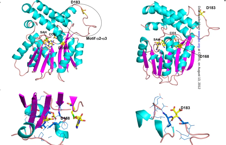
Phosphorylation Decreases PcaA Methyltransferase Activity— Multiple sequence alignments of PcaA orthologues from vari-ous mycobacterial species indicated that both Thr-168 and Thr-183 are conserved in Mtb, M. marinum, and M. leprae, whereas the second phosphorylation site (corresponding to Thr-183 in the Mtb sequence) is substituted by a proline in the M. smegmatisorthologue (Fig. 4A). Phosphorylation site map-ping on the three-dimensional structure (30) emphasized the strategic roles of both threonine residues on PcaA activity (Fig. 4B). Thr-168 is located within the -sheet of the core SAM-methyltransferase fold with the threonine side-chain facing a mobile protein segment that refolds upon cofactor-binding (33). Thr-183 was located in the so-called␣2-␣3 specific motif of mycolic acid SAM-methyltransferases seemingly involved in the interaction with the mycobacterial acyl carrier protein AcpM (30, 33). This motif, which delineates the entrance of the hydrophobic channel from the enzyme surface to the active site, displays the largest deviation when comparing the known structures of mycolic acid SAM-methyltransferases (33). The above data suggest that Thr-168 and Thr-183 are the unique phosphoacceptors in PcaA that directly influence its enzymatic
at CNRS, on August 13, 2012
www.jbc.org
activity. This prompted us to compare the methyltransferase activity of phosphorylated and non-phosphorylated isoforms using a coupled assay originally developed for CmaA2 (23). This colorimetric assay is based on the detection of S-adenosylho-mocysteine (SAH) conversion to hoS-adenosylho-mocysteine by SAH hydro-lase. Two unsaturated fatty acids were used as substrates of Mtb PcaA and MmaA2; cis, cis 11,14-eicosadienoic acid and lino-elaidic acid (Fig. 4C). Although these lipids are structurally dis-tinct from the authentic unsaturated meromycolyl-AcpM sub-strates, both PcaA and MmaA2 catalyzed cyclopropanation of double bonds in presence of either lipid (Fig. 4, D and E). This reaction was inhibited by dioctylamine as reported earlier for CmaA2 (23) (data not shown). Importantly, the activity of phos-phorylated PcaA, derived from E. coli carrying pETDuet-PcaA, was significantly reduced by 40 to 50% compared with non-phosphorylated PcaA from E. coli carrying pETPhos-PcaA, irrespective of the lipid substrate. Conversely, the MmaA2 methyltransferase activity remained unchanged whether or not it was produced from the pETPhos or pETDuet vectors, con-sistent with the fact that MmaA2 is not phosphorylated by STPKs (Fig. 1B). These results indicate that, in vitro, both PcaA and MmaA2 catalyze the introduction of cyclopropane rings on cis, cis11,14-eicosadienoic acid and linoelaidic acid and sup-port the critical role of STPK-dependent phosphorylation in regulating PcaA activity.
BCG PcaA Phosphomimetic Mutants Display Altered Phe-notypes—Acidic residues such as aspartic acid (Asp) qualita-tively mimic the phosphorylation effect with regard to func-tional activity (13–15, 17, 34). Therefore, phosphoablative (Thr to Ala replacements) and phosphomimetic (Thr to Asp replace-ments) pcaA alleles were generated and introduced into BCG mc22801 (BCG Pasteur pcaA::Tn5370) (18). In these con-structs, WT, phosphoablative (T168A/T183A) and phospho-mimetic (T168D/T183D) PcaA isoforms were fused to a HA tag and their expression was monitored by Western blotting using anti-HA antibodies. As expected, BCG ⌬pcaA transformed with the empty vector (pMV261) failed to express PcaA whereas similar amounts of PcaA_WT, PcaA_T168A/T183A and PcaA_T168D/T183D were synthesized in the correspond-ing strains (Fig. 5A), allowcorrespond-ing subsequent phenotype compari-son. Whether phosphorylation of PcaA affects de novo mycolic acid biosynthesis was next investigated by labeling the various BCG strains with [1-14C]acetate followed by mycolic acid extraction and thin layer chromatography (TLC)/autoradiog-raphy analysis. BCG mc22801 exhibited an altered␣-mycolic acid profile (18, 35), that could be restored by complementation with either WT or phosphoablative PcaA (Fig. 5B). In contrast, introduction of PcaA_T168D/T183D failed to restore the parental mycolic acid profile (Fig. 5B andsupplemental Fig. S1) indicating that the phosphomimetic isoform is unable to com-plement the lack of PcaA activity in mc22801, presumably as a consequence of its reduced methyltransferase activity (Fig. 4D). This effect was not due to a tertiary structure change, as sug-gested by modeling of the PcaA_T168D/T183D mutant struc-ture. Indeed, introduction of Asp at position 168, unaccessible to solvent, and at position 183, that is solvent exposed, does not seem to induce steric or electrostatic conflicts that could alter the protein fold (supplemental Fig. S2). Thus, it can be inferred FIGURE 3. Phosphorylation of PcaA variants with Ala-substituted
phos-pho-sites. A, purified PcaA_WT, PcaA_T168A, PcaA_T183A, and PcaA_ T168A/T183A mutants were incubated with PknF and [␥-33]ATP, separated by SDS-PAGE, stained with Coomassie Blue, or visualized by autoradiography.
B, in vitro phosphorylation of PcaA_T168A/T183A by multiple kinases.
Recom-binant STPKs encoded by the Mtb genome were expressed and purified as GST fusions and incubated with purified His-tagged PcaA_T168A/T183A and radiolabeled [␥-33]ATP. Depending on the STPK, 0.6 to 4.2 g were necessary for obtaining optimal autophosphorylation activity for each specific kinase. Samples were separated by SDS-PAGE, stained with Coomassie Blue (upper
panels) and visualized by autoradiography after overnight exposure to a film
(lower panels). C, phosphorylation of PcaA in mycobacteria. E. coli harboring pETPhos-PcaA was used as a source of non phosphorylated PcaA; the strain harboring pETDuet-PcaA coexpressing PknF and PcaA provides the phospho-rylated PcaA isoform. PcaA_WT or PcaA_T168A/T183A were produced in exponentially growing or stationary recombinant BCG strains. 3g of puri-fied His-tagged PcaA variants were probed with anti-phosphothreonine antibodies.
at CNRS, on August 13, 2012
www.jbc.org
that PcaA phosphorylation in mycobacteria reduces/abolishes its activity, supporting the view that this post-translational modification plays a key role in regulating ␣-mycolic acid cyclopropanation.
The effect of constitutive PcaA phosphorylation on the colo-nial morphology of BCG was next examined. Although no dif-ferences were observed for the different BCG strains cultured in liquid medium (Fig. 6B), mc22801 grew far more slowly than its parental strain on agar plates, as evidenced by colony size (supplemental Fig. S3). This growth defect could be fully restored by complementing the mutant strain with either
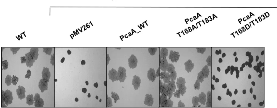
PcaA_WT or PcaA_T168A/T183A, but not with the phospho-mimetic mutant. Cording morphology, in which bacteria are intertwined into serpentine rope-like structures, is a distinctive feature of pathogenic mycobacteria. The characteristic mc22801 cord formation defect (18) was restored following complementation with either WT or phosphoablative pcaA alleles. However, the phosphomimetic construct failed to restore cord formation (Fig. 5C). Thus, PcaA phosphorylation is strongly associated with a defective mycolic acid profile, sub-sequently affecting mycobacterial growth and serpentine cord formation.
FIGURE 4. Phosphorylation of PcaA decreases methyltransferase activity. A, conservation of the phosphoacceptors in PcaA orthologues. The multiple sequence alignment of PcaA orthologues was performed using ClustalW and Espript (M_tub, Mycobacterium tuberculosis; M_mar, Mycobacterium marinum; M_lep, Mycobacterium leprae; M_sme, Mycobacterium smegmatis). Numbering of the amino acids corresponds to the PcaA protein from Mtb. Residues con-served in all species are presented in black boxes. The phosphorylated site of PcaA from Mtb and their positions are indicated. B, localization of Thr-168 and Thr-183 phospho-sites in the three-dimensional structure of PcaA. Two perpendicular views of the PcaA structure ((30); PDB entry 1L1E) are shown in ribbon
representation with␣-helices in cyan, -strands in magenta, and turns and coils in salmon. SAH, the cofactor product, and the carbonate ion found in the active
site are displayed as sticks with carbons in yellow, nitrogens in blue, oxygens in red, and sulfur in green. The two threonine side chains are shown as spheres using the same color code. For each view, insets represent the protein molecular surface. Structure visualization and analyses were carried out using the PyMOL program. C, structures of cis, cis-11,14-eicosadienoic acid or linoelaidic acid, an all-trans fatty acid homolog of linoleic acid. D, PcaA-dependent methyltrans-ferase assays. Activity of the non-phosphorylated and phosphorylated PcaA isoforms were assayed and compared using the two different lipids. Activity of the non-phosphorylated protein (pETPhos-PcaA, black bars) and activity of phosphorylated PcaA (pETDuet-PcaA) are shown in gray. Values are means⫾ S.E. of triplicates representative of three sets of experiments with independent protein preparations. E, a similar approach was used to determine the MmaA2 activity.
at CNRS, on August 13, 2012
www.jbc.org
Functional PcaA Is Required for Intracellular Survival and PMB—Although PcaA cyclopropanation is required for Mtb persistence and pathology in late stages of infection in mice (18), its role during early infection remains unclear. This prompted us to investigate the ability of BCG⌬pcaA to infect and replicate within human monocyte-derived macrophages (HMDM). Determination of intramacrophagic bacterial counts showed that survival of the mutant strain was severely compro-mised as compared with the WT strain (Fig. 6A). This was not due to an inherent growth defect as the mutant and parental strains replicated equally well in liquid medium (Fig. 6B).
Pathogenic mycobacteria survive in macrophages by residing in phagosomes which they prevent from maturing and fusing with lysosomes (2, 36). We sought out to examine the putative role of PcaA in this process. Cells were exposed to electron-dense endocytic tracers by two different approaches. In the first, small latex beads were chased to lysosomes prior to phag-ocytic uptake of the WT or the⌬pcaA strains; in the second, the endocytic tracer horseradish peroxidase (HRP) was added at selected time points after infection (1 or 5 days post-infection) with the above strains and the cells were stained for HRP by EM cytochemical methods. Phagosome processing was analyzed in terms of fusion and intermingling of contents between phago-somes and the different endomembrane compartments, namely early endosomes (eEN) and late endosomes/lysosomes (indicated as Ly in text and figures) that can be easily distin-guished from one another by the presence of latex beads (only in Ly) and the cytochemical staining pattern for HRP (37). To analyze phagosome processing, it is important to distinguish between loner and social phagosomes since, and as shown before, the PMB does not hold for social phagosomes that invariably mature and fuse with lysosomes (reviewed in Ref. 4). Therefore, when looking for the effects of PcaA on the PMB,
observations and quantitations had to be limited strictly to loner phagosomes. One must also keep in mind that non-ma-tured phagosomes fuse with eEN but are unable to fuse with Ly (4). With both strains, two types of loner phagosomes were encountered, i.e. (i) immature phagosomes either displaying a thin rim of HRP reaction product as is typical of eEN (not shown) or no marker (Fig. 6, C and F), and (ii) phagosomes that contain lysosomal material indicating that they have matured and fused with Ly (Fig. 6, D and G). As before, social phago-somes, containing two or more mycobacteria, all contained lys-osomal material (Fig. 6, E and H). A quantitative analysis showed a 2-fold increase in the percentage of loner phagosomes displaying lysosomal material for BCG⌬pcaA-containing pha-gosomes as compared with the WT strain (Fig. 6I). A more complete study done with HRP as endocytic tracer showed that the percentage of phagolysosomes remained stationary for at least 5 days post-infection (50% for the BCG⌬pcaA mutant versus about 25% for the WT strain) (Fig. 6J). Overall, these results emphasize the major contribution of PcaA in the PMB. Phosphorylation of PcaA Inhibits Intramacrophage Growth and Affects Phagosome Maturation—The above data indicate that phosphorylation of PcaA is linked to a defective cell wall-associated mycolic acid profile in BCG and affects its intracel-lular growth. We next determined the consequences of PcaA phosphorylation on both intramacrophage BCG growth (Fig. 7A) and the PMB (Fig. 7B). For both studies, HMDM were infected with the parental BCG strain, the⌬pcaA mutant or the different BCG complemented strains. Over a 6-day period the CFU count increased 4 –5-fold for the parental strain and for both BCG⌬pcaA strains complemented with the WT or phos-phoablative isoforms. The phosphomimetic strain was unable to replicate within HMDM, similarly to the⌬pcaA mutant (Fig. 7A). Concerning the PMB, only 30% of the loner phagosomes containing the⌬pcaA mutant complemented with the WT or phosphoablative PcaA contained lysosomal material, similarly to the parental WT strain. In contrast, 60% of the loner phago-somes containing the phosphomimetic strain contained lyso-somal material, as it was the case for the⌬pcaA mutant (Fig. 7B). Altogether, these results indicate that phosphorylation of PcaA prevents growth and abrogates the PMB.
DISCUSSION
The present study extends previous work showing the regu-latory role of phosphorylation on mycolic acid chain length and functionalization during its biosynthesis (12). More impor-tantly, it provides the first evidence for a Ser/Thr kinase phos-phorylation-dependent regulation of cyclopropane synthase. This mechanism is of special relevance as cyclopropanes are major contributors to the physiopathology of Mtb infection (18). Phosphorylation of Thr-168 and Thr-183 residues caused a strong decrease in methyltransferase activity in vitro. In the case of growing bacteria, a phosphomimetic mutant displayed a major alteration of the mycolic acid profile due to the lack of di-cyclopropanation of ␣-mycolic acids, similar to what is observed for the⌬pcaA mutant. Likewise, impaired cyclopro-panation was associated with altered colony morphology and inability to form serpentine cords (Fig. 8). Major phenotypical alterations have also been observed in in vivo situations. Inac-FIGURE 5. Phosphorylation of PcaA is associated with an altered mycolic
acid profile and loss of cording phenotype. A, overexpression of the vari-ous PcaA variants in BCG ⌬pcaA. BCG mc22801 was transformed with pMV261, pMV261_pcaA_WT, pMV261_pcaA_T168A/T183A, and pMV261_
pcaA_T168D/T183D constructs for constitutive expression of the various pcaA
alleles fused to a C-terminal HA epitope. 40g of crude lysates were sub-jected to immunoblotting and probed with anti-HA antibodies. B, mycolic acid profile of the various BCG transformants. Equal counts (50 000 cpm) of labeled lipids were loaded and analyzed by TLC/autoradiography using petroleum ether/acetone (95/5, v/v). Positions of␣- and keto-mycolic acid methyl ester are indicated. OAME, oleic acid methyl ester. C, cording morpho-type of BCG strains. Single BCG colonies were grown on cord-reading agar and visualized after 3 weeks. Magnification,⫻100.
at CNRS, on August 13, 2012
www.jbc.org
at CNRS, on August 13, 2012
www.jbc.org
tivation of Mtb pcaA causes attenuation of virulence and a less severe granulomatous pathology in mice (18). In addition, PcaA-dependent cyclopropanation of TDM, a direct effector of Mtbpathogenesis, regulates its inflammatory activity (18, 38). In particular, TDM purified from a PcaA mutant is hypoinflam-matory for macrophages during early stages of infection (38).
We show here that phosphorylation of PcaA not only arrests intracellular mycobacterial replication but also abrogates the PMB. In addition to the requirement for a tight apposition of the phagosome membrane to the mycobacterial surface all around (3, 4, 39), which suggests an intimate cross-talk between the mycobacterium and its host cell, a large number of both
cellular and mycobacterial molecules were seen to play a role in, or correlate with, the PMB and intracellular survival of myco-bacteria (4, 40, 41). This leads to the question of whether any one of these is sufficient by itself and, if so, why is there such redundancy (3)? Present knowledge rests upon two types of experimental approaches: either eliminating the function of the molecular factor concerned and/or reconstituting the factor in an artificial system. With these approaches, i.e. deletion of pcaA and construction of phosphomimetic or phosphoablative mutants, we have studied phagosome maturation for a suffi-ciently long time (5 days versus a few hours only in many other studies) to conclude that mycolic acid cyclopropanation by PcaA maintains the PMB and that PcaA phosphorylation, by inhibiting cyclopropanation, abrogates this block indefinitely. It is noteworthy that part of the phagosomes, however, do not mature and fuse with lysosomes upon PcaA phosphorylation, suggesting that other mycobacterial factors such as TDM (9, 42) or phospho-signaling proteins like PknG (10), the protein tyrosine phosphatase PtpA (43, 44) and the secreted lipid phos-phatase SapM (45) might also be important mycobacterial factors. Among these, only SapM and PtpA seem to interfere directly with host physiological processes leading to prevention of phagosome maturation, whereas PknG seems to affect phagosome maturation through phosphorylation of yet unknown host proteins following its secretion within macrophage phagosomes.
Previous genetic screens identified several Mtb mutants defective in PMB (8, 46) and affected in various pathways, including lipid synthesis. Of notable interest is the Mtb pcaA::Tn mutant that failed to arrest phagosome maturation and trafficked to late phagosomal compartments in bone-marrow derived macrophages (46), thus supporting the view that the data obtained with BCG in the present study are relevant to Mtb.
With the PMB being the most conspicuous survival mecha-nism of pathogenic mycobacteria, why does Mtb phosphorylate PcaA? One must keep in mind that the fate of pathogenic myco-bacteria does not rely exclusively on its own defense mecha-nisms. Host cells have developed several strategies for combat-ing invadcombat-ing pathogens which, in turn, have developed several strategies for using the host environment to their advantage. In the present case, an attractive hypothesis would be that PcaA phosphorylation is driven by cell host factors located in the phagosome membrane and hence in direct contact with the mycobacterial surface due to the close apposition between the two structures. These factors would act as a signal for trig-gering autophosphorylation of mycobacterial STPKs, inducing in turn PcaA phosphorylation (Fig. 8). Mycolic acid structural alteration could affect the close apposition between the phago-some membrane and the mycobacterial surface. As a result, phagosomes would mature and fuse with lysosomes, as observed in the present study. However, whether PcaA
phos-FIGURE 6. PcaA requirement for intracellular survival and arrest of phagosome maturation. A, replication of BCG WT and BCG⌬pcaA in HMDM infected at a MOI of 1:1. CFU were scored at the indicated time points. Results are representative of three independent experiments from three different blood donors.
B, growth curves of different BCG strains in broth medium. C–H, acquisition of endocytic tracer (C–E, HRP; F–H, latex beads) by BCG-WT- or BCG
⌬pcaA-containing phagosomes. HMDM were infected with BCG WT or BCG⌬pcaA either before or after exposure to an endocytic tracer and phagosomes were characterized in terms of presence or absence of lysosomal marker. C and F, immature loner phagosome devoid of lysosomal material; D, G, loner phagosome fusing (arrows) with lysosomes (L); E, H, social phagosome with lysosomal material. C–H, bar scale represents 0.5m. I, fold increase in percentage of BCG ⌬pcaA-containing loner phagosomes with lysosomal material with respect to BCG WT-containing loner phagosomes. J, percentage of loner phagosomes with lysosomal material (HRP) at 1 or 5 days post-infection. Both experiments (I, J) correspond to the means⫾ S.E. (n ⫽ samples from 3 different donors). FIGURE 7. Phosphorylation of PcaA inhibits intracellular growth and
abrogates PMB. HMDM were infected with different BCG strains at an MOI of 1:1 (A) or 5:1 (B). A, intracellular CFU counts enumerated at 3 h and day 6 post-infection. B, percentage of loner phagosomes containing lysosomal material after exposure to the endocytic tracer HRP added to infected cells for 90 min at day 1 post-infection. Both experiments (A, B) correspond to the means⫾ S.E. (n ⫽ samples from three different donors).
at CNRS, on August 13, 2012
www.jbc.org
phorylation is reduced/inhibited once the close apposition between the phagosome membrane and the mycobacterial sur-face is no longer maintained all around, cannot be determined with present technologies as it is not possible to separate the different types of phagosomes from one another.
For a successful infection, it must be assumed that pathogenic mycobacteria will not be destroyed in macrophages. In many situ-ations, growth is arrested in phagolysosomes but mycobacteria survive in this cytolytic environment without significant loss of viability (2– 4, 39). Our study is in full agreement with the above situations as shown by the survival curves and the morphological appearance of bacilli within phagolysosomes.
In summary, this study provides conceptual advances in our understanding of the mycolic acid metabolic adaptation and regulatory events exploited by pathogenic mycobacteria to adapt their mycolic acid cell wall content. Although very chal-lenging, future studies should now help to identify extracellular cues sensed by the different kinases and leading to PcaA phospho-rylation. This work also strengthens the biological importance of PcaA in the physiology and virulence of the bacilli and provides evidence of a Ser/Thr kinase-dependent mechanism for modulat-ing the composition of mycolic acids, a key component of the mycobacterial cell wall, and for maintaining mycobacteria in a non-matured phagosome, a hallmark for mycobacterial survival within host cells. Our results suggest that displacement of the unphosphorylated/phosphorylated PcaA balance in favor of the phosphorylated isoform rapidly leads to PMB inhibition and loss
of intracellular survival. Thus, from an applied point of view and considering that PcaA has been proposed as a target for drug development against persistent bacilli (18, 47), the selective inhi-bition of PcaA activity through constitutive phosphorylation may strongly impair Mtb survival, opening new and original perspec-tives for future anti-tuberculosis drug development. It is indeed noteworthy that small molecules, such as bryostatin (48) can acti-vate STPK, which may be of great therapeutic value in inhibiting Mtbintracellular growth.
Acknowledgments—We thank Irène Brändli in the laboratory of CdC and the members of the CIML-IBDML electron microscopy facility, Marseille, France, for expert technical assistance and wish to thank W. R. Jacobs and M. Glickman for the kind gift of BCG mc22801.
REFERENCES
1. Dye, C., and Williams, B. G. (2010) The population dynamics and control of tuberculosis. Science 328, 856 – 861
2. Armstrong, J. A., and Hart, P. D. (1971) Response of cultured macrophages to Mycobacterium tuberculosis, with observations on fusion of lysosomes with phagosomes. J. Exp. Med. 134, 713–740
3. de Chastellier, C., Forquet, F., Gordon, A., and Thilo, L. (2009)
Mycobac-teriumrequires an all-around closely apposing phagosome membrane to maintain the maturation block and this apposition is re-established when it rescues itself from phagolysosomes. Cell Microbiol. 11, 1190 –1207 4. de Chastellier, C. (2009) The many niches and strategies used by
patho-genic mycobacteria for survival within host macrophages. Immunobiology 214,526 –542
FIGURE 8. Schematic representation of the consequences of STPK-dependent PcaA phosphorylation. In response to external stimuli, STPKs are auto-phosphorylated. This induces PcaA phosphorylation on Thr-168 and Thr-183. PcaA phosphorylation decreases cyclopropane synthase activity, resulting in an altered mycolic acid profile characterized by the lack of di-cyclopropanated␣-mycolic acids. This affects colonial cording, intramacrophage replication and abrogates the PMB.
at CNRS, on August 13, 2012
www.jbc.org
5. Flynn, J. L., and Chan, J. (2003) Immune evasion by Mycobacterium
tuber-culosis: living with the enemy. Curr. Opin. Immunol. 15, 450 – 455 6. Fratti, R. A., Chua, J., Vergne, I., and Deretic, V. (2003) Mycobacterium
tuberculosis glycosylated phosphatidylinositol causes phagosome matu-ration arrest. Proc. Natl. Acad. Sci. U.S.A. 100, 5437–5442
7. Chua, J., Vergne, I., Master, S., and Deretic, V. (2004) A tale of two lipids:
Mycobacterium tuberculosisphagosome maturation arrest. Curr. Opin.
Microbiol. 7,71–77
8. Pethe, K., Swenson, D. L., Alonso, S., Anderson, J., Wang, C., and Russell, D. G. (2004) Isolation of Mycobacterium tuberculosis mutants defective in the arrest of phagosome maturation. Proc. Natl. Acad. Sci. U.S.A. 101, 13642–13647
9. Indrigo, J., Hunter, R. L., Jr., and Actor, J. K. (2003) Cord factor trehalose 6,6⬘-dimycolate (TDM) mediates trafficking events during mycobacterial infection of murine macrophages. Microbiology 149, 2049 –2059 10. Walburger, A., Koul, A., Ferrari, G., Nguyen, L., Prescianotto-Baschong,
C., Huygen, K., Klebl, B., Thompson, C., Bacher, G., and Pieters, J. (2004) Protein kinase G from pathogenic mycobacteria promotes survival within macrophages. Science 304, 1800 –1804
11. Av-Gay, Y., and Everett, M. (2000) The eukaryotic-like Ser/Thr protein kinases of Mycobacterium tuberculosis. Trends Microbiol. 8, 238 –244 12. Molle, V., and Kremer, L. (2010) Division and cell envelope regulation by
Ser/Thr phosphorylation: Mycobacterium shows the way. Mol. Microbiol. 75,1064 –1077
13. Veyron-Churlet, R., Molle, V., Taylor, R. C., Brown, A. K., Besra, G. S., Zanella-Cléon, I., Fütterer, K., and Kremer, L. (2009) The Mycobacterium
tuberculosis-ketoacyl-acyl carrier protein synthase III activity is
inhib-ited by phosphorylation on a single threonine residue. J. Biol. Chem. 284, 6414 – 6424
14. Veyron-Churlet, R., Zanella-Cléon, I., Cohen-Gonsaud, M., Molle, V., and Kremer, L. (2010) Phosphorylation of the Mycobacterium tuberculosis -ketoacyl-acyl carrier protein reductase MabA regulates mycolic acid biosynthesis. J. Biol. Chem. 285, 12714 –12725
15. Slama, N., Leiba, J., Eynard, N., Daffé, M., Kremer, L., Quémard, A., and Molle, V. (2011) Negative regulation by Ser/Thr phosphorylation of HadAB and HadBC dehydratases from Mycobacterium tuberculosis type II fatty acid synthase system. Biochem. Biophys. Res. Commun. 412, 401– 406
16. Molle, V., Brown, A. K., Besra, G. S., Cozzone, A. J., and Kremer, L. (2006) The condensing activities of the Mycobacterium tuberculosis type II fatty acid synthase are differentially regulated by phosphorylation. J. Biol.
Chem. 281,30094 –30103
17. Molle, V., Gulten, G., Vilcheze, C., Veyron-Churlet, R., Zanella-Cleon, I., Sacchettini, J. C., Jacobs Jr., W. R., and Kremer, L. (2010) Phosphorylation of InhA inhibits mycolic acid biosynthesis and growth of Mycobacterium
tuberculosis. Mol. Microbiol. 78,1591–1605
18. Glickman, M. S., Cox, J. S., and Jacobs, W. R., Jr. (2000) A novel mycolic acid cyclopropane synthetase is required for cording, persistence, and virulence of Mycobacterium tuberculosis. Mol. Cell 5, 717–727 19. Canova, M. J., Kremer, L., and Molle, V. (2008) pETPhos: a customized
expression vector designed for further characterization of Ser/Thr/Tyr protein kinases and their substrates. Plasmid 60, 149 –153
20. Molle, V., Leiba, J., Zanella-Cléon, I., Becchi, M., and Kremer, L. (2010) An improved method to unravel phosphoacceptors in Ser/Thr protein ki-nase-phosphorylated substrates. Proteomics 10, 3910 –3915
21. Jackson, M., Crick, D. C., and Brennan, P. J. (2000) Phosphatidylinositol is an essential phospholipid of mycobacteria. J. Biol. Chem. 275, 30092–30099
22. Molle, V., Kremer, L., Girard-Blanc, C., Besra, G. S., Cozzone, A. J., and Prost, J. F. (2003) An FHA phosphoprotein recognition domain mediates protein EmbR phosphorylation by PknH, a Ser/Thr protein kinase from
Mycobacterium tuberculosis. Biochemistry 42,15300 –15309
23. Barkan, D., Liu, Z., Sacchettini, J. C., and Glickman, M. S. (2009) Mycolic acid cyclopropanation is essential for viability, drug resistance, and cell wall integrity of Mycobacterium tuberculosis. Chem. Biol. 16, 499 –509 24. Kremer, L., Douglas, J. D., Baulard, A. R., Morehouse, C., Guy, M. R.,
Alland, D., Dover, L. G., Lakey, J. H., Jacobs, W. R., Jr., Brennan, P. J., Minnikin, D. E., and Besra, G. S. (2000) Thiolactomycin and related
ana-logues as novel anti-mycobacterial agents targeting KasA and KasB con-densing enzymes in Mycobacterium tuberculosis. J. Biol. Chem. 275, 16857–16864
25. Bessoles, S., Dudal, S., Besra, G. S., Sanchez, F., and Lafont, V. (2009) Human CD4⫹ invariant NKT cells are involved in antibacterial immunity against Brucella suis through CD1d-dependent but CD4-independent mechanisms. Eur. J. Immunol. 39, 1025–1035
26. de Chastellier, C., Lang, T., and Thilo, L. (1995) Phagocytic processing of the macrophage endoparasite, Mycobacterium avium, in comparison to phagosomes which contain Bacillus subtilis or latex beads. Eur J. Cell Biol. 68,167–182
27. de Chastellier, C., and Thilo, L. (1997) Phagosome maturation and fusion with lysosomes in relation to surface property and size of the phagocytic particle. Eur. J. Cell Biol. 74, 49 – 62
28. de Chastellier, C., Fréhel, C., Offredo, C., and Skamene, E. (1993) Implica-tion of phagosome-lysosome fusion in restricImplica-tion of Mycobacterium
aviumgrowth in bone marrow macrophages from genetically resistant mice. Infect. Immun. 61, 3775–3784
29. Glickman, M. S. (2003) The mmaA2 gene of Mycobacterium tuberculosis encodes the distal cyclopropane synthase of the␣-mycolic acid. J. Biol.
Chem. 278,7844 –7849
30. Huang, C. C., Smith, C. V., Glickman, M. S., Jacobs, W. R., Jr., and Sac-chettini, J. C. (2002) Crystal structures of mycolic acid cyclopropane syn-thases from Mycobacterium tuberculosis. J. Biol. Chem. 277, 11559 –11569 31. Barthe, P., Roumestand, C., Canova, M. J., Kremer, L., Hurard, C., Molle, V., and Cohen-Gonsaud, M. (2009) Dynamic and structural characteriza-tion of a bacterial FHA protein reveals a new autoinhibicharacteriza-tion mechanism.
Structure 17,568 –578
32. Canova, M. J., Kremer, L., and Molle, V. (2009) The Mycobacterium
tu-berculosisGroEL1 chaperone is a substrate of Ser/Thr protein kinases. J.
Bacteriol. 191,2876 –2883
33. Boissier, F., Bardou, F., Guillet, V., Uttenweiler-Joseph, S., Daffe, M., Que-mard, A., and Mourey, L. (2006) Further Insight into S-adenosylmethio-nine-dependent methyltransferases: Structural characterization of Hma, an enzyme essential for the biosynthesis of oxygenated mycolic acids in
Mycobacterium tuberculosis. J. Biol. Chem. 281,4434 – 4445
34. Kang, C. M., Nyayapathy, S., Lee, J. Y., Suh, J. W., and Husson, R. N. (2008) Wag31, a homologue of the cell division protein DivIVA, regulates growth, morphology and polar cell wall synthesis in mycobacteria.
Micro-biology 154,725–735
35. Alibaud, L., Alahari, A., Trivelli, X., Ojha, A. K., Hatfull, G. F., Guerardel, Y., and Kremer, L. (2010) Temperature-dependent regulation of mycolic acid cyclopropanation in saprophytic mycobacteria: role of the
Mycobac-terium smegmatis1351 gene (MSMEG_1351) in cis-cyclopropanation of ␣-mycolates. J. Biol. Chem. 285, 21698–21707
36. Frehel, C., de Chastellier, C., Lang, T., and Rastogi, N. (1986) Evidence for inhibition of fusion of lysosomal and prelysosomal compartments with phagosomes in macrophages infected with pathogenic Mycobacterium
avium. Infect. Immun. 52,252–262
37. de Chastellier, C., Lang, T., Ryter, A., and Thilo, L. (1987) Exchange kinet-ics and composition of endocytic membranes in terms of plasma mem-brane constituents: a morphometric study in macrophages. Eur. J. Cell
Biol. 44,112–123
38. Rao, V., Fujiwara, N., Porcelli, S. A., and Glickman, M. S. (2005) Mycobac-terium tuberculosis controls host innate immune activation through cy-clopropane modification of a glycolipid effector molecule. J. Exp. Med. 201,535–543
39. de Chastellier, C., and Thilo, L. (2006) Cholesterol depletion in
Mycobac-terium avium-infected macrophages overcomes the block in phagosome maturation and leads to the reversible sequestration of viable mycobacte-ria in phagolysosome-derived autophagic vacuoles. Cell Microbiol. 8, 242–256
40. Russell, D. G. (2001) Mycobacterium tuberculosis: here today, and here tomorrow. Nat. Rev. Mol. Cell Biol. 2, 569 –577
41. Nguyen, L., and Pieters, J. (2005) The Trojan horse: survival tactics of pathogenic mycobacteria in macrophages. Trends Cell Biol. 15, 269 –276
42. Vergne, I., Chua, J., and Deretic, V. (2003) Tuberculosis toxin blocking
at CNRS, on August 13, 2012
www.jbc.org
phagosome maturation inhibits a novel Ca2⫹/calmodulin-PI3K hVPS34 cascade. J. Exp. Med. 198, 653– 659
43. Wong, D., Bach, H., Sun, J., Hmama, Z., and Av-Gay, Y. (2011) Mycobac-terium tuberculosis protein tyrosine phosphatase (PtpA) excludes host vacuolar-H⫹-ATPase to inhibit phagosome acidification. Proc. Natl.
Acad. Sci. U.S.A. 108,19371–19376
44. Bach, H., Papavinasasundaram, K. G., Wong, D., Hmama, Z., and Av-Gay, Y. (2008) Mycobacterium tuberculosis virulence is mediated by PtpA de-phosphorylation of human vacuolar protein sorting 33B. Cell Host
Mi-crobe 3,316 –322
45. Vergne, I., Chua, J., Lee, H. H., Lucas, M., Belisle, J., and Deretic, V. (2005)
Mechanism of phagolysosome biogenesis block by viable Mycobacterium
tuberculosis. Proc. Natl. Acad. Sci. U.S.A. 102,4033– 4038
46. MacGurn, J. A., and Cox, J. S. (2007) A genetic screen for Mycobacterium
tuberculosismutants defective for phagosome maturation arrest identifies components of the ESX-1 secretion system. Infect. Immun. 75, 2668 –2678 47. Zhang, Y., Post-Martens, K., and Denkin, S. (2006) New drug candidates and therapeutic targets for tuberculosis therapy. Drug Discov. Today 11, 21–27
48. Shah, I. M., Laaberki, M. H., Popham, D. L., and Dworkin, J. (2008) A eukaryotic-like Ser/Thr kinase signals bacteria to exit dormancy in re-sponse to peptidoglycan fragments. Cell 135, 486 – 496
at CNRS, on August 13, 2012
www.jbc.org
K Parental BCG α α K α K K 2 + Ag 1 K UFA
BCG ΔpcaA +pMV261 BCG ΔpcaA +pMV261_PcaA_WT
BCG ΔpcaA +pMV261_PcaA_T168A/T183A BCG ΔpcaA +pMV261_PcaA_T168D/T183D
2 + Ag 1 2 + Ag 1 2 + Ag 1 2 + Ag 1 SFA
Fig. S1. Mycolic acid pattern in M. bovis BCG ΔpcaA complemented strains. Two-dimensional argentation TLC of 14C-radiolabeled mycolic acids
from M. bovis BCG Pasteur and BCG mc22801 (BCG Pasteur pcaA::Tn5370) transformed with various vectors and grown at 37°C. The arrowhead
indicates accumulation of the unsaturated α-mycolic acid precursor concomitant to the loss of α-mycolic acid synthesis. This hybrid mycolate accumulating in the ΔpcaA mutant and in the mutant complemented with pMV261_PcaA_T168D/T183D carries a cis double bond at the proximal position in place of a cis cyclopropane, as determined previously (Glickman et al. 2000). α, α-mycolates; K, keto-mycolates.
Glickman, M. S., Cox, J. S., and Jacobs, W. R., Jr. (2000) A novel mycolic acid cyclopropane synthetase is required for cording, persistence, and virulence of Mycobacterium tuberculosis. Mol Cell 5, 717-727
at CNRS, on August 13, 2012
www.jbc.org
SAH CO3 D183 D168 D168 D183 SAH CO3 Motif α2-α3 D168
B
Fig. S2. Structure modeling of the PcaA phosphomimetic T168D/T183D mutant. (A) Overall views separated by 45°. (B) Zoom-in showing the
chemical environment around D168 (left) and D183 (right): cartoon within a sphere of radius 13 Å, lines within a sphere of 8 Å, sticks within a sphere of 4 Å around these positions. The two most favorable side chain conformers of D168 and D183 are shown. SAH, the cofactor product, and the carbonate ion that was found in the active site are also displayed. α-Helices are in cyan, β-strands in magenta, turns and coils in salmon. Carbon atoms, yellow or marine; nitrogen atoms, blue; oxygen atoms, red; sulfur atoms, green. The Swiss-model program (www.swissmodel.expasy.org) (Arnold et al, 2006; Guex et al, 1997; Schwede et al, 2003) was used to deduce hypothetical structures from the sequences of PcaA mutants. The 1L1E.pdb structure was used as template and homology models were produced using the Automated mode. Structure visualization and analyses were carried out using the PyMOL program (www.pymol.org).
Arnold, K., Bordoli, L., Kopp, J., and Schwede, T. (2006) The SWISS-MODEL workspace: a web-based environment for protein structure homology modelling. Bioinformatics 22, 195-201
Guex, N., and Peitsch, M. C. (1997) SWISS-MODEL and the Swiss-PdbViewer: an environment for comparative protein modeling. Electrophoresis 18, 2714-2723
at CNRS, on August 13, 2012
www.jbc.org
BCG ΔpcaA transformed with
Fig. S3. Alteration of mycobacterial growth. M. bovis BCG ΔpcaA transformed with the various plasmids
were plated on Middlebrook 7H10 supplemented with OADC and incubated at 37°C for 2-3 weeks.
at CNRS, on August 13, 2012
www.jbc.org
Strains or Plasmids Genotype or Description Source or Reference
E. coli DH5α F-endA1 glnV44 thi-1 recA1 relA1 gyrA96 deoR
nupGΦ80dlacZΔM15 Δ(lacZYA-argF) U169, hsdR17(rK- mK+),
λ– ; used for general cloning
Invitrogen
E. coli C41 (DE3) Strain F derived from BL21(DE3), used for expression of toxic proteins
Lucigen
E. coli BL21(DE3) Star F2 ompT hsdSB(rB2 mB2) gal dcm (DE3); used to express recombinant proteins in E. coli
Invitrogen
M. bovis BCG 1173P2 Vaccine strain WHO,
Stockholm
mc²2801 BCG Pasteur pcaA::Tn5370 (1)
pETPhos pET15b (Novagen) derivative including the replacement of the thrombin site coding sequence with a tobacco etch virus (TEV) protease site and Ser to Gly mutagenesis in the Nterm His-tag
(2)
pCDFDuet-1 pET vector derivative designed for the co-expression of two proteins under T7lac promoter induction
Novagen pCDFDuet_pknF pCDFDuet-1 vector allowing expression of the PknF kinase
domain untagged
(3) pETPhos_MmaA2_WT pETPhos derivative used to express His-tagged fusion of WT
MmaA2 in E.coli
This work pETPhos_PcaA_WT pETPhos derivative used to express His-tagged fusion of WT
PcaA in E.coli
This work pETPhos_PcaA_T168A pETPhos derivative used to express His-tagged fusion of
PcaA_T168A in E.coli
This work pETPhos_PcaA_T183A pETPhos derivative used to express His-tagged fusion of
PcaA_T183A in E.coli
This work pETPhos_PcaA_T168A/T168A pETPhos derivative used to express His-tagged fusion of
PcaA_T168A/T168A in E.coli
This work pVV16 E. coli/mycobacterial shuttle vector, allows expression of C-term
His-tagged proteins, derived from pMV261 (Stover et al. 1991) (4) pVV16_PcaA_WT pVV16 derivative used to express His-tagged fusion of WT PcaA
in mycobacteria
This work pVV16_PcaA_T168A/T183A pVV16 derivative used to express His-tagged fusion of
PcaA_T168A/T183A in mycobacteria
This work pMV261 Multi-copy plasmid for E.coli and mycobacteria. Cloned genes
are under the control of the constitutive hsp60 promoter
(5) pMV261_PcaA_WT pMV261 derivative used to express HA-tagged fusion of WT
PcaA in mycobacteria
This work pMV261_PcaA_T168A/T168A pMV261 derivative used to express HA-tagged fusion of WT
PcaA_T168A/T183A in mycobacteria
This work pMV261_PcaA_T168D/T168
D
pMV261 derivative used to express HA-tagged fusion of WT PcaA_T168D/T183D in mycobacteria
This work
at CNRS, on August 13, 2012
www.jbc.org
Primers 5' to 3' Sequenceab
MmaA2 WT dir CTACCTACATATGGTCAACGACCTAACGCCGCACTTC (NdeI) MmaA2 WT rev CATGGATGCTAGCCTACTTCGCCAGCGTGAACTGGT (NheI) PcaA WT dir CTACCTACATATGTCCGTGCAGCTCACGCCGCAT(NdeII)
PcaA WT rev CAGCGAGGGATCCTTACTTTTCCAGTGTGAACTGGTCG (BamHI) PcaA T168A dir AAGATGTTGCTGCACGCCATCGTGCGCCCCACC
PcaA T168A rev GGTGGGGCGCACGATGGCGTGCAGCAACATCTT
PcaA T183A dir GGCAGGGAAAAGGGCCTGGCGTTGACCCACGAACTGGTT PcaA T183A rev AACCAGTTCGTGGGTCAACGCCAGGCCCTTTTCCCTGCC pMV261_PcaA_WT dir GTCCAGCGGTACCCGACAAGATCGGTTACGACG (KpnI)
pMV261_PcaA_WT rev CGAGGAATTCTTAAGCGTAATCTGGAACATCGTATGGGTACTTTTCCAGT GTGAACTGGT (EcoRI)
pMV261_PcaA_T168D dir CAAGATGTTGCTGCACGACATCGTGCGCCCCACC pMV261_PcaA_T168D rev GGTGGGGCGCACGATGTCGTGCAGCAACATCTTG pMV261_PcaA_T183D dir CAGGGAAAAGGGCCTGGACTTGACCCACGAACTGG pMV261_PcaA_T183D rev CCAGTTCGTGGGTCAAGTCCAGGCCCTTTTCCCTG pVV16_PcaA_WT rev CAGCGCAAGCTTCTTTTCCAGTGTGAACTGGTCG (HindIII)
a Restriction sites are underlined and specified into brackets. b Mutagenized bases are shown in bold.
References
1.
Glickman MS, Cox JS, & Jacobs WR, Jr. (2000) A novel mycolic acid cyclopropane
synthetase is required for cording, persistence, and virulence of Mycobacterium tuberculosis.
in Mol Cell, pp 717-727.
2.
Canova MJ, Kremer L, & Molle V (2008) pETPhos: a customized expression vector designed
for further characterization of Ser/Thr/Tyr protein kinases and their substrates. Plasmid
60(2):149-153.
3.
Molle V, Leiba J, Zanella-Cleon I, Becchi M, & Kremer L (2010) An improved method to
unravel phosphoacceptors in Ser/Thr protein kinase-phosphorylated substrates. Proteomics
10(21):3910-3915.
4.
Jackson M, Crick DC, & Brennan PJ (2000) Phosphatidylinositol is an essential phospholipid
of mycobacteria. J Biol Chem 275(39):30092-30099.
5.
Stover CK, et al. (1991) New use of BCG for recombinant vaccines. Nature
351(6326):456-460.
at CNRS, on August 13, 2012
www.jbc.org