Beach1 Functionally Antagonizes Rab 1 During Development and in Regulating Synaptic Morphology
by Rita Khodosh
B.S. Chemistry and Genetics University of California, Davis, 1997
Submitted to the Department of Biology
in Partial Fulfillment of the Requirements for the Degree of Doctor of Philosophy in Biology
at the
Massachusetts Institute of Technology September 2005
© 2005 Massachusetts Institute of Technology All rights reserved
Signature of Author:...
...
...
Department of Biologyrn
iiAugust
9, 2005
Certified by: ...
0'
T)"'Paul
A.Garrity
Assistant Professor of BiologyThesis Supervisor
Accepted by:... .. . . .
..-
.
-...- I - -.
.Stephen P. Bell Professor of Bioloav
MASSACHUSETTS INS
Co-Chair, Biology Graduate Committee
TABLE OF CONTENTS
Abstract ... ...
... ...
...
3
Chapter 1: Introduction ... 4
Chapter 2: Identification and characterization of Beachl, a Drosophila BEACH protein ... 39
Supplementary Materials to Chapter 2 ... 86
Chapter 3: Beach1 functionally antagonizes Rab11 ... 101
Chapter 4: Discussion ... 144
Beach1 Functionally Antagonizes Rab11 During Development and in Regulating Synaptic Morphology
By Rita Khodosh
Submitted to the Department of biology on September 6, 2005 in partial fulfillment of the requirements for the Degree of Doctor of Philosophy in
Biology ABSTRACT
BEACH proteins comprise an evolutionarily conserved family characterized by the presence of a BEACH (Beige and Chediak-Higashi) domain of unknown function. They have been shown to play a role in a number of important cellular processes, ranging from cytokinesis to synaptic transmission, and implicated in human diseases, such as Chediak-Higashi Syndrome and cancer. Analysis of several BEACH proteins suggests that they may be involved in membrane trafficking; however, little insight has been gained into their molecular mechanism of function. We identified
Drosophila Beach1 in a gain-of-function screen: beachl overexpression in
the photoreceptors drastically alters their growth cone morphology. In a subsequent genetic modifier screen, I identified rabll as a strong enhancer of the beach1 eye overexpression phenotype. Rabll is a small GTPase, which has been shown to regulate the delivery of vesicles and cargo to the plasma membrane via both the recycling and the biosynthetic pathways. Although beach1 loss-of-function mutants exhibit no obvious phenotypes, a sensitized background of a rabll mutant revealed a requirement for beach1 during development and in bristle extension. I also found that Beach1 functionally antagonizes Rab11 at the neuromuscular junction by suppressing the rabll synaptic overgrowth phenotype. Subcellular fractionation and double-labeling experiments suggest that these proteins may function in the same subcellular compartment; however, further experiments are needed to determine whether Beach1 and Rab11 interact directly, function in the same protein complex, or closely cooperate in the same molecular pathway. The interaction I found between Beach1 and Rab11 suggests a mechanism by which other BEACH proteins may be involved in vesicle trafficking.
Thesis Supervisor: Paul A. Garrity Title: Assistant Professor of Biology
Chapter 1: Introduction
Motivation for studying Beachi
Drosophila Beachl is a member of an evolutionarily conserved protein family characterized by the presence of a BEACH (Beige and Chediak-Higashi) domain. BEACH proteins are present in all eukaryotes and have been implicated in many diverse cellular processes ranging from cytokinesis to synaptic transmission. Mutations in several BEACH genes are also known to cause human disease. Lyst (lysosomal trafficking regulator), the first BEACH family gene to be discovered, is disrupted in Chediak-Higashi Syndrome (CHS)
and in its mouse model beige. CHS is an often-fatal disease characterized by
severe immunodeficiency, albinism, poor blood coagulation, and neurologic involvement (Introne et.al., 1999). Recently, another BEACH family member,
neurobeachin, has been implicated as a candidate gene for autism
(Castermans et.al, 2003). Furthermore, upregulation of LRBA (LPS-responsive and beige-like anchor) is seen in several types of cancer and appears to facilitate cancer growth (Wang, Gamsby et.al, 2004).
Since the identification of the lyst gene in 1996, some progress has been made towards the understanding of how BEACH proteins are involved in their respective cellular processes. Analyses of the loss-of-function phenotypes of several BEACH family genes suggest that they may play a role in membrane trafficking; however, little insight has been gained into their molecular mechanism of function.
BEACH proteins have been shown to be involved in a number of important cellular processes and implicated in human disease. Thus, the elucidation of their mechanism of function would be a significant advance in both biology and medicine. However, most BEACH proteins are very large, often close to, or over, 400kDa in size, making them extremely difficult to study.
In my graduate work I utilized the power of Drosophila genetics and molecular tools to try to shed some light onto the mystery of BEACH proteins.
This thesis will cover the identification of beachl in an overexpression screen in the Drosophila photoreceptors, the isolation and the characterization of the loss-of-function alleles in this gene, the identification of rabll as a modifier of the beachi overexpression phenotype, and the characterization of interactions between rabli and beachi throughout development and in regulating synaptic morphology. The main contribution of this work is in the discovery of a link between the poorly characterized BEACH proteins and the Rab GTPases--known regulators of membrane trafficking. This introduction will provide some background on these two families of proteins and the processes in which they are involved.
BEACH proteins
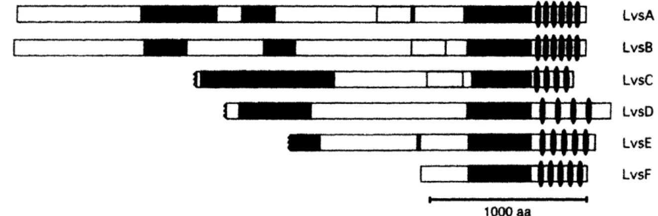
Beachl belongs to a diverse family of large proteins named after a highly conserved BEACH (Beige and Chediak-Higashi) domain of unknown function. Proteins in this family share more than just the BEACH domain; in all of these proteins the BEACH domain is preceded by a novel pleckstrin-homology (PH) domain and followed by between four and six WD40 repeats. WD40 repeats fold into a beta-propeller structure that serves as a protein-protein interaction domain, a property that has been confirmed for one of the BEACH proteins--FAN (Adam-Klages et.al.,1996). The PH domain in these proteins is only similar to the canonical PH domains in structure, not in sequence, and its unique properties will be discussed in detail in the "Crystal Structure" section bellow. As a whole, the conserved PH-BEACH-WD40 module comprises only the C-terminal 25% of the total protein length, while the remaining 75% are
~~~~~~~
----I 1"
4U
1 V V=-~~~~'l
-I
m m I 1' " ,ooo LvsA LvsB LvsC LvsD LvsE LvsFFigure 1 (Taken from De Lozanne, 2003): The domain organization of BEACH
proteins. This diagram shows the domain organization of the six Dictyostelium BEACH proteins. The BEACH domain (green) is the most conserved portion of these proteins
(50-60% identity). At the C-terminus of each protein there are multiple WD-40
repeats (ovals). Adjacent to the BEACH domain is a PH-like domain (purple). Other regions of homology (colored regions) are also shared among different Lvs proteins, but the similarity is low. The sequences of LvsC, LvsD and LvsE are truncated at the N-terminus. Accession numbers for these sequences are: LvsA, AAD52096; LvsB,
AY159038; LvsC, AY159039; LvsD, AY159040; LvsE, AY159036; LvsF, AY159037.
.At. T1812416 A. F 16M22.8 D.d LvsC 0 d. LvsD - ~_L --- - --- -O d. LvsE -' A At2945540 D,m CG1332 D.m. Akap550 Cs.e. F10F2,1 H.s. GL M m, LBA-AIDha s Nurobeachin H ., FAN Mm FAN D d. LvsF · , ttf';r ' '" - D,m CG9o11 Hs KAA1607 -- At F100312 A.1. T1OP 1.5 -- D,d, vsA S.c. BPHI S p. SPBC3H7.t16 M.m. Beige H.S, CHS D,m. CG1 814 D.,d LvsB ---- L.mr L25810511 . LI. r- Q Class-V Class-iV Class-li Class-ll lass-D.n. CG6734 150 100 Divergence Score(x 1 00)
Figure 2 (Taken from De Lozanne, ;
proteins. The BEACH and WD domains
ClustalW algorithm and the alignment The different classes of BEACH proteins
50 0
2003): Phylogenetic tree of family of BEACH
of the indicated sequences were aligned by the
was used to construct this phylogenetic tree. are indicated by the brackets on the right. *Correction: According to my sequence alignments this tree is not completely correct with respect to the human ortholog of Beachl. The protein most similar to Drosophila
Beachl in the human genome is Alfy (KIAA 0993). KIAA1607 is another homolog,
which is less similar to Beach 1 and does not have a FYVE domain.
i
971 R 250 200 f _MI I · In-c"
A a . . i I . . "'"'" - .r.. Wvit ¢^vw IArturo De Lozanne's group used the BEACH-WD40 regions of BEACH proteins from different organisms to construct a phylogenetic tree of this protein family (Figure 2). Their analysis suggests that BEACH proteins are an ancient family that diversified early, before the separation of animals and plants. The genomes of S. cerevisiae and S. pombe have one BEACH protein
each, C.elegans has three, A. thaliana and D. melanogaster have five, and D.
discodeum and H.sapiens have six BEACH proteins each. Phylogenetic analysis
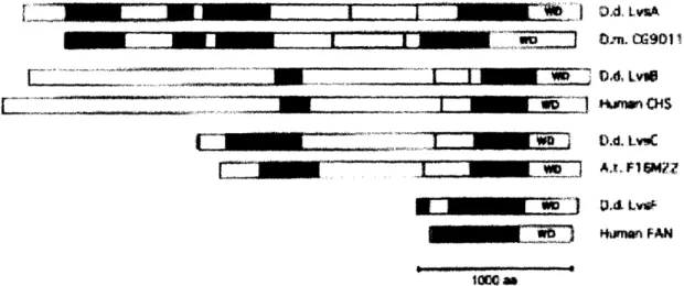
grouped BEACH proteins into five classes; interestingly, proteins within each class possess additional regions of homology within their N-terminal regions, which are not shared with proteins in other classes (Figure 3). This finding raised the possibility that proteins within each class might represent a distinct functional group (Wang, Wu et.al., 2002). To test this hypothesis De
Lozanne's group took advantage of the fact that Dictyostelium (an amoeba) has six BEACH proteins, Lvs (Large volume sphere) A-F, one belonging to each predicted class. By disrupting each Ivs gene they found that, as expected, mutants in different genes had different loss-of-function
phenotypes: IvsA nulls were unique in having defects in cytokinesis and osmoregulation, while only IvsB mutants showed defects in lysosomal traffic. The rest of the Ivs single mutants did not have any obvious phenotypic defects and, thus, were not redundant (at least as single mutants) with either IvsA or
IvsB (Wang, Wu et.al., 2002).
Given that BEACH proteins in Dictyostelium that belong to different classes have distinct cellular functions, the same is likely to hold true for
DAd l *
- Alo Fl F1MN?
Figure 3 (taken from Wang, Wu et.al., 2002): Dictyostelium Lvs proteins share
multiple domains with BEACH proteins of the same class. In addition to the BEACH domain (green) and the WD domain (WD), BEACH proteins within the same class have other domains in common. This diagram indicates in different colors the domains shared by BEACH proteins of the same class. These domains were identified as having a significant BlastP score in pairwise alignments.
Classification of BEACH proteins
According to the phylogenetic analysis, Beach1 falls into class II of BEACH proteins. However, in order to highlight the similarities and underscore the differences between different members of this protein family, it is useful to go over what is known about BEACH proteins in other predicted classes as well.
Class I
This class includes some of the more extensively studied BEACH proteins with putative roles in lysosomal membrane traffic, such as Lyst, mutated in Chediak-Higashi Syndrome (CHS) and its Dictyostelium ortholog LvsB. The hallmark of cells that lack the function of this gene, regardless of the organism, is the presence of large intracellular granules of lysosomal origin (Figure 4). For example, in patients with CHS, such large granules in
melanocytes, called melanosomes, are not properly transferred to keratinocytes, resulting in cutaneous albinism--the absence of pigment in the skin. In Cytotoxic T lymphocytes (CTLs) and natural killer (NK) cells these large lysosomes are similarly unable to undergo exocytosis, preventing the secretion of proteins used to kill infected cells (Ward, 2000). Several hypotheses about the origin of these giant lysosomes have been postulated: it has been suggested that Lyst and its homologs might regulate lysosome
fusion, fission, or motility (Stinchcombe, Page et.al. 2000, Perou et.al., 1997,
and Faigle et.al., 1998, respectively).
Figure 4 (Taken from Blood, 105(11), 2005): Bone marrow aspirate from a
17-year-old female with Chediak-Higashi syndrome is shown. Giant inclusions are present in the cytoplasm of the myeloid precursor cell (center of the image). Note also in both the granulocytes and eosinophils the multiple atypical large cytoplasmic granules that are characteristic of this disorder.
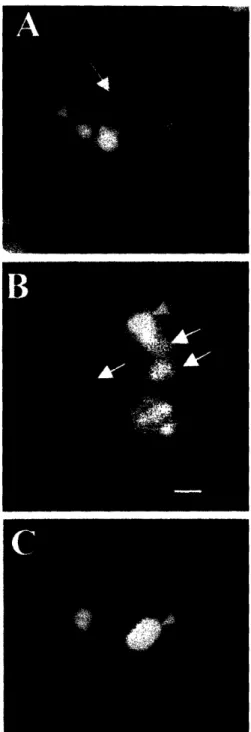
A simple pulse-chase experiment, in which populations of lysosomes were labeled with two different fluorescent dyes, helped to resolve this controversy: the oversized lysosomes in Dictyostelium IvsB mutants are likely to result from increased organelle fusion, not decreased fission (Figure 5) (Harris, Wang et.al., 2002). Although this observation clarified the way in which giant
lysosomes formed, it failed to explain how the loss of LvsB function resulted in
increased lysosomal fusion. There are many possible mechanisms that could result in such a phenotype. For instance, LvsB might regulate the rate of homotypic fusion between lysosomes, by acting as a negative regulator of SNAREs--the core executors of membrane fusion. Alternatively, LvsB could function as a regulator of lysosomal transport; its loss could allow lysosomes
to remain in closer proximity with each other, thus increasing the probability of homotypic fusion.
Interestingly, a defect in lysosomal trafficking lies at the core of another human disorder, Griscelli Syndrome, which causes symptoms that are very similar to CHS. Griscelli Syndrome (GS), like CHS, affects immune cells and melanocytes; however, the subcellular defect in GS is different from that in CHS. Lysosomes in GS are normal in size, but are unable to dock and fuse with the plasma membrane. Griscelli Syndrome can be caused by mutations in Rab27a and in a number of other genes that have one thing in common-they all interact with this Rab GTPase. Rab27a is required for the release of
lysosomal organelles from the microtubule cytoskeleton and their docking at
the plasma membrane (Stinchcombe, Barral et.al., 2001), which explains the defects in GS. Since the subcellular phenotypes caused by mutations in lyst
and rab27a are distinct, it is unlikely that Lyst interacts with this particular Rab. However, it has been suggested that Lyst, LvsB, and their homologs could interact with another member of the Rab GTPase family, one that, for example, regulates homotypic lysosome fusion (Harris, Wang et.al., 2002).
Figure 5 (Taken from Harris, Wang et.al., 2002):Large endolysosomes in the
IvsB-null cells are a result of an increase in fusion. Control NC4A2, IvsA-null, and IvsB-null
cells were pulsed with RITC-dextran, washed, pulsed with FITC-dextran, washed, chased for 5 min, fixed, and examined under a fluorescence microscope. Most of the red and green vesicles were distinct and separate from each other in the control (A),
whereas, a higher percentage of vesicles in the IvsB-null (B) fused with each other.
Examination of cells at the end of the double-pulse period revealed that little colocalization was observed in the mutant, suggesting that separately internalized
Class II
Class II BEACH proteins include the Dictyostelium LvsA, the Drosophila Beachl, and the human Alfy. Notably, each organism has at least one protein from this class, suggesting that Class II proteins might function in a basic process or processes common to all cells (De Lozanne, 2003). LvsA mutants in
Dictyostelium have defects in cytokinesis: when grown in culture they are
unable to divide and, as a result, form binucleate cells. LvsA seems to be required for the late step of cytokinesis: null mutants form the cleavage furrow normally, but are unable to complete its ingression and fail to separate
(Kwak et.al., 1999). It is noteworthy that, similarly to LvsA, Rabll has been shown to play a role in cytokinesis in C.elegans and in mammalian cells (Skop et.al., 2001, Wilson et.al., 2005, respectively). Furthermore, Drosophila rabli
mutants are defective in the process of embryonic cellularization, which is mechanistically similar to cytokinesis (Pelissier et. al, 2003 and Riggs et.al. 2003). Like mutants in IvsA, rabll mutants in different organisms have a defect in the late stages of cytokinesis. It is possible that LvsA causes a defect
in cytokinesis via its interaction with Rabil.
In addition to the cytokinesis defect, IvsA mutants are also defective in contractile vacuole (CV) function. The contractile vacuole is a set of membrane sacs and tubules that collect water and expel it by fusing with the plasma
membrane, thus allowing amoebae to survive in a hypoosmotic environment. LvsA associates with the CV only during the expulsion phase, and is otherwise present in the cytosol in a punctate pattern. In IvsA mutants the CV network is disrupted and, although some vacuoles are able to swell, they are not able to discharge normally, making these mutants osmosensitive (Gerald et.al., 2002). Notably, Rabl1 also localizes to the contractile vacuole in
Dictyostelium. Moreover, the expression of a dominant-negative Rabll
GTPase causes a somewhat opposite defect in the CV morphology than the
thicker-appearing CV network, while the network of CV tubules in IvsA mutants is diminished (Harris, Yoshida et.al., 2001). Both mutants have a defect in osmoregulation; however, their subcellular phenotypes differ. Cells lacking LvsA function have small contractile vacuoles that fuse entirely with the plasma membrane and, unlike CVs of wildtype cells, those in IvsA mutants are not able to re-form after fusion (Gerald et.al., 2002; Wu et.al., 2004). In contrast, in rabli mutant cells contractile vacuoles appear to be unable to fuse with the plasma membrane and thus accumulate within the cell (Harris, Yoshida et.al., 2001). The involvement of Rabll and LvsA in the regulation of contractile vacuole function in Dictyostelium, along with the data that implicates both Rabll and LvsA in the highly conserved process of cytokinesis, suggests that their might be a functional link between class II BEACH proteins and the Rabl GTPase.
Class III
Class III proteins, which only include members from Dictyostelium and mammals, are different from the rest in that they measure just one quarter of the length of the other BEACH proteins. Mammalian class III protein FAN has been shown to bind directly to the cytosolic tail of the tumor necrosis factor receptor,TNF-R55, via its WD40 repeats (Adam-Klages et.al., 1996). FAN is required for the activation of signaling downstream of the TNF receptor; however, the mechanism by which it does so is not understood. Furthermore, it is not clear whether, due to their much smaller size, FAN and its homologs might differ in their mechanism of function from the rest of the BEACH proteins.
Class IV
A unique feature shared by class IV BEACH proteins, which include the
Drosophila AKAP550 (A-kinase anchor protein) and the mammalian
Neurobeachin and LRBA, is a predicted binding site for the type II regulatory subunit of protein kinase A (PKA) in their N-terminal region. PKA is regulated
by cAMP and, in turn, controls many aspects of cellular function, including various membrane trafficking events (Muniz et.al., 1997; Ohashi and Huttner, 1994). AKAPs serve to target PKA function to various subcellular locations, thus, it is possible that some class IV proteins mediate their effects though
PKA. The binding of DAKAP5550 and Neurobeachin to PKA RII has been
confirmed in vitro; however, the biological significance of this interaction has not been addressed (Wang, Herberg et.al., 2000). Phenotypic analysis of mutants in several genes from this class revealed their involvement in different cellular processes. Neurobeachin appears to be involved in synaptic transmission, while DAKAP550 and LRBA are involved in signaling through the EGFR (Epidermal Growth Factor Receptor) pathway (Su et.al., 2004; Wech et.al., 2005; Wang, Gamsby et.al., 2004, respectively). Since members of this class of BEACH proteins associate with cellular membranes (Wang, Howson et.al., 2001 and Wang, Herberg et.al., 2000), it will be interesting to learn whether they mediate their respective effects by regulating some aspect of
membrane trafficking.
Class V
Class V proteins, which only include members from Dictyostelium and
Arabidopsis, have not yet been implicated in any cellular process (De Lozanne,
2003).
Structural Analysis
The BEACH domain
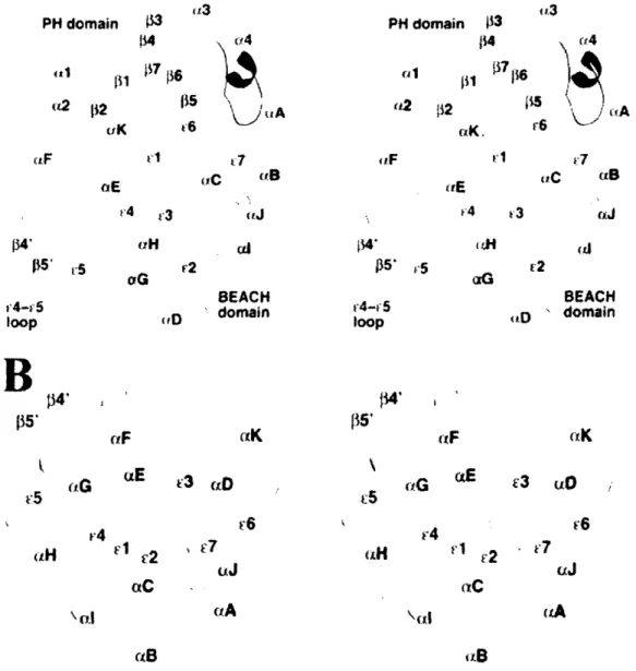
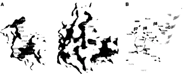
The BEACH domain--a 300 amino acid domain of unknown function, is highly conserved among BEACH family proteins and required for their function (Adam-Klages et.al., 1996; Karim et.al., 1997). Crystal structures of Neurobeachin and LRBA (Jogl et.al., 2002 and Gebauer et.al., 2004, respectively) revealed that the BEACH domain has an unusual fold, never before seen in any protein (Figure 6). The seven segments in its hydrophobic
core cannot be classified as either beta-strands or random coils. Furthermore, its secondary structure is held together by the hydrogen bonds between the main chain amides and carbonyls and the highly conserved side chains of the BEACH domain, rather than the hydrogen bonds between the main chain atoms (Jogl et.al., 2002). Since the BEACH domain fold is so unusual, the crystal structure of this domain did not provide much insight into its function.
No obvious catalytic sites were detected in the structure, although the BEACH domain, being 35kDa in size, is certainly large enough to have an enzymatic function. Assigning a protein-protein interaction role to this domain is also problematic, since the outer surfaces of BEACH domains from different
proteins are quite divergent, due to the fact that most of the conserved residues point towards the interior of the molecule, contributing to its fold (Jogl et.al., 2002). This could mean that BEACH domains of different family members have very different binding partners, or that the main function of the BEACH domain is to spatially organize the surrounding domains (De Lozanne, 2003).
The PH domain
An unexpected discovery from the crystal structure of Neurobeachin was a region with a similar backbone fold to a canonical plekstrin homology (PH) domain, found to lie directly upstream of the BEACH domain (Jogl et.al., 2002). Despite having the same structure, PH domains of BEACH proteins do
not share any sequence homology with other PH domains. Moreover, unlike the canonical PH domains, those of BEACH proteins are not able to bind phospholipids (Gebauer et.al., 2004). This finding is consistent with the information obtained from the crystal structure, showing that the sites normally involved in phospholipid binding are occupied by portions of the BEACH domain (Jogl et.al., 2002). PH domains are also known to function in protein-protein interactions, and, in the case of BEACH proteins, such an interaction appears to be with the adjacent BEACH domain (Figure 7) (Jogl et.al., 2002). In fact, biochemical binding assays show that the binding
constant for the PH-BEACH domain interaction is indicative of a specific, fully reversible protein-protein interaction (Gebauer et.al., 2004).
i'3 (12 t2 t 5 uK t'6 t7 iE r4 ,3 tiC PH domain 13 ra2 132 iK.i ,
, ,
i
i [tB ltE v4 '3 tJ tIC ciB .rJ 134' ps* r5 f4' r"F (tE (r C2 (IC (rl r2 BEACH D ' domain (xK '3 uO (J aA r(BFigure 6 (Taken from Gebauer, 2004): Structure of the PH-BEACH domains of
LRBA/BGL. (A) Schematic drawing of the structure of the PH-BEACH domains of
human LRBA/BGL. The PH domain is shown in green, and the linker is shown in
orange. For the BEACH domain, the extended segments are shown in cyan; the
a-helices, in yellow; and the loops, in purple. (B) Schematic drawing of the structure of the BEACH domain of human LRBA/BGL. The view is related to that of A by roughly a 90 degree rotation around the horizontal axis, produced with ribbons.
A
PH domain -13 1 ' rS f5' oG oiG 4-v, 5 loop BEACH domainB
f5' 4-oop5 loop p4' (iF V5 '3 ¢;D '4 AH F1 c2 aC \ C.1 (B (4H (a JBoth the PH and the BEACH domains seem to be required for BEACH
protein function, as has been demonstrated for the mammalian class III
protein FAN. Only constructs containing both of these domains were capable of rescuing TNF signaling in fan mutant mouse fibroblasts. Moreover, mutations affecting the PH-BEACH interface were shown to reduce TNFR signaling, suggesting that a close interaction between these domains is required for proper function of FAN and, likely, other BEACH proteins (Jogl et.al., 2002). Interestingly, an allele of beachl that encodes a mutation in the predicted PH-BEACH interface has a strong effect on the function of this protein.
A
;;
p7P ~~,#gr 4i /r s rh*,0P ) t) I1_4Figure 7 (Taken from Jogl, EMBO , 2002): The interface between the PH and BEACH
domains. (A) Molecular surface of the PH and BEACH domains. Residues in the PH-BEACH interface are shown in yellow for hydrophobic residues, green for polar residues, red for acidic residues, and blue for basic residues. (B) Schematic drawing of part of the interface between the PH and BEACH domains. The exposed residues of the
back sheet (5, 6 and 7) of the PH domain, shown in green, interact with the
aC-El linker of the BEACH domain (in purple). (A) was produced with Grasp (Nicholls
et al., 1991) and (B) was produced with Ribbons (Carson, 1987).
Structure-function analysis
Both the PH and the BEACH domains are vital for the function of BEACH proteins; however, structure-function analysis of the class II protein LvsA
revealed that almost the entire length of these proteins, including the unconserved N-terminus, may be required for normal function (Wu et.al., 2004). It was demonstrated that the LvsA protein missing just 689 N-terminal amino acids is only partially functional in osmoregulation, and the deletion of 1828 N-terminal amino acids results in a protein that is not functional in either cytokinesis or osmoregulation. Structure-function analysis of LvsA also suggested that it is the C-terminus of BEACH proteins that is responsible for membrane association: a truncated version of LvsA, missing all but the BEACH and WD40 domains, was still able to sediment with membranes (Wu et.al.,
2004).
BEACH proteins are putative regulators of membrane
trafficking
Localization of BEACH proteins to subcellular membranes, as well as class I and II mutant phenotypes in organelle morphology and function, suggest a role for this family of proteins in membrane trafficking. Unfortunately, phenotypic analysis of a number of BEACH mutants failed to uncover their molecular mechanism of action, nor did it reveal a common thread in how they regulate their diverse cellular processes. It seemed that biochemical and genetic studies into the nature of BEACH protein interactors were warranted to make further progress in this field. Unfortunately, biochemistry with BEACH proteins is difficult due to their enormous size and our continuing ignorance about the function of most of their protein domains. Therefore, in my research I took a genetic approach to studying Beachl, a Drosophila class II BEACH protein. I found that Beachl functionally antagonizes a known regulator of membrane trafficking-- the Rabli GTPase. The next half of the introduction will provide a brief overview of how Rab GTPases regulate vesicle trafficking, concluding with a summary of what is known about Rabli.
Membrane Trafficking--a Rab-centric view
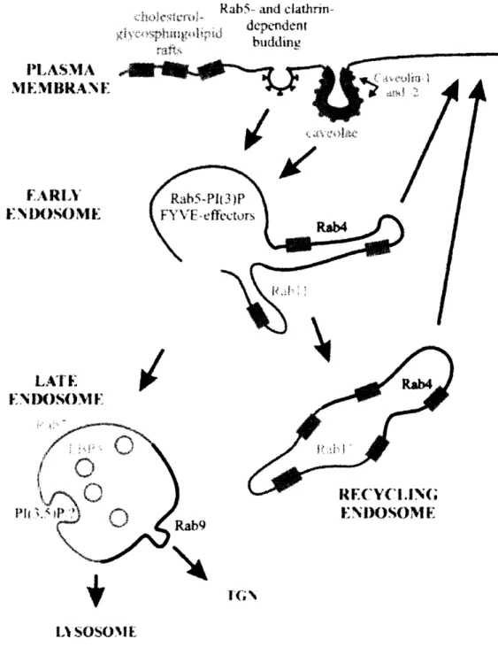
Membrane organization and trafficking are extremely important in almost every aspect of cellular function; in particular, in eukaryotic cells, compartmentalization by membranes forms the basis of functional specialization. Research in the last several years has uncovered added levels of complexity in how subcellular membranes are organized: what we previously believed to be homogeneous organelles were found to be composed of many subdomains, each with different compositions of proteins and lipids, each representing a different functional specialization. For instance, the early endosome has at least four subdomians with different functionalities: one for the fast recycling of molecules directly to the surface, another for the slow recycling via the recycling endosome, another for downregulation by targeting to the lysosome, and yet another domain responsible for homotypic endosomal fusion (Pfeffer, 2003). Similarly, the late endosome has at least two different subdomains: one for the targeting of cargo to the lysosomes,
and the other for targeting to the trans-Golgi Network (TGN) (Miaczynska and Zerial, 2002) (Figure 8).
Most of these membrane compartments and subdomains communicate with each other via different membrane trafficking pathways. For instance, in the biosynthetic pathway, vesicles carry newly made proteins from the trans side of the Golgi to their target destinations, including the plasma membrane. In the recycling pathway, molecules from the cell surface and the outside environment, such as transmembrane receptors and their ligands, are internalized and sorted for either recycling back to the surface or for lysosomal
degradation. Thus, within a cell, cargo-carrying vesicles are continuously trafficked through multiple compartments, reaching their correct destinations with astounding accuracy. At the same time, the identities of organelles and the subdomains within them are carefully maintained in the face of this constant molecular flux. From recent research efforts, Rab GTPases have
begun to emerge as both the key regulators of membrane trafficking and the important determinants of organelle identity (Figure 9) (Zerial and McBride, 2001).
Rab5- and
clathrin-depdeni t PLASMA M. 1IEMBIRAN E1t FARLYt t N iX)S)ME ENIX)SO(ME .~ > EP } > , i / INIrOSOME Rab9 Lt SOS OM[
Figure 8 (Taken from Miaczynska and Zerial, 2002): Domain organization of the
endocytic pathway. Abbreviations: ECV, endosomal carrier vesicles; LBPA,
lysobisphosphatidic acid; TGN, trans-Golgi network; PI(3)P, phosphatidylinositol 3-phosphate; PI(3,5)P2, phosphatidylinositol 3,5-diphosphate.
Rab GTPases
Rabs are the largest family of small monomeric GTPases with 63
members in humans, 29 in each flies and worms, and 11 in yeast. Like all
GTPases, Rabs function as molecular switches, they are active when
GTP-bound and inactive when GDP-GTP-bound. In addition, the activity of Rabs is critically dependent on their membrane association: a Rab protein has to be both GTP-bound and associated with its correct membrane to be in a truly activated state. These two switches: the GTP/GDP switch and the membrane in/out switch, allow Rabs to act as both spatial and temporal regulators of membrane trafficking events (Seabra and Wasmeier, 2004).
memb
(ap4aiC): Eiu3 0 :~:.;: . TON
Nattre Reviews Molecuta Cell _ olog
Rab the proteins. Summarizes intracellular localization of Rab proteins in mammalian
example, Rabl7 in epithelia) or show cell-type-specific localization (for example, Rabl3 in tight junctions). (CCV, clathrin-coated vesicle; CCP, clathrin-coated pit; EC, epithelial cells; IC, ER-Golgi intermediate compartment; M, melanosomes; MTOC,
microtubule-organizing centre; SG, secretory granules; SV, synaptic vesicles; T, T-cell
Rabs carry out their diverse functions in membrane trafficking via their many effectors--proteins that interact preferentially with the GTP-bound Rabs. Each Rab can have multiple effectors; for example, Rab5, a Rab that regulates many functions of early endosomes, has well over twenty known effectors. Since Rab GTPases are a highly conserved family, it was originally believed that their effectors would also fit nicely into a few protein families; however, this turned out not to be the case. Rab effectors identified so far interact specifically with one or a few closely related Rabs. Some effectors, however,
do have features in common, such as the FYVE domain that is present in
several Rab5 effectors. FYVE domains have been shown to bind specifically to phosphotidylinositol-3-phosphate (PtdIns(3)P), a phospholipid that is enriched in the membranes of early endosomes (Misra et.al., 2001), therefore, the presence of a FYVE domain might help a Rab5 effector to localize to early endosomes.
A possible explanation for the specificity of interaction between Rabs and their effectors was provided by the structural analysis of Rab3a bound to its effector, Rabphilin. The crystal structure revealed five regions of interaction between Rab3a-GTP and its effector. Two of these mapped onto the Switch I
and II regions-the domains in a GTPase that change confirmation upon
GDP/GTP exchange. However, the remaining three mapped onto the complementarity-determining regions (CDRs), which are divergent between different Rabs, but conserved within Rab protein subfamilies (Ostermeier and Brunger, 1999). Thus, it is thought that the CDRs impart specificity to the interaction between a Rab and its effector, while the Switch regions confer GTP dependence. In addition, it's becoming clear that, despite sequence similarity, subtle variations in shape can make different Rabs appear quite unique to their effectors. Nonconserved residues can influence the appearance of conserved regions by rendering certain amino acids sterically inaccessible or by changing the angle at which they are presented to the effector (Pfeffer, 2005).
Rabs regulate all steps of membrane trafficking
The process of vesicle trafficking can be broken down into a series of distinct steps: a vesicle must separate from its source, travel to its destination, find the correct target, and, finally, it must fuse with the target membrane. Much has been learned about the core executors of each of these steps in membrane trafficking (Figure 10). It is relatively well understood how coat proteins, such as Clathrin, mediate vesicle budding and cargo selection and how actin- and microtubule-based motors deliver vesicles to their targets (Bonifacino and Lippincott-Schwartz, 2003; Schliwa and Woehlke, 2003). We have also begun to understand how tethering complexes ensure proper vesicle docking, and have made substantial advances in the area of SNARE-mediated
membrane fusion (Scales et.al., 2000). However, we still do not have a good grasp on how the specificity of membrane trafficking is achieved.
Recently, Rabs and their effectors have been identified as regulators of each step in the vesicle trafficking cycle. During vesicle formation Rabs can help collect cargo for the inclusion in the transport vesicle. For example, Rab9 has been shown to bind to both an adaptor protein and to the Mannose-6-Phosphate Receptor during the formation of vesicles destined to travel from the late endosome to the Golgi (Pfeffer, 2003). Rabs can also link vesicles or organelles to motor proteins: Rab27a connects mature melanosomes with
Myosin Va for transport to the plasma membrane (Stinchcombe, Barral et.al., 2001). Rabs have also been shown to interact directly with tethering factors, such as the exocyst complex, which targets vesicles to sites of membrane addition. For instance, Sec4p, a yeast Rab, binds directly to one of the exocyst complex components, Secl5p and thus recruits Sec4p positive vesicles to the site of exocytosis at the plasma membrane. Finally, Rab effectors have been shown to interact directly with SNAREs, the core mediators of vesicle fusion. For instance, the binding of EEA1, a Rab5 effector, to a target SNARE Syntaxin
II
A ·. *Re ptr w Coat-complex -,, vSNARE $ tSNARE tSNARE Molecular Motor Tethering _ proteinsFigure 10 (Taken from Prekeris, 2003): Transport vesicle formation and fusion
model. Coat proteins mediate vesicle budding from the donor compartment as well as cargo selection (step 1). Actin- and microtubule-based molecular motors are responsible for delivering the transport vesicle to its final destination (step 2). Tethering proteins dock transport vesicles to target membranes (step 3). Finally, SNARE proteins, namely syntaxins and VAMPs, mediate membrane fusion and delivery of cargo to the acceptor compartment (step 4). VAMPs are usually present on transport vesicles (vSNAREs), while syntaxins are present on target membranes
(tSNAREs).
- - - ---- --·---
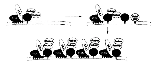
In addition to regulating vesicle trafficking, Rabs serve as domain organizers within organelles. This has been best described for Rab5, the localization of which to the early endosome is the key step in the assembly of the Rab5 microdomain, rich in Rab5 effectors and other molecules essential for early endosome function (Figure 11) (Zerial and McBride, 2001).
(a) Membrane domain
-··'--·'·ivy,
..-
-111-1-1--r -. -_m->. .
I
Figure 11 (Taken from Seabra and Wasmeier, 2004): (a) Model depicting the recruitment of several Rab5 effectors that may act in concert to generate a membrane domain. Activated Rab5 recruits PtdIns(3)K, leading to the generation of PtdIns(3)P in the endosomal membrane. The presence of the lipid then allows the binding of EEA1.
GTP-Rab5 also interacts with the Rabaptin/Rabex complex, which, through the GEF activity of Rabex, results in the activation of additional Rab5 molecules. This in turn is followed by recruitment of further effectors, leading to the formation of a functionally distinct membrane subdomain.
The Rab cycle
In order to regulate all the various aspects of membrane trafficking, Rabs themselves must be carefully regulated: they must localize to the correct membrane and their activity (GTP/GDP state) must be carefully controlled. We do not yet understand exactly how each Rab finds its target membrane; however, some pieces of the puzzle have been put into place.
Most Rabs are post-translationally modified on the C-terminus by two hydrophobic geranylgeranyl groups. This double prenyl lipid group not only
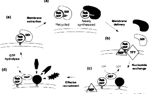
anchors Rabs to the membrane, but also is necessary for their correct localization. Mono-prenylated Rabs are unable to localize properly and are, therefore, nonfunctional (Gomes et.al., 2003). Rabs are delivered to their target membranes in a GDP-bound form by a GDI-GDP dissociation inhibitor. Once associated with the right membrane, Rabs undergo a GDP to GTP exchange with the help of GEFs-Guanine nucleotide exchange factors. Upon activation, Rabs recruit downstream effectors through which they carry out their diverse functions. After inactivation by GAPs-GTPase activating proteins, Rabs are extracted from the membrane by a GDI and recycled. They can exist in the cytosol in the GDP-bound, GDI-associated state (Figure 12) (Seabra and Wasmeier, 2004). (a) exrllncti .-Nyewly Recvyclot synteicd ~_C. .Il- · . l ICC-* _ (d)
'k,
* f ..
+4
,dJ
r,,.
I'--- GAP rsieWN , -5.N. ie~riglt - ~ _f· ,_ _ _ _ II' L- . _ _ n , __ X ·. _·UI _7 Elffctof recruitment M.mbrn '~." dIs (b)?-I JF ,1 Nuclotwde , . _. E 44 A exchlnge fur l _ .
Gurmxrd CX, -r n frl Call hAknoy
Figure 12 (Taken from Seabra and Wasmeier, 2004): Schematic representation of
the Rab cycle showing membrane recruitment and activation. (a) GDP-bound Rab
proteins form a cytosolic complex with RabGDI. (b) Membrane delivery and RabGDI
displacement are mediated by a GDF, probably aided by unidentified targeting factors (TF), followed by (c) Rab activation through GEF-catalysed nucleotide exchange. (d) GTP-bound Rab recruits effector molecules to the membrane. (e) GAP-mediated GTP hydrolysis returns the Rab to its inactive state, resulting in re-extraction from the membrane by GDI. (0) 8r:sbawll ariF· II ....---C"II-. ill 11*1*
·4·-RC
There are only two GDI proteins in the human genome and they bind indiscriminately to all Rabs. Thus, while a GDI can regulate the membrane association/dissociation cycle of Rabs, it cannot by itself specify membrane localization for different Rabs. Recently, a molecule called Yip3 (Ypt-interacting protein 3) was shown, in vitro, to catalyze the dissociation of Rab9 from GDI and its transfer to the membrane (Sivars et.al., 2003). However, this has yet to be confirmed in vivo. Yip3 is a member of a family of small integral membrane proteins that bind promiscuously to prenylated Rabs in vitro, and
are present on both endosomal and Golgi membranes in vivo (Seabra and
Wasmeier, 2004). Furthermore, the Yip family of proteins has 16 members in humans, 12 of which are ubiquitously expressed, for the 63 human Rabs, making it apparent that they cannot be the sole determinants of Rab localization (Pfeffer and Aivazian, 2004).
While it was once thought that the C-terminal hypervariable regions, which are the most divergent among Rabs, determine their membrane localization, domain-swapping experiments disproved this hypothesis. It is now clear that different regions in different Rabs are required for targeting, suggesting that diverse mechanisms might be responsible for the recruitment of different Rabs to their membranes (Seabra and Wasmeier, 2004). Thus, the
understanding of how Rabs are localized will likely require more than the identification of a family of "targeting" proteins.
We have a better grip on how the temporal activity of Rabs is regulated: GEF-- Guanine nucleotide exchange factors, turn them on and GAPs--GTPase activating proteins, turn then off. However, there are many Rabs for which these regulators have not been identified. Moreover, it is not well understood
how GEFs and GAPs are involved in the switching of a Rab between its
Rab 1
The Rabll GTPase is interesting and complex in that, unlike most other Rabs, it regulates a variety of different membrane trafficking pathways. In mammals, the Rabll family has grown to include three closely related proteins: Rabla, Rablib, Rab25a, while in flies and worms there is only one Rabli GTPase.
The recycling pathway
When Rabl1 was originally identified, it was found to localize to the pericentriolar recycling endosome and to regulate both the morphology and the function of this compartment. For instance, the expression of a dominant-negative form of Rabl1 in mammalian cells inhibits the recycling of internalized transferrin, which instead accumulates in the recycling endosome (Ren et.al., 1998 and Ullrich et.al., 1996). In Drosophila, rabl1 mutant oocytes also show a defect in transferrin recycling (Dollar et.al., 2002).
The biosynthetic pathway
In addition to the recycling endosome, Rabli has also been localized to the trans-Golgi Network (TGN) and the post-Golgi vesicles, suggesting that it plays a role in the biosynthetic exocytic trafficking pathway. Indeed, dominant-negative Rabll expression blocks the transport of vesicular stomatitis virus (VSV)-G protein to the basolateral cell surface and causes it to accumulate in the TGN (Chen et.al., 1998). Moreover, recently the Ready lab beautifully demonstrated that Rabll is required for the apical delivery of newly synthesized Rhodopsin from the TGN to the developing rhabdomere in the Drosophila photoreceptors (Satoh et.al., 2005).
Polarized membrane trafficking
As described above, Rabll plays a role in polarized membrane transport and, interestingly, in different processes it is required for vesicle trafficking to either the apical or the basolateral cell surfaces. Examples of trafficking to the
apical surface include the transport of Rhodopsin in the Drosophila photoreceptors (Satoh et.al., 2005) and the trafficking of endosomally-sequestered H+-K+ ATPase to the luminal surface of acid secreting parietal
cells in the stomach (Duman et.al., 1999). Examples of basolateral transport include the trafficking of VSV-G in kidney cells (Chen et.al., 1998) and the recycling of membrane to the expanding lateral membrane surface during
Drosophila embryonic cellularization (Pellisier et.al., 2003 and Riggs et.al.,
2003). Thus, Rabll functions in both the apical and the basolateral polarized membrane trafficking.
Specialized trafficking
Rabli also plays a role in the recycling of G protein-coupled receptors
(GPRCs) such as the CXCR2 chemokine and the M4 muscarinic acetylcholine
receptors, which seems to proceed through a specialized recycling compartment (Fan et.al., 2004 and Volpicelli et.al., 2002, respectively). It might also play a role in the important process of translocating the Glucose transporter 4 (GLUT4) from intracellular stores to the plasma membrane in response to insulin (Muller et.al., 2002).
How is the regulator regulated?
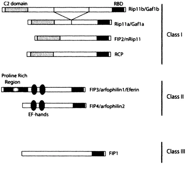
It is clear that Rabli is a key regulator of multiple vesicle trafficking pathways; however, little is known about the mechanisms by which it regulates these events. Recently, a family of Rabll interacting proteins, known as FIPS (Rabli family of interacting proteins), has been described. FIPs share a conserved 20 amino acid Rabll/25 binding protein (RBD) domain, but otherwise, different classes of FIPs are quite dissimilar (Figure 13). Unlike the canonical Rab GTPase effectors, FIPs do not require Rabl for their membrane localization, and, conversely, FIPs do not appear to be sufficient to recruit Rabl1 to the appropriate membranes (Prekeris, 2003). However, like other Rab effectors, FIPs appear to recruit proteins required for membrane trafficking to Rabl-postive vesicles and organelles. For example,
FIP2 acts as an adaptor between Rabll and the actin-based motor protein Myosin Vb, which is required for the transport of vesicles out of the pericentriolar recycling endosome compartment (Hales et.al., 2002). There is also evidence that class I FIPs might target Rabll-positive vesicles to docking sites on the plasma membrane via their C2 domains (Lindsay and McCaffrey, 2004). Finally, class II FIPs have been implicated as dual ARF/Rabll interacting proteins (Hickson et.al., 2003). Some ARF GTPases are known regulators of cytoskeletal rearrangements, thus, class II FIPs might serve to couple the function of Rabs and ARFs in the processes, such as cytokinesis, where both membrane addition and cytoskeletal rearrangements must be closely coordinated (Wilson et.al., 2005). Interestingly, Nuf, a Drosophila homolog of an ARF effector, physically associates with Rabll and, like Rabal, is required for embryonic cellularization, a process in many ways similar to cytokinesis (Rigss et.al., 2003).
In addition to FIPS, recent studies suggest that the exocyst--a tethering complex that targets vesicles to sites of membrane addition, is a Rabll effector. In mammalian cells, Sec15, a member of the exocyst, binds to Rabll in a GTP-dependent manner (Zhang et.al., 2004). Moreover, Drosophila photoreceptors mutant in sec6, an exocyst component, have an identical Rhodopsin trafficking defect to rabil mutant photoreceptors. In addition, Rabll physically interacts with Sec5, another exocyst component. These results suggest that Rabll-positive vesicles might be targeted to the plasma membrane via their interaction with the exocyst complex (Beronja et.al., 2005).
C2 domain
II
i
II , ,
RBD
- 1 Rip lb/Gaflb
:1o Rip 1 a/Gafl a
_1 ] FIP2/nRipl -'11 RCP Proline Rich Region I 1M " l EF-hands _ FP3/arfophitin/Eferin _11 FIP4/arfophilin2
_0
FP1
I Class 11 Class I11Figure 13 (Taken form Prekeris, 2003): The structure of Rabll family interacting
proteins (FIPs). Based on their domain structure, all FIPs can be divided into three main classes. Class I FIPs (Riplla, Riplib, RCP, and FIP2) contain a C2 domain at the
N-terminus end of the protein. Class II FIPs (FIP3 and FIP4) contain two EF-hands
and a proline rich region. Class III includes only one member, FIP1, which exhibits no
homology to known protein domains. RBD stands for Rabl1 binding domain. Class I m r = .a.,4 - =,'! I I n..- " , -Sl - I -· ·- ··-- ·· ·--- - ; - ---rlrlnc --- ·--- ---I_ 1 1m-7 7777Tr . . . . . i__ .---·--- --·---·· -- --- ·~~~~~~~~~--- · · -I ..
II
Remaining questions about Rab11
Although we are beginning to learn about how RablI executes its diverse functions, many questions remain. It will be important to learn how Rabll is localized to its target membranes, and furthermore, what factors are involved in its differential targeting to the TGN versus the recycling endosome. No Rabll GEFs or GAPs have been identified to date; therefore, it will be interesting to find out what they are and how they play a role in the regulation of Rabl localization and function. Rab5 is estimated to have close to forty effectors, as might Rabll. So far, only a handful of RablI effectors have been identified; it will be interesting to find other Rabl1 interactors and learn how they function in various Rabll-dependent processes.
Concluding Remarks
BEACH proteins comprise an evolutionarily conserved protein family with roles in important cellular processes and some members associated with human disease. BEACH proteins have been implicated in vesicle trafficking; however, their molecular mechanism of function is not known. In my work I took a genetic approach to studying Beach1, a previously uncharacterized Drosophila BEACH protein. Although we identified Beachl in an overexpression screen due to a strong gain-of-function phenotype in the photoreceptor growth cones, we found that loss-of-function beachl mutants had no obvious defects. However, through a genetic modifier screen, I found the beachl
overexpression phenotype to be strongly enhanced by a reduction in rabli
dosage. Rabll is a small GTPase required in several membrane trafficking pathways that culminate in the delivery of recycled or newly synthesized proteins to the plasma membrane. Analysis of beachl, rabli double mutants revealed that beachl suppresses the previously described viability and bristle defects of rabll. Furthermore, guided by the synaptic localization of Beachl and Rab1l, I found that rabll mutants also have synaptic morphology defects at the neuromuscular junction (NMJ), which are suppressed by the loss of
beachl. This confirmed that beachl is indeed involved in the regulation of the morphology of nerve terminal regions, as was originally suggested by its overexpression phenotype. Thus, my work showed that Beachl functionally antagonizes Rabl1 during Drosophila development and in regulating synaptic morphology. Furthermore, since the neuromuscular junction is highly amenable to genetic, functional, and structural analysis, future studies at the NMJ are likely to not only contribute to our understanding of how Rabl1 and Beachl function in membrane trafficking, but to also broaden our knowledge about the mechanisms regulating synaptic growth.
References
1. Adam-Klages, S., D. Adam, et al. (1996). "FAN, a novel WD-repeat
protein, couples the p55 TNF-receptor to neutral sphingomyelinase." Cell 86(6): 937-47.
2. Beronja, S., P. Laprise, et al. (2005). "Essential function of Drosophila Sec6 in apical exocytosis of epithelial photoreceptor cells." J Cell Biol
169(4): 635-46.
3. Bonifacino, . S. and . Lippincott-Schwartz (2003). "Coat proteins: shaping membrane transport." Nat Rev Mol Cell Biol 4(5): 409-14.
4. Castermans, D., V. Wilquet, et al. (2003). "The neurobeachin gene is
disrupted by a translocation in a patient with idiopathic autism." J Med Genet 40(5): 352-6.
5. Chen, ., Y. Lu, et al. (2004). "Identification and characterization of NBEAL1, a novel human neurobeachin-like 1 protein gene from fetal
brain, which is up regulated in glioma." Brain Res Mol Brain Res
125(1-2): 147-55.
6. De Lozanne, A. (2003). "The role of BEACH proteins in Dictyostelium."
Traffic 4(1): 6-12.
7. Dollar, G., E. Struckhoff, et al. (2002). "Rabll polarization of the Drosophila oocyte: a novel link between membrane trafficking, microtubule organization, and oskar mRNA localization and translation." Development 129(2): 517-26.
8. Duman, . G., K. Tyagarajan, et al. (1999). "Expression of rablla N124I in gastric parietal cells inhibits stimulatory recruitment of the
H+-K+-ATPase." Am J Physiol 277(3 Pt 1): C361-72.
9. Faigle, W., G. Raposo, et al. (1998). "Deficient peptide loading and
MHC class II endosomal sorting in a human genetic immunodeficiency disease: the Chediak-Higashi syndrome." J Cell Biol 141(5): 1121-34.
10. Fan, G. H., L. A. Lapierre, et al. (2004). "Rabil-family interacting
protein 2 and myosin Vb are required for CXCR2 recycling and
receptor-mediated chemotaxis." Mol Biol Cell 15(5): 2456-69.
11. Gebauer, D., J. Li, et al. (2004). "Crystal structure of the PH-BEACH domains of human LRBA/BGL." Biochemistry 43(47): 14873-80.
12. Gerald, N. J., M. Siano, et al. (2002). "The Dictyostelium LvsA protein
is localized on the contractile vacuole and is required for osmoregulation." Traffic 3(1): 50-60.
13. Gomes, A. Q., B. R. Ali, et al. (2003). "Membrane targeting of Rab GTPases is influenced by the prenylation motif." Mol Biol Cell 14(5):
1882-99.
14. Hales, C. M., . P. Vaerman, et al. (2002). "Rabil family interacting protein 2 associates with Myosin Vb and regulates plasma membrane recycling." J Biol Chem 277(52): 50415-21.
with and regulates the structure and function of the contractile vacuole system in dictyostelium." J Cell Sci 114(Pt 16): 3035-45.
16. Harris, E., N. Wang, et al. (2002). "Dictyostelium LvsB mutants model
the lysosomal defects associated with Chediak-Higashi syndrome." Mol
Biol Cell 13(2): 656-69.
17. Hickson, G. R., J. Matheson, et al. (2003). "Arfophilins are dual Arf/Rab 11 binding proteins that regulate recycling endosome distribution and are related to Drosophila nuclear fallout." Mol Biol Cell 14(7): 2908-20.
18. Introne, W., R. E. Boissy, et al. (1999). "Clinical, molecular, and cell
biological aspects of Chediak-Higashi syndrome." Mol Genet Metab
68(2): 283-303.
19. Jogl, G., Y. Shen, et al. (2002). "Crystal structure of the BEACH domain reveals an unusual fold and extensive association with a novel PH domain." Embo J 21(18): 4785-95.
20. Karim, M. A., K. Suzuki, et al. (2002). "Apparent genotype-phenotype correlation in childhood, adolescent, and adult Chediak-Higashi
syndrome." Am J Med Genet 108(1): 16-22.
21. Kwak, E., N. Gerald, et al. (1999). "LvsA, a protein related to the mouse beige protein, is required for cytokinesis in Dictyostelium." Mol Biol Cell 10(12): 4429-39.
22. Lindsay, A. J. and M. W. McCaffrey (2004). "The C2 domains of the
class I Rabli family of interacting proteins target recycling vesicles to the plasma membrane." J Cell Sci 117(Pt 19): 4365-75.
23. Miaczynska, M. and M. Zerial (2002). "Mosaic organization of the endocytic pathway." Exp Cell Res 272(1): 8-14.
24. Misra, S., G. J. Miller, et al. (2001). "Recognizing phosphatidylinositol
3-phosphate." Cell 107(5): 559-62.
25. Muller, H., K. Deckers, et al. (2002). "The fatty acid translocase (FAT)/CD36 and the glucose transporter GLUT4 are localized in
different cellular compartments in rat cardiac muscle." Biochem Biophys Res Commun 293(2): 665-9.
26. Muniz, M., M. E. Martin, et al. (1997). "Protein kinase A activity is
required for the budding of constitutive transport vesicles from the
trans-Golgi network." Proc Natl Acad Sci U S A 94(26): 14461-6.
27. Ohashi, M. and W. B. Huttner (1994). "An elevation of cytosolic protein phosphorylation modulates trimeric G-protein regulation of secretory vesicle formation from the trans-Golgi network." J Biol Chem 269(40): 24897-905.
28. Ostermeier, C. and A. T. Brunger (1999). "Structural basis of Rab
effector specificity: crystal structure of the small G protein Rab3A complexed with the effector domain of rabphilin-3A." Cell 96(3):
363-74.
29. Pelissier, A., J. P. Chauvin, et al. (2003). "Trafficking through Rabl endosomes is required for cellularization during Drosophila
30. Perou, C. M., J. D. Leslie, et al. (1997). "The Beige/Chediak-Higashi
syndrome gene encodes a widely expressed cytosolic protein." Biol Chem 272(47): 29790-4.
31. Pfeffer, S. (2003). "Membrane domains in the secretory and endocytic pathways." Cell 112(4): 507-17.
32. Pfeffer, S. and D. Aivazian (2004). "Targeting Rab GTPases to distinct
membrane compartments." Nat Rev Mol Cell Biol 5(11): 886-96. 33. Pfeffer, S. R. (2005). "Structural clues to Rab GTPase functional
diversity." J Biol Chem 280(16): 15485-8.
34. Prekeris, R. (2003). "Rabs, Rips, FIPs, and endocytic membrane
traffic." ScientificWorldJournal 3: 870-80.
35. Ren, M., G. Xu, et al. (1998). "Hydrolysis of GTP on rabli is required
for the direct delivery of transferrin from the pericentriolar recycling compartment to the cell surface but not from sorting endosomes." Proc
Natl Acad Sci U S A 95(11): 6187-92.
36. Riggs, B., W. Rothwell, et al. (2003). "Actin cytoskeleton remodeling during early Drosophila furrow formation requires recycling endosomal
components Nuclear-fallout and Rab11." J Cell Biol 163(1): 143-54.
37. Satoh, A. K., E. O'Tousa , et al. (2005). "Rabll mediates post-Golgi trafficking of rhodopsin to the photosensitive apical membrane of Drosophila photoreceptors." Development.
38. Scales, S. ., J. B. Bock, et al. (2000). "The specifics of membrane
fusion." Nature 407(6801): 144-6.
39. Schliwa, M. and G. Woehike (2001). "Molecular motors. Switching on kinesin." Nature 411(6836): 424-5.
40. Seabra, M. C. and C. Wasmeier (2004). "Controlling the location and activation of Rab GTPases." Curr Opin Cell Biol 16(4): 451-7.
41. Sivars, U., D. Aivazian, et al. (2003). "Yip3 catalyses the dissociation of endosomal Rab-GDI complexes." Nature 425(6960): 856-9.
42. Skop, A. R., D. Bergmann, et al. (2001). "Completion of cytokinesis in C. elegans requires a brefeldin A-sensitive membrane accumulation at the cleavage furrow apex." Curr Biol 11(10): 735-46.
43. Stinchcombe, J. C., L. J. Page, et al. (2000). "Secretory lysosome
biogenesis in cytotoxic T lymphocytes from normal and Chediak Higashi syndrome patients." Traffic 1(5): 435-44.
44. Stinchcombe, J. C., D. C. Barral, et al. (2001). "Rab27a is required for
regulated secretion in cytotoxic T lymphocytes." J Cell Biol 152(4):
825-34.
45. Stinchcombe, J., G. Bossi, et al. (2004). "Linking albinism and immunity: the secrets of secretory lysosomes." Science 305(5680): 55-9.
46. Su, Y., R. . Balice-Gordon, et al. (2004). "Neurobeachin is essential for neuromuscular synaptic transmission." J Neurosci 24(14): 3627-36. 47. Ullrich, O., S. Reinsch, et al. (1996). "Rabll regulates recycling
913-24.
48. Volpicelli, L. A., 1. J. Lah, et al. (2002). "Rablla and myosin Vb
regulate recycling of the M4 muscarinic acetylcholine receptor." J
Neurosci 22(22): 9776-84.
49. Wang, X., F. W. Herberg, et al. (2000). "Neurobeachin: A protein
kinase A-anchoring, beige/Chediak-higashi protein homolog implicated in neuronal membrane traffic." Neurosci 20(23): 8551-65.
50. Wang, . W., . Howson, et al. (2001). "Identification of a novel lipopolysaccharide-inducible gene with key features of both A kinase anchor proteins and chsl/beige proteins." J Immunol 166(7):
4586-95.
51. Wang, N., W. I. Wu, et al. (2002). "BEACH family of proteins:
phylogenetic and functional analysis of six Dictyostelium BEACH proteins." J Cell Biochem 86(3): 561-70.
52. Wang, . W., J. J. Gamsby, et al. (2004). "Deregulated expression of LRBA facilitates cancer cell growth." Oncogene 23(23): 4089-97. 53. Ward, D. M., G. M. Griffiths, et al. (2000). "Analysis of the lysosomal
storage disease Chediak-Higashi syndrome." Traffic 1(11): 816-22.
54. Wech, I. and A. C. Nagel (2005). "Mutations in rugose promote cell
type-specific apoptosis in the Drosophila eye." Cell Death Differ 12(2): 145-52.
55. Wilson, G. M., A. B. Fielding, et al. (2005). "The FIP3-Rabll protein
complex regulates recycling endosome targeting to the cleavage furrow during late cytokinesis." Mol Biol Cell 16(2): 849-60.
56. Wilson, G. M., A. B. Fielding, et al. (2005). "The FIP3-Rabll protein
complex regulates recycling endosome targeting to the cleavage furrow during late cytokinesis." Mol Biol Cell 16(2): 849-60.
57. Wu, W. I., . Yajnik, et al. (2004). "Structure-function analysis of the BEACH protein LvsA." Traffic 5(5): 346-55.
58. Zerial, M. and H. McBride (2001). "Rab proteins as membrane organizers." Nat Rev Mol Cell Biol 2(2): 107-17.
59. Zhang, X. M., S. Ellis, et al. (2004). "Secl5 is an effector for the Rabll
Chapter 2
Identification and characterization of
Beach1, a Drosophila BEACH protein
This chapter includes joint work with Adela Augsburger, who was a postdoc in the Garrity lab at the time. We worked together on the overexpression screen and the EMS screen for the isolation of beachl mutants.
Some of the work described in experiments at the neuromuscular Laboratory at Harvard Medical School by the Harvard MD/PhD program.
this chapter, specifically, localization junction, was done in the Schwarz (Children's Hospital) and funded, in part,
Introduction
Mutational analysis of the genomes of model organisms is a powerful way to gain insight into biological processes. Mutagenesis screens are
advantageous in that one needs no prior knowledge to implicate a gene in a
particular process. To create lesions in the DNA, mutagens, such as chemicals, ionizing radiation or transposable elements, are used. Most mutations generated in this fashion are loss-of-function mutations-they reduce or eliminate the function of a given gene. However, gain-of-function mutations, ones that increase the level or broaden the area of expression, have also proven useful in the understanding of how a given gene is involved in various biological processes. For example, upregulation of oncogenes leads to cancer, consistent with the normal roles of these genes in cell growth. One of the best examples of informative missexpression is probably the missexpression of homeotic genes in the Drosophila embryo that results in stunning segmental transformations, helping to elucidate the normal functions of homeotic genes in the establishment of the Drosophila body plan (Lewis, 1978 and Schneuwly et.al., 1987).
At the start of my graduate work, I was interested in the mechanisms behind axon guidance, the process by which neurons navigate to their targets using growth cones, motile structures at the end of axons that steer them along their path. At that time, several loss-of-function screens for regulators of axon guidance and growth cone morphology had already been done (Martin et.al., 1995 and Garrity et.al., 1996); therefore, we decided to use a gain-of-function approach. We hoped that, by doing an overexpression screen, we would increase our chances of identifying new genes involved in these processes.
For our screen we chose to use the compound eye of Drosophila
melanogaster, which had already been proven to be a powerful system for the