HAL Id: tel-02491142
https://tel.archives-ouvertes.fr/tel-02491142
Submitted on 25 Feb 2020
HAL is a multi-disciplinary open access archive for the deposit and dissemination of sci-entific research documents, whether they are pub-lished or not. The documents may come from teaching and research institutions in France or abroad, or from public or private research centers.
L’archive ouverte pluridisciplinaire HAL, est destinée au dépôt et à la diffusion de documents scientifiques de niveau recherche, publiés ou non, émanant des établissements d’enseignement et de recherche français ou étrangers, des laboratoires publics ou privés.
Localization and function of anionic lipids in cell
organization and plant development
Matthieu Platre
To cite this version:
Matthieu Platre. Localization and function of anionic lipids in cell organization and plant development. Subcellular Processes [q-bio.SC]. Université de Lyon, 2017. English. �NNT : 2017LYSEN098�. �tel-02491142�
Numéro National de Thèse : 2017LYSEN098
THESE de DOCTORAT DE L’UNIVERSITE DE LYON
opérée parl’Ecole Normale Supérieure de Lyon
Ecole Doctorale
N° 340Biologie Moléculaire, Intégrative et Cellulaire
Spécialité de doctorat : Sciences de la Vie
Discipline
: Biologie cellulaire des plantesSoutenue publiquement le 01/12/2017, par :
Matthieu PLATRE
Localisation et fonction des lipides
anioniques dans l’organisation
cellulaire et le développement des
plantes
Devant le jury composé de :
Robert, Stéphanie Professeure Université d’Umea Rapporteure Faure, Jean-Denis Professeur INRA-AgroParis Tech Rapporteur Boutté, Yohann CR CNRS Université de Bordeaux Examinateur Antonny, Bruno DR CNRS IMPC UMR7275 Examinateur Ingram, Gwyneth DR CNRS ENS de Lyon Examinatrice Jaillais, Yvon DR CNRS ENS de Lyon Directeur de thèse
REMERCIEMENTS
Sans nul doute cette partie de ma thèse sera lu par le plus grand nombre et pour cause les remerciements c’est important ! Mais aussi moins long et compréhensible d’entre tous comparé aux parties suivantes. C’est aussi selon moi, là, qu’en quelques paragraphes on peut sentir par le choix des mots ce qu’a signifié l’expérience d’une thèse. Dans ce but, j’ai pris le temps de m’adonner à ce délicat exercice quelques semaines après la rédaction de mon manuscrit.
Il est vrai que derrière plusieurs années de travail que représente une thèse se cache de nombreuses personnes qui ont été indispensable et qui méritent d’être remerciés. Très certainement, chacun(e) d’entre vous qui lirons ce passage chercherons assidûment par la suite leur prénom dans la liste des personnes énumérées pour voir s’ils y figurent « ou pas » ! Je tiens donc à m’excuser si par mégarde j’ai pu égarer certains prénoms car chacun(e) d’entre vous que j’ai pu croisé durant ces 5 années au RDP mérite réellement d’être remercié. Alors toi qui a pris le temps de lire cette phrase, MERCI !
Mes premiers remerciements s’adressent à celui sans qui cette thèse n’aurait pas été possible et sans qui je n’en serai pas là aujourd’hui. Comme mentionné, il y a cinq ans de cela je suis arrivé au RDP pour mon stage de fin d’étude d’ingénieur. Par mon cursus, je n’étais pas vraiment préparé à travailler en laboratoire, c’est avec patience, pédagogie et courage qu’Yvon a su me transmettre les connaissances et sa passion pour la recherche. Je mentionne le courage car je me demande encore comment as-t-il pu faire pour ne pas perdre espoir face à certaines questions de néophyte que j’ai pu lui poser ! Ainsi, je tiens à le remercier pour ces choses-là mais aussi pour la confiance et la liberté qu’il a su m’accorder. J’ai encore du mal à imaginer comment je vais pouvoir m’en sortir par la suite car il est devenu bien plus qu’un directeur de thèse. J’ai beaucoup aimé apprendre à ses côtés et espère, si la chance m’en est donné, transmettre à mon tour ce qu’il m’a appris (tout en restant bien bien souple !). Je finirai ce paragraphe en perpétuant la tradition en citant le dicton favori de Thierry qu’on apprécie dire en équipe le vendredi après-midi autour d’un
En parlant d’équipe, je remercie toute l’équipe SiCE qui est tellement « So SiCE », les discussions scientifiques à la fraiche (« ou pas ») du lundi matin, les repas chez TG, les BBQ chez Zorg, les apéros chez Yvon, j’en passe et des meilleures… J’espère que perdurera l’entraide au sein de l’équipe et remercie à cette occasion, Laia, Mathilde, Mar, Lise et Mehdi pour leur aide précieuse. J’aimerais remercier plus précisément la « ROP » loveuse de l’équipe Christine (aka kikouette) pour les longues discussions et réflexion. Mais aussi Vince pour m’avoir transmis ses talents de microscopiste et pour finir Laia pour avoir pris le soin de relire l’ensemble ma thèse.
Il est maintenant temps de parler du cadre exceptionnel dans lequel j’ai eu la chance d’évoluer, la grande famille RDPienne ! Le RDP a été pour moi ma première expérience en laboratoire et ne peux donc pas avoir d’élément de comparaison. Cela étant, il ne m’a pas fallu beaucoup de temps pour comprendre dans quel endroit j’étais tombé. Le RDP, c’est bien plus qu’un laboratoire, c’est bien plus que du développement, c’est bien plus que de la reproduction. C’est aussi des instants foyers, des repas de noël, d’innombrables pots, de multiples skiminars mais surtout des gens d’exceptions. Cette cohésion durant ces années a été une réelle chance et a participé à entretenir ma motivation chaque jour ! A travers tous ces moments j’ai vécu de beaux instants et rencontrer des amis, Léo, Steven et Jo. Une thèse ça ne se fait pas sans avoir de quoi penser à autre chose pour se changer l’esprit. A ce compte-là, je remercie tous les membres du groupe des « The Broken Teeth », Nico, Ruby et Vicky ! Nos répétitions assidues nous ont mené au top du top de la scène pop Lyonnaise :-P. Single bientôt dispo « Chez martine » la disquaire d’Oullins et chez Gilbert Joseph ! Et bien sûr dans ce paragraphe musical, je ne peux inévitablement pas oublier Ringo, qui est un bassiste de talent et avec qui j’ai eu l’immense privilège de jouer durant maintenant 10 ans (it’s been a long time bro !). Je tiens aussi à adresser mes remerciements à des objets car ils ont fait partie de l’équilibre qui m’a permis de mener cette thèse jusqu’à la fin. Mais aussi ils m’ont permis (« de force ») de faire des pauses plus ou moins prolongées durant ma thèse. Dans l’ordre chronologique, je pense à mon skate longboard, deux semaines d’arrêt pour un coude cassé, mon snowboard, un mois et demi d’arrêt pour une
clavicule cassée et pour finir mon vélo, quelques jours d’arrêt pour une omoplate/scapula cassée.
Bien entendu, je ne peux pas faire de remerciements sans m’adresser à ma famille. Rétrospectivement, au début, j’ai eu l’impression qu’ils ne comprenaient pas vraiment les raisons pour lesquelles je m’engageais dans cette aventure mais cela sans même faire oscillé leur soutien. Au fur et à mesure que le temps avançait et voyant que cela m’épanouissait, ils ont bien entendu trouvé raisons malgré l’obscurité que peut leur évoquer le but de mes recherches. Je voudrais plus particulièrement, remercier mes parents, Isabelle et François, qui ont toujours cru en moi, soutenu dans les moments difficiles et toujours su agir, selon moi, en parent exemplaire. Je pense aussi à ma sœur, Emmanuelle, mon frère, Hugo, mes grands parents, Louis et Marcelle ainsi qu’à mon oncle, Hervé. Dans ce paragraphe, je voudrais aussi m’adresser à mes amis de longue date. Plus spécialement, je voudrais remercier Thomas, Jean et Jean-Baptiste qui ont été des personnes sur lesquelles j’ai pu compter durant cette expérience et je leur signifie ici ma reconnaissance. Ringo, Simon, Damien, Lucie, Bertrand, Pierre, Paul, Thomas, François, Samy qui ont toujours étaient là pour me changer les idées. Je voudrais remercier des gens qui m’ont accompagné au jour le jour durant la rédaction de ma thèse. Leur tâche n’a pas dû être évidente à certain moment, s’échinant à rendre la mienne plus paisible. Je pense à mes super colocs, Mary, Mehdi, Thibault et Karim ainsi qu’à ma Jeannette la plus chouette.
Pour conclure, je garderai un très bon souvenir de ma thèse pour de multiples raisons. Les plus évidentes sont : l’aboutissement de nombreuses années de travail et le « début de la fin » de la formation scientifique :-P. Mais celles-ci ne sont pas des plus essentielles. La thèse c’est avant tout une vraie expérience de vie. A travers elle, on apprend à acquérir une certaine rigueur, à se tromper, persévérer mais aussi confronter ses idées ou ses convictions et à les étayer. On apprend aussi à interagir avec autrui, les écouter, réfléchir et bâtir des choses ensemble. Selon moi, cette interaction et cette construction constituent l’essence même de la science. De manière antagoniste, on comprend aussi que nous sommes le seul acteur de notre propre sujet. Bien que les discussions et les échanges soient centraux ce qui
précédemment une thèse ce n’est pas que la science, j’en garderai un très bon souvenir par l’ensemble des moments vécus et des personnes qui m’ont entouré au RDP mais pas seulement. Affectueusement, merci à tous ! Je dédicace ma thèse à tous les gens que j’aime. L'imagination est plus importante que le savoir. Le savoir est limité alors que l'imagination englobe le monde entier, stimule le progrès, suscite l'évolution. « What Life Means to Einstein », George Sylvester Viereck, The Saturday Evening Post, 26 October 1929, p. 17 »
Préambule
Ma thèse se compose en trois parties :
A. Introduction B. Résultats
C. Discussion et perspectives
L’introduction fait l’état de l’art du maintien et du rôle du champ électrostatique membranaire dans l’organisation de la cellule eucaryote. Cette propriété membranaire a été le support de mes travaux de thèse liant les trois chapitres de la partie résultats. Les deux premiers chapitres décrivent l’organisation du champ électrostatique dans la cellule végétale et son maintien par les lipides anioniques. Le troisième chapitre fait l’objet d’une introduction portant sur la voie de signalisation « non génome » de l’auxine. En effet, cette propriété membranaire s’est révélée être centrale dans la transduction du signal auxinique. Le premier chapitre ayant donné lieu à une publication, par soucis d’homogénéité ma thèse est ainsi rédigée en anglais.
A. INTRODUCTION
I. General introduction on the electrostatic field ... 3
a. Definition of the electrostatic field ... 5
b. The membrane surface charge (MSC) ... 7
i. Principle ... 7
ii. Tools to investigate PM surface charge ... 9
c. The plasma membrane is the most ionic compartment in eukaryotes ... 11
d. Anionic phospholipids present in the inner leaflet of cellular membrane ... 11
i. Phosphatidylinositol phosphates, phosphatidic acid and phosphatidylserine are anionic lipids presenting different charges and concentration in cellular membrane ... 11
ii. Regulation, turnover and localization of anionic phospholipids in yeast and mammals ... 15
II. Anionic lipids in the maintenance of the PM electrostatic field in mammals…23 a. Anionic lipids are required to maintain the plasma membrane electrostatic field ... 23
b. Involvement of PIPs in the plasma membrane surface charge ... 25
c. Evidence that PS contributes to the PM electrostatic field ... 29
III. Anionic lipids in the maintenance of the PM electrostatics field in yeast ... 33
IV. Membrane surface charges defines an electrostatic territory corresponding to PM-derived organelles ... 35
a. In mammals, endocytic compartments are electrostatic. ... 35
b. Membrane electrostatic of intracellular compartments in yeast ... 37
V. Why electrostatism? ... 41
a. Tuning the electrostatic field by adjusting the spatio-temporal control of anionic phospholipid enrichment ... 43
i. Example 1: Anionic lipid remodeling controls protein dynamics during phagocytosis ... 43 ii. Example 2: Phosphatidylserine generates a charge gradient along the plasma membrane to
i. Encoding cationic regions with a variety of charges ... 47 ii. Postranslational modification on proteins and the electrostatic switch hypothesis ... 47 c. The ion shielding effect links membrane potential and membrane electrostatics and
localy organize the plasma membrane electrostatic field ... 51
VI. The limits of the electrostatic framework ... 53
a. Membrane targetting: a combination of electrostatic, hydrophobic interactions and
trafficking. ... 53 b. Polybasic regions may have specificity for lipid head group ... 55
VII. General conclusion and problematic ... 56
B. RESULTS
I. Chapter I: A PtdIns(4)P-driven electrostatic field controls cell membrane
identity and signalling in plants……….53
II. Chapter II: Combinatorial PA, PS and PI4P cooperation determines the
electrostatic territory in plants………99
III. Chapter III: A tunable lipid rheostat steers Rho-mediated auxin
signaling………151 a. General introduction on Rho of plant GTPases. ... 53
b. ROPGTPase as a central regulator of the ‘non-genomic’ auxin signaling pathway 155
i. Auxin influence the interdigitating status of leaf pavement cells ... 157 ii. Auxin activates ROPs to orchestrate cytoskeletal rearrangement required to establish pavement cell polarity. ... 157 iii. TRANSMEMBRANE RECEPTOR KINASEs (TMKs) acts upstream of ROP signaling in the “non-genomic” auxin signaling pathway ... 159 iv. “Non-genomic” auxin signaling pathway regulates the gravitropic response through
ROPGTPase ... 171 v. Auxin controls microtubule organization during the gravitropic response, which may
impact differential cell elongation ... 175 vi. Activated ROPs are localized in the so called detergent resistant membrane (DRM) to trigger proper signaling ... 178
c. A tunable lipid rheostat steers Rho-mediated auxin signaling………182
C. DISCUSSION AND PERSPECTIVES I. The membrane electrostatic field ... 243
a. Maintenance of the electrostatic field ... 243
i. By phosphatidylinositol 4-phosphate ... 243
ii. By phosphatidylserine ... 249
iii. By phosphatidic acid ... 253
b. Why does a cooperation of three anionic lipids sustain the plasma membrane electrostatic field in plants? ... 254
c. PS and PI(4)P sustain the gradient of electrostatic all along the endocytic pathway ... 257
II. ROPs nanoclustering ... 260
a. How are PS platforms up set up? ... 261
b. How is ROPs nanoclustering regulated? ... 263
ABA Abscisic acid
AFB AUXIN SIGNALING F-BOX
AKT PROTEIN KINASE B
ALA AMINOPHOSPHOLIPID TRANSLOCASE
ALPS Amphipathic lipid packing sensor
AP ADAPTATOR
Arf ADP-ribosylation factor
Arl ADP-ribosylation factor-like
ASM ACID SPHINGOMYELINASE
ATP Adenosine triphosphate
BFA Brefeldin A
C2LACT C2 Lactadherine
CDC CELL DIVISION CONTROL
CDP-DG CYTIDINE DIPHOSPHATE DIAGLYCEROL
CDS PHOSPHATIDATE CYTIDYLTRANSFERASE
CLC2 CLATHRIN LIGHT CHAIN 2
DGK DIACYLGLYCEROL KINASE
DNF DRS2 NEO FAMILY
DRM Detergent resistant membrane
DRP1 DYNAMYN RELATED PROTEIN 1
DRS2 DEFICIENCY OF RIBOSOMAL SUBUNITS
ED Electrostatic domain
EE Early endosomes
ER Endoplasmic reticulum
EVCT2 EVECTIN 2
FAPP1 FOUR PHOSPHATE ADAPTOR PROTEIN 1
GEF Guanine nucleotide exchange factors
FKBP FK506 binding protein
FM4-64
N-(3-Triethylammoniumpropyl)-4-(6-(4-(Diethylamino)Phenyl)Hexatrienyl)pridinium dibromide
FRB FKBP-rapamycin binding
FRET Förster resonance energy transfer
GAP GTPase-activating proteins
GCS1 GROWTH COLD SENSITIVE 1
GDI Guanosine nucleotide dissociation inhibitor
GDP Guanosine diphosphate
GIPC Glycosyl Inositol Phospho Ceramides
GPI Glycosylphosphatidylinositol
GTP Guanosine triphosphate
HRV Hyper variable region
INPP5E INOSITOL 5 POLYPHOSPHATASE PHOSPHATASE
K-Ras V-KI-RAS2 KIRSTEN RAT SARCOMA VIRAL ONCOGENE HOMOLOG
KAT KATANIN
LE Late endosome
MARCKS MYRISTOYLATED ALANINE-RICH C-KINASE SUBSTRATE MD Molecular dynamics
MSC Membrane surface charge
MT Microtubule
MVB Multi-vesicular body
Myr Myristoylation
NAA Naphthaleneacetic acid
NADPH Nicotinamide adénine dinucléotide phosphate
Nir2 PYK2 N-TERMINAL DOMAIN-INTERACTING RECEPTOR
ORP OSBP-RELATED PROTEIN
OSBP OXYSTEROL BINDING PROTEIN
OSH OXYSTEROL BINDING PROTEIN HOMOLOGUE
P4MSidM PI4P binding of SidM PA Phosphatidic acid
PAH PHOSPHATIDIC ACID HYDROLASE
Palm Palmytoylation
PAT PROTEIN S-ACYL TRANSFERASE 4
PBR Polybasic region
PC Phosphatidylcholine
PCs Pavement cells
PDE
RETINAL ROD RHODOPSIN cGMP 3',5'-CYCLIC PHOSPHODIESTERASE SUBUNIT DELTA
PE Phosphatidylethanolamine
PH Pleckstrin homology
PIN AUXIN EFFLUX CARRIER
PIP Phosphatidylinositol phosphate
PIP2 Phosphatidylinositol biphosphate
PIP3 Phosphatidylinositol triphosphate
PIS PHOSPHATIDYLINOSITOL SYNTHASE
PKC PROTEIN KINASE C PLC PHOSPHOLIPASE C PLD PHOSPHOLIPASE D PM Plasma membrane Pre Prenylation PS Phosphatidylserine PSD PHOSPHATIDYLSERINE DECARBOXYLASE PSS PHOSPHATIDYLSERINE SYNTHASE PVC Prevacuolar compartment RE Recycling endosomes
Rin RIC-LIKE IN NEURONS
Rit RIC-LIKE EXPRESSED THROUGHOUT THE ORGANISM
RLK RECPETOR-LIKE KINASE
ROP RHO OF PLANT
Sac SUPPRESSOR OF ACTIN
Scr SARCOMA
Sec14 SECRETORY PROTEIN 14
SM Soluble membrane
SPK SPIKE
SptPALM Single particule tracking photo activated localization microscopy TAG Triacylglycerol
TAGL TRIACYLGLYCEROL LIPASE
TGN trans-Golgi network
TIRF Total internal reflexion microscopy
TIR TRANSPORT INHIBITOR RECEPTOR 1
TMK TRANSMEMBRANE KINASE
I. General introduction on the electrostatic field
A wide range of processes, including endocytosis, exocytosis and signaling occur at the cell surface through the reversible association of proteins from the cytosol. Some rare lipids are enriched in specific compartments and thereby contribute to the identity of cell organelles by acting as biochemical landmarks. Lipids also influence membrane biophysical properties, which emerge as an important feature in specifying cellular territory. Such parameters are crucial for signal transduction. In broad terms, eukaryotic cellular membranes can be categorized in two main territories: a territory of loose lipid packing that corresponds to ER-derived organelles and an electrostatic territory that specifies post-Golgi membranes (Bigay and Antonny, 2012) (Figure 1A). The cytosolic leaflet of ER derived membranes is characterized by its low electrostatic property (as the vast majority of anionic phospholipids in the ER are orientated toward the lumen) and by its high occurrence of lipid packing defects, which are promoted by unsaturated lipids and the presence of small lipid head groupe (Bigay and Antonny, 2012) (Figure 1B). By contrast, PM-derived organelles have few packing defects but are electrostatic, as they accumulate anionic phospholipids. In this electrostatic territory, anionic membranes recruit proteins with polybasic regions to the membrane surface and as such participate in the localization of a large number of cellular factors at the cell surface and along the endocytic pathway (Jackson et al., 2016) (Figure 1C).
During my PhD thesis, I studied the characteristics, properties and functions of the electrostatic territory in plants. In this introductory chapter, I will define membrane electrostatics, review how this property was studied in vivo (using the known examples in yeast and mammalian cell lines) and discuss some physiological implications of membrane electrostatics on cellular organization and cell signaling.
a. Definition of the electrostatic field
Association and dissociation of peripheral proteins from membranes fine-tune cellular signaling. Parameters such as hydrophobic and electrostatic interactions controlled protein-membrane association. Hydrophobic interactions are provided by the insertion of aromatic amino acids into the hydrophobic core of the lipid bilayer and by protein post translational modifications such as lipid modifications (eg. palmitoylation, myristoylation and farnesylation; Figure 2). Electrostatic protein-membrane interactions are often highly reversible compare to hydrophobic interaction. An electric potential at the inner leaflet of the plasma membrane is generated by negatively-charged phospholipids and attracts cations and proteins-containing polybasic motifs.
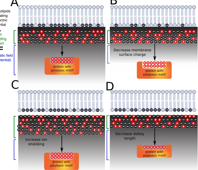
To be entirely clear, membrane surface charge (MSC) (also refered to as surface/electric potential or electrostatic field) and membrane potential are two different concepts. The first one refers to charge distributed within the same surface (cytosolic leaflet). Membrane potential (also refered to as transmembrane potential or membrane voltage) corresponds to the difference in electric potential across the membrane (e.g. between the inner and outer leaflet of the plasma membrane) and is mostly driven by cation pumps. I will discussed how these two concepts are linked (see section V.c) but for the rest of the introduction, I will focus on membrane surface charge of the inner, cytosol-facing, leaflet of membranes.
At the inner membrane leaflet, anionic lipids concentration determine the surface charge of the membrane. However, the effective electric potential (refered as electrostatic field) depends not only on the surface charge density but also on the concentration of counter ions in the solution. Cations (eg. K+ and Na+) are attracted to the charge surface to form a layer of positively charged ions. The consequent recruitment of cations creates the so called ion shielding effect which substantially reduces the apparent membrane surface charge. The zeta potential reflects the apparent electrostatic field (net negative membrane charge minus ion shielding; Figure 3A). The zeta potential has been investigated by physicists and is described by the Gouy-Chapman-Stern theory of the electrical double bilayer. In addition, the net electrostatic effect of a charged surface onto molecules in solution can be quantified by the Debye length, which is inversely proportional to the square root of the ionic strength of the solution. When a protein senses the
zeta potential depending on its own Debey length, the protein will be attracted by the negatively charged membrane and remove the ion shield to interact directly with the membrane (Figure 3B). This theoritical framework is well understood from a physics point of view since the eighties (McLaughlin, 1989), however tools to study these properties in vivo have been developped in the past decade. Bellow, I review how membrane electrostatic properties were dissected in vivo and how these methodological developments led to the discovery of the electrostatic territory and helped to describe how this territory is established and maintained and what are its functions.
b. The membrane surface charge (MSC) i. Principle
While the concept of MSC for protein localization was postulated long ago, tools to sense this predicted feature were only developed during the last decade via the generation of genetically encoded biosensors (sensors/probes; Figure 4A). These biosensors are based on peptides or protein domains that binds to anionic phospholipids (based on their negative charge and irrespective of their head group) and fused to a fluorescent protein. At least ten independent probes have been described to act as sensors of anionic membranes. Each one contains cationic amino acids required to interact with anionic lipids. However, purely electrostatic interactions are not sufficient for membrane binding, which often requires additional hydrophobic interactions. Hydrophobicity can be provided either by a lipid anchor or hydrophobic/aromatic residues. In the following paragraphs, I will discuss the design of some of these “electrostatic” sensors and how they were validated in vitro and in vivo.
ii. Tools to investigate PM surface charge
Peptide-based MSC sensors: The first type of peptide-based MSC sensor was described by Yeung et al., and is composed of a polybasic peptide containing a signal for lipid anchoring. It includes, the N-terminal tail of c-Src (Nt-Src), the C-terminal tail of V-Ki-ras2 Kirsten rat sarcoma viral oncogene homolog (K-Ras tail), the polybasic region of K-Ras containing a signal for myristoylation (myr-K), a mutated version of K-Ras tail to prevent serine phosphorylation (K-pre) and a K-pre version where every lysine is replaced by arginine (R-(K-pre)). These probes are based on the N- or C-terminal tails of small GTPases or kinases. The lipid anchor can be either in N-terminal (e.g. Nt-Src, myr-K-) or C-terminal (e.g. K-Ras tail, K-pre, R-pre) and the polybasic region (PBR) contains from 6 to 8 cationic residues (i.e. either lysines (K) or arginines (R); Figure 4B).
The second peptide-based MSC sensor is composed of a PBR in conjugation with hydrophobic amino acids (W, Y, L, F). This includes, the synthetic sequence (KRf), the C-terminal tail of the small GTPases ric-like in neurons (Rin) and ric-like expressed throughout the organism (Rit) and the myristoylated alanine-rich C-kinase substrate electrostatic-domain (MARCKS-ED)). These peptides form an amphiphathic helix required for membrane interaction (Figure 4B).
Domain-based MSC sensors: The KA1 membrane-associated domain binds acidic phospholipids without discrimination. This is the first domain known to bind unselectively anionic phospholipids. Structure of beta and alpha helix is crucial for binding as well as cationic residues associated with the helices (Figure 4B).
FRET MCS sensor: This sensor was named, MCS+ for membrane charge sensor +, and is based on Förster resonance energy transfer (FRET). This sensor is composed of three main region: a first part (MA1) is a membrane attachment unit (myristoylation and palmitoyltion lipid anchors) that allows the anchoring of the sensor to the plasma membrane (PM) independently of its electrostatic field. A second part (FPs) is made of two fluorescent proteins, mVENUS (a yellow FP variant) and mCHERRY (a red FP variant) to quantify the energy transfer. A third part (MA2) corresponds to the entity which sense the electrostatics field and that is a synthetic PBR region,
which is loosely inspired by the C-terminal tail of K-Ras. The principle is the following: in the case of a low electrostatic field at the PM, the MA2 part will be less associated with the PM and consequently the FRET signal will be decreased due to a higher average distance between the two fluorescent proteins in the FPs part. Inversely, in case of high electrostatic field, the MA2 will be more associated with the PM resulting in the emission of higher FRET signal due to a close proximity of the two fluorescent proteins. This MCS+ sensor has the advantage to be quantitative and more sensitive than the previously described probes (as it can report changes of 10 to 20% of the PM electrostatic field). However, the design of such probe first requires to know which membrane is electronegative (as it required the membrane targeting MA1 anchor). In this case, the PM was first determined to by highly electronegative (thanks to the reporters described above), which then allowed the rational design of the FRET-based reporter. Therefore, these tools are complementary, with the direct reporter binding (peptide and domain based reporters) being important to map membrane electrostatic properties within the cell and quantitative reporters (here FRET-based reporter) to explore more physiological changes within a membrane (note that this remark is valid for the study of the electrosatic field, but also to study other membrane parameter such as local lipid enrichment; Figure 4B).
Membrane integrity sensors: A way to investigate anionic phospholipids participation in MSC is to modulate lipid pools. However, lipids are crucial for membrane organization. Studying membrane surface charge therefore requires a number of controls to verify that lipid modification does not have unwanted side effects on membrane integrity and other non-targeted lipids. Such controls include monitoring the localization of PM proteins that are not targeted to membrane based on electrostatic interactions. These controls will thereafter be referred to as “membrane integrity sensors” and include both integral transmembrane and lipid anchored proteins, that resides in both the raft and non-raft fraction of the PM. For example, the C-terminal tail of N-ras is used as a marker of non-lipid raft portion of the membrane, while glycosylphosphatidylinositol (GPI) and H-ras are used as raft resident proteins and the transmembrane of GT46 protein as a non-diffusive PM protein.
c. The plasma membrane is the most ionic compartment in eukaryotes
Even though the above-mentioned probes have different mechanistic anchoring and present some variation in their net positive charges (from 5 to 13 positive charges), each individual probe interacts in vitro with anionic phospholipids.
When fused to a fluorescent protein, these probes label strictly the plasma membrane in all eukaryotic cell types analyzed including yeast, and animals (Hammond et al., 2012; Heo et al., 2006; Moravcevic et al., 2010; Yeung et al., 2006, 2008) (Figure 5). This common feature highlights a unique signature of the plasmalemma as the most anionic compartment in the cell versus intracellular membranes. However what are the lipids that powers this high PM electrostatic field in different organisms?
d. Anionic phospholipids present in the inner leaflet of cellular membrane i. Phosphatidylinositol phosphates, phosphatidic acid and
phosphatidylserine are anionic lipids presenting different charges and concentration in cellular membrane
Most phospholipids are zwitterionic, meaning that they form a dipole with both positive and negative charges and that their overall charge at physiological cytosolic pH is neutral. These lipids, are often referred to as structural lipids, and they form the bulk of plasma membrane phospholipids (which themselves corresponds to about 30% of total PM lipids), with phosphatidylcholine (PC) and phosphatidylethanolamine (PE) representing up to 50% and 35% of plasma membrane phospholipids, respectively. However, as aforementioned, negative charges at the membrane are carried by anionic phospholipids. By contrast to zwitterionic phopholipids, anionic phospholipids contain a negatively charged head group, the negative charges being notably carried by phosphate groups. From the least to the most anionic phospholipids, we find phosphatidylserine (PS) and phosphatidylinositol (PI) (1 negative charge), phosphatidic acid (PA,
2 negative charges), and phosphoinositides (phosphatidylinositol phosphates or PIPs from 3 to 7 negative charges; Figure 6B). Concerning PIPs, the more the inositol ring is phosphorylated, the more negatively charged the lipid is. Consequently, a phosphatidylinositol 3,4,5,-trisphosphate (PI(3,4,5)P3,overall charge -7) is more charged than a phosphatidylinositol 4,5,-bisphosphate
(PI(4,5)P2, overall charge -5), which is itself more anionic than phosphatidylinositol 4-phosphate
(PI4P, overall charge -3). Anionic lipids contain different negative charges, but their concentration in cell also differ. In human erythrocyte, PIPs represent about 0.1% of total lipids (0.05% of P(4,5)P2, 0.05% of PI4P and less than 0.005% of other PIPs), PI about 1%, PA about
1.5% and PS about 8.5% (Figure 6B). In budding yeast, PA is the most abundant phospholipid representing about 12%. PI is about 3%. PI4P and PI(4,5)P2 are the two most abundant PIPs close to 5% of total PIs (which are approximately 10–20% of total glycerophospholipids) (Payrastre et al., 2001). PS accounts for 1.5% of total lipids. However, PS concentration have a high propensity to fluctuate depending on growth phase and environmental conditions, reaching about 7% in media supplemented with different source of carbon such as glucose (Klose et al., 2012).
Because anionic phospholipids have different net negative charges, but also accumulate at different levels, it is difficult to predict a priori, which lipids will contribute significantly to the membrane electrostatic field. For example, PS is the most abundant anionic lipid in animal cells, but its charge is only -1, while PI(3,4,5)P3 is present at far lower concentration (0.0001% of total
lipids) but is highly electronegative (-7). Which one is more likely to contribute to membrane electrostatics? Given the set of numbers I introduced in the previous paragraph (Figure 6B), one would assume that PS should be a major contributor as compared to PI(3,4,5)P3, since it is
present almost four order of magnitude higher than PIP3. However, these set of numbers may be
deceiving because I presented bulk lipid measurements, which does not take into account the local lipid enrichment and their position in the inner (cytosolic) or outer (luminal/extracellular) membrane leaflets. Indeed, only PS present at the PM inner leaflets is relevant for PM electrostatics, while bulk measurements also include PS molecules that are present in luminal/outer membrane leaflet and organelle membranes (e.g., mitochondria). In addition, while PIP3 molecules are rare, they may be clustered and thereby form patches of highly electrostatic
membranes. To conclude, while the number presented in Figure 6B are informative, they do not bypass the requirement to experimentally analyze the subcellular localization of each individual lipid species and their respective role in membrane electrostatics.
ii. Regulation, turnover and localization of anionic phospholipids in yeast and mammals
1. Phosphatidylinositol (PI) and phosphatidylinositol phosphates (PIPs)
Phosphatidylinositol phosphates (PIPs) possess an inositol ring facing the cytosol that can be phosphorylated and dephosphorylated in position 3, 4 and 5 by appropriate kinases or phosphatases. This property can give rise up to seven PIP species including phosphatidylinositol monophosphate (PI(3)P, PI(4)P, PI(5)P), phosphatidylinositol biphosphate (PI(3,4)P2, PI(3,5)P2,
PI(4,5)P2) and phosphatidylinositol triphosphate PI(3,4,5)P3. PIPs derive from
phosphatidylinositol (PI) that is generated facing the cytosol in the endoplasmic reticulum (ER) for de novo synthesis by phosphatidylinositol synthase (PIS) from CDP-diacylglycerol and L-myo-inositol or by sac1 phosphatase from PI(4)P, in yeast and mammals (A) (Figure 7A) (Bochud and Conzelmann, 2015). The PIS enzyme localizes in the ER and in an ER-derived highly mobile “organelle” that may serve as a dynamic PI distribution device to several organelles (Kim et al., 2011). PI is then distributed throughout the cell presumably by several PI transfer proteins (PITPs) and possibly via vesicular trafficking (Figure 7B). In particular, PI is extracted from the ER at membrane contact sites by PI transfer protein such as secretory protein 14 (sec14) or Pyk2 N-terminal domain-interacting receptor 2 (Nir2) to reach trans-golgi network (TGN) or the plasma membrane, respectively (Bankaitis et al., 2010; Kim et al., 2015). Sec14 exchanges PI from the ER to trans-golgi network and in counterpart exchanges PC located at the TGN to the ER, while Nir2 exchange PI from the ER to the plasma membrane and phosphatidic acid (PA) in the way back (PM -> ER)12,13 (Figure 7C; Bankaitis et al., 2010; Kim et al., 2015). PIPs can be transported through the cellular membrane by regular trafficking such as endocytosis or exocytosis (Balla, 2013). Depending on the location and enzymatic specificity for a given PIP
species, kinases and phosphatases (Mayinger, 2012) generate the large range of PIPs in different subcellular compartments (e.g. plasma membrane, early endosomes, trans-golgi network, Golgi, late endosomes and lysosomes/tonoplast). The enrichment of a PIP in a given subcellular compartment is used as a landmark for protein targeting and signaling (Platre and Jaillais, 2017) (Figure 8). The spatiotemporal PIPs dynamics is highly regulated by phosphatases and kinases and their constant interconversion confers a high potential for phosphoinositides to be involved in the regulation of membrane surface charge.
2. Phosphatidylserine
In mammals, PS is produced by two enzymes: PS synthase1 (PSS1) and PSS2. These two genes encode exchange type enzymes that generate PS by exchanging the choline or ethanolamine head group from PC or PE with a serine. PSS1 uses preferentialy PC as a substrate, while PSS2 uses preferentialy PE. While PSS1 carries the major PS enzymatic activity in cells, accounting for 60 to 70% of PS production, both enzymes are redundant and the corresponding double mutant is embryonic lethal (Sousa et al., 2013) (Figure 9B). PSS enzymes are integral transmembrane protein that localize in the ER and produce PS in the luminal leaflet (Figure 9B). Based on immunogold labelling in mammals, PS distribution differs not only among organelles but frequently also between the two leaflets of the membrane suggesting regulations by “flip-flop” mechanisms(Hankins et al., 2015). Flippases are aminophospholipid translocases that are able to transport PS, from the extracellular or the luminal leaflet of an organelle to the cytosolic side and are localized at the TGN, early endosomes and plasma membrane. Unlike flippases, which transport lipids unidirectionally, plasma membrane localized-scramblases are bidirectional and function to randomize or at least reduce the asymmetry of phospholipids in membranes and are particularly active during apoptosis and blood clotting (Hankins et al., 2015) (Figure 9C). Historically, PS was thought to follow a secretion route from the ER to the Golgi/TGN and then the PM (Figure 8). In this scenario, specific lipid flippase would flip PS either at the trans-face of the Golgi or at the PM from the outer to the inner membrane leaflet (Hankins et al., 2015). While such pathway might account in part for the cellular distribution of PS, recent work revealed that
the major pathway to bring PS at the PM is through lipid transfer at ER/PM contact sites (Chung et al., 2015; Filseck et al., 2015; Maeda et al., 2013; Sohn et al., 2016). In this later model, it is probable that PS is flipped directly at the ER before being transferred to the PM. In counterpart, PI4P is transported back to the ER from the cell surface to be degraded into PI by a phosphatidylinositol 4-phosphatase (sac1). This process links PI4P and PS metabolism in regulating its concentration to the cell surface (Figure 9D). However, the exact nature of the flippase involved is still unkown. PS production is finely tuned since PSS proteins are inhibited by their own product. This negative feedback regulation is critical for PSS1 activity and normal development. A rare genetic disease named Lenz Majewski Syndrome (LMS) is caused by gain of function mutations in PSS1 that alleviates PS-feedback inhibition of PSS1 activity. LMS syndrome is characterized by osteosclerosis, intellectual disability, characteristic facies and distinct craniofacial, dental, cutaneous and distal-limb anomalies(Sousa et al., 2013). In addition, removal of PSS1 autoinhibition alters PI4P spatial organization. In yeast, PS degradation is required to yield phosphatidylethanolamine. This reaction is catalyzed by two PS decarboxylases (PSD1 and PSD2), which are localized in the mitochondria and Golgi complex/vacuole membranes, respectively. A single gene, PSD1 has been reported in mammals and localized to the mitochondrial membrane. But, PS can also be hydrolyzed by two phospholipases (phospholipase A1 and A2) located in the plasmalemma (Leventis and Grinstein, 2010). PS high abundance and its concentration regulation between inner and outer leaflet by flippases argue for au plausible role of PS in the maintenance of the intracellular electrostatic field.
3. Phosphatidic acid
Phosphatidic acid (PA) is a backbone lipid since it is an essential substrate for enzymes participating in the synthesis of phospholipids and triacylglycerol (TAG; Figure 10A). Phospholipids generation from PA involves CDP-diacylglycerol synthase (CDS) while TAG involves PA phosphatases (PAP) enzymes (Athenstaedt and Daum, 1999). The de novo synthesis of PA is catalyzed by two different pathways corresponding to the Gro3P (glycerol 3-phosphate) pathway, and the GrnP (dihydroxyacetone phosphate) pathway. Two other pathways are involved
in PA production. The first one, uses phospholipids as substrat through the action of phospholipase D (PLD) and the second one diacylglycerol through the activity of diacyglycerol kinases (DGKs) that is generated from phospholipids by Phospholipase C (PLC) or triacylglycerol by triacyglycerol lipase (TAGL) (Figure 10B). Based on the subcellular localization of enzymes involved in PA biosynthesis, PA is thought to be present in mitochondria, ER and PM but also in lipid droplet in yeast and peroxisomes in mammals (Hermansson et al., 2011). Genetically-encoded biosensor sensing PA reveals that most cells do not accumulate significant level of free PA in the cytosolic leaflet of the plasma membrane (Bohdanowicz et al., 2013) (Figure 8). However, PA is acutely produced by PLD in response to receptor kinase activation (Zhang et al., 2014). In addition, it is constitutively produced at significant level (i.e. sufficient to trigger the constitutive PM association of PA sensors) in phagocytic cells (e.g. Macrophages and dentritic cells) by PM localized DGK enzymes (Bohdanowicz et al., 2013). As mentioned previously, PA is exchanged at membrane contact sites by Nir2, which extracts PI from the ER to the PM and transports back to ER PM-associated PA. PA is generated from diacylglycerol (DAG), itself generated by PLC activity, which hydrolyzes PI(4,5)P2 at the plasma membrane. In this case, PA synthesis depends on PI(4,5)P2
and PLC activity at the PM. However, PI(4,5)P2 production requires PI(4)P that itself required PI
synthesis. The production of PI occurs in the ER and depends on CDP-diacylglycerol, which is generated from PA by CDP-diacylglycerol synthase. To sum up, we have a kind of “schizophrenic” system, where PA is localized at the PM and requires ER-generated PI and conversely PI synthesis in the ER depends on PA production which is localized at the plasma membrane. Nir2 lipid transfer protein play a central role in this lipid synthesis and homeostasis as an ER-PM lipid exchanger (Figure 7C) (Kim et al., 2015). PA regulation is complex as it is a highly dynamic phospholipids, which can be produced by many different pathways in different compartments. Although I highlighted earlier the regulation of PS localization in inner and outer membrane leaflets by lipid scramblase and flippase, it worth noting that the presence (and regulation) of PA in inner vs outer leaflets in not well documented and is probably important for its activity and availability.
PA is an anionic phospholipid (net charges -2) present at the PM. However, to my knowledge, its potential role in the establishment/maintenance of the PM electrostatic field has not been explored in yeast and animals. I will therefore describe below how the role of PIPs and PS in
membrane electrostatics has been studied but will not discuss further the potential role of PA. However, I believe this could be an interesting avenue of future research, notably in yeast and mammalian cells with active phagocytic activities.
II. Anionic lipids in the maintenance of the PM electrostatic field in mammals a. Anionic lipids are required to maintain the plasma membrane electrostatic
field
The first study to analyze the role of anionic lipids in MSC-establishment was published by the group of Sergio Grinstein in 2006 (Yeung et al., 2006). In this seminal paper, Yeung et al., described and validated the first set of MSC-reporters and showed that the cytosolic leaflet of the PM is highly electrostatics. They then perturbed anionic phospholipid pools using pharmacological approaches to determine their roles for the generation of the PM electrostatic field.
Ionomycin elevates cytosolic calcium, which induces PI(4,5)P2 hydrolysis through activation of
phospholipase C (PLC). At the same time, the increase of cytosolic calcium activates lipid scramblase, which results in the translocation of PS from the inner (cytosolic facing) leaflet of the PM to the outer leaflet. Therefore, ionomycin induces the concomitant loss of PI(4,5)P2 and PS in
the inner PM leaflet. Ionomycin treatment delocalized the cationic probes (K-pre, Krphy, K-myr), while membrane integrity sensors (GPI, GT46 and Palmitoylation) were not affected (Figure 11A). A more recent study (Ma et al., 2017) using the MCS+ based-FRET sensor verified this observation. Indeed, the concomitant depletion of PI(4,5)P2 and PS following ionomycin
treatment induced a decrease of the FRET signal Figure 11B). Dibucaine promotes PS flipping from the inner to the outer leaflet independently of PIPs metabolism and induced a delocalization of cationic probes into the cytosol and a decrease FRET-ratio of the MSC+ probe. In addition, the drug fendiline sollubilizes PS and impacts PM MSC (Ma et al., 2017) (Figure 11A-B). Taken together, these results suggest that a decrease of PI(4,5)P2 and PS concentration at the PM inner
However, these results must be taken with some care given the side effects of the chemical compounds used. Indeed, ionomycin induces a massive calcium entry into the cell, which might contribute in part to the release of cationic probes from the PM by increasing ion shiedling. In addition, dibucaine is cationic, and therefore its own positive charges could participate in displacing cationic probes from the membrane. Moreover, this experiment lacks control on other PM-associated anionic phospholipids such as PI4P, PI(3,4)P2, and PI(3,4,5)P3. Finaly, fendiline
inhibits acid sphyngomyelinase (ASM), which decreases the level of ceramide and increase those of sphyngomyelin, leading to a depletion of PM cholesterol and PS (Cho et al., 2015),(van der Hoeven et al., 2013). Why variations of ceramide/sphyngomyelin impact PS biosynthesis is unknown and suggest an indirect mode of action.
b. Involvement of PIPs in the plasma membrane surface charge
The drugs mentioned above are expected to have pleiotropic effects. It is therefore impossible to exclude that they might induce a large-scale remodeling of global cell physiology and membrane lipids thereby affecting the localization of MSC sensors. In parallele to pharmacological approaches, a number of genetic strategies were implemented to modify anionic lipid pools. Most of these tools are based on the targeting of lipid phosphatase activities at the PM. For example, overexpression of Inp54p, a 5-specific phosphatidylinositol 4,5-bisphosphate phosphatase (5-phosphatase) induced a significant reduction in the FRET efficiency of the MCS+ reporter. However, in this case, Inp54 is constitutively overexpressed, leading to chronic PI(4,5)P2
depletion. Such chronic depletion may also have side effects and therefore it is difficult to pin point the change in the FRET efficiency of the MCS+ probe to the sole depletion of PI(4,5)P2
(Ma et al., 2017).
To overcome these drawbacks, (Heo et al. 2006), designed an elegant method based on the inducible recruitment of phosphoinositide phosphatases at the PM. This inducible phosphatase recruitment is built on genetically encoded PM-localized FK506-binding protein (FKBP12)-rapamycin-binding (FRB) construct and a cytosolic inositol polyphosphate 5-phosphase (Inp54p)
enzyme conjugated with FKBP12 (CF-Inp; Figure 12). Rapamycin treatment induces CF-Inp translocation from the cytosol to the PM by chemical heterodimerization that triggers Inp54p activity at the PM and thereby inducible and rapid (i.e. minutes) depletion of PI(4,5)P2
specifically to this membrane. By contrast to chronic depletion of PI(4,5)P2, rapamycin-induced PM-Inp54p recruitment did not affect cationic probes localization (MARCKS-ED, Rin and Rit tail; Figure 13A-B). This result suggests that PI(4,5)P2 is not required, by itself, for MSC and
that another anionic phospholipid(s) might act redundantly with PI(4,5)P2 to control PM MSC.
A limitation of the approach from Heo et al., was that dephosphorylation of PI(4,5)P2 by Inp54p
induces the production of PI4P, another PM phosphoinositide which is also anionic, albeit to a lesses extent (roughly -3 vs -5 net charges, for PI4P and PI(4,5)P2, respectively). This could
explain the absence of effect of PI(4,5)P2 depletion on cationic probes, since only part of the
charges carried by PI(4,5)P2 are depleted with this technique. Hammond et al., in 2012 found that
PM PI4P is not only a precursor of PI(4,5)P2 biosynthesis but also an important regulator of PM
identity. Gerry Hammond indeed built a rapamycin–triggered system, which allows the inducible recruitment at the PM of a chimeric synthetic enzyme, composed of 4- and 5-phosphatase catalytic activities. He named this enzyme “pseudojanine” by analogy to the protein synaptojanin, which naturaly carries 4- and phosphatase catalytic activities. The 4- and 5-phosphatases catalytic domains of pseudojanin came from the yeast Sac1p and human INPP5E proteins, respectively. Point mutations within the Sac1p or INPP5E catalytic domains shut down either the 4-phosphatase or 5-phosphatase activity or both and are used as a control. Depending on the mutations, this system allows altering either PI4P, PI(4,5)P2, both, or none (when both
phosphatase domains are mutated). Using an optimized immunolocalization protocol (Hammond et al., 2009), they validated their system and showed that the rapamycin-inducible PM-targeted 5-phosphatase had no effect on PI4P, but depleted PI(4,5)P2 from the PM. Similarly, decreasing the
PM-PI4P pool by PM-targeted 4-phosphatase had no effect on the PM PI(4,5)P2 abundance. This
unexpected observation suggested a relative independence of the PI4P and PI(4,5)P2 pools at the
PM, even though PI(4,5)P2 is made from PI4P. Depletion of either PI4P or PI(4,5)P2 had no
effect on the PM targeting of various MSC probes (including Kras-tail, MARCKS-ED, Rit tail and the KA1 domain; Figure 13A-B). Therefore, PI4P and PI(4,5)P2 are not required (on their
own) to maintain PM surface charge. However, the concomitent depletion of both PI4P and PI(4,5)P altered the PM localization of MSC reporters but did not affect membrane integrity
probes.
PI(3,4,5)P3 is a highly anionic phospholipid (net charge -7) localized at the PM. It is present in
minute amount but its synthesis is acutely induced upon growth factor stimulation. Heo et al. reported that the stimulation of PI3Kinase activity at the PM increased PI(3,4,5)P3 PM level, with
a concomitant recruitment of the cationic probe Rin tail at this membrane. This suggested that the PM-associated PI(3,4,5)P3 could be involved in PM MSC. The subcellular localization of cationic
probes and the FRET signal of the MCS+ reporter were only slightly affected by LY294002 or wortmanin treatment, two pharmacological inhibitors which prevent PI(3,4,5)P3 production by
inhbiting PI3-Kinases. Therefore, similar to PI(4,5)P2, PI(3,4,5)P3 might also control PM MSC
together with other anionic lipids. Application of PI3-kinase inhibitors coupled to PM-inducible recruitment of a 5-phosphatase (Inp54) concomitantly decreased PI(4,5)P2 and PI(3,4,5)P3 levels
and induced the PM-dissociation of cationic probes (MARCKS-ED, Rin and Rit tail) but not that of membrane integrity sensors (Figure 13C-D). This indicated that PI(4,5)P2 and PI(3,4,5)P3 also
act redundantly to regulate PM MSC. Altogether, PI(4,5)P2 seems to be critical to define PM
membrane surface charge in human cells but is not sufficient and acts redundantly with PI(3,4,5)P3 and/or PI4P.
c. Evidence that PS contributes to the PM electrostatic field
PIPs are highly anionic but represent only about 1% of total phospholipids in living cells. Other less anionic lipids might also contribute to MSC, notably due to higher abundance. In animals, PS represents about 10 to 20% of PM-phospholipids but PS is less anionic than phosphoinositides (net charge -1). The pharmacological experiments described above from Yeung et al., (2006) (part II.A.) suggested that PS might take part in PM surface charge. However, the exact distribution of intracellular PS was unknown due to the lack of appropriate tools. Two years later7, Yeung et al., (2008) investigated the role of PS in MSC by setting up a specific intracellular PS probe. By contrast to the C2 domain of annexin-V, the C2 domain of bovine lactadherin synthase (C2LACT) binds selectively to PS independently of calcium and can be used
as a genetically encoded PS sensor when fused to a fluorescent protein. Based on co-localization analyses and immunogold electron-mycroscopy, PS was found at the plasma membrane, endosomes and lysosomes and more enriched at the PM than in late endosomal compartments (Yeung et al., 2008;Fairn et al., 2011a) (Figure 14A).
To investigate the relative role of PS compared to PIPs in PM electrostatic field maintenance the authors inhibited ATP synthesis by using a concomitant treatment of antimycin and deoxyglucose. This treatment concomitantly inhibits mitochondrial respiration and glycolysis, thereby depleting cellular ATP. The rational of this experiment is the following: among other effects, ATP depletion in the cell should trigger the rapid depletion in phosphoinositides because in the absence of ATP, lipid kinases are not making any new PIPs, while lipid phosphatases are still constantly dephosphorylating these lipids. However, PS maintenance does not require a kinase and the PS pool should not be affected, at least under short-term treatment with antimycin. To verify this assumption Yeung et al., monitored the localization of PI(4,5)P2 and PI(3,4,5)P3
probes and that of their newly described PS sensor (C2LACT) following antimycin treatment. Antimycin depleted PH-PLC and PH-AKT probes from the PM, but had no effect on C2LACT, suggesting a decrease of cellular PIPs but not PS. Next, they examined the localization of the MSC probe K-Ras tail and found that following antimycin treatment, it was delocalized in intracellular compartments, although a portion was still associated with the PM (Figure 14B). This experiment showed that in the absence of PIP, the PM looses its electrostatic signature, which confirms the importance of phosphoinositides in this particular PM property (as described by Heo et al., (2006) and Hammond et al., (2012)). However, a portion of the K-Ras tail probe was retained at the PM in the absence of phosphoinositides, suggesting that other anionic lipids, likely PS, are involved in this PM electrostatics. Next, Yeung et al., investigated, in which cellular compartment K-Ras tail MCS sensors relocalized in the absence of PIPs (i.e. following antimycin treatment). In this condition, K-Ras tail resides almost exclusively in PS containing organelles (as labeled by C2LACT, i.e. endosomes and lysosomes), showing a near perfect correlation between the putative presence of PS and negative charges (Figure 14B). Together, these results argue that (1), that PIPs are required for PM electrostatic field and (2) that PS might contribute to MSC both at the PM but also along the endocytic pathway (point discussed below). Altogether, Yeung et al., suggested that PS is a regulator of the PM electrostatic field. However,
due to limitations in the pharmacological approaches described in the previous paragraph and the lack of direct evidence, it is still not fully demonstrated that PS indeed participate in PM electrostatics in mamalian cells. Loss of function experiments that would consist of depleting the PS cellular content and analyze its effect on MSC has not been conducted to my knowledge.
III. Anionic lipids in the maintenance of the PM electrostatics field in yeast
By contrast to mammals, yeast has a single PS synthase gene, called cho1. Cho1p has a different catalytic activity than the mamalian PSS1/PSS2 enzymes as it produces PS via a CDP-diacylglycerol:l-serine O-phosphatidyltransferase activity (Figure 15A). In laboratory conditions, when grown in rich medium, Cho1p is not critical for yeast viability. However, biochemically, the cho1 mutant does not contain PS and the PS sensor C2LACT becomes cytosolic when expressed in cho1. While the catalytic activity of Cho1p is different than PSS1/PSS2, there are extensive paralellism between PS synthesis in yeast and animals: PS is produced in the ER and then transferred to the PM at membrane contact sites by evolutionary conserved proteins19,20 (Maeda et al., 2013; Moser von Filseck et al., 2015). However, the site of PS subcellular accumulation are different in yeast and animals. In mammals, C2LACT localizes at the PM but also in PM-derived organelles along the endocytic pathway. In yeast however, C2LACT is exclusively localized at the PM and virtualy no intracellular compartments are labelled by this probe (Yeung et al., 2008; Moravcevic et al., 2010; Fairn et al., 2011b; Filseck et al., 2015; Maeda et al., 2013) (Figure 15B). This suggests that by contrast to animal cells, PS is predominantly accumulated at the PM in yeast, and therefore might have a predominant role for PM electrostatics.
The K-Ras based MSC probe is not restricted to the PM in S. cerevisiae. This prevented Yeung et al., to obtain direct comparison of MSC reporter localization in WT vs cho1 mutant. However, in 2010, Moravcevic et al. identified a new MSC reporter by characterizing the Kinase associated 1 (KA1) domain. KA1 domains have been identified in both yeast and mammalian proteins involved in kinases regulation. Biochemical assay, crystallography and in vivo experiments define the KA1 domain as a membrane-associated domain that binds all acidic phospholipids,
regardless of their respective head group. Similarly, to the PS probe C2LACT, KA1 domains localized strictly to the plasmalemma in yeast. Next, the authors addressed, whether PS, PIP or both participate in KA1 domains localization. In cho1, KA1 lost its specific PM localization (i.e. it became soluble and associated with a much broader membrane domain including both PM and intracellular compartments; Figure 15C). This results suggested a major contribution of PS in setting up PM surface charges in yeast. Thermo-sensitive mutations in the genes encoding the PI4-kinases that generate PI4P at the plasma membrane (Stt4p) and Golgi (Pik1p) deplete PI4P from these mutants at restrictive temperature. In the other hand, thermo-sensitive mutation in the only gene that codes for PI4P-5-kinase (Mss4p) inhibits PI(4,5)P2 production. Mss4p mutant is
depleted of PI(4,5)P2 but not PI4P at restrictive temperature, while the double mutant stt4p;pik1p
(that lack a PI4-kinase), lack both PI4P and PI(4,5)P2. Surprisingly, KA1 domains remains
strictly localized at the PM in all these yeast mutant strains (Figure 15D), suggesting that unlike in animals, PIPs do not play a major role in PM MSC and that PS is the major anionic lipids of the yeast plasmalemma inner leaflet (importantly, the yeast S. cerevisiae does not produce any PI(3,4)P2 and PI(3,4,5)P3, making PI4P and PI(4,5)P2 the only phosphoinositide at the cell
surface). Altogether, these results suggest either no or minor role of PIPs in plasmalemma surface charge, while PS is the main anionic lipid driving the PM electrostatic potential in yeast.
IV. Membrane surface charges defines an electrostatic territory corresponding to PM-derived organelles
a. In mammals, endocytic compartments are electrostatic.
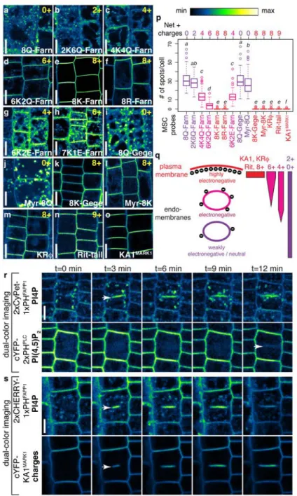
With the idea that not only the PM is an anionic membrane, Yeung et al., altered the strength of electrostatic associations (decrease of the Debey length) by changing the charges of the PBR of MSC reporters. To this end, they mutated the K-Ras tail by substituting its charged amino acids (i.e. Lysines) into neutral residues (i.e. Glutamine) to decrease its net positive charge. They obtained a set of probes containing from 0 to 8 positive charges called 0+ to 8+ (Figure 16A). In macrophages, 0+ sensor reports an intracellular localization that corresponds to the default
localization driven by the lipid anchor. However, the 8+ sensor reports PM-labeling as previously described. Intermediate charge sensors have dual-localization, at the PM and in other intracellular compartments. Therefore, the more cationic the sensor is, the more it reports PM-labeling and less an intracellular labeling. Conversely, neutral sensors are less localized at the PM and more in intracellular membranes. Therefore, sensors with intermediate charges (e.g. 4+) report the existence of intracellular compartments of intermediate charges, that are not as electronegative as the PM but that are not neutral either. Colocalization studies suggested that these compartment of intermediate electronegativity corresponds to endosomes (including early and late endosomes, as well as lysosomes; Figure 16B). In addition, MSC sensors relocalized to PS containing organelles in the absence of PIPs (i.e. following antimycin/deoxyglucose treatment), suggesting that PS is important for the electrostatic properties of endosomes.
While it is likely that PS contributes to the overall charge of PM-derived organelles in animals, it is worth noting that this conclusion is essentially based on correlations (i.e. colocalization between a PS sensor and a charge sensor in the absence of PIP) observed in antimycin treated cells. Depletion in cellular ATP is expected to have a myriad of effects on cell physiology, including stopping of all intracellular trafficking, kinases reactions and membrane potential. All this effects could also affect the localization of MSC in antimycin treated cells, independent of PS localization. Again, it would be interesting in the future to analyze the localization of MSC reporters in PS depleted cells.
b. Membrane electrostatic of intracellular compartments in yeast
In the yeast Saccharomyces cerevisiea, the C2LACT sensor is strictly localized at the PM, suggesting that the cell surface massively accumulates PS at the expense of its intracellular localization (Yeung et al. 2008; Filseck et al., 2015;Fairn et al., 2011a;Moravcevic et al., 2010). While this is an excellent argument for PS as a driver of PM electrostatics in this yeast species, it does not argue in favor of PS being important for intracellular compartments electrostatics. In the litterature, only one paper reported PS localization in intracellular compartments (Xu et al.,
2013). In this paper, Xu et al., used the C2LACT reporter and were able to detect it at the surface of the TGN. The reason for the discrepencies between the usualy reported localization of C2LACT in yeast and this study are unclear.
Xu et al., investigated the role of PS in defining the electrostatic field of the TGN (Xu et al., 2013). They identified a motif corresponding to an amphiphatic lipid packing sensor (ALPS) that are positively charge (+ALPS) in an ArfGAP protein (Gcs1). ALPS motifs are able to sense lipid packing defects that are present (notably) in highly curved membranes and are composed of an hydrophobic and of an hydrophilic face (Figure 17A-B). Because ALPS motifs are able to senses curved membranes (diameter 50nm), theirs localizations are restricted to the cis-golgi. Historically, the ALPS motif has been identified in the protein ArfGAP1 and is responsible for ArfGAP1 targetting and function in the cis-golgi for vesicular sorting. Colocalization analysis between the +ALPS motif and trans-golgi network (TGN; Tlg1 or Sec7) clearly demonstrates its localization beyond the Golgi. Mutations in the +ALPS positively charged amino acids restrict its localization to the cis-golgi in vivo (Figure 17C). In vitro experiments showed that +ALPS and mutated +ALPS bind more and less PS-containing liposomes, respectively; this result implied that charged amino acids could drive the +ALPS motif out of the Golgi. The authors speculated that the extended localization to the TGN of the +ALPS motif may be due to negative surface charges of this compartment and argued that the electrostatic territory is not limited to the PM in yeast. Consistent with this hypothesis, in their hand, C2LACT colocalized with the +ALPS motif which itself colocalized with TGN markers (Tgl1 and sec7). In addition, the localization of Gcs1P +ALPS motif was significantly affected in the cho1Δ mutant.
Drs2 is a P4-ATPase that is flipping PS from the luminal to the cytosolic leaflet of the TGN. In Drs2 thermo-sensitive mutant at restrictive temperature, the +ALPS localization was significantly dispersed throughout the cytoplasm (Figure 17D). Therefore, the authors proposed that Drs2-dependent PS flipping at the TGN induces negative charges on the surface of this compartment. This mechanism allows the specific recruitment of +ALPS motif containing ARF-GAP to the TGN, as they are both highly curved and electrostatic membranes. Because, the localization of the +ALPS motifs was unchanged in vps34 and fab1 mutants, which are impaired in PI3P and PI(3,5)P2 synthesis, respectively, it is likely that intracellular phosphoinositides play a minor