Christoph A. Karlo Paul Stolzmann Sandra Habernig Lukas Müller Traudel Saurenmann Christian J. Kellenberger Received: 12 February 2010 Revised: 25 March 2010 Accepted: 7 May 2010 Published online: 18 June 2010 # European Society of Radiology 2010
Size, shape and age-related changes
of the mandibular condyle during childhood
Abstract ObjectiveTo determine age-related differences in the size and shape of the mandibular condyle in children to establish anatomical reference values. MethodsA total of 420 mandibular condyles in 210 children (mean age, 7years) were retrospectively analysed by using computed tomography (CT) imaging. The greatest left–right (LRD) and anterior–posterior (APD) diameters and the anteversion angles (AA) were measured by two readers. An APD/ LRD ratio was calculated. The shape of the condyles was graded into three types on sagittal images. Correlations of parameters with the children’s age were assessed by using Pearson’s correlation analyses.ResultsThe LRD (mean, 14.1±2.4mm), APD (mean, 7.3±1.0mm) and LRD/APD ratio (mean, 1.9±0.3) increased (rLRD=
0.70, p<0.01; rAPD=0.56,
p<0.01; rrat=0.28, p<0.01) while the
AA (mean, 27±7°) decreased signi
fi-cantly (rantang=−0.26, p<0.001) with age. The condylar shape as deter-mined on sagittal images correlated significantly with age (r=0.69, p< 0.05). Boys had significantly higher anteversion angles (p<0.01), greater LRDs (p<0.05) and greater mean ratios (p<0.05).ConclusionThe mandibular condyle is subject to significant age-related changes in size and shape during childhood. As the size of the condyles increases with age, the anteversion angles decrease and the shape of the condyle turns from round to oval.
Keywords Temporomandibular joint . Computed tomography . Paediatric . TMJ . Mandibular condyle
Introduction
The temporomandibular joint (TMJ) is frequently subject to inflammatory disorders in children especially within the disease pattern of juvenile idiopathic arthritis (JIA) [1–9]. Arthritis of the TMJ, if not detected early and treated properly, may lead to bone destruction and osseous deformation of the mandibular condyle resulting in growth disturbances and dysmorphic facial features [2, 10–12]. Further on osseous deformations lead to an earlier onset of TMJ arthrosis. In order to detect abnormalities of the shape and size of the mandibular condyles in children of
all ages it is necessary to be familiar with the normal appearance of the mandibular condyle in cross-sectional diagnostic imaging. However, to the best of our knowl-edge, only few data can be found on the normal size of the mandibular condyle in recent literature [13,14] evaluating symptomatic TMJ disorders or including not only children but also adults in their study populations.
Therefore the purpose of our study was to evaluate the size, shape, gender- and age-related changes of the mandibular condyle in asymptomatic children in a retro-spective analysis of multidetector computed tomography (CT) examinations.
C. A. Karlo
:
S. Habernig:
C. J. KellenbergerDepartment of Diagnostic Imaging, University Children’s Hospital Zurich, Zurich, Switzerland
C. A. Karlo ())
:
P. StolzmannInstitute of Diagnostic and Interventional Radiology,
University Hospital Zurich,
Raemistrasse 100, 8091 Zurich, Switzerland e-mail: [email protected]
Tel.: +41-44-2552900 Fax: +41-44-2554443 L. Müller
Clinics for Orthodontics and Paediatric Dentistry,
University of Zurich, Zurich, Switzerland T. Saurenmann
Department of Rheumatology, University Children’s Hospital Zurich, Zurich, Switzerland
Materials and methods
Patient population
A total of 210 children (mean age, 7 years; range, 0– 17 years; 81 girls, 129 boys) were included in this cross-sectional study. The children underwent computed tomog-raphy (CT) of either the skull base (n=20, 9.5%), the cervical spine (n=30, 14.3%) or the head (n=160, 76.2%) between December 2008 and August 2009. None of the children included in this study underwent CT imaging for known or suspected disorders of the TMJ. The medical history of all children was reviewed and revealed that none of them was affected by rheumatological diseases or TMJ disorders. Indications for CT imaging included suspected intracranial bleeding or fractures after trauma to the head or cervical spine (n=96, 45.7%), suspected non-traumatic cerebral haemorrhage (n=29, 13.8%), eval-uation for sinusitis or assessment of anatomical variants of the nasal cavity (n=38, 18.1%), follow-up or initial evaluations of hydrocephalus (n=31, 14.8%), suspected cerebral sinus venous thrombosis (n = 4, 1.9%) and complicated mastoiditis (n=12, 5.7%).
All patients or their legal guardians gave consent consisting in a general waiver which allows the use of data from the hospital chart for retrospective scientific analysis. This approach is approved by the Institutional Ethics Review Board.
Computed tomography imaging
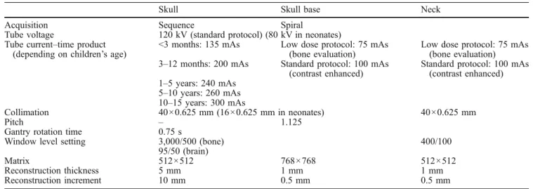
All CT examinations were performed on a commer-cially available 40-detector-row CT system (Brilliance CT 40, Philips Medical, Eindhoven, the Netherlands) with all children in the supine position, head first and with their mouth closed. For the CT protocols applied see Table 1.
Image analysis
Image analysis was performed upon multi-planar recon-structions (MPR) in transverse and sagittal imaging planes derived from axial-source CT images with a reconstruc-tion slice thickness of 1.0 mm and a reconstrucreconstruc-tion increment of 0.5 mm.
Two radiologists (R1 and R2 with 4 and 3 years experience, respectively, of musculoskeletal and paediatric cross-sectional image interpretation) performed all image analysis on a commercially available post-processing software tool connected to the CT workstation (Philips Medical, Eindhoven, Netherlands).
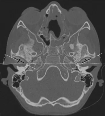
At the level of the greatest left–right diameter of the mandibular condyles in the transverse imaging plane the greatest left-to-right diameter (LRD), the greatest anterior-to-posterior diameter (APD) and the anteversion angle (AA) of the mandibular condyles were measured (Fig. 1) separately for the left and right sides using an electronic calliper tool. The shape of the mandibular condyle was evaluated on sagittal imaging planes tilted to the long axis of the mandibular ramus and classified into three types (Fig.2), which were defined by a panel of paediatric and musculoskeletal radiologists. In cases of disagreement, consensus reading was appended after 1 week. An APD/ LRD ratio (RAPD/LRD) was calculated to classify the shape
of the condyles in the transverse imaging plane into roundish or oval. A screen shot of all measurements was archived into the hospital’s PACS (picture archive and communication system) for documentary purposes.
Statistical analysis
All quantitative variables are described as mean ± stand-ard deviation. The data were descriptively reviewed and statistically analysed by using the Kolmogorov–Smirnov’s test for normality.
Table 1 Imaging parameters for the acquisition of CT examinations of the skull, the skull base and the neck
Skull Skull base Neck
Acquisition Sequence Spiral
Tube voltage 120 kV (standard protocol) (80 kV in neonates) Tube current–time product
(depending on children’s age)
<3 months: 135 mAs Low dose protocol: 75 mAs (bone evaluation)
Low dose protocol: 75 mAs (bone evaluation) 3–12 months: 200 mAs Standard protocol: 100 mAs
(contrast enhanced)
Standard protocol: 100 mAs (contrast enhanced) 1–5 years: 240 mAs 5–10 years: 260 mAs 10–15 years: 300 mAs Collimation 40×0.625 mm (16×0.625 mm in neonates) 40×0.625 mm Pitch – 1.125
Gantry rotation time 0.75 s
Window level setting 3,000/500 (bone) 400/100
95/50 (brain)
Matrix 512×512 768×768 512×512
Reconstruction thickness 5 mm 1 mm 1 mm
The interobserver agreements for continuous variables (i.e. the LRD, APD and AA) were assessed according to the method of Bland and Altman and determined as the mean differences (bias) that are presented with corre-sponding limits of agreement.
Correlation between both readers’ measurements was assessed by Pearson’s correlation analysis. The
interob-server agreement for the ordinal variable of condylar type (i.e. types I, II or III) was assessed by using k statistics and interpreted as follows: a k value greater than 0.81 corresponded to excellent agreement, a k value of 0.61 to 0.80 corresponded to very good interobserver agree-ment, a k value of 0.41 to 0.60 corresponded to good interobserver agreement, and a k value of 0.21 to 0.40 corresponded to moderate interobserver agreement.
Student’s t test for independent samples was used to test for gender-specific differences between measurements of boys and girls.
Correlation between children’s age and LRD, APD and AA measurements was assessed by using Pearson’s correlation analysis. Results were demonstrated on scatter plots using cubic fitting. Correlation between children’s age and condylar types on sagittal image reconstructions was assessed by using Spearman’s correlation analysis. P values less than 0.05 were considered statistically signi fi-cant. All statistical analyses were performed using commercially available software (SPSS, release 17.0, Chicago, IL, USA).
Results
Interobserver agreements of CT measurements
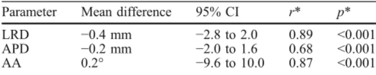
Bland–Altman analysis for testing the degree of agree-ment between the two readers revealed minimal mean differences, and all measurements were within close limits of agreement for all parameters. Correlation coefficients ranged from 0.68 to 0.89, as shown in Table 2. Because of excellent agreement regarding CT measurements, the means of both readers’ measure-ments were taken for further analyses. Very good interobserver agreement was found for grading the condylar types with a k value of 0.67.
Fig. 1 Transverse computed tomography imaging plane showing the left-to-right (LRD, longer black line) and anterior-to-posterior (APD, shorter black line) diameters as well as the anteversion angle (AA) measurements (white dotted lines) of the mandibular condyles
Fig. 2 Reformatted sagittal computed tomography images demon-strating the three different types of condylar shape as described in our study: type I (left) showing a smooth, round shape, which is most frequently seen in children aged 0–5 years; type II (centre)
shows the beginning of the development of an anterior beak, which is mostly seen in type III shapes (right) together with aflattening of the condyle’s anterior surface which can primarily be observed in children aged 10 and older
Condylar measurements and types
No gender-specific differences were found regarding anterior-to-posterior measurements. However, boys had significantly higher anteversion angles (p<0.01), greater left-to-right diameters (p < 0.05) and greater mean RAPD/LRD(p<0.05) compared to girls.
Statistically significant correlations were found between all measurements and the children’s age. The LRD (rLRD=
0.70, p < 0.01), APD (rAPD= 0.56, p < 0.01) and the
RAPD/LRD (rrat= 0.28, p < 0.01) increased significantly
while the anteversion angle decreased (rantang=−0.26,
p<0.001) with increasing age (Fig.3).
The shape of the condyles on sagittal imaging planes was graded into three types by both readers (frequencies; R1/R2):
&
Type I: R1, n=48 (11.5%); R2, n=52 (12.3%)&
Type II: R1, n=156 (37.1%); R2, n=152 (36.2%)&
Type III: R1, n=216 (51.4&); R2, n=216 (51.4%)Children’s age was significantly correlated with the condylar types observed (consensus evaluation: r=0.69, p<0.05).
Discussion
The purpose of this study was to evaluate the size, shape, gender- and age-related changes in the mandib-ular condyles in children without known or suspected disorders of the TMJ to create a standard of reference for future evaluations of osseous deformations of the mandibular condyles. We have found gender- and age-related changes of the mandibular condyles regarding their size, shape and position (by assessing the anteversion angle). Therefore we were able to demon-strate dynamic changes of the mandibular condyles during childhood growth and to establish reference values for the assessment by CT.
Deformations of the mandibular condyles represent well-known disorders in the elderly patient due to age-related osseous degeneration [15–18]. However with an increasing incidence of TMJ arthritis in children suffer-ing from JIA [1,19], erosions and osseous deformations are becoming more common in the TMJ of paediatric patients. Therefore, a description of the dynamic changes of the mandibular condyles’ shape and size may be of interest to establish a reference for the evaluation of deformations. In the medical literature Table 2 Interobserver agreement for temporomandibular joint
me-asurements of both readers
Parameter Mean difference 95% CI r* p* LRD −0.4 mm −2.8 to 2.0 0.89 <0.001 APD −0.2 mm −2.0 to 1.6 0.68 <0.001 AA 0.2° −9.6 to 10.0 0.87 <0.001
LRD left-to-right diameter, APD anterior-to-posterior diameter, AA
ante-version angle, CI confidence interval, *Pearson’s correlation analysis
Fig. 3 Scatter plots correlating the mandibular condyle’s anteversion angle (a), anterior-to-posterior diameter (APS, b), ratio (c) between the LRD and APD and left-to-right diameter (LRD, d) to the children’s age
only few data can be found on the physiological size and shape of the mandibular condyle in children [13, 14]; however, these studies investigated the roof of the mandibular fossa in symptomatic patients [13] or evaluated the dynamic growth changes in the paediatric mandible without assessing for changes of the man-dibular condyle [14], whereas multiple studies have discussed the articular eminence, the temporal fossa, differences between the paediatric and adult TMJ and growth-related remodelling of the TMJ in adults [13, 14, 20–25]. Some of the first to describe the shape and size of the mandibular condyle on CT were Christiansen et al. [26, 27] in 1987 who described the dimensions of the mandibular condyle in 53 adult patients. Meng et al. [20] discovered significant differences in TMJ morphol-ogy between child cadavers and adult volunteers, describing the shape of the paediatric condyles as rather round and small compared with the glenoid fossa. We are able to confirm this finding in our study with the ratios we have calculated.
Cascone [28] and his group presented a 3D model of the TMJ using simple software to perform recon-structions from CT and MRI data with an emphasis on anatomy and function. Ueda et al. [29] performed curvature analyses in 317 patients suffering from inner or middle ear disease with an age range from 4 to 89 years. These authors proposed curvature analyses as being a valuable tool for the depiction of the morphology of the mandibular condyles of various types. However no details of additional time require-ments for this procedure were provided and its implementation into clinical routine may be associated with additional time-related expenses when reviewing or reconstructing CT images. We chose standard imaging planes to facilitate the assessment of the morphology of the mandibular condyles in clinical routine at no substantial extra time-related expense. Yale et al. were among the first to describe the shape of the mandibular condyle as one of four types: flattened, convex, angled and rounded [30–32]. A similar classification of the sagittal shape of the mandibular condyle was reported by Lemke et al. [33] in 2005, who investigated 320 temporomandibular joints in 184 patients (age range 12–86 years) upon MRI data and described four types of mandibular condyles in the sagittal imaging plane: round (normal), flat, osteophytic and with cortical thickening. However, they included patients between 12 and 86 years old with a mean age of 38.5 years. In our study, we have established three types of shape of mandibular con-dyles in the sagittal imaging plane, and were able to show that the shape of the condyle changes signi fi-cantly during childhood growth (Fig. 2). Ishibashi [16]
and his group evaluated 34 right-sided mandibular condyles obtained from autopsy, covering an age range of 16–92 years including only four specimens derived from patients under the age of 20 years. However no information was supplied about possible TMJ disorders in that study. In our study, none of the children was suffering from or was even suspected of suffering from TMJ disorders.
Our results demonstrate that the mandibular con-dyles are subject to significant changes in size and shape during childhood growth. As the size of the condyles increases, the anteversion angle decreases and therefore the position of the condyle within the TMJ changes. By calculating the ratio between the AP and LR diameters we were able to show that the shape of the mandibular condyles, as assessed on transverse images, turns from a round into an oval configuration. With our data obtained in healthy children, we are the first—to the best of our knowledge—to establish gender- and age-specific reference values for CT of the mandibular condyles. These age-related changes of the mandibular condyle need to be taken into consid-eration when assessing mandibular condyles using CT imaging.
Limitations
In this study we have not investigated the mandibular fossa, which might also undergo significant age-related changes. Also we have not assessed the angle between the mandibular condyle and the mandibular ramus in coronal imaging planes because the region of interest on our CT data sets was mostly limited to the skull base and therefore the mandibular ramus was not always included.
Conclusion
The mandibular condyle is subject to significant age-related changes in size and shape during childhood. As the size of the condyles increases with age, the anteversion angles decrease and the shape of the condyle turns from round to oval.
Conflict of interest All of the above mentioned authors have nothing to disclose.
References
1. Gare BA (1996) Epidemiology of rheumatic disease in children. Curr Opin Rheumatol 8:449–454 2. Karhulahti T, Ylijoki H, Ronning O
(1993) Mandibular condyle lesions related to age at onset and subtypes of juvenile rheumatoid arthritis in 15-year-old children. Scand J Dent Res 101:332–338
3. Kuseler A, Pedersen TK, Herlin T, Gelineck J (1998) Contrast enhanced magnetic resonance imaging as a method to diagnose early inflammatory changes in the temporomandibular joint in children with juvenile chronic arthritis. J Rheumatol 25:1406–1412 4. Martini G, Bacciliero U, Tregnaghi A,
Montesco MC, Zulian F (2001) Isolated temporomandibular synovitis as unique presentation of juvenile idiopathic arthritis. J Rheumatol 28:1689–1692 5. Mayne JG, Hatch GS (1969) Arthritis of
the temporomandibular joint. J Am Dent Assoc 79:125–130
6. Pedersen TK, Jensen JJ, Melsen B, Herlin T (2001) Resorption of the temporomandibular condylar bone according to subtypes of juvenile chronic arthritis. J Rheumatol 28:2109– 2115
7. Ronning O, Valiaho ML, Laaksonen AL (1974) The involvement of the temporomandibular joint in juvenile rheumatoid arthritis. Scand J Rheumatol 3:89–96
8. Scolozzi P, Bosson G, Jaques B (2005) Severe isolated temporomandibular joint involvement in juvenile idiopathic arthritis. J Oral Maxillofac Surg 63:1368–1371. doi:10.1016/j. joms.2005.05.300
9. Twilt M, Mobers SM, Arends LR, ten Cate R, van Suijlekom-Smit L (2004) Temporomandibular involvement in juvenile idiopathic arthritis. J Rheumatol 31:1418–1422. doi:0315162X-31-1418
10. Larheim TA, Haanaes HR (1981) Micrognathia, temporomandibular joint changes and dental occlusion in juvenile rheumatoid arthritis of adolescents and adults. Scand J Dent Res 89:329–338 11. Ronning O, Valiaho ML (1981)
Progress of mandibular condyle lesions in juvenile rheumatoid arthritis. Proc Finn Dent Soc 77:151–157
12. Svensson B, Larsson A, Adell R (2001) The mandibular condyle in juvenile chronic arthritis patients with mandibular hypoplasia: a clinical and histological study. Int J Oral Maxillofac Surg 30:300–305
13. Kijima N, Honda K, Kuroki Y, Sakabe J, Ejima K, Nakajima I (2007) Relationship between patient characteristics, mandibular head morphology and thickness of the roof of the glenoid fossa in symptomatic temporomandibular joints.
Dentomaxillofac Radiol 36:277–281. doi:10.1259/dmfr/56344782 14. Smartt JM Jr, Low DW, Bartlett SP
(2005) The pediatric mandible: I. A primer on growth and development. Plast Reconstr Surg 116:14e–23e. doi:10.1097/ 01.PRS.0000169940.69315.9C
15. Alexiou K, Stamatakis H, Tsiklakis K (2009) Evaluation of the severity of temporomandibular joint osteoarthritic changes related to age using cone beam computed tomography. Dentomaxillofac Radiol 38:141–147. doi:10.1259/dmfr/ 59263880
16. Ishibashi H, Takenoshita Y, Ishibashi K, Oka M (1995) Age-related changes in the human mandibular condyle: a morphologic, radiologic, and histologic study. J Oral Maxillofac Surg 53:1016– 1023. doi:0278-239190117-5,
discussion 1023–1014
17. Wiberg B, Wanman A (1998) Signs of osteoarthrosis of the temporomandibular joints in young patients: a clinical and radiographic study. Oral Surg Oral Med Oral Pathol Oral Radiol Endod 86:158– 164. doi:S1079-210490118-4
18. Widmalm SE, Westesson PL, Kim IK, Pereira FJ Jr, Lundh H, Tasaki MM (1994) Temporomandibular joint pathosis related to sex, age, and dentition in autopsy material. Oral Surg Oral Med Oral Pathol 78:416–425 19. Arabshahi B, Cron RQ (2006)
Temporomandibular joint arthritis in juvenile idiopathic arthritis: the forgotten joint. Curr Opin
Rheumatol 18:490–495. doi:10.1097/ 01.bor.0000240360.24465.4c
20. Meng F, Liu Y, Hu K, Zhao Y, Kong L, Zhou S (2008) A comparative study of the skeletal morphology of the temporo-mandibular joint of children and adults. J Postgrad Med 54:191–194
21. Katsavrias EG (2002) Changes in articular eminence inclination during the craniofacial growth period. Angle Orthod 72:258–264
22. Nickel JC, McLachlan KR, Smith DM (1988) Eminence development of the postnatal human temporomandibular joint. J Dent Res 67:896–902
23. Solberg WK, Hansson TL, Nordstrom B (1985) The temporomandibular joint in young adults at autopsy: a morphologic classification and evaluation. J Oral Rehabil 12:303–321
24. Wish-Baratz S, Hershkovitz I, Arensburg B, Latimer B, Jellema LM (1996) Size and location of the human temporomandibular joint. Am J Phys Anthropol 101:387–400
25. Xiang XL, Chen Y, Dai QC, Chen MY (2001) Morphologic survey of
temporomandibular joint an autopsy investigation. Shanghai Kou Qiang Yi Xue 10:142–144
26. Christiansen EL, Chan TT, Thompson JR, Hasso AN, Hinshaw DB Jr, Kopp S (1987) Computed tomography of the normal temporomandibular joint. Scand J Dent Res 95:499–509
27. Christiansen EL, Thompson JR, Zimmerman G et al (1987) Computed tomography of condylar and articular disk positions within the
temporomandibular joint. Oral Surg Oral Med Oral Pathol 64:757–767 28. Cascone P, Rinaldi F, Pagnoni M, Marianetti TM, Tedaldi M (2008) Three-dimensional temporomandibular joint modeling and animation. J Craniofac Surg 19:1526–1531. doi:10.1097/SCS.0b013e31818ac1f0 29. Ueda M, Yonetsu K, Ohki M, Yamada
T, Kitamori H, Nakamura T (2003) Curvature analysis of the mandibular condyle. Dentomaxillofac Radiol 32:87–92
30. Yale SH (1969) Radiographic evaluation of the temporomandibular joint. J Am Dent Assoc 79:102–107 31. Yale SH, Ceballos M, Kresnoff CS,
Hauptfuehrer JD (1963) Some observations on the classification of mandibular condyle types. Oral Surg Oral Med Oral Pathol 16:572–577 32. Yale SH, Rosenberg HM, Ceballos M,
Haupt-Fuehrer JD (1961)
Laminagraphic cephalometry in the analysis of mandibular condyle morphology. A preliminary report. Oral Surg Oral Med Oral Pathol 14:793–805 33. Lemke AJ, Griethe M, Peroz I, Lange
KP, Felix R (2005) Morphometric analysis of the temporomandibular joint with MRI in 320 joints. Rofo 177:217– 228. doi:10.1055/s-2004-813871