Biol. Chem. 2015; 396(6-7): 813–825
*Corresponding author: Stephanie Schwalm, Pharmazentrum Frankfurt/ZAFES, Klinikum der Johann Wolfgang Goethe-Universität, Theodor-Stern-Kai 7, D-60590 Frankfurt am Main, Germany; and Institute of Pharmacology, University of Bern, Friedbühlstrasse 49, CH-3010 Bern, Switzerland, e-mail: [email protected] Tankica Maneva Timcheva, Iuliia Filipenko, Uwe Zangemeister-Wittke and Andrea Huwiler: Institute of Pharmacology, University of Bern, Friedbühlstrasse 49, CH-3010 Bern, Switzerland
Mahsa Ebadi and Josef Pfeilschifter: Pharmazentrum Frankfurt/ ZAFES, Klinikum der Johann Wolfgang Goethe-Universität, Theodor-Stern-Kai 7, D-60590 Frankfurt am Main, Germany
Lotte P. Hofmann: Pharmazentrum Frankfurt/ZAFES, Klinikum der Johann Wolfgang Goethe-Universität, Theodor-Stern-Kai 7, D-60590 Frankfurt am Main, Germany; and Institute of Pharmacology, University of Bern, Friedbühlstrasse 49, CH-3010 Bern, Switzerland
Stephanie Schwalm*, Tankica Maneva Timcheva, Iuliia Filipenko, Mahsa Ebadi,
Lotte P. Hofmann, Uwe Zangemeister-Wittke, Josef Pfeilschifter and Andrea Huwiler
Sphingosine kinase 2 deficiency increases
proliferation and migration of renal mouse
mesangial cells and fibroblasts
Abstract: Both of the sphingosine kinase (SK) subtypes
SK-1 and SK-2 catalyze the production of the bioactive lipid
molecule sphingosine 1-phosphate (S1P). However, the
subtype-specific cellular functions are largely unknown.
In this study, we investigated the cellular function of
SK-2 in primary mouse renal mesangial cells (mMC) and
embryonic fibroblasts (MEF) from wild-type C57BL/6 or
SK-2 knockout (SK2ko) mice. We found that SK2ko cells
displayed a significantly higher proliferative and
migra-tory activity when compared to wild-type cells, with
concomitant increased cellular activities of the classical
extracellular signal regulated kinase (ERK) and PI3K/Akt
cascades, and of the small G protein RhoA. Furthermore,
we detected an upregulation of SK-1 protein and S1P
3receptor mRNA expression in SK-2ko cells. The MEK
inhib-itor U0126 and the S1P
1/3receptor antagonist VPC23019
blocked the increased migration of SK-2ko cells.
Addi-tionally, S1P
3ko mesangial cells showed a reduced
pro-liferative behavior and reduced migration rate upon S1P
stimulation, suggesting a crucial involvement of the S1P
3receptor. In summary, our data demonstrate that SK-2
exerts suppressive effects on cell growth and migration
in renal mesangial cells and fibroblasts, and that
thera-peutic targeting of SKs for treating proliferative diseases
requires subtype-selective inhibitors.
Keywords: fibroblasts; migration; proliferation; renal
mesangial cells; sphingosine 1-phosphate; sphingosine
kinase.
DOI 10.1515/hsz-2014-0289
Received December 1, 2014; accepted March 2, 2015; previously published online March 9, 2015
Introduction
One major function of sphingolipids is the structural
support of cellular membranes. However, it has become
clear that some sphingolipid subspecies also exert
important signaling functions and thereby regulate a
variety of physiological and pathophysiological
pro-cesses including cell growth and differentiation, cell
sur-vival, migration and inflammation (Huwiler et al., 2000;
French et al., 2003; Huwiler and Pfeilschifter, 2006;
Kunkel et al., 2013).
In particular, ceramide and sphingosine 1-phosphate
(S1P) have attracted a lot of interest, as they build a
cellu-lar rheostat that finally determines whether a cell grows or
dies (Huwiler et al., 2000; Huwiler and Pfeilschifter, 2006;
Kunkel et al., 2013). These two lipids are rapidly
intercon-verted by the action of two enzyme classes, i.e., the
ceram-idases that deacylate ceramide to form sphingosine, and
sphingosine kinases (SK) that phosphorylate sphingosine
to form S1P.
So far, two subtypes of, SK, SK-1 and SK-2, have been
cloned and characterized (Alemany et al., 2007). Both
subtypes of SK are ubiquitously expressed in most tissues,
but show differential subcellular localizations (Alemany
et al., 2007). Although both enzymes catalyze the same
reaction and produce S1P, the physiological or
pathophys-iological functions of the two subtypes and variants are
presently still unclear. Many studies have addressed the
functions of SK-1 and there is a broad consent on its key
function in cell proliferation and migration (Shida et al.,
2008; Pyne and Pyne, 2010). This is further supported by
act with Bcl-X
Land thereby inhibit the protective potential of
Bcl-X
Lconsequently resulting in enhanced apoptosis.
Addi-tionally, the overexpression of SK-2 in various murine and
human cell lines resulted in reduced DNA synthesis (Igarashi
et al., 2003; Okada et al., 2005). In line with this, previous
data on mouse mesangial cells isolated from SK-2 knockout
mice revealed that lack of SK-2 protects from stress-induced
apoptosis (Hofmann et al., 2008). However, recently it was
suggested that also SK-2 positively contributes to cancer cell
growth, as the putative selective SK-2 inhibitor ABC294640
reduced cancer cell growth in vitro and in a tumor
xeno-graft model in mice (French et al., 2010).
In the present study, we used mouse renal mesangial
cells (mMC) and mouse embryonic fibroblasts (MEF)
iso-lated from SK-2 deficient mice or siRNA against SK-2, and
investigated changes in cellular responses such as cell
migration and proliferation. We show that the loss of SK-2
correlates with a higher proliferative and migratory
phe-notype of the cells. Conversely, when using the putative
SK-2 inhibitor ABC294640, a reduced proliferation rate of
renal mesangial cells was seen, which, surprisingly, was
also detected in SK-2ko cells, suggesting that this
com-pound is non-specific and affects cell proliferation
unre-lated to SK-2 inhibition. On the molecular level, we found
that SK-2 depleted cells have a compensatory upregulated
SK-1 protein expression, an increased activity of the small
G protein RhoA, increased activities of the classical ERK
and PI3K/Akt cascades, and a strongly upregulated S1P
3receptor expression. All these alterations may contribute
to the observed increased cell migration and proliferation.
Results
Loss of SK-2 increases proliferation and
migration of mouse mesangial cells and
fibroblasts
As the cellular function of SK-2 is still poorly
under-stood, we used renal mesangial cells and MEF isolated
from either wild-type mice or SK-2 gene deficient mice to
unravel a possible contribution of SK-2 to physiological
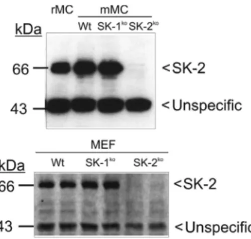
Figure 1: Sphingosine kinase 2 protein expression in mouse renal mesangial cells and embryonic fibroblasts.
Protein lysates of quiescent mouse mesangial cells (mMC) and mouse embryonic fibroblasts (MEF) isolated from either C57BL/6 mice (Wt), SK-1 deficient mice (SK-1ko), SK-2 deficient mice (SK-2ko) or rat mesangial cells (rMC) were separated by SDS-PAGE, trans-ferred to nitrocellulose and subjected to Western blot analysis using a specific antibody against mouse SK-2 at a dilution of 1:1000. Bands were visualized by the ECL method according to the manufac-turer’s instructions.
reduced in the SK-2 depleted cells, thus suggesting a likely
unspecific staining.
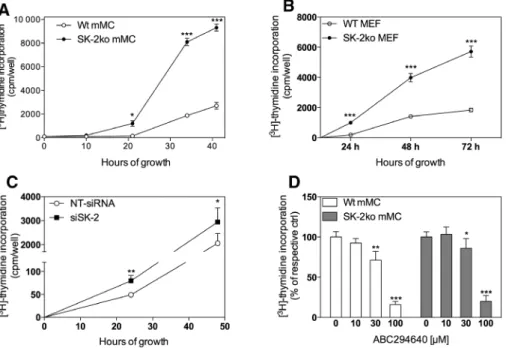
Under normal cell culture conditions, it became
obvious that SK-2ko cells proliferated much faster than the
corresponding wild-type cells. To confirm that DNA
synthe-sis was enhanced, [
3H]thymidine incorporation was
meas-ured in cells plated in equal numbers. As seen in Figure
2A, over the time period of 2 days, SK-2ko mMC showed a
several-fold increase of DNA synthesis. A similar increase
was also seen in SK-2ko MEFs when compared to the
wild-type cells (Figure 2B) and in mesangial cells transiently
transfected with siRNA against SK-2 (Figure 2C).
Addition-ally, we tested the putative SK-2 inhibitor ABC294640 on
renal mesangial cell proliferation and detected a
reduc-tion of cell proliferareduc-tion at a concentrareduc-tion of 30 μm and
even more pronounced at 100 μm. Strikingly, the same
effect was obtained when using SK-2 knockout mesangial
cells (Figure 2D, grey bars) suggesting an unspecific effect
of ABC294640 not related to SK-2 inhibition.
S. Schwalm et al.: SK-2 deficiency leads to migration and proliferation of mesangial cells
815
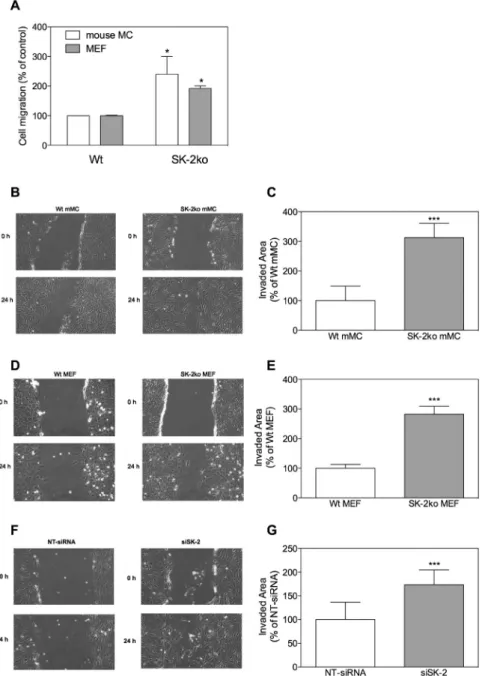
In a next step, the migratory capacity of cells was
determined. To this end, either an adapted Boyden
chamber assay (Figure 3A) or a scratch assay (Figure 3B–G)
was performed. In the adapted Boyden chamber assay,
equal numbers of cells were plated onto a Transwell filter
with 8 μm pores and allowed to migrate through the filter
for 7 h. Thereafter, non-migrated cells were scraped away
from the surface of the filter and cells that had migrated
into the filter were visualized by DAPI staining. Figure 3A
shows again that SK-2ko cells migrated faster than
wild-type cells. Another method to measure cell migration is
the so-called scratch assay. However, this assay cannot
discriminate between migrating cells and proliferating
cells and results reflect more a mixture of migration and
proliferation. Therefore, to minimize the amount of cell
proliferation, we performed this assay under strongly
reduced serum concentrations [1% fetal bovine serum
(FBS)]. Again, our data reveal that SK-2ko mesangial cells
(Figure 3B, right panels) and SK-2ko MEFs (Figure 3D,
right panels) or cells transfected with siRNA against SK-2
(Figure 3F, right panels) migrated faster than the
corre-sponding wild-type (Figure 3B and D, left panels) or
non-target siRNA transfected cells (Figure 3F, left panels).
Loss of SK-2 activates ERK and Akt signaling
in mesangial cells and fibroblasts
We further investigated whether the increased
prolif-eration and migration of SK-2ko cells was the result of
specifically activated signaling cascades, such as the
well-known proliferation and migration regulating
clas-sical mitogen-activated protein kinase (MAPK)/ERK
cascade. To this end, Western blot analyses of mesangial
cell lysates were performed by staining for
phosphoryl-ated and thus activphosphoryl-ated p44-ERK1 and p42-ERK2.
Quies-cent wild-type cells that were incubated for 6 h in a very
low serum concentration (1% FBS) expressed low levels of
phospho-ERK1/2, whereas the SK-2ko cells showed a
sig-nificantly increased ERK1/2 phosphorylation (Figure 4A).
Another signaling cascade that has been linked to cell
growth and survival, but also to cell migration, is the
PI3K/Akt cascade (Vivanco and Sawyers, 2002). We found
that also phospho-Akt was enhanced in SK-2ko mesangial
cells (Figure 4B), and similar data were obtained in MEFs
(Figure 4C and D). All these data suggest that in SK-2ko
cells the classical MAPK/ERK and the PI3K/Akt cascades
are hyperactivated.
Figure 2: Effect of SK-2 deficiency on proliferation of mouse mesangial cells and embryonic fibroblasts.
104 mouse mesangial cells (A, C, D, mMC) or mouse embryonic fibroblasts (B, MEF) isolated from either wild-type C57BL/6 mice (Wt, open
circles) or SK-2 deficient mice (SK-2ko, closed circles) or mesangial cells transiently transfected with a non-target siRNA (C, NT-siRNA, open circles) or siRNA against SK-2 (C, siSK-2, closed circles) were plated and grown for the indicated time periods in growth medium supple-mented with [3H]thymidine alone (A–C) or with the indicated concentrations of ABC294640 for 24 h (D) and processed as described in the
Methods section. [3H]thymidine incorporated into DNA was measured in a β-counter. Data are expressed as cpm/well (A, B) or % of control
(C, D) and are mean±SD (n = 3–5). *p < 0.05, **p < 0.01, ***p < 0.001 considered statistically significant compared to the corresponding Wt values (A, B) or to the respective control values (C, D).
Figure 3: Effect of SK-2 deficiency on migration of mouse mesangial cells and embryonic fibroblasts.
(A) 105 serum-starved mouse mesangial cells (open columns) or mouse embryonic fibroblasts (closed columns) isolated from either
wild-type (Wt) or SK-2 deficient mice (SK-2ko) were subcultured onto Transwell filters and allowed to migrate for 7 h. Migrated cells were determined as described in the Methods section. (B–G) Quiescent wild-type (Wt) or SK-2 deficient (SK-2ko) mesangial cells (B, C, mMC), quiescent mouse embryonic fibroblasts (D, E, MEF) or transiently transfected mouse mesangial cells with either non-target siRNA (F, G, NT-siRNA) or siRNA against SK-2 (F, G, siSK-2) were subjected to a scratch and then allowed to recover for 24 h in DMEM containing 1% FBS. Light microscopy pictures were taken at time point 0 h and 24 h after the scratch (B, D, F show representative samples). Data in C, E, G show the quantification of the scratch area after 24 h and are expressed as invaded area from time point 0 h to 24 h as % of the Wt or NT-siRNA control and are mean±SD (n = 6–9). ***p < 0.001 is considered statistically significant compared to the Wt or the NT-siRNA control.
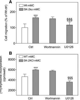
To see whether the ERK and PI3K/Akt pathways, are
involved in the increased migration and proliferation seen
in Sk-2ko cells, we performed scratch assays and measured
DNA synthesis of mouse mesangial cells in the absence
or presence of inhibitors of these signaling pathways.
Figure 5A and B shows that the specific MEK inhibitor
U0126 (Wymann et al., 1996; Favata et al., 1998), but not
the PI3K inhibitor wortmannin (Wymann et al., 1996)
inhibited migration and proliferation of SK-2ko mMC.
Furthermore, it is well-known that small G proteins
are important molecular regulators of cell migration and
also proliferation (Hall, 1990). We therefore investigated
S. Schwalm et al.: SK-2 deficiency leads to migration and proliferation of mesangial cells
817
whether small G proteins are altered in SK-2ko cells. Small
G protein activity was measured by G-LISA
®, which
spe-cifically detect the activated forms of the indicated small
G proteins (Keely et al., 2007). As shown in Figure 6, basal
RhoA activity was significantly increased in SK-2ko
mesan-gial cells and this was further enhanced after S1P
treat-ment. In contrast, Ras, Rac and cdc42 were not altered in
SK-2ko mesangial cells (data not shown).
Loss of SK-2 is associated with enhanced
expression of SK-1 and the S1P
3receptor
In a next step, we tested both cell types for a possible
compensatory upregulation of SK-1 expression. By using
Western blot analysis, we indeed found an enhanced
expression of SK-1 protein in SK-2ko mesangial cells
and MEF. Even a partial and transient reduction of SK-2
with SK-2-siRNA resulted in a higher SK-1 protein
expres-sion compared to non-target siRNA transfected cells
(Figure 7A). Furthermore, we screened the cells for S1P
receptor subtype expression and, interestingly, found that
in SK-2ko mMCs (Figure 7B) and MEFs (Figure 7C) S1P
3specifically was highly upregulated on the mRNA level.
When using SK-2 siRNA, we also observed an increased
mRNA expression of S1P
3in comparison to the
control-transfected cells (Figure 7D). In addition, we
quanti-fied cellular S1P in the cells and found that SK-2ko MEF
contain significantly more cellular S1P than the wild-type
cells (Figure 7F). Despite a similar upregulation of SK-1
protein in SK-2ko mesangial cells, the S1P concentration
in whole-cell lipid extracts of mMCs did not vary between
wild-type and SK-2ko cells (Figure 7E). Sphingosine and
C16-ceramide accumulated in SK-2ko cells in both cell
types (Figure 7E and F). To address if the altered S1P
receptor expression profile affected cell motility, we
per-formed a scratch migration assay in the presence of the
S1P receptor antagonists W146 (S1P
1antagonist), JTE-013
(S1P
2antagonist) and VPC23019 (S1P
1/3antagonist). Our
data revealed that a significant reduction of cell migration
was only detected in SK-2ko cells when using the S1P
1/3inhibitor VPC23019, but not in the presence of W146, thus
excluding a role of S1P
1in the increased migration
capac-ity of SK-2ko cells (Figure 8). The S1P
2inhibitor JTE-013
enhanced the migratory response of wild-type and SK-2ko
cells, thus demonstrating a potentially inhibitory role of
S1P
2in mesangial cell migration (Figure 8). These data
make it tempting to speculate that the observed enhanced
Figure 4: Effect of SK-2 deficiency on the activation state of ERK1/2 and PKB/Akt in mouse mesangial cells and fibroblasts.Quiescent mouse mesangial cells (A, B) and mouse embryonic fibroblasts (C, D) isolated from either wild-type (Wt, white bars) or SK-2 defi-cient mice (SK-2ko, grey bars) were incubated for 6 h in DMEM containing 1% FBS. Thereafter, cell lysates containing 50 μg of protein were separated by SDS-PAGE, transferred to nitrocellulose and subjected to Western blot analysis using antibodies against phospho-ERK1/2 and total ERK1/2 (A and C), and phospho-Ser473-PKB/Akt and total PKB/Akt1 (B, D). Bands were visualized by the ECL method according
to the manufacturer’s instructions and bands were densitometrically evaluated. Data are expressed as % of controls and are mean±SD (n = 3). *p < 0.05, **p < 0.01, ***p < 0.001 considered statistically significant compared to the corresponding Wt values. The insets show representative immunoblots.
Figure 5: Involvement of the PI3K and ERK pathway in the SK2ko-mediated increased migration and proliferation in mesangial cells. (A) Quiescent wild-type (Wt, white bar) or SK-2 deficient (SK-2ko, grey bars) mesangial cells (mMC) were subjected to a scratch and then allowed to recover for 24 h in DMEM containing 1% FBS in the absence (Ctrl) or presence of wortmannin (200 nm) or U0126 (10 μm). Light microscopy pictures were taken at time point 0 h and 24 h after the scratch and analyzed using the Image J software (Wayne Rasband, NIH, USA) by measuring the scratch area. Data are expressed as area reduction from time points 0 h to 24 h as % of the Wt control and are mean±S.D. (n = 4). (B) Wild-type (Wt, white bars) or SK-2 deficient (SK-2ko, grey bars) mesangial cells (mMC) were plated and grown for the indicated time periods in growth medium supplemented with [3H]thymidine alone (Ctrl) or in the presence of
wortmannin (200 nm) or U0126 (10 μm) and processed as described in the Methods section. [3H]thymidine incorporated into DNA was
measured in a β-counter. Data are expressed as cpm/well and are mean±SD (n = 4–6). ***p < 0.001 considered statistically significant compared the Wt ctrl, §§§p < 0.001 considered statistically significant
compared to the SK-2ko ctrl value.
Figure 6: Effect of SK-2 deficiency on RhoA activity in mouse mesangial cells.
Quiescent mouse mesangial cells isolated from either wild-type (Wt, white bars) or SK-2 deficient mice (SK-2ko, grey bars) were stimulated for 10 min with either vehicle (Ctrl) or sphingosine 1-phosphate (S1P, 1 μm) in the presence of 1% FBS. Thereafter, cell lysates were subjected to G-LISA® RhoA activation assay according
to the manufacturer’s instructions. Data are expressed % of Wt ctrl and are mean±SD (n = 3). **p < 0.01, ***p < 0.001 considered statisti-cally significant compared to the vehicle treated Wt value, §§p < 0.01
considered statistically significant compared to the S1P-stimulated Wt value.
migration and proliferation of SK-2ko cells was the result
of an autocrine S1P action through the S1P
3receptor,
although a further increase of S1P in renal mesangial cells
seems dispensable.
Finally, we investigated the contribution of S1P
3to
mesangial cell proliferation and migration by using cells
isolated from S1P
3knockout mice. The data in Figure 9A
show that the growth of S1P
3ko mMCs in normal growth
medium containing 10% FBS was only slightly but still
significantly reduced when compared to wild-type cells.
When migration of these cells was analyzed in a scratch
assay, we found that stimulation with S1P triggered
increased migration in wild-type cells (Figure 9B), which
was abolished in S1P
3ko cells (Figure 9B). In addition,
the stimulation of RhoA activity by S1P detected in
wild-type cells was clearly reduced in S1P
3ko cells (Figure 9C).
Together, these data suggest that S1P
3indeed contributes
to mesangial cell proliferation and migration.
Discussion
In this study we show for the first time that renal
mesan-gial cells and embryonic fibroblasts isolated from SK-2
deficient mice have a higher capacity to proliferate and
migrate when compared to wild-type cells. Since the
suc-cessful cloning of SK-2 in 2000 (Liu et al., 2000a), the few
published studies have reported rather controversial data
on possible cellular functions of SK-2.
On the one hand, overexpression studies in various
cell types revealed that SK-2 is localized in the nucleus and
can inhibit DNA synthesis (Igarashi et al., 2003). Moreover,
Liu et al. (2003) reported that overexpression of SK-2 had
a pro-apoptotic effect on cells because of the existence of
a BH3 domain that interacts with the anti-apoptotic factor
Bcl-X
Land thereby blocks Bcl-X
L’s anti-apoptotic function.
In line with this, our own previous studies revealed that
a loss of SK-2 rendered mesangial cells less sensitive to
stress-induced apoptosis (Hofmann et al., 2008).
On the other hand, SK-2 was activated by epidermal
growth factor (EGF) (Hait et al., 2007) due to ERK-mediated
S. Schwalm et al.: SK-2 deficiency leads to migration and proliferation of mesangial cells
819
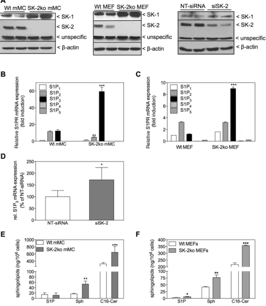
Figure 7: Effect of SK-2 deficiency on SK-1 protein expression, S1P receptor subtype mRNA expression, and on cellular sphingolipid levels in mouse mesangial cells and embryonic fibroblasts.
(A) Quiescent mouse mesangial cells and mouse embryonic fibroblasts isolated from either C57BL/6 mice (Wt) or SK-2 deficient mice (SK-2ko) or mouse mesangial cells transiently transfected with either non-target siRNA (NT-siRNA) or siRNA against SK-2 (siSK-2) were subjected to WB analysis using specific antibodies against SK-1 (upper panels), SK-2 (middle panels) or β-actin (lower panels). (B–D) Quiescent mouse mesangial cells (B) and mouse embryonic fibroblasts (C) isolated from either C57BL/6 mice (Wt) or SK-2 deficient mice (SK-2ko) or tran-siently transfected mouse mesangial cells with either non-target siRNA (D, NT-siRNA) or siRNA against SK-2 (D, siSK-2) were taken for RNA extraction and subjected to quantitative PCR analysis using mouse primers of S1P1-5 (B, C) or S1P3 (D) as described in the Methods section.
Data were obtained according to the ΔΔCT method and are expressed as fold induction of S1P1 expression level (B, C) or as% of NT-siRNA (D)
and are mean±SD (n = 3–4). *p < 0.05, **p < 0.01, ***p < 0.001 considered statistically significant compared to the corresponding Wt values. (E, F) Lipids were extracted from quiescent mouse mesangial cells (E) and mouse embryonic fibroblasts (F) from wild-type (Wt, white bars) and SK-2 deficient mice (SK-2ko, grey bars) and taken for LC-MS/MS to quantify sphingosine 1-phosphate (S1P), sphingosine (Sph) and C16-ceramide (C16-Cer). Results are expressed as ng lipid per 106 cells and are mean±SD (n = 3). *p < 0.05, **p < 0.01, ***p < 0.001 considered
statistically significant compared to the corresponding Wt values.
hyperphosphorylation in breast cancer cells. Such an
activation of SK-2 by growth factors is hardly consistent
with a growth inhibiting and pro-apoptotic effect of SK-2.
In fact, we previously showed that in the breast cancer
cell line MCF-7, EGF and estrogen rather down-regulated
SK-2 mRNA expression (Döll et al., 2005, 2007), which
Figure 8: Effect of S1P receptor antagonists on the increased migra-tion of mouse mesangial cells.
Quiescent wild-type (Wt, white bars) or SK-2 deficient (SK-2ko, grey bars) mesangial cells (mMC) were subjected to a scratch and then allowed to recover for 24 h in DMEM containing 1% FBS in the presence of either vehicle (Ctrl) or VPC23019 (VPC, 10 μm), W146 (10 μm) or JTE-013 (JTE, 10 μm). Light microscopy pictures were taken at time point 0 h and 24 h after the scratch and analyzed using the Image J software (Wayne Rasband, NIH, USA) by measur-ing the scratch area. Data are expressed as area reduction from time points 0 h to 24 h as % of the Wt ctrl and are mean±SD (n = 4). **p < 0.01, ***p < 0.001 considered statistically significant com-pared to the Wt ctrl, §§p < 0.01 considered statistically significant
compared to the SK-2ko ctrl value.
Figure 9: Effect of S1P3 knockout on proliferation and migration of mouse mesangial cells.
(A) 105 mesangial cells isolated from either BalbC mice (Wt, empty
circles) or S1P3-deficient mice (S1P3ko, black circles) were plated and
grown for the indicated time periods in growth medium supplemented with [3H]thymidine and processed as described in the Methods
section. [3H]thymidine incorporated into DNA was measured in a
β-counter. Data are expressed as cpm/well and are mean±SD (n = 3). *p < 0.05, ***p < 0.001 considered statistically significant compared to the corresponding control values. (B) Quiescent control mouse mesangial cells (Wt, white bars) or S1P3-deficient mesangial cells (S1P3ko, grey bars) were subjected to a scratch and allowed to recover
for 24 h in DMEM containing 1% FBS the presence of either vehicle (Ctrl) or S1P (1 μm). Light microscopy pictures were taken at time point 0 h and 24 h after the scratch and analyzed using the Image J software (Wayne Rasband, NIH, USA) by measuring the scratch area. Data are expressed as area reduction from time points 0 h to 24 h as % of the Wt ctrl value and are mean±SD (n = 4–6). **p < 0.01 considered statisti-cally significant compared to the Wt ctrl value. (C) Quiescent mouse mesangial cells isolated from either BalbC mice (Wt, white bars) or S1P3-deficient mice (S1P3ko, grey bars) were stimulated for 10 min with either vehicle (Ctrl) or sphingosine 1-phosphate (S1P, 1 μm) in the presence of 1% FBS. Thereafter, cell lysates were subjected to G-LISA®
RhoA activation assay according to the manufacturer’s instructions. Data are expressed as % of Wt ctrl value and are mean±SD (n = 4). *p < 0.05, ***p < 0.001 considered statistically significant compared to the vehicle treated Wt value, §§§p < 0.001 considered statistically
significant compared to the S1P stimulated Wt value.
occurred concomitantly to an upregulation of SK-1 by
these factors.
Recently, a catalytic inhibitor of SK-2 ABC294640
(French et al., 2010; Antoon et al., 2011) was
devel-oped and used to investigate the cellular function of
SK-2. Although the in vitro IC
50for SK-2 was rather high
with 50 μm (French et al., 2010), oral administration of
ABC294640 to mice bearing mammary adenocarcinoma
xenografts resulted in a dose-dependent antitumor
activ-ity associated with depletion of S1P levels in the tumors
and progressive tumor cell apoptosis (French et al.,
2010; Antoon et al., 2011). These data suggested that SK-2
similar to SK-1 also drives cancer growth and progression.
In the present study, treatment of mesangial cells with 30
and 100 μm ABC294640 decreased proliferation, which,
however, was also detected in SK-2ko cells (Figure 2C).
These data clearly demonstrate that ABC294640 elicits
an anti-proliferative effect independent of SK-2
inhibi-tion, thereby questioning the specificity of ABC294640.
Recently, ABC294640 was shown to exert anti-estrogenic
effects by directly antagonising estrogen receptor (ER)
activation (Antoon et al., 2010). Molecular modelling
studies revealed a direct docking of ABC294640 into the
ligand binding site of the ERα. This estrogen antagonistic
potential may explain on the one hand the
anti-prolifer-ative effect and on the other hand why especially
estro-gen-sensitive breast cancer cells were strongly affected
by the compound. The comparison of in vivo effects of
S. Schwalm et al.: SK-2 deficiency leads to migration and proliferation of mesangial cells
821
ABC294640 with the effect of genetic SK-2 knockout mice
also revealed inconsistent results. Whereas the inhibitor
aggravated inflammatory arthritis, SK-2 knockout mice
showed no change of disease severity compared to
wild-type mice (Baker et al., 2013). Moreover, Liang et al. (2013)
showed that SK-2 deficient mice develop more severe
chronic intestinal inflammation and colitis-associated
cancer, which, in terms of mechanism, was proposed to
involve a counter-upregulation of SK-1 and S1P.
Our study also showed a compensatory
upregula-tion of SK-1 protein in SK-2ko cells and SK-2-siRNA
trans-fected cells, although the exact mechanisms leading to
the observed enhanced SK-1 expression are not known.
However, using LC-MS/MS we detected a rise of
cellu-lar S1P only in SK-2ko MEF but not in SK-2ko mMCs. The
reason for this discrepancy might be the result of a
detec-tion limit, as we only measured S1P in whole-cell lipid
extracts but not in subcellular fractions. Another
possibil-ity is that produced S1P is secreted more efficiently from
mesangial cells than from fibroblasts. However, so far we
were not able to measure an increased level in cell culture
supernatants (data not shown). Further experiments are
needed to clarify this issue.
In addition, our data revealed that specifically the S1P
3receptor was upregulated upon loss of SK-2 (Figure 7B–D).
We therefore speculate that increased proliferation and
migration observed in SK-2ko cells may be due to this
enhanced S1P
3expression. We obtained evidence that S1P
3is involved in mesangial cell migration and proliferation
from S1P
3knockout mesangial cells as (i) the S1P
3ko cells
showed a slower proliferation rate in comparison to
wild-type cells (Figure 9A) and (ii) S1P was able to induce cell
migration only in wild-type but not in S1P
3ko cells (Figure
9B). Furthermore, the S1P
1/3antagonist VPC23019, but not
the S1P
1antagonist W146, reduced migration in mMC. This
effect was only observed in SK-2ko cells, which showed a
higher S1P
3mRNA expression, and not in Wt cells (Figure 8).
However, as mesangial cell proliferation was only partially
reduced in S1P
3-deficient cells and also showed normal
migration behavior under control conditions, we conclude
that other receptor subtypes are involved too. Katsuma
et al. reported that extracellular S1P triggers mesangial
cell proliferation by involving both S1P
2and S1P
3(Katsuma
et al., 2002). Similarly, proliferation of hepatoma cells (An
et al., 2000) and satellite cells (Calise et al., 2012) also
depended on both S1P
2and S1P
3. Concerning cell
migra-tion, we observed an increased migration of mesangial cells
when inhibiting the S1P
2receptor with JTE013, suggesting a
potentially inhibitory role of S1P
2in cell migration, which
was previously also reported in other cell types (Osada
et al., 2002; Arikawa et al., 2003; Goparaju et al., 2005).
Similar to S1P
2, the involvement of S1P
3in a migratory
event is also conflicting. In this respect, Okamoto et al.
(2000) showed that overexpression of S1P
1and S1P
3in
Chinese hamster ovary (CHO) cells resulted in more
S1P-stimulated migration. A similar situation was reported for
human endothelial cells where S1P-triggered migration
was abolished by antisense oligonucleotides against S1P
1and S1P
3(Paik et al., 2001), and in lung cancer cells where
either a S1P
3knockdown by RNAi or Rho kinase inhibition
by a catalytic inhibitor prevented a S1P-stimulated
migra-tory/invasive response (Zhang et al., 2013).
By generating S1P receptor knockout mice, it turned
out that S1P
1deficiency is embryonically lethal, as
vas-cular maturation failed due to a defect in smooth muscle
cells migration (Liu et al., 2000b). Consistently,
embry-onic fibroblasts (MEF) isolated from these S1P
1deficient
mice also exhibited a defect in chemotaxis towards S1P
(Liu et al., 2000b). These data clearly support an in vivo
relevant function of S1P
1in smooth muscle cell and
fibro-blast migration. In contrast, S1P
3deficient mice showed
no vascular maturation deficit, suggesting that S1P
3is
not involved in smooth muscle cell migration and
conse-quently vessel maturation (Liu et al., 2000b; Ishii et al.,
2001).
Although these data do not allow a general
conclu-sion, they somehow stress the possibility that S1P
3, likely
in cooperation with S1P
1, is involved in S1P-stimulated cell
migration in a cell-type specific manner.
Our data further demonstrate that not only the basal
but also the S1P-stimulated RhoA activity is enhanced
in SK-2 deficient cells. RhoA is one of three members of
the Rho GTPase family that plays a crucial role in the
regulation of cell migration. There seems to be a
spati-otemporal dynamic of RhoA action driving not only
mem-brane protrusions at the leading edge of the cells but also
tail retraction (Pertz et al., 2006). Although previously
regarded as an anti-migratory GTPase, the finding that
RhoA localizes at the leading edge of cells where it is
important for membrane ruffling and lamellae formation
confirms other studies describing RhoA as pro-migratory
factor (for review, see O’Connor and Chen, 2013). As we
were only able to measure RhoA activity in whole-cell
lysates, we cannot decipher the exact cellular
localiza-tion of the enhanced RhoA activity in SK-2ko cells to allow
a final conclusion on the specific impact of RhoA acting
pro-migratory on the leading edge or anti-migratory by
tail retraction in mesangial cells. However, RhoA is
acti-vated by extracellular S1P in many cell types including
CHO cells (Okamoto et al., 2000), endothelial cells (Paik
et al., 2001), and bladder cancer cells (vom Dorp et al.,
2011), and this seems to be mandatory for cell migration.
lations whether another sphingolipid mediator besides
S1P contributes to the effects observed in this study. In
this regard, we and others have shown that ceramide can
directly bind to and activate c-Raf (Huwiler et al., 1996),
PKC-α (Huwiler et al., 1998) and PKC-ζ (Muller et al., 1995)
but inhibit PKC-δ (Huwiler et al., 1998; Bessa et al., 2013).
Further studies are needed to evaluate the exact
contribu-tion of the various sphingolipid-derived mediators to the
observed phenotype of SK-2ko mesangial cells.
Although our study has only addressed the effect
of SK-2ko in mesangial cells, it will be interesting to see
whether in vivo in a renal disease model SK-2 deficient
mice also respond with increased proliferation and
migra-tion. In this context, in a renal ischemia/reperfusion injury
(IRI) model in SK-2ko mice it was reported that loss of SK-2
led to more severe kidney injury as indicated by elevated
plasma creatinine levels, increased tubular cell
necro-sis, dilation of tubules, and cast formation in the outer
medulla. In addition, kidneys of IRI-exposed SK-2ko mice
not only showed more neutrophil infiltration but
remark-ably also increased S1P
3expression (Jo et al., 2009), which
supports our data obtained in renal mesangial cells and
fibroblasts.
Altogether, our data show for the first time that loss
of SK-2 in renal mesangial cells and fibroblasts may have
considerable impact on cell growth and migration, and
that SK-2 exerts a suppressive effect on these cellular
responses that are crucial in many renal diseases, such as
renal cancer and chronic inflammatory kidney diseases.
Materials and methods
Chemicals
[6–3H]methyl-thymidine (specific activity: 14.5 Ci/mmol was from
American Radiolabeled Chemicals Inc., St. Louis, MO, USA. The secondary anti-rabbit and anti-mouse horseradish peroxidase-coupled antibodies, HyperfilmMP and the enhanced chemilumi-nescence (ECL) reagents were from GE Health Care Systems GmbH, Freiburg, Germany. Sphingolipid standards for LC/MS, W146, JTE-013 and VPC23019 were from Avanti Polar Lipids Inc., Alabaster, AL, USA; U0126, and wortmannin were from Merck Biosciences, Schwalbach, Germany. Antibodies against phospho-Ser473-PKB/Akt,
Peptide synthesis and antibody generation was performed by Euro-gentec s.a. (Seraing, Belgium). Two synthetic peptides based on the sequence of the mouse SK-1 (accession number: NM_011451) (-CPS GRD SRR GPP PEE P-COOH and -LEP RSQ-RGV FSV DGE C-CONH2) or
on the sequence of the mouse SK-2 (accession number: NM_020011) (-CTL LTG PAG QKP QA-COOH and -CPI AEG PPE MPA SSG F-CONH2)
were synthesized and coupled to keyhole-limpet hemocyamin, and used to immunize two rabbits. The antibodies from terminal bleeds were purified by affinity chromatography by using an anti-peptide-coupled sepharose column.
Cell culture
Primary cultures of mouse renal mesangial cells were isolated from C57BL/6 mice (Wt) or SK-2 knockout (ko) mice exactly as previously described (Klawitter et al., 2007; Hofmann et al., 2008). Outgrown mesangial cells were subcultured and further used up to passage 20. Cells were cultured in RPMI medium containing 15% FBS, 10 mm HEPES, pH 7.4, 100 units/ml penicillin, 100 μg/ml streptomycin, 6 μg/ml bovine insulin, 5 μg/ml transferrin, 5 ng/ml sodium selenite, 4.5 μg/ml β-mercaptoethanol. Mouse embryonic fibroblasts (MEF) were isolated from C57BL6 mice or SK-2 deficient mice as previously described (Conner, 2001). Cells were cultured in Dulbecco’s modified Eagle medium (DMEM) containing 10% FBS, 10 mm HEPES, pH 7.4, 100 units/ml penicillin, 100 μg/ml streptomycin.
siRNA transfection
siRNA transfection was conducted by using the Amaxa™ Mouse/Rat Hepatocyte Nucleofector™ Kit (Lonza, Cologne, Germany) according to the Amaxa™ protocol for mouse embryonic fibroblasts. Briefly, mouse mesangial cells were harvested by trypsinization, centrifuged and resuspended in 100 μl Nucleofector™ solution. After addition of either 0.5 μm non-target siRNA (Thermo Fisher Scientic, Waltham, MA, USA) or 0.5 μm SK-2 siRNA (antisense sequence: GCC CUA CAC AUA CAG CGA C) transfection was performed in a cuvette using proto-col N-024. The cells were immediately transferred to the culture plate and cultured at 37°C for 1–2 days prior to use in further experiments.
Cell stimulation and Western blot analysis
Confluent cells in 100 mm diameter dishes were rendered quiescent by incubating for 24 h in DMEM containing 0.1 mg/ml of fatty acid-free bovine serum albumin (BSA). Thereafter, cells were treated as
S. Schwalm et al.: SK-2 deficiency leads to migration and proliferation of mesangial cells
823
indicated. To stop the stimulation, the medium was withdrawn andcells were homogenized as previously described (Rölz et al., 2002). Equal amounts of protein were separated by SDS-PAGE, transferred to nitrocellulose membranes, and subjected to Western blot analysis as previously described (Rölz et al., 2002).
Transwell migration assay
Cell migration was measured by using an adapted Boyden chamber assay. In brief, the ability of cells to migrate through a Transwell filter (6.5 mm diameter, 8 μm pore size) was analyzed. After serum starva-tion, cells were detached by trypsinization and seeded into Transwell filters at 1 × 10⁵ cells in 100 μl starvation medium. 500 μl of starvation
medium were placed in the lower compartment and the cells were left to migrate for the indicated time periods. Thereafter, the Transwell filters were removed and processed as previously described (Huwiler et al., 2006). Migrated cells were determined by counting the DAPI-stained cells on the filters in five random areas per sample using a fluorescence microscope.
Scratch assay for cell migration
Equal number of cells were seeded in growth medium in PS35 and cultured for 6 h to allow adherence followed by a starvation period in DMEM containing 1% of FBS for 18 h. Thereafter, a ‘scratch’ was created on the confluent cell monolayer using a p200 pipet tip and the dish was immediately photographed under a phase-contrast microscope at specific reference points. A second image was taken 24 h after previous alignment along the reference points to ensure assessment of the same scratch area. The images were analyzed using the Image J software (Wayne Rasband, NIH, USA) by measuring the scratch area at 0 h and 24 h after incubation. Data are expressed as area reduction from time points 0 h to 24 h.
[
3H]Thymidine incorporation
5000 cells were plated per well of a 24-well plate and incubated for 20 h. Thereafter, cells were incubated in growth medium containing 0.2 μCi/ml of [3H]methyl-thymidine. After different time periods, the
medium was removed and cells were washed twice with PBS and incubated with ice-cold 5% trichloroacetic acid (TCA) for 30 min. Cells were washed twice with 5% TCA, the DNA was solubilized in 1 m NaOH for 30 min at 37°C, and the radioactivity was counted in a β-counter.
Quantitative PCR analysis
Real-time PCR was performed using SYBRgreen® and a BioRad iQ
iCycler Detection System. Primer sequences were as follows. Mouse S1P1: forward: CTG ACC TTC CGC AAG AAC ATC T; reverse: CTT CAG
CAA GGC CAG AGA CTT C; mouse S1P2: forward: GAG CTC ATC ACC
TCT TCA TCC TAT C; reverse: GAA GAT GCA GTA AGA GTA CCC AGG A; mouse S1P3: forward: TGT AGC TTC ATC GTC GTC TTG GAG; reverse:
GCC GAT GAA AAA GTA CAT GCG G; mouse S1P4: forward: GGC TAC
TGG CAG CTA TCC TG; reverse: AAG GCC ACC AAG ATC ATC AG; mouse S1P5: forward: GGA GGG ACT CTC CTG GAT TC; reverse: TTC
CTC TGT AGC CAG CCA CT. IQ™5 Optical System Software (Version 2.0) was used to analyze real-time and endpoint fluorescence. One microgram of total RNA isolated with TRIZOL® reagent was used for
reverse transcriptase-PCR (First Strand Synthesis Kit, MBI Fermen-tas, St-Leon-Roth, Germany); a random hexamer primer was utilized for amplification. The fold induction values were obtained using to the ΔΔCT method, after normalization to the housekeeping gene 18S
RNA.
RhoA activity assay
The activity of the small G protein RhoA was determined by using a specific G-LISA™ activity assay kit (Cytoskeleton Inc., Denver, CO, US) exactly as described in the manufacturer’s manual.
Lipid quantification by mass spectrometry
Confluent cells in PS35 wells were either taken for lipid extraction and LC-MS/MS analysis exactly as previously described (Hofmann et al., 2008) or taken for cell counting for lipid equalization.
Statistical analysis
Statistical analysis was performed by unpaired t-test and two-tailed p-values for the comparison of two groups, and by one-way analysis of variance (ANOVA) with Bonferroni post-test for the comparison of three and more groups.
Acknowledgments: This work was supported by the Swiss
National Science Foundation (3100A0–111806), and the
German Research Foundation (SFB1039/TP02). We thank
Isolde Römer and Simone Albert for technical assistance.
References
Alemany, R., van Koppen, C.J., Danneberg, K., Ter Braak, M., and Meyer Zu Heringdorf, D. (2007). Regulation and functional roles of sphingosine kinases. Naunyn-Schmiedeberg’s Arch. Pharmacol. 374, 413–428.
An, S., Zheng, Y., and Bleu, T. (2000). Sphingosine 1-phosphate-induced cell proliferation, survival, and related signaling events mediated by G protein-coupled receptors Edg3 and Edg5. J. Biol. Chem. 275, 288–296.
Antoon, J.W., White, M.D., Meacham, W.D., Slaughter, E.M., Muir, S.E., Elliott, S., Rhodes, L.V., Ashe, H.B., Wiese, T.E., Smith, C.D., et al. (2010). Antiestrogenic effects of the novel sphin-gosine kinase-2 inhibitor ABC294640. Endocrinology 151, 5124–5135.
Baker, D.A., Eudaly, J., Smith, C.D., Obeid, L.M., and Gilkeson, G.S. (2013). Impact of sphingosine kinase 2 deficiency on the devel-opment of TNF-a-induced inflammatory arthritis. Rheumatol. Int. 33, 2677–2681.
Bessa, C., Pereira, C., Leao, M., Maciel, C., Gomes, S., Goncalves, J., Corte-Real, M., Costa, V., and Saraiva, L. (2013). Using yeast to uncover the regulation of protein kinase Cdelta by ceramide. FEMS Yeast Res. 13, 700–705.
Calise, S., Blescia, S., Cencetti, F., Bernacchioni, C., Donati, C., and Bruni, P. (2012). Sphingosine 1-phosphate stimulates prolifera-tion and migraprolifera-tion of satellite cells: role of S1P receptors. Biochim. Biophys. Acta. 1823, 439–450.
Conner, D.A. (2001). Mouse embryo fibroblast (MEF) feeder cell preparation. Curr. Protoc. Mol. Biol. Chapter 23, Unit 23 22. Döll, F., Pfeilschifter, J., and Huwiler, A. (2005). The epidermal
growth factor stimulates sphingosine kinase-1 expression and activity in the human mammary carcinoma cell line MCF7. Biochim. Biophys. Acta. 1738, 72–81.
Döll, F., Pfeilschifter, J., and Huwiler, A. (2007). Prolactin upregu-lates sphingosine kinase-1 expression and activity in the human breast cancer cell line MCF7 and triggers enhanced proliferation and migration. Endocr. Relat. Cancer 14, 325–335. Favata, M.F., Horiuchi, K.Y., Manos, E.J., Daulerio, A.J., Stradley,
D.A., Feeser, W.S., Van Dyk, D.E., Pitts, W.J., Earl, R.A., Hobbs, F., et al. (1998). Identification of a novel inhibitor of mitogen-activated protein kinase kinase. J. Biol. Chem. 273, 18623– 18632.
French, K.J., Schrecengost, R.S., Lee, B.D., Zhuang, Y., Smith, S.N., Eberly, J.L., Yun, J.K., and Smith, C.D. (2003). Discovery and evaluation of inhibitors of human sphingosine kinase. Cancer Res. 63, 5962–5969.
French, K.J., Upson, J.J., Keller, S.N., Zhuang, Y., Yun, J.K., and Smith, C.D. (2006). Antitumor activity of sphingosine kinase inhibi-tors. J. Pharmacol. Exp. Ther. 318, 596–603.
French, K.J., Zhuang, Y., Maines, L.W., Gao, P., Wang, W., Beljanski, V., Upson, J.J., Green, C.L., Keller, S.N., and Smith, C.D. (2010). Pharmacology and antitumor activity of ABC294640, a selec-tive inhibitor of sphingosine kinase-2. J. Pharmacol. Exp. Ther. 333, 129–139.
Goparaju, S.K., Jolly, P.S., Watterson, K.R., Bektas, M., Alvarez, S., Sarkar, S., Mel, L., Ishii, I., Chun, J., Milstien, S., et al. (2005). The S1P2 receptor negatively regulates platelet-derived growth factor-induced motility and proliferation. Mol. Cell. Biol. 25, 4237–4249.
Hait, N.C., Bellamy, A., Milstien, S., Kordula, T., and Spiegel, S. (2007). Sphingosine kinase type 2 activation by ERK-mediated phosphorylation. J. Biol. Chem. 282, 12058–12065.
Hall, A. (1990). The cellular functions of small GTP-binding proteins. Science 249, 635–640.
Histamine increases sphingosine kinase-1 expression and activity in the human arterial endothelial cell line EA.hy 926 by a PKC-alpha-dependent mechanism. Biochim. Biophys. Acta. 1761, 367–376.
Huwiler, A., Fabbro, D., and Pfeilschifter, J. (1998). Selective cera-mide binding to protein kinase C-a and -d isoenzymes in renal mesangial cells. Biochemistry 37, 14556–14562.
Huwiler, A., Kolter, T., Pfeilschifter, J., and Sandhoff, K. (2000). Physiology and pathophysiology of sphingolipid metabolism and signaling. Biochim. Biophys. Acta. 1485, 63–99. Huwiler, A. and Pfeilschifter, J. (2006). Altering the
sphingosine-1-phosphate/ceramide balance: a promising approach for tumor therapy. Curr. Pharm. Des. 12, 4625–4635.
Igarashi, N., Okada, T., Hayashi, S., Fujita, T., Jahangeer, S., and Nakamura, S. (2003). Sphingosine kinase 2 is a nuclear protein and inhibits DNA synthesis. J. Biol. Chem. 278, 46832–46839. Ishii, I., Friedman, B., Ye, X., Kawamura, S., McGiffert, C., Contos,
J.J., Kingsbury, M.A., Zhang, G., Brown, J.H., and Chun, J. (2001). Selective loss of sphingosine 1-phosphate signaling with no obvious phenotypic abnormality in mice lacking its G protein-coupled receptor, LP(B3)/EDG-3. J. Biol. Chem. 276, 33697–33704.
Jo, S.K., Bajwa, A., Ye, H., Vergis, A.L., Awad, A.S., Kharel, Y., Lynch, K.R., and Okusa, M.D. (2009). Divergent roles of sphingosine kinases in kidney ischemia-reperfusion injury. Kidney Int. 75, 167–175.
Katsuma, S., Hada, Y., Ueda, T., Shiojima, S., Hirasawa, A., Tanoue, A., Takagaki, K., Ohgi, T., Yano, J., and Tsujimoto, G. (2002). Signalling mechanisms in sphingosine 1-phosphate-promoted mesangial cell proliferation. Genes Cells 7, 1217–1230. Keely, P.J., Conklin, M.W., Gehler, S., Ponik, S.M., and Provenzano,
P.P. (2007). Investigating integrin regulation and signaling events in three-dimensional systems. Methods Enzymol. 426, 27–45.
Klawitter, S., Hofmann, L.P., Pfeilschifter, J., and Huwiler, A. (2007). Extracellular nucleotides induce migration of renal mesangial cells by upregulating sphingosine kinase-1 expression and activity. Br. J. Pharmacol. 150, 271–280.
Kunkel, G.T., Maceyka, M., Milstien, S., and Spiegel, S. (2013). Tar-geting the sphingosine-1-phosphate axis in cancer, inflamma-tion and beyond. Nat. Rev. Drug Discov. 12, 688–702. Liang, J., Nagahashi, M., Kim, E.Y., Harikumar, K.B., Yamada, A.,
Huang, W.C., Hait, N.C., Allegood, J.C., Price, M.M., Avni, D., et al. (2013). Sphingosine-1-phosphate links persistent STAT3 activation, chronic intestinal inflammation, and development of colitis-associated cancer. Cancer Cell 23, 107–120. Liu, H., Sugiura, M., Nava, V.E., Edsall, L.C., Kono, K., Poulton, S.,
Milstien, S., Kohama, T., and Spiegel, S. (2000a). Molecular cloning and functional characterization of a novel mammalian
S. Schwalm et al.: SK-2 deficiency leads to migration and proliferation of mesangial cells
825
sphingosine kinase type 2 isoform. J. Biol. Chem. 275,19513–19520.
Liu, Y., Wada, R., Yamashita, T., Mi, Y., Deng, C.X., Hobson, J.P., Rosenfeldt, H.M., Nava, V.E., Chae, S.S., Lee, M.J., et al. (2000b). Edg-1, the G protein-coupled receptor for sphingo-sine-1-phosphate, is essential for vascular maturation. J. Clin. Invest. 106, 951–961.
Liu, H., Toman, R.E., Goparaju, S.K., Maceyka, M., Nava, V.E., Sankala, H., Payne, S.G., Bektas, M., Ishii, I., Chun, J., et al. (2003). Sphingosine kinase type 2 is a putative BH3-only pro-tein that induces apoptosis. J. Biol. Chem. 278, 40330–40336. Muller, G., Ayoub, M., Storz, P., Rennecke, J., Fabbro, D., and
Pfizenmaier, K. (1995). PKC zeta is a molecular switch in signal transduction of TNF-a, bifunctionally regulated by ceramide and arachidonic acid. EMBO J. 14, 1961–1969.
O’Connor, K. and Chen, M. (2013). Dynamic functions of RhoA in tumor cell migration and invasion. Small GTPases 4, 141–147. Okada, T., Ding, G., Sonoda, H., Kajimoto, T., Haga, Y.,
Khosrow-beygi, A., Gao, S., Miwa, N., Jahangeer, S., and Nakamura, S. (2005). Involvement of N-terminal-extended form of sphingo-sine kinase 2 in serum-dependent regulation of cell prolifera-tion and apoptosis. J. Biol. Chem. 280, 36318–36325. Okamoto, H., Takuwa, N., Yokomizo, T., Sugimoto, N., Sakurada, S.,
Shigematsu, H., and Takuwa, Y. (2000). Inhibitory regulation of Rac activation, membrane ruffling, and cell migration by the G protein-coupled sphingosine-1-phosphate receptor EDG5 but not EDG1 or EDG3. Mol. Cell. Biol. 20, 9247–9261.
Osada, M., Yatomi, Y., Ohmori, T., Ikeda, H., and Ozaki, Y. (2002). Enhancement of sphingosine 1-phosphate-induced migration of vascular endothelial cells and smooth muscle cells by an EDG-5 antagonist. Biochem. Biophys. Res. Commun. 299, 483–487. Paik, J.H., Chae, S., Lee, M.J., Thangada, S., and Hla, T. (2001).
Sphingosine 1-phosphate-induced endothelial cell migration requires the expression of EDG-1 and EDG-3 receptors and Rho-dependent activation of avb3- and b1-containing integrins. J. Biol. Chem. 276, 11830–11837.
Pertz, O., Hodgson, L., Klemke, R.L., and Hahn, K.M. (2006). Spati-otemporal dynamics of RhoA activity in migrating cells. Nature 440, 1069–1072.
Pyne, N.J. and Pyne, S. (2010). Sphingosine 1-phosphate and can-cer. Nat. Rev. Cancer 10, 489–503.
Rölz, W., Xin, C., Ren, S., Pfeilschifter, J., and Huwiler, A. (2002). Interleukin-1 inhibits angiotensin II-stimulated protein kinase B pathway in renal mesangial cells via the inducible nitric oxide synthase. Eur. J. Pharmacol. 442, 195–203.
Ruckhäberle, E., Rody, A., Engels, K., Gaetje, R., von Minckwitz, G., Schiffmann, S., Grösch, S., Geisslinger, G., Holtrich, U., Karn, T., et al. (2008). Microarray analysis of altered sphingolipid metabolism reveals prognostic significance of sphingosine kinase 1 in breast cancer. Breast Cancer Res. Treat. 112, 41–52.
Shida, D., Takabe, K., Kapitonov, D., Milstien, S., and Spiegel, S. (2008). Targeting SphK1 as a new strategy against cancer. Curr. Drug Targets 9, 662–673.
Vivanco, I. and Sawyers, C.L. (2002). The phosphatidylinositol 3-Kinase AKT pathway in human cancer. Nat. Rev. Cancer 2, 489–501.
vom Dorp, F., Sanders, H., Boergermann, C., Lummen, G., Rub-ben, H., Jakobs, K.H., and Schmidt, M. (2011). Inhibition of Rho-kinase abrogates migration of human transitional cell carcinoma cells: results of an in vitro study. Urol. Int. 86, 220–227.
Wymann, M.P., Bulgarelli-Leva, G., Zvelebil, M.J., Pirola, L., Van-haesebroeck, B., Waterfield, M.D., and Panayotou, G. (1996). Wortmannin inactivates phosphoinositide 3-kinase by covalent modification of Lys-802, a residue involved in the phosphate transfer reaction. Mol. Cell Biol. 16, 1722–1733.
Zhang, W., Zhao, J., Lee, J.F., Gartung, A., Jawadi, H., Lambiv, W.L., Honn, K.V., and Lee, M.J. (2013). ETS-1-mediated transcriptional up-regulation of CD44 is required for sphingosine-1-phosphate receptor subtype 3-stimulated chemotaxis. J. Biol. Chem. 288, 32126–32137.

![[PDF] Catalogue de formation bureautique PDF | Cours Bureautique](data:image/gif;base64,R0lGODlhAQABAIAAAP///wAAACH5BAEAAAAALAAAAAABAAEAAAICRAEAOw==)