HAL Id: inserm-02479963
https://www.hal.inserm.fr/inserm-02479963
Submitted on 14 Feb 2020HAL is a multi-disciplinary open access archive for the deposit and dissemination of sci-entific research documents, whether they are pub-lished or not. The documents may come from teaching and research institutions in France or abroad, or from public or private research centers.
L’archive ouverte pluridisciplinaire HAL, est destinée au dépôt et à la diffusion de documents scientifiques de niveau recherche, publiés ou non, émanant des établissements d’enseignement et de recherche français ou étrangers, des laboratoires publics ou privés.
Agnes Kane, Didier Jean, Sakari Knuutila, Marie-Claude Jaurand
To cite this version:
Agnes Kane, Didier Jean, Sakari Knuutila, Marie-Claude Jaurand. Malignant Mesothelioma: Mech-anism of Carcinogenesis. Occupational Cancers, Springer London, pp.299-319, 2014, �10.1007/978-1-4471-2825-0_17�. �inserm-02479963�
1
CHAPTER 22
2
Mechanism of mesothelial carcinogenesis
3 4
Agnes B. KANE1, Didier JEAN2,3, Sakari KNUUTILA4,5, Marie-Claude JAURAND2,3. 5
6
1 Department of Pathology and Laboratory Medicine, Brown University, Providence, RI, U.S.A.
7
2 INSERM, UMR-S 674, Paris, F-75010, France.
8
3 Université Paris Descartes, Sorbonne Paris Cité, UMR-S674, Paris, F-75010, France.
9
4 Department of Pathology, Haartman Institute and HUSLAB, University of Helsinki, Helsinki, Finland.
10
5 Helsinki University Central Hospital, Helsinki, Finland.
11 12
Corresponding author: Marie-Claude JAURAND – INSERM U674 – 27, rue Juliette Dodu – 75010 13
PARIS – FRANCE 14
Phone: +33 1 53 72 51 88 - Fax: +33 1 53 72 51 92 - Email: marie-claude.jaurand@inserm.fr 15
16 17
18
1. Introduction 19
Our present knowledge of the mechanism of mesothelial carcinogenesis results from 20
pathophysiological and toxicological research carried out in vivo in rodents, in 21
mammalian cells in culture, and from biological and molecular studies of malignant 22
mesothelioma (MM) tissue samples and cell lines from humans and experimental 23
animals. In this latter context, most experimental studies have been based on the 24
cellular and/or animal responses to asbestos fibers and in genetically modified mice. 25
These investigations have provided a body of data on the cellular and molecular 26
effects of asbestos fibers on mesothelial cells and the mesothelium, including genomic 27
and genetic changes, and alterations of regulatory and signaling pathways. Human 28
MM has been characterized at the genomic, genetic, epigenetic, and physiological 29
levels, with the development of large-scale analyses allowing global integration of the 30
networks involved in transformation of the mesothelial cell. The aim of the present 31
work is to propose a potential mechanism of mesothelial carcinogenesis by integrating 32
data based on cellular and molecular effects of asbestos fibers on mesothelial cells, 33
with altered physiological and molecular features of malignant mesothelioma cells. 34
35
2. Mechanism of action of asbestos fibers 36
a. Translocation
37
The initial route of entry of asbestos fibers is by inhalation and deposition in 38
the tracheobronchial regions, distal airways, and alveolar spaces of the lungs 39
[1]. While particles and fibers are readily cleared from the tracheobronchial 40
airways by mucociliary transport, clearance from distal airways and alveoli 41
is slower and mediated by phagocytosis by alveolar macrophages. Fiber 42
length impairs macrophage-mediated clearance, especially for fibers that 43
exceed the diameter of alveolar macrophages (10-25 μm). Impaired 44
clearance may result in penetration of fibers through the alveolar epithelium 45
and subsequent translocation to the pleura and distant sites [2]. Fibers that 46
enter the interstitium may cross the visceral pleural by paracellular migration 47
or by direct penetration [3]. An alternative route of translocation to the 48
pleural space is transport via lymphatics or the bloodstream [4]. 49
The parietal pleura lines the chest wall and the superior surface of the 50
diaphragm and the visceral pleura covers the lungs. The pleural space in 51
humans is lined by a single layer of mesothelial cells approximately 1 μm 52
thick resting on a basement membrane and underlying connective tissue and 53
blood vessel [5]. The major route of drainage of fluid, protein, particulates, 54
and cells from the pleural space is lymphatic stomata that open between 55
mesothelial cells on the parietal pleural lining [6,7]. The diameter of 56
lymphatic stomata (~ 10-12 μm) limits clearance of long fibers from the 57
pleural space [4]. 58
Systemic dissemination of fibers through lymphatics and the blood stream 59
has been described in humans following autopsy [8-10]. Asbestos fibers and 60
asbestos bodies have been noted in the liver, mesentery, spleen, and 61
abdominal lymph nodes [11,12]. Diffuse peritoneal malignant mesothelioma 62
is also associated with exposure to asbestos fibers [13,14]; fibers may reach 63
the peritoneal mesothelial lining via diaphragmatic lymphatics that connect 64
the pleura and peritoneal spaces or following systemic vascular and 65
lymphatic dissemination. 66
b. Experimental studies on biological effects of asbestos fibers
67
As this volume is devoted to occupational cancer, the studies reported here 68
will focus on asbestos as the only known etiological factor associated with 69
MM. However, other types of fibers are associated with MM following 70
environmental exposure, and other fibers used for industrial or commercial 71
applications have been found to produce MM in animals, including man-72
made mineral fibers and more recently carbon nanotubes. Their effects will 73
be discussed separately in subsequent paragraphs related to the fiber 74
parameters related to carcinogenicity (see paragraphs in 22-2.c). 75
i. Effects of asbestos fibers in animals 76
Epidemiological studies have clearly linked mesothelial carcinogenesis with 77
asbestos exposure. Nevertheless, no history of exposure can be found in 78
about 10-20% of MM cases [15-18]. This relationship between 79
mesothelioma and asbestos has also been well demonstrated by numerous 80
experimental studies carried out in rodents. It must be noted that in animals, 81
other types of fibers also induce MM. Some samples of asbestos fiber 82
substitutes, refractory ceramic fibers (RCF) and glass fibers have induced 83
MM after inhalation by rats or hamsters. These data have been described in 84
detail in several IARC monographs, and summarized in peer reviews [19]. 85
Other routes of exposure by intracavitary pleural or peritoneal injection have 86
illustrated the carcinogenic potency of these mineral fibers. Both types of 87
exposure have been used to assess fiber parameters modulating the 88
oncogenic response in the pleura. It can be emphasized here that fiber-89
induced MM show similar morphological features in rodents as in humans 90
[20-23]. 91
Some studies have investigated pleural responses to asbestos fibers after 92
deposition in the lung. An inflammatory reaction characterized by the 93
recruitment of inflammatory cells and the presence of growth factors in the 94
pleural fluid was demonstrated [24]. These growth factors were able to 95
induce proliferation of mesothelial cells in culture. This inflammatory 96
response may be triggered by fiber translocation to the pleura as 97
demonstrated in rodents exposed to glass fibers or to RCF [25,26]. Several 98
studies have demonstrated the presence of asbestos fibers in the human 99
pleura [9,10,27]. Hypotheses on the mechanism of asbestos translocation 100
have been recently discussed [3,4] (see paragraphs 22.2.a).
101
Several fiber parameters are of importance in the mechanism of asbestos 102
toxicity. They are discussed in paragraphs 22-2.c. In animal experiments, it 103
was generally found that the fiber dimensions were important, with a greater 104
carcinogenic potency of long and thin fibers in comparison with shorter 105
fibers. 106
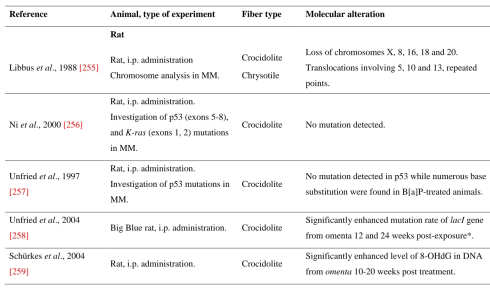
Mutations in malignant mesotheliomas have been investigated in animals, 107
after in vivo exposure to asbestos fibers. Table 22-1 summarizes genomic 108
alterations in MM identified in asbestos-exposed animals. Although few 109
studies have been performed, these results are consistent with observations 110
made in human MM. Chromosome rearrangements were observed in wild-111
type animals exposed to asbestos. Mutations and base hydroxylation have 112
been detected within several weeks after asbestos administration. At the gene 113
level, no or few mutations were found in the tumor suppressor gene (TSG) 114
Tumor protein p53 (Trp53), both in wild-type rats and heterozygous NF2 115
mice. Interestingly, genes at the Ink4a locus were deleted, as found in human 116
MM. In MM from genetically modified mice, gene inactivation occurred by 117
loss of heterozygosity (LOH). These studies suggest that asbestos fibers are 118
genotoxic, and can produce DNA strand breaks and chromosomal 119
recombination. 120
ii. Effects of asbestos fibers on mesothelial cells in culture. 121
While early studies have been carried out with cells of different species and 122
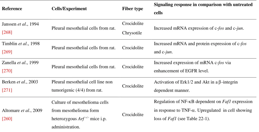
tissues, rat and human mesothelial cells have been most widely used to study 123
the response of mesothelial cells to asbestos fibers. Detailed data can be 124
found in a several reviews [28,29]. 125
Various types of asbestos fibers have been found to cause cytotoxic and 126
genotoxic defects in primary cell cultures and in animals exposed to fibers 127
[30]. Typically, chromosomal breaks, centromeric and telomeric alterations 128
as well as aneuploidy (an lower number of chromosomes in comparison with 129
normal cells), polyploidy (twice or several times the normal number of 130
chromosomes) and heteroploidy (an abnormal number of chromosomes) due 131
to spindle defects, are seen. Because of chromosomal breaks, as well as 132
spindle and centrosomal damage, micronucleus formation is a typical feature 133
of asbestos-induced genotoxicity, whereby genotoxic endpoints are 134
quantitated by scoring the number micronuclei [31]. 135
Table 22-2 summarizes genomic alterations in mesothelial cells in culture 136
treated with asbestos fibers. Briefly, when exposed to asbestos fibers, 137
mesothelial cells demonstrate phagocytic properties. Within hours, responses 138
to oxidant stress, activation of the Mitogen-Activated Protein Kinase 139
(MAPK) pathway, and induction of transcription factors are detected. Table 140
22-3 summarizes activation of various signaling pathways in mesothelial 141
cells in culture exposed to asbestos fibers. When incubated in the absence of 142
serum or in low levels of serum concentration, cell proliferation was 143
observed [32,33]. In proliferating mesothelial cells, asbestos provoked a 144
p53- and p21-dependent cell cycle arrest consistent with the induction of a 145
DNA damage-induced response [34]. P53 was also induced in serum-146
deprived G0 synchronized mesothelial cells exposed to asbestos, but failed 147
to block cell cycle progression [35]. However, genotoxicity was also found 148
suggesting that the DNA repair mechanism was incomplete, error-prone, or 149
impaired. 150
Several types of genetic damage have been found in asbestos-exposed 151
mesothelial cells (Table 22-2). Briefly, DNA damage was demonstrated 152
directly by the occurrence of DNA breakage [36-39], and indirectly by the 153
induction of DNA repair [40,41]. Oxidation of deoxyguanosine has been 154
reported in several studies. Notably, recurrent chromosome abnormalities 155
have been reported. These consist in numerical and structural changes, 156
including aneuploidy and polyploidy, micronucleus formation, and 157
chromosomal missegregation [42-48]. Comparison between different studies 158
showed that significant effects were found with doses of 0.5 - 1 µg/cm2 [29]. 159
These studies demonstrate that asbestos fibers are genotoxic for mesothelial 160
cells, able to produce base hydroxylation, DNA breakage, and numerical and 161
structural chromosomal changes in mesothelial cells. DNA repair processes 162
are stimulated in asbestos-treated mesothelial cells. The consequences of 163
DNA damage will be dependent on the efficiency and fidelity of repair. 164
When genomic damage is extensive, an apoptotic program should be 165
induced. As discussed previously, life-or-death decisions may be at the heart 166
of malignant transformation and defective mechanisms of arrest or apoptosis 167
may be critical to development of malignancy [49]. Several studies with 168
mesothelial cells in culture have emphasized the occurrence of apoptosis, 169
which should be beneficial for the mesothelium. However, some cells can 170
survive with gene alterations that can be inherited in daughter cells. In that 171
context, it is remarkable that mesothelial cells show both cell cycle arrest 172
and mitotic abnormalities, suggesting that the cells could pass through cell 173
cycle checkpoints with unrepaired DNA and chromosomal damage. 174
According to our knowledge, no data on epigenetic changes in asbestos-175
exposed cells in culture, or in animals have been reported. Further 176
investigations would be of great interest for our understanding of the 177
mechanism of action of asbestos fibers in carcinogenesis. 178
iii. MM in genetically modified mice 179
Several models of MM have been developed using genetically modified 180
mice exposed to mineral fibers. One study was based on mice carrying a 181
heterozygous mutation in the TSG Trp53 (Trp53+/-), and others on mice 182
heterozygous for a mutation on the neurofibromin 2 gene (NF2), a TSG 183
known to be inactivated in human MM (Nƒ2+/- mice). Interestingly MM cells
184
obtained from Trp53+/- mice exhibited Trp53 LOH and polyploidy [50]. 185
LOH of the Nƒ2 gene was found in Nƒ2+/- mice suggesting a common
186
mechanism for loss of the wild type (WT) allele [23,51]. Moreover, in NF2 +/-187
mice, two other TSG, cyclin-dependent kinase inhibitor 2a (p16/cdkn2a) and 188
cyclin-dependent kinase inhibitor 2b (p15/cdkn2b) were deleted at a high 189
rate, similar to human MM, while Trp53 was mutated at a much lower rate 190
[51,52]. In studies carried out by one of us (MCJ), Nƒ2 and Trp53 were 191
exclusively inactivated. Spontaneous MM in the absence of asbestos 192
exposure have been generated in double mutants Nf2+/-;Trp53-/- and Nf2 +/-193
;Ink4a/Arf-/- mice. MM developed rapidly and at a high incidence [53]. These 194
results suggest that MM development can be associated with inactivation of 195
TSG involving several pathways including Trp53 or Nƒ2 and genes at the 196
Ink4a locus, the two latter genes being more specific targets of asbestos
197
effects. Murine MM closely mimicked the human disease characterized by 198
peritoneal ascites, a long latency between fiber injection and MM 199
development, and histological subtypes, epithelioid, sarcomatoid and 200
biphasic, similar to human MM. The results obtained with genetically 201
modified mice show that MM progression could follow several routes 202
involving different TSG, and are in good agreement with (i) specific clinical 203
features and molecular alterations in human MM, and (ii) the role of tobacco 204
smoke in cancer development. It is generally accepted that MM is not related 205
to smoking, and that p53 mutation is a signature of tobacco smoke, 206
consistent with no signature of tobacco smoke in MM development. 207
Nevertheless, this strongly suggests that other carcinogens targeting p53 that 208
could reach the pleura would be able to induce MM. 209
c. Fiber properties in relation to the biological effects and carcinogenic potency
210
This chapter will discuss the biological mechanisms leading to development 211
of diffuse malignant mesothelioma focusing on the physiochemical 212
properties of asbestos fibers, carbon nanotubes, and other engineered high 213
aspect ratio nanomaterials relevant for the pathogenesis of this cancer. The 214
reader is referred to the comprehensive reviews cited above for a detailed 215
summary of the toxicological studies related to biological activity of carbon 216
nanotubes. 217
i. Mineral fibers 218
Asbestos and erionite are naturally-occurring fibrous minerals that have been 219
associated with the development of diffuse malignant mesothelioma in 220
epidemiological studies [54,55]. Asbestos fibers are fibrous silicates and are 221
classified into two groups based on their crystal structure and chemical 222
composition: serpentine asbestos which is called chrysotile and amphibole 223
asbestos which includes crocidolite, amosite, tremolite, actinolite, and 224
anthophyllite [56,57]. Erionite fibers are a form of the mineral zeolite 225
characterized by a high internal surface area [58]. These naturally-occurring 226
fibrous minerals are variable with respect to chemical composition, 227
associated minerals, and trace contaminants depending on their geographic 228
origin [59]. Asbestos fibers may contaminate other mineral deposits, for 229
example, talc [54,60] and vermiculite from Libby, Montana [60,61] and 230
exposure to these mixed materials have also been linked with diffuse 231
malignant mesothelioma [58,62]. The physiochemical properties of mineral 232
fibers associated with biological activity include shape and dimensions, 233
surface chemistry and reactivity, and biopersistence [19]. 234
ii. Shape and dimensions 235
Elongated fibers with a high aspect ratio, defined as a length: diameter 236
ratio of 3:1 or greater, are characteristic of the crystalline structure of the 237
mineral. Asbestos fibers occur as bundles of individual crystals or fibrils that 238
split longitudinally at the silicate layers. Fiber length and diameter determine 239
respirability and site of deposition in the lungs and fiber length is related to 240
efficiency of phagocytosis by alveolar macrophages and rate of clearance 241
from the lungs [19]. 242
Titanium dioxide nanorods have been shown to induce frustrated 243
phagocytosis and activation of the Nalp3 inflammasome [63] similar to 244
asbestos fibers [64]. Carbon nanotubes have also been shown to induce 245
frustrated phagocytosis by macrophages in vitro [65]. In rodents, long rigid 246
carbon nanotubes have been shown to translocate to the subpleural regions 247
of the lungs [66-69] and to induce inflammation, frustrated phagocytosis, 248
and granulomas similar to asbestos fibers following intraperitoneal injection 249
[65]. Direct intraperitoneal [70] or intrascrotal injection [71] of some 250
commercial carbon nanotubes induced diffuse malignant mesothelioma in 251
heterozygous p53-deficient mice and wild type rats, respectively. However, 252
short multiwalled carbon nanotubes (< 1 μm long) did not induce 253
mesotheliomas in rodents following intraperitoneal injection [72]. 254
iii. Surface chemistry and reactivity 255
Serpentine or chrysotile asbestos is a magnesium silicate (Mg3 Si2O5(OH)4);
256
Al3+ or Fe2+ may substitute for Si4+ or Mg2+. Amphibole asbestos fibers are 257
double-chain silicates containing a variety of cations including Fe2+, Fe3+,
258
Mg2+, Al3+, Ca2+, and Na+. Surface chemistry determines interactions 259
between the fiber, physiological fluids, and cells with possible proton 260
transfer, oxidation-reduction reactions, and adsorption of biological 261
macromolecules [58]. Broken chemical bonds at the fiber surface are highly 262
reactive with molecular oxygen and can generate free radicals in aqueous 263
fluid [73]. Surface Fe2+ and Fe3+ ions on amphibole asbestos fibers are 264
bioavailable and catalyze formation of reactive oxygen species (ROS) [74]. 265
Erionite fibers can acquire Fe2+ and Fe3+ ions and become redox active in the 266
presence of intracellular chelators or reductants such as citrate or ascorbate 267
[75]. Iron-catalyzed redox activity has been associated with biological 268
effects of mineral fibers including lipid peroxidation, oxidative DNA 269
damage, and activation of intracellular signaling pathways [76,77]. 270
Genotoxicity of natural and man-made fibers has been linked with surface 271
reactivity, especially redox activity, as detected using acellular assays for 272
free radical generation [78], induction of micronuclei [28], and mutagenicity 273
using a hamster-human hybrid cell line [79]. Amphibole and chrysotile 274
asbestos fibers show strong activity using these assays, while silicon carbide 275
fibers show no free radical activity [78]. Refractory ceramic fibers contain 276
bioavailable iron and are active in the salicylate assay to detect release of 277
hydroxyl radicals [78]. Chrysotile asbestos fibers, tremolite (an amphibole 278
fiber that contaminates chrysotile deposits), and erionite are mutagenic in the 279
hamster-human hybrid cell line, while refractory ceramic fibers are non-280
mutagenic [79]. 281
The ability of carbon nanotubes to generate free radicals is controversial. 282
Some commercial carbon nanotube samples have not been shown to 283
generate carbon or oxygen-centered free radicals using spin-trapping and 284
electron spin resonance [68,80]. In fact, carbon nanotubes can scavenge 285
hydroxyl and superoxide radicals which has been attributed to defects in the 286
graphene sidewalls creating gaps in the carbon lattice and dangling bonds 287
[81]. Multiwalled carbon nanotubes are not directly mutagenic in bacterial 288
reverse mutation assays [82]. Agglomerated multiwalled carbon nanotubes 289
are also negative in this assay and do not induce chromosome aberrations in 290
the V79 cell assay [83]. Long multiwalled carbon nanotubes, but not short 291
multiwalled carbon nanotubes or long single-walled carbon nanotubes, 292
induced DNA strand breaks in human lung epithelial cells [84]. Multiwalled 293
carbon nanotubes also induced micronuclei in rat lung epithelial cells in 294
culture and in animals [85]. Single-walled carbon nanotubes, carbon 295
nanofibers, and graphite nanofibers induced micronuclei in V79 cells [86] 296
and human bronchial epithelial cells [87]. Both single-walled and 297
multiwalled carbon nanotubes have been shown to induce oxidative stress, 298
DNA damage, and activation of intracellular signaling pathways in cultures 299
of human mesothelial cells [88,89]. 300
Direct generation of ROS at the surface of asbestos or erionite fibers may be 301
amplified by secondary generation of reactive oxygen and nitrogen species 302
by target cells, including inflammatory cells and mesothelial cells in the 303
pleural lining [76,90]. Target cells generate endogenous ROS and reactive 304
nitrogen species during the process of phagocytosis [91], disruption of 305
mitochondrial electron transport [77], and activation of inducible nitric oxide 306
synthase generating nitrogen-derived radicals [92]. These exogenous and 307
endogenous reactive oxygen and nitrogen species have multiple effects on 308
target cells in the pleura that amplify the inflammatory response, activate 309
inflammatory cells to release chemokines, cytokines, and other mediators, 310
stimulate cell proliferation, and induce cell injury and apoptosis [64,76]. 311
Fiber length has also been associated with induction of aneuploidy and 312
chromosomal damage due to direct physical interference with the mitotic 313
apparatus [28,93] or by binding to cell cycle regulatory proteins [94]. 314
Induction of chromosomal breaks and aneuploidy has been shown for single-315
walled carbon nanotubes and carbon nanofibers in V79 cells [86] and for 316
single-walled and multiwalled carbon nanotubes in rat [85] and human [87] 317
lung epithelial cells. These direct physical effects of long, thin fibers on 318
target cells in the lungs and pleura raise concern about potential 319
carcinogenicity of man-made mineral fibers that have been developed as 320
asbestos substitutes [19] or engineered fibrous nanomaterials including 321
carbon nanotubes [4,95] and metal and metal oxide nanorods or nanowires 322
[63]. Although these man-made fibers and engineered nanomaterials may 323
not have intrinsic redox activity, other surface properties (e.g., structural 324
defects or carbonaceous residues on the surface of carbon nanotubes) may 325
generate oxygen-derived radicals. 326
iv. Biopersistence 327
A major determinant of fiber pathogenicity is biopersistence in the lungs 328
[19]. If long fibers are not efficiently cleared or destroyed by physical 329
breakage, splitting or chemical dissolution in the lungs, they are called 330
biopersistent [19]. Differences in biopersistence of asbestos fibers have been 331
linked with carcinogenic potency, as biopersistent fibers could sustain a local 332
inflammatory response [96]. Amphibole asbestos fibers are more potent than 333
chrysotile asbestos fibers due to their increased biopersistence in the lungs. 334
However, fiber biopersistence in the pleura is not documented; in particular, 335
there are no data on the relationship between biopersistence in the lung and 336
translocation of fibers from the lung to the pleura, nor on the pleural 337
clearance of fibers following inhalation [97,98]. 338
Biopersistence of natural and man-made fibers in the lungs [99] or 339
peritoneal cavity [100] is an important characteristic of fibrous materials that 340
induce lung cancer and diffuse malignant mesothelioma in rodents following 341
inhalation [19]. Man-made mineral fibers developed as asbestos fiber 342
substitutes, especially refractory ceramic fibers [26] and silicon carbide 343
whiskers, have been shown to be biopersistent [101] following inhalation by 344
rodents. Following inhalation by hamsters, refractory ceramic fibers 345
translocated to the pleura and induced mesothelial cell proliferation and 346
fibrosis [26]. Refractory ceramic fibers also induced pleural malignant 347
mesotheliomas after chronic inhalation by rats and hamsters [19]. 348
Intrapleural [102] or intraperitoneal injection of silicon carbide whiskers 349
[103] also induced diffuse malignant mesothelioma in rats. Although no 350
malignant mesotheliomas have been reported in worker cohorts involved in 351
manufacturing and application of refractory ceramic fibers, the rodent 352
carcinogenicity assays raise concern that long thin biopersistent mineral 353
fibers may be carcinogenic [104]. Erionite fibers are very potent in induction 354
of malignant mesotheliomas following intrapleural injection [105] or 355
inhalation [106]. 356
Natural and man-made fibers are not unique in induction of rodent 357
malignant mesotheliomas following intraperitoneal or intrapleural injection. 358
A variety of chemicals, radionuclides, SV40 virus, and metallic nickel 359
particles are also carcinogenic in this rodent bioassay [107]. From a 360
mechanistic viewpoint, ferric saccharate, nitrilotriacetic acid, nickel 361
particles, and alpha- or beta- emitting radionuclides are notable in their 362
abilities to generate reactive oxygen species [108]. 363
Unfunctionalized carbon nanotubes are bioperistent when assessed in 364
acellular assays [109]; however, carboxylated single-walled carbon 365
nanotubes are susceptible to enzymatic [110] or oxidative degradation [111]. 366
In principle, carbon nanotubes could be engineered to alter their 367
physiochemical properties in order to decrease their biological reactivity and 368
potential carcinogenicity. 369
High aspect ratio and biopersistence [4,112] have been hypothesized to be 370
important properties of engineered nanomaterials that raises concern about 371
their potential to be translocated to and retained in the pleural following 372
inhalation. So far, this hypothesis has not been tested in any long-term 373
inhalation studies of high aspect ratio engineered nanomaterials in rodents. 374
v. Unique characteristics of nanomaterials 375
Additional features of engineered carbon nanomaterials that may alter their 376
biological activity include their purity, rigidity, hydrophobicity, and 377
agglomeration state. Carbon nanotubes are frequently produced 378
commercially in the presence of metal catalysts including nickel, iron, 379
cobalt, and yttrium [113]. Other potential contaminants include combustion-380
derived products such as polycyclic aromatic hydrocarbons [113]. 381
Amorphous carbon residues at the graphenic surface of carbon nanotubes 382
may also contribute to surface reactivity [85]. Bioavailability of metal 383
catalyst residues is variable depending on the purity of carbon nanotubes; 384
redox active metal catalyst residues can generate reactive oxygen species 385
leading to cell toxicity, inflammation, activation of intracellular signaling 386
pathways involving the MAPK and the nuclear factors NF-B and AP-1 387
[76], and genotoxicity [95]. Carbon nanotubes can be highly variable in 388
length ranging from 1nm to 1mm. Although short nanotubes and nanofibers 389
less than 5 μm in length should be more easily phagocytized and cleared 390
following inhalation [4], they may behave as needles and penetrate into cells 391
and the nucleus where they could directly damage chromosomes and DNA 392
[95]. Unfunctionalized carbon nanomaterials are very hydrophobic and tend 393
to form agglomerates or bundles called nanoropes, although individual 394
carbon nanotubes have been detected in aerosols [114]. Hydrophobic 395
nanomaterials may interact differently with biological macromolecules in 396
comparison with hydrophilic crystalline mineral fibers [115]. Very thin, 397
hydrophobic carbon nanotubes may bend and agglomerate to form spherical 398
aggregates that are more readily phagocytized than long, rigid multiwalled 399
carbon nanotubes that have been shown to induce frustrated phagocytosis 400
resulting in impaired clearance and translocation to the pleura [4,116]. The 401
extent of agglomeration has also been shown to influence cell toxicity: rope-402
like agglomerates of carbon nanotubes were shown to be more toxic than 403
crocidolite asbestos fibers using a mesothelioma cell line [115]. Finally, 404
structural defects at carbon nanotube surfaces attributed to imperfections in 405
the graphene lattice or defects leading to surface oxidation and increased 406
hydrophilicity have been shown to contribute to acute toxicity and 407
genotoxicity of even short multiwalled carbon nanotubes [85]. 408
The potential of engineered carbon nanotubes to induce pathological 409
reactions (lung inflammation, fibrosis, and diffuse malignant mesothelioma) 410
similar to asbestos fibers has generated significant controversy and concern 411
for occupational safety and health [112,117]. Occupational exposures via 412
inhalation, skin contact, and ingestion are possible during the synthesis, 413
handling, and fabrication steps of engineered carbon nanotubes; airborne 414
mass concentrations in the range of 0.7 – 430 μg/m3 have been detected at 415
eight worksites and research laboratories [118]. Several recent reviews have 416
summarized the numerous in vitro cellular and rodent toxicology studies 417
investigating biological activity and potential toxicity of carbon nanotubes 418
[90,114,116,118]. 419
d. Summary hypotheses on the mechanism of action of asbestos fibers to generate
420
mesothelioma
Development of diffuse malignant mesothelioma is a complex, multistage 422
process that is governed by the physicochemical properties of crystalline 423
mineral fibers and their propensity to migrate to the pleural and peritoneal 424
linings as summarized in Figure 22-1. The most important properties of 425
asbestos fibers related to carcinogenicity are fibrous shape and dimensions, 426
surface chemistry and reactivity, and biopersistence [19]. Long, rigid 427
biopersistent fibers that are translocated to the pleura are trapped on the 428
parietal pleura lining at the sites of lymphatic openings [27] and incite a 429
persistent inflammatory response [4]. The pleura is covered by a thin, single 430
layer of mesothelial cells that have lower antioxidant defenses than lung 431
epithelial cells [119]. 432
Interactions between mesothelial cells and fibers can cause genetic and 433
chromosomal changes. There is a great body of evidence that 1) asbestos 434
fibers can directly interfere with chromosomes and the mitotic spindle [120-435
122], and 2) that they induce formation of reactive oxygen species (ROS) 436
resulting in DNA breaks, oxidation, and mutations [123]. Further, 3) the 437
physical interaction of fibers with target cells causes persistent inflammation 438
and, consequently, modulation of inflammatory and immune responses. 439
ROS have been clearly indicated to cause genetic damage including 440
chromosomal breaks and mutations [123]; and they are well shown to initiate 441
signal transduction pathways that are, in turn, linked to inflammation, 442
proliferation, and apoptosis [124]. Free radical scavengers have reported to 443
decrease genotoxic endpoints such as micronucleus formation induced by 444
fibers [125]. Further, there is clear-cut evidence that antioxidant enzymes 445
can protect cells against genotoxicity induced by chrysotile fibers [126]. 446
Prolonged interaction between pleural inflammatory cells and adjacent 447
mesothelial cells causes persistent release of chemokines and cytokines, 448
inflammatory mediators, reactive oxygen and nitrogen species, and growth 449
factors that trigger repeated episodes of inflammation resulting in 450
mesothelial cell injury, death, and proliferation [127]. In this chronic 451
inflammatory microenvironment, genomic instability and acquired genetic 452
and chromosomal alterations in mesothelial cells may lead to altered cell 453
cycle and growth regulation, resistance to apoptosis [128], impaired repair of 454
DNA and chromosomal damage induced directly or indirectly by asbestos 455
fibers [28,93], and activation of oncogenes and inactivation of tumor 456
suppressor genes [129]. Persistent inflammation has also been linked with 457
altered gene methylation patterns identified in diffuse pleural malignant 458
mesotheliomas in humans [130]. DNA methylation leads to epigenetic gene 459
silencing and has been linked to inflammation-mediated damage to cytosine 460
[131] or endogenous generation of methyl radicals [132]. This persistent 461
inflammatory microenvironment in combination with oxidative stress 462
generates a strong selective force for mesothelial cells that have acquired 463
genetic and epigenetic changes that promote their survival, proliferation, and 464
tumor progression [133]. 465
466
3. Mesothelial cells and malignant mesothelioma 467
a. The mesothelial cell in situ. Normal cells.
The mesothelium consists of a monolayer of mesothelial cells lying on a basement 469
membrane and supported by connective tissue stroma containing fibroblasts. 470
Mesothelial cells provide a protective barrier for frictionless interface for the free 471
movement of apposing organs and tissues, and in fluid transport across the pleura 472
[134]. Mesothelial cells may have specialized functions at different anatomical sites, as 473
demonstrated by morphological studies at the ultrastructural level [135]. Mesothelial 474
cells play a role in the resolution of inflammation and tissue repair after pleural injury. 475
Fibrosis is a potential outcome of chronic inflammation. These processes are of 476
particular interest in investigating the mechanism of action of asbestos fibers in the 477
pleura. 478
So far, the mechanism of mesothelial cell regeneration is poorly understood, mostly in 479
the context of serosal injury following dialysis; however some controversial 480
hypotheses have been formulated. Recent comprehensive reviews summarize our 481
present knowledge of these potential mechanisms [136,137]. The regeneration process 482
has been studied experimentally following mechanical, chemical or heat injury of the 483
peritoneal serosa. Briefly, six mechanisms have been suggested to replace the injured 484
mesothelial cells : (i) centripetal migration of adjacent mesothelial cells, (ii) exfoliation 485
of mature or proliferating mesothelial cells that replicate on the wound surface, (iii) 486
pre-existing free-floating serosal cells having the capability to differentiate into new 487
mesothelium, (iv) macrophage transformation into mesothelial cells, (v) 488
submesothelial mesenchymal precursors that migrate to and differentiate at the 489
mesothelium surface, and (vi) bone marrow-derived circulating precursors [137]. 490
The origin of these new mesothelial cells has not yet been confirmed, but according to 491
Mutsaers et al. [136], mesothelial regeneration is not dependent on subserosal cells, but 492
more likely results from implantation, proliferation, and incorporation of free-floating 493
mesothelial cells [138]. 494
b. The malignant mesothelioma cell
495
i. Role of gene mutations in the neoplastic transformation of mesothelial 496
cells 497
Carcinogens provoke several types of somatic gene mutations, consisting of 498
DNA and chromosome alterations. Some mutations are the signature of past 499
exposure to one or several given carcinogens. Somatic mutations in tumors 500
are of interest both to determine the mechanism of action of carcinogens, and 501
to elucidate their adverse consequences on cellular homeostasis. 502
In malignant pleural mesothelioma (MPM), there is a limited number of 503
genes known to be recurrently mutated. Mutations in TSG cyclin-dependent 504
kinase inhibitor 2A (P16/CDKN2A), an alternative open reading frame of 505
CDKN2A generating a distinct protein (P14/ARF), cyclin-dependent kinase
506
inhibitor 2B (P15/CDKN2B) and NF2 have been reported in a high 507
percentage of MM, and TP53 (tumor protein p53) has been found mutated at 508
a lower rate in comparison with other human cancers [139-141]. These genes 509
play a role in cell cycle regulation at different levels. The CDKN2A locus 510
encodes two different proteins, p16INK4A and p14ARF while P15/CDKN2B 511
encodes one protein p15INK4B. Both p16INK4A and p15INK4B are inhibitors of 512
the kinase function of cyclin/cdk complexes involved in cell cycle 513
progression. TP53 encodes a protein, p53, which is activated in response to 514
DNA damage and is a regulator of apoptosis. The protein p14ARF has indirect 515
function on cell cycle regulation, by positively regulating the level of p53 516
through interaction with p53 inhibitors. Consequently, cells with damaged 517
DNA can proliferate and survive in the absence of p14ARF. Interestingly, all 518
of these genes carry different types of mutations. The most frequent 519
alteration at the P16/CDKN2A and P15/CDKN2B loci is homozygous 520
deletion in about 70% of MM cases [141]. In murine models of asbestos-521
induced mesothelioma, the orthologous genes, p16/Cdkn2a and p15/Cdkn2b, 522
were also inactivated by deletion [51,52,142]. It can be also noted that 523
P16/CDKN2A deletions have been considered as a marker of asbestos
524
exposure in a study of non small cell carcinomas [143]. However, in MM, 525
DNA methylation of P16/CDKN2A and P15/CDKN2B has been reported at a 526
frequency of 13% (9 patients) and 4% (3 patients) respectively, and 527
positively correlated with asbestos body counts in the lung [130,144]. The 528
average methylation frequency of these gene in the literature is about 10% 529
[52,144-149]. It was also suggested that mesotheliomas express microRNA 530
(miRNA) that could inhibit P16/CDKN2A expression, based on in silico 531
analysis for miRNA target gene prediction [150]. 532
Point mutations are the main types of alterations of TP53 in MM. Six point 533
mutations are indicated in the IARC p53 database, five missense mutations 534
and one stop mutation [151]. So far, no specific type of mutation in TP53 535
has been related to asbestos exposure. In lung cancer, G:C-to-T:A 536
transversions are generally interpreted as mutagenic fingerprints of tobacco 537
smoke [152]. This base substitution can be due to the formation of 8-OH-538
deoxyguanosine generated by oxidative damage, which in turn causes 539
primarily G-to-T transversions. A few studies have reported TP53 mutations 540
in relationship to asbestos exposure. In lung cancer, the frequency of 541
mutations was diminished in lung adenocarcinomas of asbestos-exposed 542
subjects in comparison with unexposed patients but the difference was not 543
significant [153]. G-to-T transversions in asbestos-exposed lung cancers 544
have been reported, but not in all studies, and G:C-to-A:T transitions are rare 545
[153]. 546
TP53 mutations reported in MM consisted of different types of base
547
substitution and base deletion but G:C-to-A:T transitions seems to be the 548
most frequent [151] (unpublished data from MCJ). In animal models of MM, 549
the mutated status of Trp53 was investigated in mice exposed to mineral 550
fibers by intraperitoneal inoculation. In C57Bl/6 p53+/- mice, a strain having 551
one allele mutated in the gene Trp53, loss of the wild type allele was found 552
at a high rate in MM induced by asbestos fibers [154]. In Nƒ2WT and Nƒ2
+/-553
FVB mice, Trp53 alterations were infrequent. Two point mutations A:T-to-554
C:G were detected in mice exposed to asbestos, and two point mutations, 555
A:T-to-G:C and A:T-to-T:A, and a duplication of 12 bases in MM were 556
found in mice exposed to ceramic fibers [52,142]. Frequency of alteration in 557
the chromosomal region of the Trp53 locus was infrequent [155]. These 558
results suggest that deletions would be more likely a consequence of the 559
mechanism of action of asbestos, while p53 point mutations could be related 560
to “spontaneous” gene alterations in this model. 561
Alterations of NF2 TSG are frequently found in about 50% of MPM 562
[156,157]. This gene encodes merlin, a protein found in cell-cell junctions 563
and microvilli and regulating contact-dependent cell proliferation [158,159]. 564
NF2 has pleiotropic functions, being involved in regulation in cell
565
proliferation, apoptosis and endocytic trafficking, and acting upstream of the 566
Hippo signaling pathway [160]. Mutations in NF2 consist of both point 567
mutations and deletions [129]. So far, there is no explanation for the high 568
level of alterations in NF2 in MPM. However, some hypotheses can be 569
formulated and will be discussed below (see 4. Concluding remarks). 570
571
ii. Role of genomic alterations in the neoplastic transformation of mesothelial 572
cells 573
Chromosome banding, fluorescence in situ hybridization, flow cytometry, 574
Southern blotting and chromosome and array comparative genomic 575
hybridization, single nucleotide polymorphism (SNP) array and representational 576
oligonucleotide microarray analysis (ROMA) as well as second generation 577
sequencing analyses all indicate complex genomic alterations in MM [161-578
171]. Typically, chromosomal abnormalities are very complex, even chaotic, 579
that is involving alterations both in chromosome structure and number 580
[161,163-165,172-174]. It is characteristic for this disease that the 581
chromosome number is mostly hypodiploid (less than 46 chromosomes, the 582
normal number of chromosomes in human), but it varies greatly within a 583
specimen, as a given tumor can exhibit a variety of hypodiploid metaphases 584
[161]. Similarly, polyploid forms (with a number of chromosomes twice or 585
more the number of chromosomes present in the parental cell) of the 586
hypodiploid clone are commonly encountered. Other cytogenetic alterations 587
may be observed such as diplochromosomes of endoreduplication which are 588
a signature of alteration of the mitotic process. The polyploidization and 589
non-disjunction type of aneuploidy are due to fiber-induced damages to the 590
structures involved in cell division, i.e. the middle spindle, centrosome, 591
centriole, cleavage furrow, and cell membrane. 592
Similar to numerical chromosomal aberrations, structural aberrations in MM 593
are highly variable. Typically, translocations, deletions, insertions and 594
inversions are seen. Occasionally, double minutes and a homogenously 595
staining region, representing cytological manifestations of gene 596
amplification are also observed. So far, translocations are mainly unbalanced 597
and no recurrent chromosomal translocations with known fusion genes have 598
been reported. Due to chaotic nature of these aberrations and methodological 599
difficulties, the detection of specific chromosomal aberrations has been very 600
difficult in karyotypic analysis. Novel next generation sequencing methods, 601
such as exome sequencing, has facilitated overcoming the above-mentioned 602
problems and, for the first time, fusion genes have been described in MM 603
[169]. 604
These described structural changes are mainly due to DNA breaks. The 605
mechanism known as breakage-fusion-bridge phenomenon nicely explains 606
severe chromosomal imbalances and intratumoral heterogeneity in MM 607
[175]. As already mentioned, asbestos fibers are capable of causing DNA 608
breaks either directly or indirectly through ROS generation. Whether there 609
are hot spots in the genome for DNA breaks caused by fibers is still largely 610
an open question. However, experiments with cells in culture have indicated 611
that chromosome aberrations induced by fibers may be recurrent. Certain 612
numerical chromosomal abnormalities have been reported to be over-613
represented in human pleural cell cultures exposed to asbestos [176]. Even 614
though no distinct hot spots were seen in this study, chromosome 1 seemed 615
to be involved more often than other chromosomes. Interestingly, we have 616
previously reported that structural aberrations in the short arm of 617
chromosome 1 and loss of material in the short arm chromosomes 1 and 4 618
were associated with high asbestos fiber burden in MM [177,178]. More 619
recently, one of us (DJ) identified a recurrent region of chromosome loss, 620
14q11.2-q21, in MM from asbestos-exposed patients that was not found in 621
unexposed patients. The syntenic region that also lost in fiber-induced MM 622
in mice, suggesting that this region might be a target of action of mineral 623
fibers [155]. 624
Very recent experiments from one of us (SK), carried out with cell lines and 625
with lung tumor tissues (not mesotheliomas) of patients who had been either 626
exposed or unexposed to asbestos fibers, indicated a couple of asbestos 627
associated chromosomal areas. These findings are described in detail in 628
Chapter 24. Even though chromosomal aberrations in MM are complex, they 629
are not random and they are clonal in nature originating from one cell. 630
Chapter 24 describes, in more details, these recurrent aberrations and their 631
clinical significance. 632
The chromosomal alterations characteristic of MM, such as hypodiploid 633
chromosomes number as well as deletions and losses in chromosome 14 and 634
10, are not seen in lung adenocarcinoma, which helps in differential 635
diagnosis of these malignancies [179,180]. Interestingly, chromosomal 636
aberrations in gastrointestinal stromal tumors (GIST) resemble those seen in 637
MM [181]. To our knowledge GIST is not, however, an asbestos related 638
tumor, but the similarities of chromosomal alterations may, instead, be 639
related to similarities in mesenchymal stem cells from which the tumors 640
originate. 641
To conclude, asbestos fibers cause a wide variety of chromosomal 642
imbalances. Even though some of these changes may be recurrent, most of 643
them are random. Various genetic changes caused by asbestos fibers offer a 644
versatile genomic aberration reservoir, from which the aberrations promoting 645
uncontrolled growth and malignant transformation are selected during the 646
long initiation and progression (latency) period before tumor diagnosis. 647
Variable chromosomal aberrations together with multifocal clonal evolution 648
are consistent with this mechanism. 649
iii. Role of epigenetic alterations in the neoplastic transformation of 650
mesothelial cells 651
Altered gene expression in MPM could also be due to epigenetic 652
mechanisms. MPM show specific patterns of gene methylation as compared 653
to normal pleura or other tumors [130,145,148,182,183]. Data on 654
methylation profiles of MPM will be described in detail chapter 24. Several 655
studies suggested that DNA methylation at specific gene loci could be 656
correlated with asbestos exposure. Significant associations between asbestos 657
exposure and DNA methylation were first described in genes encoding 658
heavy metal binding proteins, MT1A and MTA2, with a positive association 659
for MT1A, but not for MT2A. Asbestos exposure does not seem to be an 660
independent variable in this study [184]. A trend towards an increasing 661
number of methylated cell cycle control genes (APC, CCND2, CDKN2A, 662
CDKN2B, HPPBP1 and RASSF1) and increasing asbestos body counts was
663
observed [144]. These findings were confirmed in a more recent, high-664
throughput methylation analysis underlining distinct methylation profiles 665
between MPM from asbestos-exposed and unexposed patients, and a 666
significant positive association between asbestos fiber burden and 667
methylation status of CDKN2A, CDKN2B, RASSF1 and MT1A and about 668
one hundred other loci [130]. 669
MiRNAs are small (around 22 base pairs in size) RNAs that have a crucial 670
role in posttranscriptional gene regulation. Their biosynthesis and functions 671
have been described in more detail in Chapter 24. It has been demonstrated 672
that MPM has a characteristic miRNA profile and that different MPM 673
histopathological subtypes can be discriminated according to their profiles 674
(see Chapter 24). Even though significantly differentially expressed miRNAs 675
discriminated MPM patients according to smoking habit, this did not 676
significantly discriminate asbestos-exposed patients versus unexposed [150]. 677
The reason for this may be the low number of non-smoking patients. On the 678
other hand, it is possible that patients classified into the unexposed category 679
were actually exposed to asbestos fibers. Recent results provide evidence 680
that a group of miRNAs differentiates asbestos associated lung 681
adenocarcinomas from the non-associated tumors [185]. The results of these 682
lung carcinoma studies are presented in detail in Chapter 24. As the 683
mechanisms of the miRNA regulation are yet poorly understood, it is 684
premature to speculate how asbestos fibers cause miRNA dysregulation seen 685
in MM and in lung carcinomas. Nevertheless, some of them could be lost, as 686
their loci are located in chromosomal regions frequently altered in MPM and 687
possibly linked to asbestos exposure, as was demonstrated for miR31 which 688
is close to the CDKN2A locus [186]. So far, no experiments using cell 689
cultures or experimental animals have been published that investigate 690
miRNA profiles in asbestos-exposed cells or animals. Further investigations 691
are needed to elucidate the mechanisms responsible for miRNA 692
dysregulation and function in MM. In two MPM cell lines lacking either 693
miR31 or miR29C, overexpression by transfection of these miRNAs 694
decreased proliferation, migration, invasion, and colony formation 695
[186,187]. 696
The molecular mechanisms responsible for epigenetic changes in MPM are 697
poorly understood and it is not known whether they are directly induced by 698
asbestos or they are indirect effects. Nevertheless, as with chromosomal 699
imbalances, they most likely play a role in mesothelial carcinogenesis. 700
iv. Pathways involved in the neoplastic transformation of mesothelial cells 701
Constitutive activation of several signaling pathways has been demonstrated 702
in MPM by the occurrence of mutations and/or deregulated expression of 703
specific regulators in comparison with normal mesothelial cells. These 704
studies have been carried out in primary tumor samples but also in malignant 705
mesothelial cell cultures developed from tissue samples. Pathway activation 706
in MM has been shown by gene expression profiling. So far, the relationship 707
between pathway activation and asbestos exposure has not been specifically 708
investigated in MM. The effects of asbestos on mesothelial cells are 709
discussed in paragraph 22-2.b.ii. 710
The Hippo pathway is of special interest regarding the high frequency of 711
mutations detected in merlin encoded by the NF2 gene. Merlin negatively 712
regulates cell proliferation. Its activity is affected by interaction between 713
extracellular signals and membrane proteins, and activated merlin transduces 714
signals suppressing the transcriptional activity of YAP coactivator [141]. In 715
a recent study, another negative regulator of the hippo pathway, LATS2, was 716
found to be deleted in 3 out of 6 MM cell lines and in 1 out of 25 tumors by 717
DNA sequencing analyses [188]. Merlin exists in two forms, active 718
unphosphorylated or inactive phosphorylated. This later form is found in 719
MPM cells possibly accounting for another mechanism for deregulation of 720
the hippo pathway in these cells[189]. 721
Cell cycle. Alterationof CDK inhibitor genes located at the INK4 (CDKN2A 722
and CDKN2B) locus, as mentioned above, contributes to uncontrolled cell 723
proliferation. However, cell cycle control can be affected in MM cells not 724
only by the loss of other negative regulators, but also by the overexpression 725
of cyclin-dependent kinases (CDKs), cyclins (CCNs), and regulators of the 726
mitotic checkpoints. These alterations have been shown by gene profiling 727
analyses using microarrays [190-192]. Overexpressed genes were involved 728
in the regulation of all phases of the cell cycle, cell replication and control of 729
cell cycle progression: cyclin-dependent kinase 1 (CDK1/CDC2); cell 730
division cycle 6 (CDC6), a regulator of replication; cyclin-dependent kinase 731
inhibitor 2C, p18, (CDKN2C); cyclin H (CCNH); cyclin B1 (CCNB1), 732
controlling the cell cycle at the G2/M transition; forkhead box M1 733
transcription factor (FOXM1), a regulator of gene expression in the G2 734
phase. Others are more specific of a response to DNA damage such as 735
checkpoint kinase 1 (CHEK1). The protein encoded by this gene, Chk1, is 736
required for checkpoint-mediated cell cycle arrest in response to DNA 737
damage. Underexpression of cyclin D2 (CCND2), a regulator of Cdk4 and 738
Cdk6, which controls the cell cycle at the G1/S transition), was also detected 739
[190]. 740
Several genes involved in the control of entry in mitosis and mitosis 741
progression were also detected. Overexpression of aurora kinases has been 742
reported in several studies [191,193]. Stathmin, a gene involved in the 743
regulation of the microtubule dynamics, by inhibiting the formation of 744
microtubules and/or promoting their depolymerisation, was strongly 745
overexpressed in MPM, resulting in protein overexpression [194,195].
746
These results can account for the complex, even chaotic chromosomal 747
alterations mentioned above, as a result of defective control of cell cycle 748
progression through different phases of the cell cycle, including 749
dysregulation of mitosis. 750
Signaling pathways. The MAPK signaling pathway controls cell 751
proliferation and differentiation, survival, apoptosis and Wnt signaling [196]. 752
In normal cells, the MAPK pathway is triggered by the activating 753
phosphorylation of tyrosine kinase receptors (RTKs), followed by a protein 754
kinase cascade. Downstream networks from RTKs can be activated by RTK 755
mutation or sustained signaling through autocrine or paracrine mechanisms. 756
The MAPK signaling pathway is constitutively activated in MM as 757
demonstrated by the phosphorylation and activation of downstream proteins 758
of the MAPK cascade, extracellular-regulated kinases (ERKs), Jun amino-759
terminal kinases/stress-activated kinases (JNKs/SAPKs), and p38 MAPK 760
[197,198] and inhibition of cell proliferation and induction of apoptosis by 761
inhibitors of the pathway [199]. RTKs activation can be achieved by a 762
variety of growth factors, EGF family, PDGF, FGF, HGF/SF and cytokines 763
such as TGF-ß, TNF and IL1. In a recent study, the relative levels of tyrosine 764
phosphorylation of 42 distinct RTKs was determined in MM cell lines 765
established from surgical specimens. Coordinated activation of several 766
RTKs: EGFR, ERBB3, AXL and MET was found [200]. 767
MPM cells express both vascular endothelial growth factor (VEGF) and the 768
VEGF receptors (fms-related tyrosine kinases, FLT1 and FLT4, and fetal 769
liver kinase, KDR/FLK1) [201-204]. VEGF expression was enhanced in a 770
large proportion of MPM in comparison with nonneoplastic specimens 771
[205]. An autocrine role for VEGF in cell proliferation has been suggested 772
[203,206]. 773
MM cell growth may also be linked to autocrine or paracrine stimulation by 774
platelet-derived growth factor (PDGF), and the regulation by PDGF appears 775
to be complex in MM cells. PDGF has been suggested as a regulatory factor 776
for proliferation of MM cells, either directly or indirectly via the 777
hyaluronan/CD44 pathway [207,208]. Human MM cells express high levels 778
of PDGF-A, and PDGF-B and PDGFR-B while normal human mesothelial 779
cells express low levels of PDGF-A mRNA chain and PDGFR-A [209,210]. 780
PDGF-A could contribute to tumor formation via a paracrine mechanism 781
[211,212]. 782
Epidermal growth factor receptor (EGFR) is over-expressed in 44–97% of 783
MM as found by immunohistochemical studies, but no mutation was 784
detected in contrast with others types of cancer [213]. 785
Human MM cells express insulin growth factor (IGF) and insulin growth 786
factor receptors (IGFR), and the activation of IGFR activates downstream 787
signaling [214,215].IGF-I appears to function as an autocrine growth factor 788
in human mesothelial cells [216]. IGFBPs also regulate IGF-dependent 789
growth [215,217,218].
790
Hepatocyte growth factor receptor (MET) is a proto-oncogene. It is the 791
receptor for the ligand hepatocyte growth factor/scattering factor (HGF/SF). 792
Mutation in the MET gene has been detected in a few MM cell lines 793
[219,220]. Both MET and HGF/SF proteins are expressed in some MPM 794
[221,222]. In vitro HGF/SF increases spreading, motility and/or invasiveness 795
of mesothelial cell lines and inhibition of MET reduced cell proliferation 796
[219,223,224]. The activation status of MET and other RTKs, EGFR family
797
(Erb1, Erb2, Erb3), PDGF-A and PDGFR-B has been investigated in 20 798
MPM cell lines and 23 primary specimens of MPM, and the effect of MET-799
specific inhibitors (MET-shRNA interference vector and RTK inhibitors) 800
was investigated on cell lines [220]. The results showed that inhibition of a 801
single RTK was not sufficient to obtain a tumor suppressor effect but that 802
inhibition of multiple RTK was required [220]. 803
Activation of RTKs also induces activation of other downstream signaling 804
cascades including phosphatidylinositol-3-kinase (PI3K-AKT) pathway, 805
regulating cell survival and proliferation, cell migration and apoptosis. 806
Phosphorylation of AKT protein, the active form of the protein, and 807
activation of the Akt pathway have been demonstrated in MM cells 808
[225,226]. In PTEN, a TSG and negative regulator of the PI3K-AKT 809
pathway, homozygous deletion has been reported in a small subset of MPM 810
cell lines [227,228]. 811
The Wnt signaling pathway regulates developmental processes, cell 812
proliferation, and cell polarity and its activation prevents beta-catenin 813
inactivation, a coactivator of transcription, allowing the expression of a 814
variety of genes exerting pleiotropic effects [229]. However, cell growth 815
inhibition and apoptosis of MPM cells was observed according to a beta-816
catenin-independent inhibition of Wnt signaling [230,231]. In MPM, the 817
Wnt pathway could be altered as a result of promoter hypermethylation of 818
regulatory genes[230,232,233]. Gene expression profiling of MM cell lines, 819
primary MPM tumors and normal pleural tissue demonstrated that numerous 820
Wnt and Wnt-related genes were upregulated and that some WNT 821
antagonists were downregulated [234]. These results suggest that 822
deregulation of the Wnt signaling pathway is involved in mesothelial 823
carcinogenesis. 824
Apoptosis. Deregulation of signaling pathways likely plays an important role 825
in dysfunction of apoptosis in MPM. Moreover specific regulators can 826
contribute to MM resistance to apoptosis. In MM cells, apoptosis alteration 827