HAL Id: tel-00721757
https://tel.archives-ouvertes.fr/tel-00721757
Submitted on 30 Jul 2012
HAL is a multi-disciplinary open access
archive for the deposit and dissemination of
sci-entific research documents, whether they are
pub-lished or not. The documents may come from
teaching and research institutions in France or
abroad, or from public or private research centers.
L’archive ouverte pluridisciplinaire HAL, est
destinée au dépôt et à la diffusion de documents
scientifiques de niveau recherche, publiés ou non,
émanant des établissements d’enseignement et de
recherche français ou étrangers, des laboratoires
publics ou privés.
Découverte d’une nouvelle famille de protéine kinases
bactériennes : mécanismes de fonctionnement et rôle
cellulaire de YdiB, un archétype chez Baccillus subtilis
Hien-Anh Nguyen
To cite this version:
Hien-Anh Nguyen. Découverte d’une nouvelle famille de protéine kinases bactériennes : mécanismes
de fonctionnement et rôle cellulaire de YdiB, un archétype chez Baccillus subtilis. Sciences agricoles.
Université de Grenoble, 2012. Français. �NNT : 2012GRENV017�. �tel-00721757�
Université Joseph Fourier / Université Pierre Mendès France / Université Stendhal / Université de Savoie / Grenoble INP
THÈSE
Pour obtenir le grade de
DOCTEUR DE L’UNIVERSITÉ DE GRENOBLE
Spécialité : Chimie-Biologie
Arrêté ministériel : 7 août 2006Présentée par
Hien-Anh NGUYEN
Thèse dirigée par le Dr. Jean-Michel JAULT
préparée au sein de
l’Institut de Biologie Structurale J.-P. Ebel,
et du CEA de Grenoble
dans l'École Doctorale Chimie et Sciences du Vivant
Découverte d’une nouvelle famille de protéines kinases
bactériennes :
Mécanisme de fonctionnement et rôle cellulaire de
YdiB, un représentant chez
B. subtilis
Thèse soutenue publiquement le 23 mai 2012 devant le jury composé de :
Mme. Patricia DOUBLET
RapporteurProf. de Bactériologie Moléculaire, Université Lyon 1, Directrice de recherche, MVRL, CNRS UMR 5240
M. Ivan MIJAKOVIC
Rapporteur
Prof. de Biologie Systemique, AgroParisTech, Rapporteur Directeur de recherche, Micalis, AgroParisTech-INRA UMR 1319
Mme. Patricia RENESTO
ExaminateurDirectrice de recherche UVHCI, CNRS-EMBL-UJF UMI 3265
M. Harald PUTZER
Examinateur
Directeur de recherche, IBPC, CNRS UPR 9073
M. Christophe GRANGEASSE
ExaminateurDirecteur de recherche, IBCP, CNRS UMR 5086
M. Jean-Michel JAULT
Examinateur
Université Joseph Fourier / Université Pierre Mendès France / Université Stendhal / Université de Savoie / Grenoble INP
Remerciements
NGUYEN Hien-Anh
Les travaux présentés dans cette thèse ont été princialement réalisés dans le laboratoire de
« Membrane & Pathogènes » à l’Institut de Biologie Structurale Jean-Pierre Ebey de Grenoble,
mais également dans le laboratoire de Chimie et Biologie des Métaux au CEA de Grenoble
ainsi que dans le laboratoire de « Phosphorylation des Protéines et Pathogénie Bactérienne »
à l'Institut de Biologie et Chimie des Protéines de Lyon. Je remercie donc ces trois
établissements pour m’avoir accueillie et ainsi permis de réaliser mon doctorat.
Ce travail, qui faisait partie du projet « P-loop proteins » dans le cadre de l’ANR blanc 2008,
a été financé par une bourse du Programe Doctoral International d’Irtelis de la Direction des
Sciences du Vivant du CEA (trois années) et par le CNRS sur budget d’unité (trois mois).
Je tiens tout particulièrement à remercier le professeur Eva Pebay-Peyroula, qui était
membre du jury d’Irtelis, pour m’avoir offert la chance de réaliser ma thèse dans le domaine
de la biochimie alors que j’avais une formation de Génie des Procédés et Technologie
Alimentaire, et m'avoir accueillie dès mes premiers pas dans son « Laboratoire des Protéines
Membranaires », le précédesseur du « Membrane & Pathogènes ».
Je remercie très chaleureusement les professeurs Patricia Renesto, Harald Putzer, Christophe
Grangeasse, Patricia Doublet et Ivan Mijakovic pour l'honneur qu'ils me font en participant
au jury en charge de mon évaluation ce 23 mai 2012. Je voudrais remercier en particulier
Patricia Doublet et Ivan Mijakovic pour avoir accepté de se pencher aussi sérieusement sur
mon travail ainsi que pour leurs précieuses suggestions.
Je remercie très sincèrement mon directeur de thèse, le Dr. Jean-Michel Jault, tout d’abord
pour avoir pris le risque d’encadrer en thèse une ingénieur de technologie alimentaire, qui
n’avait qu’une connaissance très vague en biochimie et aucune formation en biologie
moléculaire. Je le remercie surtout pour sa patience, sa disponibilité illimitée et son
enthousiasme scientifique. Je le remercie également pour sa bonne humeur et ses
encouragements quotidiens qui contribuèrent à son excellent encadrement pendant ces
années.
Je remercie toutes les personnes de l’IBS avec qui j’ai interagi au cours de ma thèse et plus
particulièrement les membres de « Conserved GTPase/ATPase Team » de Jean-Michel. Merci
à Anne-Emmanuelle, Catherine, et particulièrement Johanna pour avoir entamné le sujet de
YdiB. Merci à Maria et Bastien pour votre grande contribution et à Anne-Emmanuelle pour
la continuation. Merci également à Eric, Carmen, Sandrine, Jean, Benjamin, Jonathan, Argel,
Pankaj, Shahid, Isabelle, Lina, Céline, Stéphanie, Fréderique, Charlotte et Emilie pour leur
presence, leurs sourires, leurs aides et supports quotidiens.
Catherine, je te remercie pour ta gentillesse et ta sensibilité. C’ést toujours toi qui est venue
vers moi pour m’aider lorsque tu sentais que j’en avais besoin, même si je ne le disais pas.
Merci de m’avoir considérée comme une petite soeur et merci pour toutes nos discussions
partagées autour de la musique.
Remerciements
NGUYEN Hien-Anh
Un grand merci à Christophe Grangeasse et également à son équipe, pour m’avoir accueillie
et m’avoir consacré une grande attention durant chacun de mes séjours à l’IBCP. Son
support scientifique, en tant que d’une part notre direct collaborateur et d’autre part
membre du comité de ma thèse, a été indispensable à la réalisation de ce travail, mais
également à ma construction en tant que scientifique. C’était dans son laboratoire, avec les
aides précieuses de Sébastien Guiral, Emmanuelle Bechet et Aurore Fleurie que les premières
démarches sur YdiB en tant que kinase ont été réalisées.
Toute ma reconnaissance va également à Anne Marie Di Guilmi du groupe « Pneumococcus
» de l’IBS pour sa disponibilité en tant que membre de comité de thèse et particulièrement
pour ses précieux conseils concernant des techniques d’immunofluorescentes, tout comme
un généreux support de materiel du Pr. Errington Jeffrey à l’Université de Oxford.
Je tiens à remercier également sincèrement Patrice Catty, Martine Cuillel et tous les membres
de l’équipe Biologie des Métaux du CBM au CEA Grenoble avec qui j’ai intéragi au cours de
mes séances de développement des radioactives membranes.
Un merci particulier à Alain Grunberg, Georges Eminet, Chantal Robesson, Blandine Caidou,
Odile Rossignol et tous les membres de l’équipes administrative et informatique que je ne
peux pas citer intégralement ici, pour leur accueil, leur travail et leur responsabilité qui ont
beaucoup facilité la vie scientifique des étudiants étrangers, moi incluse.
Enfin, mon séjour en France durant ces années de thèse n’aurait pas été aussi enrichissant si
je n’avais pas eu des amis proches à mes côtés. Merci à Phương, Minh, anh Minh, anh Nhật,
cô Hương, chị Ngọc, chị Thùy, Trân
, Quỳnh, Christophe, Laurent, Nina, Brice, Fréderic.
Merci à Marie-Laure, Alain, Muriel, Guillaume, Karine, Damien, Cécile, Gérard et les cinq
petits chérubins, avec qui je me sens bien adoptée.
A mes parents, pour leur amour sans conditions et sans limites
A Benjamin, avec qui je me retrouve et me renouvelle
Avant-propos
NGUYEN Hien-Anh
« The only thing that interferes with my learning is my education »
(Albert Einstein)
« For actually the earth had no roads to begin with,
but when many men pass one way, a road is made »
(Lu Hsun, My old home, 1921)
«… Nobody said it was easy
No one ever said it would be so hard
I'm going back to the start… »
(Coldplay music band, The Scientist)
Avant-propos
NGUYEN Hien-Anh
After all…, tomorrow is another day…
(Margaret Mitchell, Gone with the wind, 1936)
Abstract & Key words
NGUYEN Hien-Anh
Abstract
:Genome sequencing data has revealed genes encoding uncharacterized protein family UPF0079 which are exclusively found in bacteria; broadly distributed in this kingdom and possess an ATP-binding motif in their sequences. Biochemical characterization and physiological role elucidation of UPF0079 will undoubtedly increase our fundamental biology knowledge, and also remain a prerequisite towards the development of new antimicrobial compounds. Our investigation on YdiB, an archetype of this family in Bacillus subtilis revealed both autophosphorylating and protein phosphotransferase activities. The dual-specificity Ser/Thr and Tyr kinase activity of YdiB seems to require oligomerization is upregulated by basic molecule activators such as natural polyamines or poly-L-lysine. The 10 most conserved residues were studied to gain insights into molecular mechanism of the kinase YdiB. To characterize the function of phosphorylation events linked to YdiB, starting with the B. subtilis ydiA-B-C-D-E operon we showed that YdiB and YdiC function as cognate protein kinase/phosphatase towards two ribosome-related protein substrates YdiD and YdiE. Some co-localization between YdiB and ribosomes were observed. Furthermore, YdiB is capable of phosphorylating both the ribosomal 50S and 30S subunits as well as two ribosome-binding GTPases EngA and EngB. We also demonstrated that phosphorylated EngA by YdiB is an in vitro substrate of the phosphatase YdiC. Finally, based on the phosphoproteome pf Bacillus subtilis, peptides mimicking the in vivo phosphorylation sites were used. Some of them were found to be phosphorylated in vitro by YdiB, including two peptides which belong to the superoxide dismutase SodA. The activity of purified SodA was then shown to be upregulated via phosphorylation by YdiB. We furthermore found that B. subtilis cells lacking ydiB become more sensitive to oxidative stress-causing agents such as paraquat or norfloxaxin. We propose that in vivo, YdiB functions as a protein kinase involved in ribosome function in normal condition; and in protecting cells from oxidative stress damage.
Keywords
: kinase, phosphatase, phosphorylation, YdiB, Bacillus subtilis, ribosome, oxidative stressRésumé
:Les données de séquençage des génomes ont révélé la famille UPF0079 comprenant des protéines de fonction inconnue qui sont exclusivement présentes chez les bactéries; largement distribuées dans ce règne et possèdent le motif A de Walker dans leur séquence. La caractérisation biochimique et l'élucidation du rôle physiologique de cette famille contribueront à élargir nos connaissances en biologie fondamentale, et sont également un préalable vers le développement de nouveaux composés antimicrobiens. Notre étude sur YdiB, un archétype de cette famille chez Bacillus subtilis a révélé à la fois l’autophosphorylation de YdiB et son activité de protéine kinase. L’activité kinase de double spécificité Ser/ Thr et Tyr de YdiB semble nécessiter son oligomérisation et semble être stimulée par des molécules basiques telles que des polyamines naturelles ou la poly-L-lysine. Les 10 résidus les plus conservés chez cette famille ont été étudiés afin de mieux comprendre le mécanisme moléculaire de YdiB. Concernent la caractérisation fonctionnelle de la phosphorylation liée à YdiB, l’étude de l’opéron ydiA-B-C-D-E de B. subtilis nous a permis de montrer que YdiB et YdiC fonctionnent comme un couple de protéine kinase/phosphatase de deux protéines substrats dont les fonctions seraient liées aux ribosomes, YdiD et YdiE. Une co-localisation partielle entre YdiB et les ribosomes a été observée. En outre, YdiB est capable de phosphoryler des protéines ribosomiques appartennant aux deux sous-unités 50S et 30S, ainsi que deux GTPases liées aux ribosomes, EngA et EngB. Nous avons également démontré que EngA phosphorylée par YdiB est un substrat in vitro de la phosphatase YdiC. Enfin, basé sur le phosphoprotéome de Bacillus subtilis, des peptides mimant des sites de phosphorylation in vivo ont été utilisés. Certains d’entre eux ont été trouvés à être phosphorylés in vitro par YdiB. Deux de ces peptides appartient à la superoxyde dismutase SodA dont l'activité in vitro et après purification est régulée positivement via la phosphorylation par YdiB. Nous avons ensuite constaté que les cellules de B. subtilis dépourvues du gène ydiB sont plus sensibles aux agents oxidants tels que le paraquat ou la norfloxaxin. Nous proposons que, in vivo, YdiB fonctionne comme une protéine kinase impliquée dans la fonction des ribosomes dans des conditions physiologiques normales, et son activité kinase contribuerait à protéger les cellules contre les dommages du stress oxydatif.
Abstract & Key words
Tables of contents
NGUYEN Hien-Anh
TABLES of CONTENTS
INTRODUCTION --- 1
I. LITERATURE REVIEW --- 3
I.1. Protein phosphorylation in prokaryotes --- 3
I.1.1. Overview and Historical background --- 3
I.1.2. Phosphoproteomics in bacteria --- 4
I.1.3. Classification of bacterial protein kinases --- 6
I.1.3.1. Two-component systems (TCS) --- 6
I.1.3.2. Eukaryotic-like Ser/Thr kinases --- 8
I.1.3.3. Two-component system-like Ser/Thr/Tyr kinase --- 11
I.1.3.4. Eukaryotic-like Tyr kinases (eYK) --- 12
I.1.3.5. Isocitrate Dehydrogenase Kinase/Phosphatase (IDHK/P) --- 12
I.1.4. Classification of bacterial protein phosphatases --- 14
I.1.4.1. PPP family --- 14
I.1.4.2. PPM Family --- 17
I.1.4.3. Phospho Tyr Phosphatase superfamily (PTPs) --- 18
I.2. Phosphorylation in prokaryotes by Walker-motif-protein kinases --- 20
I.2.1. Overview of Walker motifs and Walker motif-protein kinases --- 20
I.2.2. HPrK/P --- 21
I.2.3. Bacterial Tyrosine Kinases (BYks) --- 24
II. Results-Discussion --- 28
II.1. A new class of protein kinases in bacteria --- 28
II.1.1. Evidence for a new class of enzymes present exclusively in bacteria --- 28
II.1.2. Structural analysis reveals a new fold among ATP-binding proteins --- 30
II.1.3. Auto-kinase and Phosphotransferase activity of YdiB --- 31
II.1.4. Evidence for a new class of protein kinases --- 32
II.2. Enzymatic characterization of YdiB kinase activity --- 33
II.2.1. Phosphorylation site analysis --- 33
II.2.2. Implication of conserved residues on kinase activity --- 34
II.2.3. Effect of basic molecule on kinase activity --- 41
II.2.4. Effect of oligomerization on kinase activity --- 45
II.3. Identification of YdiB potential cellular partners --- 49
II.3.1. YdiB and other proteins within its operon --- 49
II.3.2. YdiB and ribosomal proteins --- 53
II.3.2.1. Colocalization between YdiB and ribosome --- 53
II.3.2.2. Phosphorylation of B. subtilis ribosomes by YdiB --- 54
II.3.2.3. Phosphorylation by YdiB of conserved GTPases associated with ribosome --- 56
II.3.3. YdiB and B. subtilis phosphorylatable peptides --- 60
II.4. Implication of YdiB in oxidative stress --- 63
II.4.1. Activity of SodA is regulated upon phosphorylation by YdiB --- 65
II.4.2. Implication of ydiB gene in oxidative stress caused by paraquat --- 67
MATERIAL-METHODS --- 72
General methods --- 72
Molecular biology methods --- 72
Plasmids and strains construction --- 72
Competent cells and plasmid transformation --- 74
Biochemistry methods --- 74
Recombinant protein over-expression and purification --- 74
Size-exclusion chromatography --- 75
Ribosome purification --- 76
Protein co-purification with GST-YdiB-His6 --- 76
Kinase assay --- 76
Phosphatase assay --- 77
Superoxide dismutase assay --- 77
Phosphoamino acid analysis --- 78
In vitro peptide phosphorylation (Biotide assay) --- 78
Bioinformatic methods --- 80
Protein alignment and Phylogenetic analysis --- 80
Modelization --- 81
Cell biology methods --- 81
Sub-cellular localization under immunofluorescence microscopy --- 81
Growth under oxidative stress condition --- 81
SUPPLEMENTARY DATA --- 83
Co-purification of YdiB and other B. subtilis proteins --- 83
Up-regulation of SOD activity of SodA upon phosphorylation --- 84
Tables & Figures
NGUYEN Hien-Anh
TABLES and FIGURES
Table 1: Historical landmarks in the elucidation of prokaryotic signalling --- 3
Table 2: Phosphoproteomics, the systematic approach has been developed for almost one decade --- 5
Table 3: Classification of bacterial kinases by kinase sequence & phosphate acceptors --- 6
Table 4: Bacterial Phosphorelay architectures --- 7
Table 5: Bacterial eSTKs and their substrates --- 8
Table 6: Classification of bacterial phosphatases --- 15
Table 7: Bacterial protein phosphatases--- 15
Table 8: Topological comparison of 5 subfamilies of PTPs --- 18
Table 9: Potential partners of YdiB revealed by Biotide assay --- 61
Table 10: Strains and plasmids used in this study --- 72
Table 11: Primers and restriction enzymes used in this study --- 73
Table 12: Buffers used in this study --- 75
Table 13: Biotides used in this study --- 79
Table S1: Potential partners of YdiB revealed by Pull-down coupled with Mass spectrometry --- 83
Figure 1: Reversible phosphorylation, and nature of Phosphate acceptor & linkage product --- 3
Figure 2: Two-component & Phosphorelay transduction system --- 7
Figure 3: Dual regulation of MrpC by S/T-kinase cascade and H/D-TCS in M. xanthus --- 10
Figure 4: Catalytic core of Hanks-type kinases --- 10
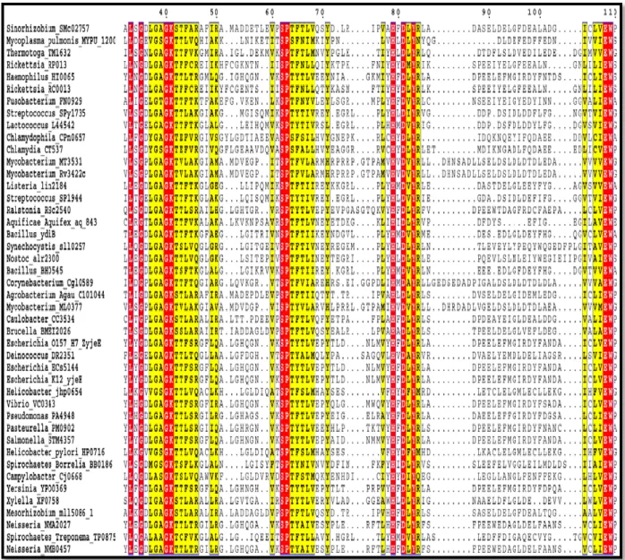
Figure 5: Phylogenetic and sequence alignment analysis of 18 bacterial “Histidine” kinases --- 11
Figure 6: Role of IDHK/P in Glyoxylate bypass --- 12
Figure 7: Structure of AceK --- 13
Figure 8: Phylogenetic and sequence alignment analysis of AceK and 17 eukaryotic protein kinases --- 13
Figure 9: Structural comparison of PPP and PPM families --- 17
Figure 10: Walker-A loop and Walker-B motif in nucleotide-binding proteins --- 20
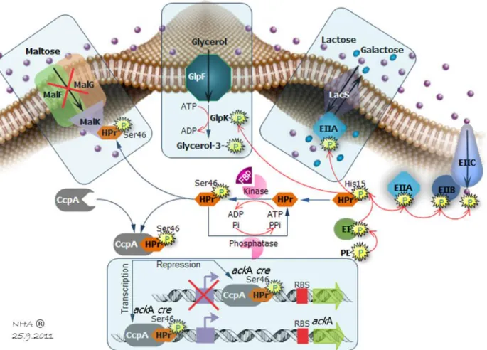
Figure 11: Carbohydrate transport and phosphorylation by the PTS and their coupling to glycolysis --- 21
Figure 12: Phylogenetic and sequence alignment analysis of HPrK/P active-sites in 9 Gram (+) and 3 Gram (-) organisms --- 22
Figure 13: Kinase and phosphorylase catalytic mechanism from HPrK/P towards HPr --- 22
Figure 14: Role of HPrK/P in CCR/CCA, PTS transport activity and inducer exclusion --- 23
Figure 15: CcpA/HPr/ackA-cre structure complex --- 23
Figure 16: Phylogenetic and sequence alignment analysis of BY kinases in 5 Gram (+) and 7 Gram (-) organisms --- 24
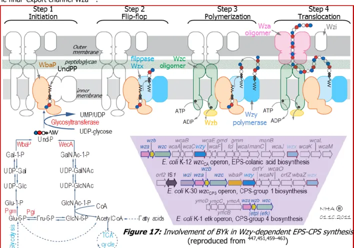
Figure 17: Involvement of BYk in Wzy-dependent EPS-CPS synthesis --- 25
Figure 18: Involvement of BYk in different cellular pathways --- 26
Figure 19: Autophosphorylation mechanism of WzcCA in E.coli K12 via two-step process --- 26
Figure 20: Overview on autophosphorylation mechanism in BYks --- 27
Figure 21-A: Widespread distribution and conservation of COG0802 family in the Kingdom of bacteria --- 28
Figure 21-B: Potential sequence alignment of COG0802 family in the Kingdom of bacteria --- 29
Figure 22: Modelization of Bacillus subtilis YdiB based on Haemophilus influenzae YjeE --- 30
Figure 23: New fold of YdiB family among Walker A-containing family --- 30
Figure 24: Auto-kinase activity and Phosphotransferase activity of YdiB --- 31
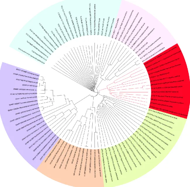
Figure 25: Phylogenetic analysis reveals new branch of bacterial protein kinases --- 32
Figure 26: In vitro phospho-amino acid analysis of YdiB and MBP --- 33
Figure 27: Implication of conserved residues on kinase activity of YdiB --- 35
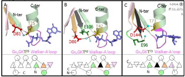
Figure 28: Structural analysis focused on Walker A-lysine residue--- 36
Figure 29: Structural analysis focused on Walker A-threonine residue --- 37
Figure 30: Structural analysis focused on SPT motif --- 37
Figure 31: Structural analysis focused on conserved histidine & glutamic acid --- 38
Figure 32: Structural analysis focused on conserved tyrosine and arginine residue --- 39
Figure 33: R83A mutant forms stable dimers --- 39
Figure 34: Structural analysis focused on glutamic acid and tryptophan in “hypothetical” added strand --- 40
Figure 35: Effect of basic molecules on YdiB autophosphorylation --- 41
Figure 36: Electrostatic potentials of models of MBP, YdiB and complex kinase-substrate YdiB-MBP --- 42
Figure 37: Polyamines in prokaryote: biosynthesis, uptake and export --- 43
Figure 38: Oligomerization and autokinase activity of YdiB --- 45
Figure 39: Model of H. influenzea YjeE tetramer and implication of conserved residues on oligomerization --- 46
Figure 40: Model of B. subtilis YdiB tetramer and implication of conserved residues in oligomerization --- 47
Figure 41: Genomic and Functional genomic context of ydiB gene --- 49
Figure 42: Phosphatase activity of YdiC --- 50
Figure 43: Phosphorylation of YdiD by YdiB in presence of poly-L-lysine --- 51
Figure 44: YdiE is substrate of both YdiB and YdiC --- 52
Figure 45: Subcellular localization of YdiB, ribosomes and nucleoids in B. subtilis --- 53
Figure 46: Structure of bacterial ribosome and phosphorylation of ribosomal proteins --- 54
Figure 47: Phosphorylation of purified B. subtilis ribosomes by YdiB --- 55
Figure 48: Involvement of conserved prokaryotic GTPases in ribosome biogenesis and function --- 56
Figure 49: Phosphorylation of EngA and EngB by YdiB and dephosphorylation of EngA by YdiC --- 58
Figure 50: In vitro phosphorylation by YdiB of peptides containing in vivo phosphosites in B. stubilis --- 60
Figure 51: Oxidative stress in aerobic bacteria: origin, defense and damage --- 63
Figure 52: Superoxide dismutase activity of SodA is increased upon phosphorylation by YdiB --- 66
Figure 53: Involvement of YdiB in oxidative stress in B. subtilis caused by paraquat --- 67
Figure 54: Working models of YdiB’s cellular function(s) in B. subtilis during unstressed and under oxidative stress conditions 71 Figure 55: Principle of SOD activity assay --- 787
Figure 56: Biotides assay schema --- 78
Figure S1: Co-purification of GST-YdiB-(His6) with other B. subtilis revealed by Pull-down method --- 83
functioning mechanism and cellular role of YdiB, an archetype from Bacillus subtilis
Introduction
NGUYEN Hien-Anh
functioning mechanism and cellular role of YdiB, an archetype from Bacillus subtilis
Introduction
functioning mechanism and cellular role of YdiB, an archetype from Bacillus subtilis
Introduction
1
INTRODUCTION
If the year 1995 marked the first sequenced genome of a free-living organism, Haemophilus influenza, at the end of 2011, full genome sequencing becomes normal, even ‘blasé’ science with the expected efficiency of 1000 genomes per month1. The major task that the scientific community is now facing is to turn data into
knowledge2. In the first step, computational methods including sequence comparison, phylogenetic patterns
and gene neighborhoods are inevitable to establish homolog network of each putative proteins in order to retrace its possible cellular function. However on average, there is no clear functional prediction for at least one-third of genes in most genomes3.
One of the most intriguing uncharacterized protein families regroups those found in many distantly related organisms. One such case includes a conserved family of small proteins, homologous to YjeE in E. coli, called UPF0079 in UniProtKB4 or COG0802 in the COG database5. This family was mentioned for the first time by
Galperin in 20016 with the purpose of illustrating the power and limitations of comparative genomics in
deducing functions of uncharacterized proteins. Based on the conserved Walker A motif (Gx4GKT/S) which is a
fingerprint to detect nucleotide-binding proteins, this protein family has been annotated as ‘probable ATP-binding protein’ in UniProtKB, and ‘predicted ATPase or kinase’ in COG database.
At the writing time of this manuscript, of the total 3237 UFP0079-homologs, it is noteworthy that still none of them comes from eukaryotes, rendering this family a promising target in antimicrobial drug development7.
The widespread distribution and conservation of this family of unknown function is implicit evidence for our lack of understanding of some basic cellular process. Bioinformatics can give some predicted clues for the general biochemical properties of these proteins. However, most of the time this approach is not sufficient and follow-up biochemical characterization is required to pinpoint the physiological function(s) of these new conserved proteins.
In 2002, Teplyakov & al.8 solved the crystal structure of YjeE in Haemophilus influenza, the first and still the
only structure of UPF0079 family. This structure revealed a unique fold, evidence of a new class of nucleotide-binding proteins which possesses characteristics of both Kinase/GTPase and ATPase folds.
Since 2002, several groups have been reporting biochemical data on YjeE from H. influenzae, E. coli and for the past few years, our group has been focusing on YdiB, the counterpart from B. subtilis. An agreement was found on a very weak ATPase activity of these proteins8–10. The question of whether the real physiological
function of this family is an ATPase arose when J. Karst in our group observed the in vivo oligomerization of YdiB which disfavorized its inherently weak ATPase activity10.
In this manuscript we report that YdiB is in fact a new kinase with both autophosphorylating and protein phosphotransferase activities. To our knowledge, YdiB represents the first bacterial dual-specificity Ser/Thr and Tyr kinase family distinguishable from eukaryotic-like Ser/Thr/Tyr kinases. Along with the five major well-known prokaryotic protein kinase families (including Histidine kinases of two-component systems11–13,
IDHK/P14–16 and HPrK/P17–20 discovered in the 1980’s, eukaryotic like-Ser/Thr kinases of Hank’s type21–23 in the
1990s’; and the recently discovered branch of BYks24–27 in the 2000’s), the discovery of YdiB-like family by our
group in collaboration with C. Grangeasse’s team in the 2010’s therefore opens a new chapter on prokaryotic protein kinases. Given the fact that this new family is broadly conserved in bacterial kingdom, we propose to name it Ubk for “Ubiquitous bacterial kinase”.
Enzymatic characterization showed that oligomerization favorizes the kinase activity of YdiB and that this activity is increased by basic molecules such as natural polyamines or poly-L-lysine. We also provided insights into molecular mechanism of the kinase YdiB by studying the implication of the 10 most conserved residues on the autophosphorylating and protein phosphotransferase activities of YdiB.
The next important task is to characterize the function of phosphorylation events linked to YdiB, or in other words, we are dealing with the question: in which in vivo biochemical pathway does YdiB take place. Cellular partner “hunting” is one of the strategies to cope with this challenge. Starting with the B. subtilis ydiA-B-C-D-E operon we showed that YdiB, YdiC, YdiD and YdiydiA-B-C-D-E might participate in the same physiological pathway where YdiB and YdiC function as cognate protein kinase/phosphatase towards two putative ribosome-related protein substrates YdiD and YdiE. The link between YdiB and ribosome was furthermore strengthened by the phosphorylation by YdiB of ribosomal proteins of both 50S and 30S subunits as well as of two ribosome-binding GTPases EngA and EngB. We also demonstrated that phosphorylated EngA by YdiB is an in vitro substrate of the phosphatase YdiC. Given that either YdiBCDE, EngA and EngB are well conserved proteins, from an evolutionary point of view, these proteins might have co-evolved with ribosomes to secure the biogenesis and/or function of the protein-making machinery.
Using immunofluorescent technique, we demonstrated some co-localization between ribosomes and YdiB. This co-localization however, is not stricto sensus especially when cells were treated with antibiotics, since YdiB was also found in zones without ribosomes. We thus assume that YdiB might be a protein of multifunction that participates in more than one physiological pathway. We then showed that YdiB indeed is capable of phosphorylating a number of peptides which previously have been shown to be phosphorylated in vivo in B.
functioning mechanism and cellular role of YdiB, an archetype from Bacillus subtilis
Introduction
2
subtilis by Macek & al.28 in 2007. In light of these results, a list of corresponding potential substrates of YdiB is
now being under construction in order to confirm their in vitro phosphorylation by YdiB. We anticipate that YdiB is a protein kinase which may have global importance in B. subtilis thanks to its capacity of phosphorylating proteins involved in fundamental cellular processes such as protein synthesis, central carbohydrate metabolism, stress resistance, etc… Opening the YdiB puzzle might therefore leads to uncover a bigger picture where different separate pieces communicate via phosphorylation and signal transduction. The last result reported in this manuscript focus on the involvement of YdiB in oxidative stress resistance via phosphorylation of the superoxide dismutase SodA. The superoxide scavenger capacity of this later protein against oxidative stress is upregulated via phosphorylation by YdiB. B. subtilis cells lacking ydiB therefore become more sensitive towards oxidative stress-causing agents such as paraquat or norfloxaxin.
In conclusion, the finding of the protein kinase activity of YdiB, which possibly plays important role in ribosome function in normal physiological condition and in protecting cells from oxidative stress damage; as well as insights into its molecular mechanism involving oligomerization status and basic molecule effect, therefore contribute towards a more comprehensive understanding of this attractive yet previously poorly characterized protein.
Literature review
Literature review
3
I. LITERATURE REVIEW
I.1. Protein phosphorylation in prokaryotes
I.1.1. Overview and Historical background
Asearlyasthe19thcenturyitwasknownthatphosphatescouldbeboundtoproteins.Mostexamplesof these phosphoproteins(
P
i-proteins) werefoundinmilk(caseins)andeggyolk(phosvitin)andweresimply considereda biological method of providing phosphorus as a nutrient. Therefore, the existence of
P
i-proteins wasconsidered a consequence of metabolic reactions, and nothing more, for almost a century after their discovery29.
In the 1950s this all began to change as
P
i-proteins emerged as key regulators of cellular life. An initiatingfactor of this emergence occurred in 1954, when an enzyme activity was observed that transferred a phosphate onto another protein30 - a biological reaction called phosphorylation. The protein responsible was a
liver enzyme that catalyzed the phosphorylation of casein and became known as a protein kinase (Fig. 1). A year later, the role of phosphorylation became more interesting as Fischer & Krebs31, and Wosilait &
Sutherland32, showed that an enzyme involved in glycogen metabolism was regulated by the addition or
removal of a phosphate, suggesting that reversible phosphorylation could control enzyme activity.
Figure 1: Reversible phosphorylation, and nature of Phosphate acceptor & linkage product 33–37
Today, it is thought that about 30% of human proteins contain covalently bound phosphate, and more than 500 protein kinases and a third of that number of protein phosphatases are encoded by the human genome. Phosphorylation or dephosphorylation can affect the function of a protein in every conceivable way: increasing or suppressing activity, marking a protein for destruction (apoptose), allowing it to move from one sub-cellular compartment to another (motility, organelle trafficking, ion channels and membrane transport) 38–41, or
enabling it to interact with or dissociate from other proteins...42. The simplicity, flexibility and reversibility of
this post-translational modification 43, coupled with the availability of ATP as a phosphoryl donor, is presumably
why it has been adopted to regulate so many biological processes.
Despite its obvious virtue as a vehicle for regulating protein function in eukaryotic cells, the existence and nature of protein phosphorylation-dephosphorylation in prokaryotes had long been the subject of controversy44. Early attempts during the 1960s and 1970s to detect protein phosphorylation in microbial
organisms, by using the techniques that had been successful with mammals, proved negative. Many scientists concluded that protein phosphorylation-dephosphorylation was the exclusive province of higher organisms, a relatively late evolutionary invention devised to meet the special demands of organisms composed of multiple, differentiated cells - one for which simple organisms had no need and hence may do without43, 45.
It was only in the late 1970s, with the pioneering work of Wang and Koshland46, Garnak and Reeves14, and
Manai and Cozzone47, which pointed out persuasive evidence that bacteria do contain specific protein kinases,
confirming the universal virtue of protein phosphorylation. The following table summarizes the big landmarks in the history of prokaryotic protein phosphorylation.
Table 1: Historical landmarks in the elucidation of prokaryotic signaling
Year Prokaryotic protein kinases/phosphatases
1969
-Kuo & Greengard48 reported the phosphorylation of exogenous histones by a cAMP-dependent kinase in E. coli
extracts. However, E. coli extracts harbor a polyphosphate kinase49 and an acyl-phosphate kinase50 that requires
histones for maximum activity. The evidence of bacterial protein kinase was thus still unclear.
1973
-Rahmsdorf51 showed that a cyclic nucleotide-independent protein kinase appeared in E. coli cells upon infection with
bacteriophage T7. However, enzyme activity in uninfected cells is negligible. This protein kinase is, in fact, coded for by a specific viral gene, since its appearance is prevented by ultraviolet irradiation of the phage genome but not by that of the host genome52.
1978 ÷ 1979
-Wang & Koshland46 provided the first persuasive evidence for prokaryotic protein phosphorylation. At least 4
proteins have been found to be serine/threonine-phosphorylated in Salmonella typhimurium.
-Manai and Cozzone47 also provided evidence for protein phosphorylation in E. coli. Phosphorylation occurs at the level
of threonine and serine residues.
-Garnak & Reeves14 reported the first endogenous substrate for protein phosphorylation in bacteria. E. coli
isocitrate dehydrogenase (IDH) was shown to be regulated by phosphorylation.
Literature review
4
1980 -Spudich & Stoeckenius54 reported the first examples of archaeal protein phosphorylation. Two of them are light-
regulated reversible phosphorylation – having its origin in retinal synthesis.
-Acid and hydroxylamine resistance of the phosphate bonds in both proteins suggests phosphoSer/Thr linkages.
1981 -Wang & Koshland55 detected the first protein phosphatase in Salmonella typhimurium.
1982 -Laporte & Koshland56 characterized the first bifunctional IDH kinase/phosphatase (IDHK/P) in bacteria
1983 -Deutscher and Saier17 found that the phosphotransferase activity of HPr was regulated by phosphorylation on serine,
being reversibly controlled by a Pi-(Ser)-HPr-kinase of 20 kDa and a Pi-(Ser)-HPr-phosphatase of 70 kDa.
1986 ÷ 1988
-Nixon & al. 11 proposed a model for transduction of environmental signals by some pairs of regulatory gene products,
which thereafter became the TCS paradigm (i.e. two-component regulatory system).
-Aspartate kinase activity of Histidine kinases 57 and auto-phosphatase activity of response regulators characterized 58.
1987 ÷ 1988
-Stueland & al.59 found an ATPase activity of IDHK/P
-Cortay & al.60 sequenced the gene aceK coding for IDHK/P
-Cohen & al.61 reported PP-λ in bacteriophage λ, the first eukaryotic-like PPP-family protein phosphatase.
1989 -Hurley & al.62 solved the structure of IDH.
1990 -Guan & Dixon 63 discovered YopH from Yersinia, the first eukaryotic-like PTP family protein phosphatase.
1991 -Muñoz-Dorado & al. 21 discovered Pkn1, the first eukaryotic-like Ser/Thr kinase (eSTK) in Myxcococcus xanthus
1993 -Min & al. 64 identified SpoIIAB from B. subtilis as a protein- Ser/Thr kinase with homology to TCS
-Chang & al. 65 and Maeda & al.66 discovered the first TCS in plant and in yeast.
-Potts & al.67 discovered IphP, the first dual-specific PTP-family protein phosphatase in Nostoc commune UTEX584.
1994 -Matsumoto & al.68 reported the first evidence of phosphorylation of an exogenous protein (AfsR), by a 'eukaryotic'-like
kinase (AfsK) from Streptomyces coelicolor.
1995 -Duncan & al. 69 identified SpoIIE from B. subtilis as the first bacterial PPM-family protein-Ser phosphatase.
1996 -Li & al. 70 characterized PtpA from S. coelicolor, as the first bacterial LMW-PTP-family protein-Tyr phosphatase.
-Duclos & al. 24 detected the first BY (bacterial tyrosine) autokinase of about 81 kDa in Acinetobacter johnsonii
1997 -Two PPP-family protein phosphatases, PrpA and PrpB, characterized from E. coli71.
-Grangeasse & al.72 identified ptk as the gene encoding the first previously detected BY autokinase in A. johnsonii
1998 -Galinier & al73. identified B. subtilisyvoB as the gene encoding the ATP-dependent HPr-kinase
1999 -Ilan & al.74 showed the first kinase activity from the BY-Ptk homolog, Etk towards exogenous substrate
-Wu & al.75 characterized the first crosstalk between TCS and bacterial tyrosine kinases, DivL from C. crescentus.
2001 -Fieulaine & al.18 reported the first crystal structure of the HPr Kinase/Phosphatase (HPrK/P) in Lactobacillus casei, showing an ATP-binding loop of Walker A motif 76,77.
2002 -Morona & al. 78 discoverd the first PPM-family protein phosphatase in S. pneumoniae
2003 -Ortiz-Lombardia & al.79 presented the first crystal structure of an eSTK, PknB in M. tuberculosis.
2004 -Pullen & al.80 described the first crystal structure of a eukaryotic-like Ser/Thr protein phosphatase, PstP/Ppp in M. tuberculosis 81.
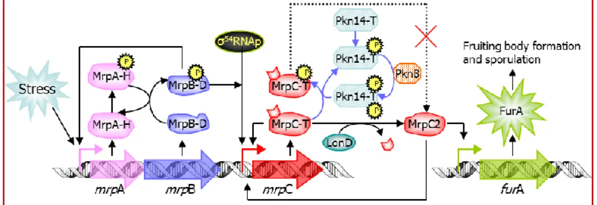
2005 -Nariya & Inouye
22 demonstrated the first functional bacterial Ser/Thr kinase cascade. The Pkn8–Pkn14 kinase
cascade negatively regulates mrpC expression by phosphorylating MrpC during vegetative growth in M. xanthus. Not only MrpC is involved in eSTK signaling pathway, it is also regulated by the TCS MrpA-MrpB 82,83.
2007 ÷ 2008
-Collins & al. provided the first structural insights of E. coli Wzc, a BY kinase, alone84 or in complex25 with the
capsular polysaccharide translocon Wza.
-Structures of E. coli Etk85 (Proteobacteria) and S. aureus CapA/B86 (Firmicutes) BY kinases were solved.
2010 -Zheng & Jiain complex with its IDH substrate. 16 solved the first crystal structure of IDH K/P AceK in its AMP-bound and AMP-free forms; alone or
I.1.2. Phosphoproteomics in bacteria
Phosphoproteomics is a relatively new branch of proteomics that identifies, catalogs, and characterizes proteins containing a phosphate group as a post-translational modification.
Before 2007, traditional bacterial phosphoproteomics relied on 2D-gel separation of bacterial proteomes, 32P
radiolabelling or immunodetection, and detection of Pi-proteins by low-resolution mass spectrometry (MS). 128 phosphoproteins (
Pi
-proteins) from E. coli87, 29 from B. subtilis88, 41 from C. glutamicum89 were found by suchapproach, but no phospho-sites (
Pi
-sites) were identified.The combination of phosphoprotein enrichment and 2D-gel followed by MS analysis of trypsin digested spots however enabled identification of 36
P
i-proteins including 3 on Tyr in C. jejuni90. Recently this approach allowedidentification of 16
P
i-sites from 63P
i-proteins of M. pneumoniae91 and 51 novelP
i-proteins from N. Meningitidis92.One of the advantages of 2D gel based phosphoproteomics is the relative ease with which changes in e.g. different growth conditions can be analyzed, but there are still some technical problems with this approach
28,93,94. Starting from 2007, gel-free approach appeared, leading to the publication of site-specific Ser/Thr/Tyr
bacterial phosphoproteomes for a number of species (Tab. 3). This approach, in short, involves digestion of crude extracts with endoprotease (e.g. trypsin) followed by phosphopeptide enrichment. The mixture is then separated by liquid chromatography (LC) that is coupled to a high resolution MS. A technique known as Stable Isotope Labeling by Amino acids in Cell culture (SILAC) is the latest development in the field of quantitative proteomics/ /phosphoproteomics94 that enables comparison of two strains (e.g. wild type and kinase or
phosphatase mutant) or one strain cultured in two different growth conditions95.
The recent site-specific phosphoproteomes have greatly extended the list of known Pi-proteins and Pi-sites. Around 80÷100 Pi-sites on average were reported in each bacterial phosphoproteome, except in M. tuberculosis with more than 500 sites, possibly due to its large STPKs number (11)96. These data nonetheless
Literature review
5
Table 2: Phosphoproteomics, the systematic approach has been developed for almost one decade
Color code: Green (Latest results in each organism), Gray (Studies using gel-free approach)
Bacterium Year Phosphoproteomic analysis
C.glutamicum 2003 Bendt & al.89 used two methods: (33P radiolabelling then autoradiography) and (immunostaining with phospho amino acid-specific antibodies), followed by 2D-gel and MS, to identify 41
phosphoproteins.
C.bacterjejuni 2007 Voisin & al.90 analyzed the cytoplasmic fraction of C. jejuni by Fe-IMAC for phosphoprotein enrichment + 2D-gel for protein separation + in-gel tryptic digestion and MALDI-ToF MS for mass
fingerprinting. The results revealed 58 phosphopeptides from 36 proteins.
Bacillus subtilis
Before
2006
-On establishing the complete genome of B. subtilis, Kunst & al. 97 showed 34 genes encoding response regulators (most of which have adjacent genes encoding histidine kinase to create
two-component regulatory systems), and 11 aspartate phosphatase genes (involved in dephosphorylation of response regulators, that do not seem to have counterparts in Gram (-) bacteria such as E. coli). -Only 16 Pi-sites on 8 of its proteins have been in vitro determined 28, including PrkC (or YloP, 8 Pi-sites), SsbA, Idh, PtkA (or YwqD), Udg (also YwqF or TuaD, substrate of PtkA).
2006 Lévine & al.
88, by using 32P radiolabelling with one-unit pH on 2D-gel combined with MS, indentified a total of 29 phosphoproteins, with 19 identified for the first time, however this study did not give
any information on the location of Pi-sites. 2007 -Macek & al.
28 performed gel-free analysis consisting of biochemical enrichment of phosphopeptides and high accuracy MS, identified 78 phosphoproteins with 78 Pi-sites (Ser:Thr:Tyr = 54:16:8).
-Of 16 Pi-sites from 8 proteins reported so far, they detected 7 sites from 6 proteins.
-Only 1 of 8 Pi-sites on PrkC was found. None of Pi-sites were found on SsbA, Idh and PtkA. By contrast, Ugd was identified, although Ugd phosphorylation in vitro is less efficient than that of PtkA. E. coli 1986 Cortay & al.
87, by using only 32P radiolabelling with 2D-gel, showed 128 Ser/Thr/Tyr protein spots. They were mainly located in the cytosolic fraction of cells. However most of them were only described
in terms of pI and molecular weight87 and, surprisingly, were never identified98.
1986 ÷
before
2008
-No homologs of eukaryotic Ser/Thr kinases had so far been found in E. coli, except a preliminary description of a protein kinase C-like activity99. E. coli possesses 2 well characterized Ser/Thr kinases:
the IDHK/P100 and the YihE kinase101. Other E. coli proteins were also reported to autophosphorylate on Ser/Thr residues, including DnaK, the molecular chaperone heat-shock protein102,103; Era, an
essential Ras-like GTPase104, and the nucleotide diphosphate kinase (NDK)105.
-E. coli possesses 3 BY-kinases, BipA93,106,107,108, Wzc and Etk84,108,109, which are capable of auto- and substrate phosphorylation.
-At least 62 open reading frames (ORFs) were identified as putative members of the two-component signal transducers in E. coli. Among them, 32 were identified as response regulator and 23 others as orthodox sensory kinases. In addition, E. coli has five hybrid sensory kinases110.
-Available biochemical data include 12 phosphorylation sites on 6 proteins in E. coli 107, 111.
2008 Macek & al.98 performed the same method used in 2007 28 and identified 79 proteins with 81 Pi-sites (Ser:Thr:Tyr = 55:19:7).
2009 Soung & al.
112 used either phosphorylation specific visualization techniques, (Pro-Q diamond staining) or specific phospho-Ser, Thr, Tyr antibodies followed by MS/MS after phosphopeptide enrichment
through immobilized Metal Affinity Chromatography and by peptide fractionation through Strong cation eXchange Chromatography (SXC), to analyze phosphorylated proteins in E. coli ribosomes. Of the 54 proteins in the 70S ribosome, they found 24 to be steady-state phosphorylated. 9 of them belong to the small subunit 30S, the rest are in the large subunit 50S. Only 5 of these ribosomal phosphoproteins were previously detected in the phosphoproteome by Macek & al 98.
Lactococcus
lactis 2008 -Soufi & al.
113 performed the same method used by Macek & al.28,98 to identify 73 Pi-sites distributed over 63 different proteins.
-The presence of several multiply phosphorylated proteins, as well as over-representation of phospho-Thr seems to be the distinguishing features of the L. lactis phosphoproteome. Klebsiella
pneumoniae 2009 -Lin & al.
114 developed the technique previously used by Soung & al.112 through a gel-free analysis consisting of peptide fractionation by SXC + phosphopeptide enrichment by TiO
2 affinity and by
phospho-tyrosine specific antibodies + LC-LTQ-OrbitrapMS/MS analysis. They identified 81 proteins with 93 Pi-sites (Ser:Thr:Tyr:His:Asp= 29:14:24:12:14). -Using phospho-tyrosine specific antibodies for enrichment led to the identification of 17 additional Tyr phosphorylated peptides out of 24 in total.
Pseudomonas
spp 2009 -Ravichandran & al.
115 developed a novel phospho-enrichment method using aliphatic Hydroxy Acid-Modified Metal Oxide Chromatography (HAMMOC) with titania and zirconia.
-By Ti/Zr HAMMOC-based nanoLC-MS approach, they founded 53 Pi-sites of P. putida (Ser:Thr:Tyr = 28:21:4) and 55 Pi-sites of P. aeruginosa (Ser:Thr:Tyr = 29:18:8) -Interestingly, the GTP-binding protein EngA was found to be phosphorylated in P. putida at Ser-34 in this study.
Strepto- -coccus pneumoniae 2010
-Sun & al.116 used TiO
2 enrichment and LC-MS/MS to reveal 102 unique phosphopeptides corresponding to 84 proteins and 163 Pi-sites with distributions of (Ser:Thr:Tyr = 48:45:9)
-A striking characteristic of S. pneumoniae phosphoproteome is the large number of multiple species-specific phosphorylated sites, indicating that high level of protein phosphorylation may play important roles in regulating many metabolic pathways and bacterial virulence. EngA was found to be phosphorylated in S. pneumoniae at Thr-5 in this study.
M.pneumoniae 2010 -Schmidl & al. 91 used 2D-gel followed by MS to identify 16 Pi-sites (Ser:Thr = 8:8) from 63 proteins.
S. coelicolor 2010 -Parker & al. 117 performed gel-free analysis consisting of protein and peptide fractionation by IEF and SXC chromatography respectively + phosphopeptide enrichment by TiO
2 affinity chromatography +
phosphopeptide identification by LC-ESI-LTQ-Orbitrap MS. The result revealed 46 novel Pi-sites on 40 proteins with a Thr-preferred profile (Ser:Thr:Tyr = 16:24:6)
Myco- -bacterium tuberculosis
Before
2010
-If B. subtilis and E. coli have each more than 30 two-component systems (TCS) 97, 110, M. tuberculosis has only 11 complete pairs of sensor histidine kinases and response regulators, and a few isolated
kinase and regulatory genes 118.
-This relatively paucity of TCS is offset by the presence of many eukaryotic-like Ser/Thr protein kinases (eSTKs) which function as part of a phosphorelay system. M. tuberculosis has only 3 eukaryote-like protein phosphatase (PstP, PstA, PstB) but 11 eSTKs (PknA to PknL) 119.
-Mycobacteria with larger genomes such as M. marinum or M. smegmatis STPKs outnumber TCS, suggesting that the bulk of signal transduction is via Ser/Thr phosphorylation120.
2010 -Prisic & al.
121 used a gel-free approach followed by LC-MS/MS to identify 516 Pi-sites inside 301 with a Thr-preferred profile (Ser:Thr = 60%:40%).
-The motif XααααTX(X/V)ϕ(P/R)I (where α is an acidic residue and ϕ a large hydrophobic residue) was proposed to be the specific recognition substrate-kinase motif for 6 eSTKs (PnkA, B, D, E, F, H). This common motif explains why multiple kinases can target a single protein, (e.g GarA and Rv1422).
Literature review
6
and environmentally dependent kinome (*)93, on the other hand the technical imperfections. However, in term
of evolution, one can anticipate right now a lack of (or extremely low) conservation of
Pi
-sites96122, since onlyone single protein, the phosphosugar mutase (ManB in M. pneumoniae or GlmM in B. subtilis), is phosphorylated on a conserved Ser residue in all studied organisms, from archaea and bacteria to man. For most other proteins (e.g. DnaK or Hsp70), even if they are phosphorylated in different species, the actual
Pi
-sites are different. This suggests that site-specific phosphorylation is a form of adaptation of the bacteria to the specific needs of their particular ecological niche 113, 122.An alternative to experimental identification of
Pi
-sites is in silico prediction. Originally trained on eukaryotic data, the most widely phosphorylation predictor NetPhos fell short of performance on bacterial proteins (e.g when it was evaluated on the published phosphoproteome of B. subtilis 94). This led to the development of abacteria-specific version NetPhosBac 123 that out-competed the eukaryotic predictors as well as the bacterial -
- specific mode of DISPHOS predictor 96. NetPhosBac is currently dedicated to Ser and Thr phosphorylation due
to the limited number of known Tyr
Pi
-sites, but it is only a question of time before enough data for a Tyr specific version is available.In term of functional insights obtained from phosphoproteomic studies,
Pi
-proteins are seemingly over-represented in central carbon metabolism (especially among glycolytic enzymes) 28,94,98, as well as inhouse-keeping processes including the virulence functions of bacterial pathogens93. But the physiological roles of
these phosphorylation events are still largely unknown. Furthermore, a number of
Pi
-proteins of unknown function were also comprised in each published phosphoproteome. To link protein kinases and their substrates more than ever remains the main challenge ahead. At this point, along with the improvement of analytical tools (e.g. SILAC), one can expect a new wave of bacterial kinomes (including wild type as well as kinase/phosphatase-knockout strains) in different growth conditions. Such data should allow us to infer connections between kinases/phosphatases and their substrate and hopefully their physiological role(s), a stepping stone towards bacterial systems biology96. Interestingly, a recent study applied this approach in M.pneumoniae with the surprising conclusion that only 5 out of 63 phosphoproteins are targeted by the two known kinases pointing towards the presence of novel classes of kinases91.
I.1.3. Classification of bacterial protein kinases
The hypothesis towards the presence of novel kinases undetected by analytical phosphoproteomic tools has become more and more apparent. The lack of a significant bacterial protein kinase classification system based on conserved features and deduced phylogeny of the catalytic domains (as the eukaryotic one established by Hanks & al.124), in fact does not help to accelerate the characterization of these kinases of unknown function.
The first framework for classification of prokaryotic Ser/Thr kinases was proposed by Tyagi & al.125, but is still
far from convenient utilization.
Traditional classification proposed by Hunter34 (on the basis of phosphate acceptor amino acids, fig. 1) in
combination with the kinase sequence analysis (based on the presence of the phosphate-binding motif Gx4GKT[S], so-called Walker A motif76,77) will be developed in the table 4, on purpose of systematization of
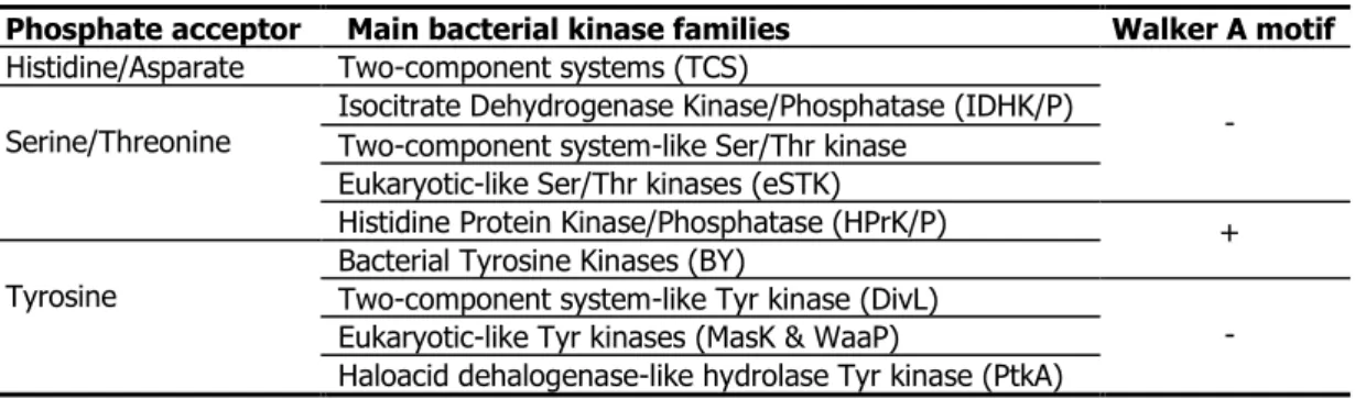
representatives of bacterial protein kinases. Only kinases which do not contain a Walker A motif will be shortly described inside this section, aiming to show the highly divergence phosphorylation in bacteria, which is in fact a proper versatile attribute in order to adapt to environmental stimulus, but not acquired from eukaryote by horizontal gene transfer. Other protein kinases containing the Walker A motif (including YdiB family which is present exclusively in prokaryote but not in eukaryote - objective of this thesis) will be introduced in I.2.
Table 3: Classification of bacterial kinases by kinase sequence & phosphate acceptors
I.1.3.1. Two-component systems (TCS)
The first model of TCS was proposed by Nixon & al.11 in 1986, based on the chemotaxis previously observed in several E. coli sensory transducers91, 92. It has become clear that TCS is widespread, although not ubiquitous, in
bacteria and archaea (30 to 40 complete TCS was found in E. coli, B. subtilis, or Synchosystis ssp.,more than 100 in Nostoc puctiformis128, but none inMycoplasma fenitaliul orMethanococcus jannaschii129). TCSwasfound
latelyinplant65and in yeast66, but isseeminglyabsentinanimals, worm andfly129,12.
Phosphate acceptor Main bacterial kinase families Walker A motif Histidine/Asparate Two-component systems (TCS)
- Serine/Threonine Isocitrate Dehydrogenase Kinase/Phosphatase (IDHK/P) Two-component system-like Ser/Thr kinase
Eukaryotic-like Ser/Thr kinases (eSTK)
Histidine Protein Kinase/Phosphatase (HPrK/P) +
Tyrosine Bacterial Tyrosine Kinases (BY) Two-component system-like Tyr kinase (DivL)
- Eukaryotic-like Tyr kinases (MasK & WaaP)
Haloacid dehalogenase-like hydrolase Tyr kinase (PtkA)
Literature review
7
TCS consists of a signal recognition sensor kinase (SK) that autophosphorylates on a Histidine, usually in response to the presence of a signal; and a response regulator (RR) transcription factor that activates or represses gene expression when phosphorylated on an Aspartate by the cognate sensor kinase.
Figure 2: Two-component & Phosphorelaytransduction system (based on 97,98,13)
SK generally contains a signal input domain coupled to an autokinase domain (which can be divided into a His-phosphotransferase subdomainand anATP-bindingsubdomain distinguishable from Walker A motif131, Fig. 5).
RR has a regulatory domain that controls the activity of the output domain. Detection of the stimulus by the SK induces the transfer of γ-phsophate to His residue. The phosphoryl group from the His of the SK will be transferred to the Asp of the regulatory domain of RR128. Finally, the auto-dephosphorylation of this RR96, 97 will
return to the initial form by releasing enough energy to trigger on/off the output signal (Fig. 2).
More-complex variants of TCS, termed phosphorelays, are used in bacteria for pathways responding to multiple signal inputs 133. Phosphorelays in fact are four-component systems with an additional regulatory domain and a
phosphotransferasesubdomain.ThesignaltraductionpathwaydirectsphosphoryltransferinH-D-H-D sequence. In thesporulation phosphorelay,these domainsoccuronseparateproteins134–136, butinotherphosphorelays,
one or more subdomains might be attached to the sensor kinases as a polydomain protein12,128,130,134,135,137–139.
Table 4: Bacterial Phosphorelay architectures (updated from 140 and 141)
25 years have passed since the first model proposed on the basis of NtrB/NtrC pair in E. coli. Up to now what do we know and do not know about this TCS-phosphorelay system in bacteria? 130, 139
We know:
TCS is strain specific but also conserved among strains to respond to the same type of stimulus 149. TCS is
widely distributed but not universal94,129; 2 different mechanisms were proposed to explain the evolution of TCS
through recruitment & co-evolution149.
TCS participate in lots of pathways in response to a myriad of signals132,150.
The structure of regulator and output domains of RR, the structure of ATP domains and phosphotransferase domains of histidine kinases129.
A limited appreciation of the mechanism of phosphotransfer between the two proteins128.
We do not know:
The structure of membrane-based signal input domains for the majority of kinases. Is membrane location important for their function?
How signals activate kinases? How regulator domains control output domains? (except few examples)
How phosphorylation interferes with this control, although we have some suspicions about how this works, through several modeling studies139.
What are the functional differences between simple single-step TCS and the various phosphorelays? What
are the general principles that may account for the use of one architecture over another? Future research:
Modelization and experimental validation in larger-scale.
Interactions between TCS and other regulatory networks in the cell.
Development of new methods to follow histidine kinase and response regulator activity in vivo139.
Two-component systems and phosphorelays as targets for therapeutic intervention13.
Phosphorylay architectures Representative Description
EnvZ-OmpR
(E. coli)
Typical TCS, E is an effector protein mediating DNA binding and transcriptional control of target genes 142–144
BvgS-BvgA
(B. pertussis)
The first regulator domain RR1 and the second
Histidine-phosphotransfer domain HPt, are fusioned to the hybrid SK
KinA-Spo0F-Spo0B-Spo0A
(B. subtilis)
Typical phosphorelay as described above, except that Spo0B is not a typical HPt domain 145, 146
CheA-CheY
(E. coli)
The sensory receptor is detached from the His-kinase CheA. The RR consists of only a regulatory domain CheY 107, 108
Literature review
8
I.1.3.2. Eukaryotic-like Ser/Thr kinases
The eukaryotic Ser/Thr protein kinase paradigm (Hanks-type)124 was first challenged in 1991 by pkn1 from
Myxococcus xanthus – an ORF whose predicted product Pkn1 resembles eukaryotic Ser/Thr kinases21. Three
years after, the first evidence of phosphotransfer to a protein substrate (AfsR) by a 'eukaryotic'-like kinase (AfsK) from Streptomyces coelicolor, was reported68. With the advent of genome sequencing, scientists began to trace
the distribution of ORFs encoding deduced protein kinases111,151,152. Of 27 bacterial genomes analyzed, 85%
contained ORFs for potential eukaryotic-like protein kinases43. The following Tab. 5 updates 33 biochemically
studied Hanks-type Ser/Thr kinases from 13 bacteria.
Table 5: Bacterial eSTKs and their substrates (updated from 23, 153, 154, 155)
Bacillus subtilis Function Methodology-References Year
Substrates PrkC PrkD YabT Comment
YabT +
PrkC and PrkD are respectively so-called
YolP and YbdM
Sporulation, biofilm, germination, cell wall KA, MS, 3D structure156–159 2002
AlsD + α-Acetolactate decarboxylase; central metabolism Kinase assay (KA) and Mass spectrometry (MS)160
2010 GlnA + + Glutamine synthetase; central metabolism
Icd + +
Hpr + Central metabolism
YwjH + Kinase, phosphotransferase system
CpgA + Transaldolase; central metabolism KA, mutagenesis Phosphoamino acid analysis (PAA), 2D gel161
2009 YezB + GTPase; peptidoglycan sacculus deposition
EF-Tu + Stressome protein
EF-G + GTPase; Elongation factor; protein translation Immunoprecipitation, KA157,162,163 2002
PrkD + GTPase; Elongation factor; protein translation 156, 160 2002
DegS + + Cytosolic sensor kinase of TCS of DegS/U, swarming KA, mutagenesis164 2011 Chlamydia trachomatis Function Methodology-References Year
Substrates Pkn1 PknD Comment
Pkn1 + + Unknown Bacterial two-hybrid, immunoprecipitation, KA1652003
IncG + Virulence PknD + Unknown
Corynebacterium glutamicum Function Methodology-References Year Substrates Kinases Pkn Comment
A B G L
Both FtsZ and OdhI are substrates of the phosphatase Ppp
MurC + Cell wall synthesis KA, MS, mutagenesis166 2009
FtsZ + + + Cell division KA, MS, in vitro and in vivo 2D gel167, 1682006
PknG + Soluble eSTK
OdhI + + + + FHA protein, Glutamate catabolism
Mycobacterium tuberculosis Function Methodology-References Year Substrates A B D E F G H I J K L Kinases PknA to PknL
PknA + Cell elongation / division KA169 2009
PknB + + Elongation/division, peptidoglycan synthesis KA, MS, 3D structure 169–171 2003
PknD β-propeller, PQQ domain, phosphate transport KA172 1997
PknF + Membrane transporter KA173 2004
PknG + Trx & TPR motif, glutamte metabolism KA, MS174 2008
PknH + AfsK like, arabian metabolism, NO stress KA, PPA175 2003
PknI + Abnormal Asn in active site, cell division KA176,177 2004
PknJ + - KA, PPA, mutagenesis178–180 2006
PknK + Transcription, 2nd metabolism KA, PPA,179,181 2009
EmbR + + + + Arabinan synthesis, cell wall KA, PPA, Phosphor-chip175,180 2003
PBPA + Cell wall synthesis KA, MS, mutagenesis182 2006
GlmU + + Cell wall (peptidoglycan) synthesis KA, MS, mutagenesis171
2009 2009 DacB1 + Penicillin-binding protein, cell wall
FabH + + + + + Mycolic acid pathway; cell wall biosynthesis In vivo KA, MS, mutagenesis183 2009
FabD + + + Mycolic acid biosynthesis KA, PAA179
2006 KasA + + + + + + + Mycolic acid biosynthesis
KasB + + + + + + + Mycolic acid biosynthesis
MabA + + + + + Mycolic acid biosynthesis KA, MS, mutagenesis184 2010
MmA4 + Mycolic acid biosynthesis KA, Phospho-chip, MS1802010
PepE + DNA-binding protein, cell wall/cell division
FibA + Cell division under H2O2 oxidative stress In vivo KA, MS, mutagenesis185 2010
FtsZ + Cell division KA, MS185,186 2006
MurD + Cell division KA187 2008
Wag31 + Cell division, orthologue of DivIVA In vivo KA, MS, mutagenesis169 2005
Mmpl7 + Membrane transporter; resistance, cell division In vivo 2D gel, MS188 2006
Rv1747 + + + Putative ABC transporter KA173,189 2004
Rv0020c + + FHA-containing protein KA189 2005
Rv2175 + Dipeptidase, DNA-binding protein 2D gel, PAA, MS, KA190,191 2008
Rv1422 + + - In vivo KA, MS, mutagenesis169 2005
PykA + Pyruvate kinase A, Glycolysis KA178 2009
GarA + + Glycogen recycling, tricarboxylic acid cycle KA, MS, SPR174 2008
SigH + Oxidative, nitrosative, heat stress σH factor In vivo KA, MS, mutagenesis192 2008
RshA + Oxidative, nitrosative, heat stress anti-σH factor
Rv0156 + Anti-anti-sigma factor In vivo KA, MS, mutagenesis193 2007
VirS + Transcriptional regulator KA, PPA179,181 2009
Rv0681 + TetR class transcription factor KA, mutagenesis194 2007