Université de Montréal
‘Evo1ution ofC2H2-Zinc finger genes in mammalian genomes”
par
“Hamsa Dhwani Tadepally”
“Département de Biochimie” “Faculté de Médecine”
Thèse présentée à la Faculté des études supérieures en vue de l’obtention du grade de Maitrise
En Biochimie
“July 2007”
© “Hamsa Dhwani Tadepally” ,2007
w
I IjJt
O
Université
de Montréal
Direction des bibliothèques
AVIS
L’auteur a autorisé l’Université de Montréal à reproduire et diffuser, en totalité ou en partie, par quelque moyen que ce soit et sur quelque support que ce soit, et exclusivement à des fins non lucratives d’enseignement et de recherche, des copies de ce mémoire ou de cette thèse.
L’auteur et les coauteurs le cas échéant conservent la propriété du droit d’auteur et des droits moraux qui protègent ce document. Ni la thèse ou le mémoire, ni des extraits substantiels de ce document, ne doivent être imprimés ou autrement reproduits sans l’autorisation de l’auteur.
Afin de se conformer à la Loi canadienne sut la protection des renseignements personnels, quelques formulaires secondaires, coordonnées ou signatures intégrées au texte ont pu être enlevés de ce document. Bien que cela ait pu affecter la pagination, il n’y a aucun contenu manquant.
NOTICE
The author of this thesis or dissertation has granted a nonexclusive ticense allowing Université de Montréal to reproduce and publish the document, in part or in whole, and in any format, solely for noncommercial educational and research purposes.
The author and co-authors if applicable retain copyright ownership end moral rights in this document. Neithet the whole thesis or dissertation, flot substantial extracts from it, may be printed or otherwise reproduced without the author’s permission.
In compliance with the Canadian Privacy Act some supporting forms, contact information or signatures may have been removed from the document. While this may affect the document page count, it does flot tepresent any loss of content from the document.
Université de Montréal faculté des études supérieures
Cette thèse intitulée
“Evolution of C2H2-Zinc finger genes in mammalian genomes”
Présentée par: “Hamsa Dhwani Tadepally”
a été évaluée par un jury composé des personnes suivantes:
“Martine Raymond” Président-rapporteur “Muriel Aubry” Directrice de recherche “Gertraud Burger” Co-directrice “Nicolas Lartillot” Membre dujmy
Résumé
Les gènes de doigt de zinc de C2H2/Kruppel (C2H2-ZNF) encodent la plus grande classe des facteurs de transcription chez Phomme. Ces gènes constituent une des plus grandes familles de gène chez les mammifères et sont souvent trouvés sous forme de regroupements de gènes juxtaposés sur les chromosomes. Par une recherche extensive basée sur des similitudes de séquences visant à d’identifier l’ensemble des gènes C2H2-ZNF du génome humain, nous avons assemblé un répertoire complet de 718 gènes C2H2-ZNf humains. Les gènes C2H2-ZNF ont été classifiés en sous-familles en fonction des domaines effecteurs N-terminaux aux quels ils sont associés. Nous avons constaté que la sous-famille encodant un domaine KRAB comprend 45% de tous les gènes C2H2-ZNF et est par conséquent fa plus grande sous-famille de gènes à motifs doigt de zinc. De plus, nous avons identifié 81 regroupements de gènes C2H2-ZNf qui correspondent à 70% de tous les gènes C2H2-ZNf. Presque 90% des gènes C2H2-ZNF appartenant aux sous-familles KRAB et SCAN sont trouvés sous forme de regroupements. Pour mieux comprendre l’évolution des gènes C2H2-ZNF, nous avons par la sUite assemblé un répertoire complet de tous les regroupements de gènes C2H2-ZNF humains ainsi que de leurs contre-parties dans les régions synténiques des génomes de chimpanzé, de souris, de rat et de chien. Une analyse systématique de ce répertoire chez ces mammifères a révélé qu’il existe une variation dans le nombre de regroupements et de gènes faisant partie de ces regroupements parmi les primates, les rongeurs et les canins. Cette variation suggère que ces gènes ont évolué de façon différentielle chez les mammifères. Des études phylogénétiques de plusieurs regroupements de gènes C2H2-ZNf choisis indiquent qu’outre une duplication
différentielle, la perte de gènes dans certaines espèces a condujt à des répertoire différents de gènes C2H2-ZNF chez les mammifères. En plus des variations spécifiques aux espèces dans le nombre de gènes, nous avons également mis en évidence une variation chez des orthologues dans le nombre de motifs de doigt de zinc et la présence de domaines effecteurs, ces derniers étant souvent perdus par dégénération. En conclusion, sur la base principale de ces résultats et de l’étude de la structure exon-intron des gènes C2H2-ZNF, nous proposons un nouveau modèle pour lévolution de leurs sous-familles selon lequel les sous-familles les plus anciennes seraient dans l’ordre SCAN> SCAN-KRAB >KRAB.
Abstract
The C2H2/Kruppel zinc finger genes (C2H2-ZNF) encode the largest ciass of transcription
factors in hurnans. These genes constitute one of the largest gene families in mammals and
are often found in ciusters. Using an extensive similarity search on the hurnan genorne to
identify ail C2H2-ZNF genes, we assembled a comprehensive repertoire of 718 human
C2H2-ZNF genes. The genes were grouped into subfamilies based on the N-terminal effector domains they were associated with. We found that the KRAB-domain encoding
subfarnily constitutes 45% of the total C2H2-ZNF genes and hence is the largest
subfamiiy of zinc finger genes. In addition to this, we also identified 8 1 C2H2-ZNF clusters
which constitute 70% of the total genes. Almost 90% of the C2H2-ZNF belonging to the
KRAB and SCAN subfamilies were found in ciusters. We then assembled a comprehensive
repertoire of ail the hurnan C2H2-ZNF clusters and their syntenic counterparts in
chimpanzee, mouse, rat and dog genomes. A systernatic analysis of ah the syntenic clusters reveaÏed a variation in the numbers of clusters and the genes within clusters among primates, rodents and canines indicating differential pattems of evolution in mammals.
Evolutionary analysis of few selected C2H2-ZNf syntenic clusters in the five mammals
studied suggested that not only differential duplication, but also gene ioss has led to different repertoires in mammahian genomes. In addition to lineage- and species-specific variation in the number of genes, we aiso find a variation among orthologs in the number of zinc finger motifs and in the presence of the effector domains, the later being often lost by
sequence degeneration. finally, based on the above resuits and on the analysis of the exon
their subfarnilies suggesting that the more ancient subfarnilies are in sequential order SCAN> SCAN-KRAB > KRAB.
Keywords: C2H2/Kruppel, zinc finger, gene farnily, tandem repeats, gene duplication, gene loss, evolution.
List of abbreviations
DNA: Deoxyribonucleic Acid RNA: Ribonucleic AcidBIB: Broad-Cornplex, Tramtrack and Bric-a-bric
POZ: Pox virus and Zinc finger KRAB: Kmppel Associated Box
SCAN: SRE-ZBP,CTfin5l, AW-l andNumberl8 cDNA KRI motif: KRAB Interior motif
1g: Immunoglobulin
ZNF45: Zinc finger 45 (protein or gene) ZNF91: Zinc finger 91 (protein or gene) BLAST: Basic Local Alignment Search Tool
MUSCLE: Multiple Sequence Comparison by Log-Expectation OR: Olfactory Receptor
VH and VL domains: Heavy & Light chains ofthe Variable domain oflmmunoglobulin molecule
KRAB C2H2-ZNF: C2H2-Zinc finger proteins associated with a KRAB domain SCAN C2H2-ZNF: C2H2-Zinc fingerproteins associated with a SCAN dornain
BIB C2H2-ZNF: C2H2-Zinc finger proteins associated with a BTB domain
KAP-1: KRAB associated protein 1
List of definitions
Homology: This is a concept that signifies common ancestly.
Orthologs: Genes in different species, which are similar to each other and originatedfrom
a common ancestor, regardless oftheir functions through a speciation event.
Paralogs: Genes that are derived from a duplication event, in the sarne species or different species. They may or may not have the same function.
Gene duplication: Duplication ofa region ofDNA that contains a gene; it may occur as an en-or in homologous recombination, a retrotransposition event, or duplication of an entire chromosome.
Phylogenetic tree: This is also called an evolutionary tree, and shows the evolutionary interrelationships arnong various species or other entities that are believed to have a common ancestor.
Acknowledgements
Questions and Answers are what life at the university seems to be about. Whiie tiying to answer the questions about zinc fingers during my thesis, I also seem to have leamt a lot about myseif These three years at UdeM have been a wonderffil leaming experience both academically and personally.
First and foremost, I would like to thank rny thesis supervisor, Muriel Aubiy for accepting and offering me the chance to be her student and work on zinc fingers and for ah the guidance and encouragement. For teaching me that what you leam during the whole process of research is as important as the end resuit. For supporting me when I was struggiing with rny courses in French. For ail the days and nights of constant guidance she gave me for the thesis and for ahi the weekends at North Hatley. for giving me the opportunity to go to the SMBE 07, which by far has been the most exciting experience of my life. Dr.Muriel, thank you very rnuch foi- eveiything. This experience has made me the confident person I am today.
Gertraud Burger, my co-supervisor for lier vahuabie guidance and suggestions. Foi- giving
me the opportunity to interact with everyone from the Bioinformatics group and supporting
me to letme continue in the Masters program.
Franz.B.Lang, Nicolas Lartihlot, Herve Philippe, Henner Brinkrnann and Amy Hauth for ail the helpful guidance, discussions and constructive comments. Ahian Sun for the assistance with the hardware and software problems I had.
My labrnates, Patricia, Deiphine, Xavier, Imene, Phuong and Hadrian for ail the help, for being so nice and ahways making me feel welcome in the iab.
I would like to thank my friends Uma, Reena and Ekta for supporting me during the
difficuht times I had. Karthik for being my computer guru. My girls Lakshmi, Gayatri, Sujata, Shivani and Ramaa for taking care of me and putting up with me during the difficuit times of my thesis. Siva for helping me out at the university every tirne I had a probiem. Nagu who always let me take my frustrations and bad rnoods on him and for aiways being there to talk. Preethi and Kavitha for just being rny friends.
Last but not the least; this entire experience would be at most an unftilfihled dream were it not for my loving family. I would like to express my gratitude to rny parents, Dr.Nagender Swamy and Vijaya Lakshmi for supporting my dreams and aspirations, for ietting me take my own decisions. make mistakes, leam and grow. My sister Vamsee Priya, my brother Charan for always being there for me and aiways taking care of me no matter what and rny brother-in-law Sanjay.
Table of contents
Identification of the Jury ii
Résumé iii
Abstract y
Table of contents vii
List of figures ix
List of Supplementary f igures xi
List of Tables xii
List of Supplementaiy Tables xiii
List ofabbreviations xiv
Acknowledgernents xvi
Chapter 1 iNTRODUCTION
1.1 Transcription factors 2
1.2 The C2H2 zinc finger gene farnily 6
1.2.2 The tandemly organized C2H2 zinc finger motif 7
1.2.3 The N-terminal regulatory dornain of C2H2 zinc finger proteins 9
1.3.Gene farnilies and Gene duplication 15
1.3.1 GeneFamilies 15
1.3.2 Gene Duplication and Gene Loss: Two important evolutionary mechanisms
guiding the evolution of gene families in mammals 1$
1 .4 Infening gene duplication and gene Ioss 25
1.6 Hypothesis and Objective . 30
Chapter 2. ARTICLE 32
Evolution ofC2H2-zinc finger genes inmammals: Species-specific duplication and loss at
the level ofclusters, genes and their frmnctional dornains 33
Chapter 3. DISCUSSION 145
3.1 The C2H2-ZNf genes in the human genome 147
3.2 Variation in the numbers ofC2H2-ZNF genes in mammalian clusters 148
3.3 Evolution of C2H2-ZNF genes in mammals through differential expansion and loss 150
3.4 Evolution ofthe C2H2-ZNf genes through duplication or loss of zinc finger and N
terminal effector motifs 152
3.5 Birth and Death model ofevolution 153
3.7 A few concems to the study 156
3.8 Merits ofthe study 159
3.9 Perspectives 160
List of Figures
INTRODUCTION and DISCUSSION
Figure 1: The basic structural unit ofa C2H2 zinc finger protein 8
Figure 2: The Regulatoiy domains associated with C2H2 Zinc finger proteins 14
Figure 3: Darwin’s evolutionary tree 20
figure 4: Schernatic representation ofspeciation and duplication 22
Figure 5: Schematic representation of different evoÏutionaiy processes shaping the gene
farnilies in different species 24
Figure 6: Inferring gene duplication and loss events from a gene treein comparison with the species tree
27
Figure 7: Birth-and-death model ofevolution 154
Figure 8: Plot of the amino acid sequence lengths of ail the C2H2-ZNF in the human genome
158
ARTICLE
Figure 1: Flowchart of the analysis procedure of C2H2-ZNF genes and clusters 69 Figure 2: Distribution of ail the singletons and clustered genes from the various human
C2H2-ZNF sub-farnilies and gene composition ofthe C2H2-ZNf clusters 70
Figure 3: Differential expansion and loss of C2H2-ZNF clusters in five mammalian genomes
72
Figure 5: Phylogenetic analysis ofC2H2-ZNf genes in cluster 19.12 ofhuman and its
syntenic counterparts in other mammals 76
Figure 6: Physical maps showing the organization of the hurnan C2H2-ZNF from cluster 19.12 localized on 19q13.4 and its syntenically homologous counterparts in other mammals
7$
Figure 7: Variation in the numbers of zinc finger motifs in mammals and in the presence of
consewed N-terminal dornains in orthologs 80
Figure 8: Model for the evolution ofthe SCAN, SCAN-KRAB andKRAB C2H2-ZNF
List of Supplementary Figures
ARTICLESupplernentaiy Figure 1: Distribution of intergenic distances between 71$ C2H2-ZNF in
the human genome 87
Supplernentary Figure 2: Comparison ofthe number ofC2H2-ZNF genes in the 40 human clusters containing at least 3 C2H2-ZNF and their syntenic counterparts in four other
List of Tables
INTRODUCTION
List of Supplementary Tables
ARTICLE
Supplementaiy Table Si: Comprehensive catalogue of the 718 C2H2-ZNf genes in the
human genome 91
Supplernentaiy Table $2: Comprehensive surnmary ofthe organization of ail C2H2-ZNF found as singletons or in clusters on each human chromosomes and classified with respect
to the various C2H2-ZNF sub-farnilies 112
Supplementary Table $3: Gene organization of the 81 hurnan C2H2-ZNF clusters 113 Supplementary Tabie S4: Comprehensive catalogue ofthe C2H2-ZNF genes from the 81 human clusters and their syntenic counterparts from other mammalian genomes
1.1 Transcription Factors
A veiy important problem in biology is trying to understand the mechanisms by
which particular genes are expressed in a temporal or a tissue-specific manner. The process through which a DNA sequence is copied by an RNA polymerase enzymatically to produce compÏementary RNA is called Transcription.
The transcription process in prokaryotes and eukaryotes differs in the fact that an RNA polyrnerase alone can initiate transcription in prokaryotes. In contrast, eukaryotes have a much more complex transcriptional regulatory mechanism. In addition to the RNA polymerase, eukaryotic genes need an initial assernbly of transcription factors at the promoter (Pabo and Sauer 1992).
Transcription factors are proteins involved in the regulation of gene expression by binding to the promoter elernents upstream of genes. They are composed mainly of two functional regions 1) a DNA-binding dornain and 2) an Effector domain.
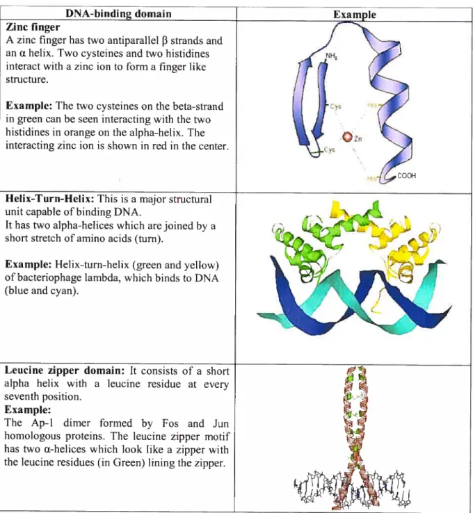
The DNA-binding dornain consists of amino acids that recognize specific DNA bases generally near the start of transcription. Based on its structure, the DNA-binding domain is classified into different types as detailed in Table I.
1. Zinc finger
2. Helix-tum-helix
3. Leucine zipper domain
4. Winged helix
6. Helix-loop-helix
7. Immunoglobulin fold
In addition to a DNA-binding dornain, transcription factors also contain an effector domain. This domain often interacts with proteins to either inhibit or activate transcription. Transcription factors can thus act as transcriptional activators or repressors that control gene expression by acting directly on the RNA-polymerase-containing complex bound at proxirnity of the transcription initiation sites and/or on proteins involved in the assembly of chromatin, the complex of DNA and proteins that make up chromosomes (Roberts 2000). Transcription factors bring about these changes either by themselves or indirectly by recruiting co-factors that are called co-repressors or co-activators (Roberts 2000) depending on their effect on transcription. Co-repressors or co-activators do not bind DNA directly, but are recruited to the gene by the effector domain of transcription factors.
ETS domain: This dornain is 85-90 arnino acids long. It was discovered in the ETS oncogene. Three aipha-helices and a 4-strand beta sheet fold into a domain. The third helix is the recognition helix.
Example:
The Elki-E74DNA complex, where Elk-1 is a member of a large group of eukaryotic
transcription factors with ETS domain. Alpha helices are in blue and beta-strands in yellow
Hellx-Loop-Hellx: This motif has two alpha helices connected by a loop. Generally transcription •factors with this ioop are dimeric. A smailer helix allows dimerization while the other larger helix facilitates DNA binding. Example:
Iwo alpha helices (in Red) connected by a loop (in Green) to form a domain.
Immunoglobulin fold: This is also called an ail f3 protein fold, which has a 2-layer sandwich of 7 antiparallel f3-strands arranged in two f3-sheets. Example:
Hurnan Tenascin with its immunoglobulin fold, fibronectin type Iii, coloured from Blue (N terminus) to red (C-terminus).
Winged helix: This motif has 110 amino acids. Each dornain bas four alpha-helices and two beta-sheet strands.
Example: Alpha helices are in purple and beta strands are in yellow.
4
Table 1: Different types ofDNA bïnding domaïns
DNA-bindin Zinc linger
A zinc finger hastwo antiparallel 13 strands and
an a helix. Two cysteines and two histidines interact with a zinc ion to forma finger like
structure.
Example: The two cysteines on the beta-strand in green can be seen interacting with the two histidines in orange on the aipha-helix. The interacting zinc ion is shown in red in the center.
llellx-TurnHeiïx: This is a major structural
unit capable of binding DNA.
It has twoaipha-helices which are joined by a
short stretch ofamino acids (turn).
Example: Helix-turn-helix (green and yellow) of bacteriophage lambda, which binds to DNA (blue and cyan).
Leucine zipper domain: it consists of a short alpha helix with a leucine residue at every seventh position.
Example:
The Ap-1 dimer formed by Fos and Jun homologous proteins. The leucine zipper motif
bas two Œ-helices which look like a zipper with
the leucine residues (in Green) lining the zipper.
1.2 The C2H2 zinc finger gene famïly
0f the rnany large families encoding transcription factors that have been identifled,
zinc finger genes of the C2H2 type constitute the largest one (Schuh, Aicher et al. 1986; Bellefroid, Lecocq et al. 1989). The C2H2 motif encoded in these genes typically includes two cysteines and two histidines coordinating a zinc ion. This motif was first identified in the TFIIIA of Xenopus leavis and later in the Krtippel drosophila segmentation gene (Miller, McLachlan et al. 1985; Schuh, Aicher et al. 1986). Thus, the C2H2 zinc finger
genes are often refeired to as TFIIIA/Kmppel type of zinc finger genes.
Known to constitute one of the ten largest gene families LPfam databasel, these zinc finger genes are found not oniy in eukaiyotes but also in prokaiyotes. Members of C2H2 zinc finger family have now been identified in ail kingdoms of life i.e. eubacteria, archaebacteria, protists, ftingi, animais and plants (Bouhouche, Syvanen et al. 2000; Moreira and Rodriguez-Valera 2000). Throughout evolution, there bas been a massive expansion in the numbers of the C2H2 zinc finger genes (Lander, Linton et al. 2001; Venter, Adams et al. 2001). Noticeably, human beings are predicted to have more than 700 zinc finger genes often found in a ciustered organization (Bellefroid, Lecocq et al. 1989; Looman, Abrink et ai. 2002).
While most of the C2H2 zinc finger genes characterized have been described as genes encoding transcription factors which bind to DNA, some are also known to encode RNA binding proteins that may thus participate in RNA metabolisrn or maturation (Theunissen, Rudt et ai. 1992; Grondin, Bazinet et al. 1996).
1.2.1 Structure of the proteins encoded by C2H2 ZNf genes
The C2H2 zinc finger transcription factors generaÏly consist of two essential regions
1) the C2H2 zinc finger region containing in most instances several zinc finger motifs
organized in tandem and 2) the N-terminal regulatoiy domain
1.2.2 The tandemly organized C2H2 zinc finger motif
The C2H2 zinc finger proteins are composed of zinc finger motifs which form the zinc finger region of the protein. Each motif is a highly conserved sequence of 28 amino acids (CX24CX3FX5LX2HX34HTGEKPYX, where X is any amino acid). Each motif is separated from the following one by a highly conserved linker region (TGEKPYX, where X is any arnino acid) (Miller, McLachlan et al. 1985; Wolfe, Nekiudova et al. 2000; Loornan, Abrink et al. 2002). The basic conserved C2H2 zinc finger structural unit includes
two cysteines and two histidines which interact with a zinc ion and are essential for the proper folding ofthe motif into a finger like structure (See f igure 1) (Looman, Abrink et al.
2002). C2H2 zinc finger proteins are composed of one or more tandemly organized zinc finger motifs. The number of zinc finger motifs in the protein varies from one to more than 30 in a few cases (Ruiz i Altaba, Peny-O’Keefe et al. 1987).
()
o
J
o
o
Ç)
o
‘© (vk) Ac
________ Bo
(F)J
o
u
n
o
rn-
-ÇÇ)cm—
tH C) tH)’®
©( ®®
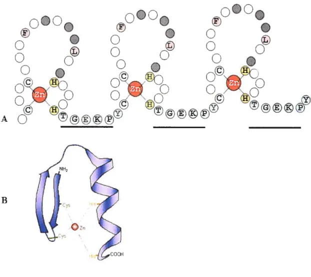
Figure 1: The basic structural unit of a C2112 zinc linger protein.
(A) The C2H2 zinc finger motif is present in tandem in the protein. Three zinc linger motifs arc connected by a conserved Iinker region (TGEKPY). The two cysteines and two histidines which interact with a zinc ion inc]uding die other conserved residues are shown with their single letter codes. The residues involved in DNA binding are shown in grey.
(B) The three-dimensional structure of a zinc finger binding domain. Two anti-parallel f3-strands and one Œ-heÏix interact with a zinc ion as shown in the figure.
Nuclear magnetic resonance spectroscopy (NMR) was used to determine the three dimensional structure of the C2H2 zinc finger motif (Lee, Gippert et al. 1989; Omichinski, Clore et al. 1990). Two beta strands and one alpha helix form an independently folded domain with a compact globular structure (See f igure 1). The zinc ion, that is tetrahedrally coordinated betwcen two invariant pairs of cysteines and histidines, connects the 3-sheet and the a-helix. Four amino acids on the surface of the a-helix in the zinc finger motif make base specific contacts with three to four bases in the major groove of the DNA helix (frankel, Berg et al. 1987; Panaga, Horvath et al. 1988; Omichinski, Clore et al. 1992; Krishna, Majumdar et al. 2003). Aithougli the zinc finger domain has been described as nucleic acid binding domain, not ail the zinc finger motifs are involved in DNA or RNA binding. For example, in ZBRK1 zinc finger protein, only the first few fingers are involved DNA binding and ail the others in protein-protein interactions (Zheng, Pan et al. 2000).
1.2.3 The N-terminal regulatory domain of C2112 zinc finger
proteins
In addition to the zinc finger region, C2H2 zinc finger proteins are also associated with an N-terminal regulatoiy domain (f igure 2), which regulates subcellular localization and the gene expression by acting as either a repressor or an activator by itself or by associating with other factors (Collins, Stone et al. 2001).
The regulatoiy domains associated with C2H2 Zinc finger proteins are j. BTB/POZ domain
ii. KRAB domain iii. SCAN dornain
j. The BTB domain
The BTB domain (Broad-Cornplex, Tramtrack and Bric-a-bric) also known as the POZ domain (Pox virus and Zinc finger) is a 120 arnino acid conserved dornain found to be associated with both DNA and actin-binding proteins. The 3TB domain is involved in protein-protein interactions (Collins, Stone et al. 2001). As a part of DNA binding proteins, the BTB/POZ domain is known to be a dirnerization domain which, in a few cases also recmits co-repressors (such as N-CoR, STN3A or SMRT) and acts a repression domain. When found in association with C2H2 zinc finger transcription factors, the BTB domain is generally located N-terminal to the zinc finger region. Thus, by mediating oligornerization and in some instances interaction with co-factors, the BTB domain can lead to chromatin remodeling and change in gene expression (Melnick, Carlile et al. 2002).
ii. The Kruppel-Associated Box (KRAB) domaïn
Another well known example of an N-terminal regulatory domain associated with C2H2 zinc finger proteins is the Kruppel-Associated Box or the KRAB domain (Bellefroid, Poncelet et al. 1991; Rosati, Marino et al. 1991). KRAB domains are almost
aiways associated with C2H2 zinc finger proteins. An exception to this scenario is the SSX family ofproteins. These proteins are associated with a “SSX KRAB dornain” which is distantly related to the KRAB domain from C2H2 zinc finger proteins
(
49% similar) but are flot associated with zinc fingers (Collins, Stone et al. 2001; Urrutia 2003).Unlike the C2H2 zinc finger proteins which are present in organisms ranging from bacteria to humans, the KRAB dornain as seen in C2H2 zinc finger proteins is present only in vertebrates, more specifically in tetrapods (Looman, Abrink et al. 2002). However, a recent study identified a sea urchin homolog to the mammalian Meistez protein which includes a tandem array of C2H2 zinc finger motifs, a SET dornain and a sequence with some sirnilarity to the “SSX-KRAB domain” (Birtie and Ponting 2006). This suggests the presence of the KRAB domain in the common ancestor of echinoderms and vertebrates. A further study of these proteins in ftingi and plants identified a 26 amino acid motif called the KRI motif which was found to be similar to the aipha-helical regions of KRAB and present in ail eukaryotes. This indicated that the KRI motif was present in the last common ancestor of animais, plants and fungi and is the progenitor of the KRAB dornain.
The KRAB domain is most abundant in mammals (Lander, Linton et al. 2001; Venter, Adams et al. 2001; Waterston, Lindblad-Toh et al. 2002). For example, about one third of the mouse C2H2-ZNF are associated with KRAB (Benn, Antoine et al. 1991; Waterston, Lindblad-Toh et al. 2002). The KRAB domain is mostly associated with more than 5 C2H2 zinc finger motifs in a protein, justifying the name “Multifingered protein” (Bellefroid, Poncelet et al. 1991). Many genes encoding the KRAB containing proteins are found in a clustered organization as opposed to the ones found as singletons (Bellefroid,
Marine et al. 1993; Shannon, Kim et al. 199$; Chung, Schafer et al. 2002; Rousseau Merck, Koczan et al. 2002; Hamilton, Huntley et al. 2003).
The KRAB domain is 75 amino acids long and is divided into two boxes, Box A (-38 amino acids) and Box B (32 amino acids) (Looman, Abrink et al. 2002; Urrutia 2003). A variant of the B box, called b box also exists. Some C2H2 zinc finger proteins
have another box following the A box, called the C box (21 amino acids). Each of these boxes is encoded by different exons and separated by introns of vaiying lengths (Loornan, Heilman et al. 2004). The KRAB domain functions as a potent repressor of transcription (Margolin, Friedman et al. 1994). The KRAB A box plays an important role in repression by binding to co-repressors, while the KRAB B box doesn’t have transcriptional activity but is known to enhance the repression activity of the A box (Witzgall, O’Leary et al. 1994). The process of transcription repression is mediated by KAP-1, also called transcription intenriediary factor 13 (Tlflt3) which is a co-repressor that interacts with
KRAB A (Friedman, Fredericks et al. 1996; Germain-Desprez, Bazinet et aI. 2003). The KRAB domain of C2H2 zinc finger proteins recruits the KAP I co-repressor to DNA, which results in the formation of a heterochromatin like complex and leads to gene silencing (Pengue, Calabro et al. 1994; Kim, Chen et al. 1996; Moosrnann, Georgiev et al.
1996; Pengue and Lania 1996).
iii. The SCAN domain
The SCAN domain like the KRAB domain is another vertebrate specific domain only associated with C2H2 zinc finger proteins (Williams, Khachigian et al. 1995; Looman,
Abrink et al. 2002). The SCAN domain was estimated to be associated with 10% of the total C2H2-ZNF present in the human genome (Collins, Stone et al. 2001; Edeistein and Collins 2005). Also known as the LeR domain because of its leucine rich primaiy structure, the SCAN domain is named after the four proteins it was initially identifled (SRE-ZBP, CTfin5l, AW-1 and Numberl8 cDNA) (Urrutia 2003). In addition to being associated with the C2H2 zinc finger proteins, the SCAN domain containing proteins are sometimes associated with a KRAB domain having the organization SCAN-KRAB (C2H2) or in very few cases KRAB-SCAN-KRAB-(C2H2) (Edeistein and Collins 2005; Huntley, Baggott et al. 2006).
Structural studies on the SCAN domain indicate that it has 84 arnino acids and is found to have three to five a-helices which are delineated by one or more proline residues. Proline residues are also present before and after the SCAN domain (Stone, Maki et al. 2002). The SCAN domain is a homo and hetero-dimerization domain mediating protein protein interactions by self association and formation of heterodimers between SCAN family members (Sander, Haas et al. 2000; Schumacher, Wang et al. 2000). The importance of the dimerization for the transcriptional activity of SCAN-C2H2 zinc finger protein lias flot been clearly established.
Regiilatory Dornain Spacer Zinc linger region _ - flUBA VTFED5AVYFSQEEWGLLDPAQRNLYRDvLENY RNLVSL
—FJT---
-J. KRAB b bHQLFJOEDX I sQLEREEKLWMMIxATQRGDS S>’ k !. nU SCAN PDPEIFRQRFRQFCYQETPGPREALSR LRELCHQ WLRPEVHTKEQILEL LVLEQF LTI LPKELQAWVQ EIfflPESGEEAVTLLEDLERELDEPGQQV . LQNPSIWTGLLCKANQMRLAGTLCDVVIMVDSQE FEFTILiCTSK14FEILFRRNSQHiTLDFLSPK ., ., . TFQQILEYAYTATLQAKAEDLDDLLYAAEILEIE Y LEEQC LKM L BFigure 2: The Regulatory domains associated with C2H2 Zinc linger proteins.
(A) The different combinations of dornains associated with zinc finger proteins are shown. Zinc finger proteins generally have three main regions: The Regulatory domain, the Spacer and the Zinc finger region. (B) The consensus sequence of the domains KRAB (A, B, b and C boxes), SCAN and BTB. The residues essential for binding KAPI and thus for repression are shown in KRAB A underlined in red.
1.3
Gene familles and Gene duplication
1.3.1 Gene Families
A gene family colTesponds to a set of genes that are grouped based on their shared homology, biologicai or biochemical activity, sequence motifs or similarities in stntcture. Because they consist of a large number of genes, gene families are the most informative systems to study evolutionary dynamics of genes. Nuclear genomes have many multigene families and their studies provide dues to the evolutionaiy forces that have shaped these genomes (Ohta 2000; Thomton and DeSalle 2000). Mammalian genornes in particular have large numbers of genes organized in gene families (Demuth, Bie et al. 2006). Some gene families have uniforrn copy numbers of genes in ail species (Thomton and DeSalle 2000), while there are gene families like the Immunoglobulin gene family, the Olfactory receptor gene family and the C2H2 zinc finger gene family which have a large variation in the number of genes across different species . The variation in the gene numbers of these
families and diversity in ftinction, suggests that gene duplication and/or gene ioss have played an important role in shaping differentmammalian genomes.
I. The Olfactory Receptor gene family
Olfactoiy receptor (OR) genes form the largest known multigene family in mammalian genomes (Glusman, Bahar et al. 2000) and code for membrane receptors that are responsible for olfaction, the sense ofsmell. OR genes are present in various vertebrates ranging from lampreys to humans. The OR proteins belong to the G-protein coupÏed
receptor family which have seven transmembrane dornains. OR genes are divided into 2 classes based on their protein sequence similarity (Glusman, Bahar et al. 2000; fuchs, Glusman et al. 2001). 0f the two classes, Class I genes first identified in fish but also found in mammals are specialized in water-soluble odorants and the Class II genes specialized for airbome odorants are specific to tetrapods.
The number of the OR genes is quite varied in different genomes. Rodents have nearly twice as many as the number present in human, chimpanzee or dog (Niimura and Nei 2005). The Human genome has more than half of the -900 OR genes as pseudogenes. In contrast, the mouse genome bas l300 OR genes of which only one-fourth are pseudogenes. $tudies on the human, chimpanzee and mouse OR gene repertoires indicate that there are species-specific expansion and pseudogenization signifying different selection pressures in humans, chimpanzees and mouse owing to their different sensoiy requirements (Sharon, Glusman et al. 1999; Glusman, Yanai et al. 2001; Lapidot, Pilpel et al. 2001; Gilad, Man et al. 2005; Niimura and Nei 2005). Evolutionary analysis of the human, mouse and chimpanzee datasets indicate the presence of clustered organization which is generally well conserved in these genornes. Analyses of the clusters indicate that there are tandem arrays of the OR genes which appear to have arisen by tandem duplication and several chromosornal rearrangements. The difference in the numbers of OR genes in hurnan and mouse has been attributed to gene duplication and loss events (Sharon, Glusman et al. 1999; Niimura and Nei 2005).
ii. The Immunoglobtilin gene family
The immunoglobulin gene family represents an example where its two subfamilies, the immunoglobulin heavy variable region sub-farnily and immunoglobulin light chain variable region subfamily, have co-evolved by valying in gene number and extent of diversity in different species ($itnikova and Sti 1998). An immunoglobulin molecule is a tetramer with two identical heavy chains and two identical light chains which forrn a Y shaped structure. Each of these chains has a variable (V) and constant (C) domain. The VH and VL domains have the complementarity determining regions, called the CDRs which form the sites of interaction with antigens. Analyses of these two sub-farnilies of genes from various species of amniotes identified that these gene families have diversified throughout the course of evolution (Sitnikova and Su 1998). Different coordinated loss and duplication events have led to different species-specific gene repertoires.
iii. The C2112 zinc finger gene family
In addition to the above mentioned gene families, the C2H2 Zinc finger gene farnily is another example of a large multigene family with varying number of genes in different species. Over the course of evolution, this gene famlly bas expanded drastically in mammalian genornes (e.g. — 400 in mouse and 700 in human) (Venter, Adams et al.
2001; Waterston, Lindblad-Toh et al. 2002). Several studies involving these genes in the human genome have indicated that tandem duplication events are responsible for the
clustered organization of this family (Shannon, Kim et al. 199$; Elemento and Gascuel 2002; Elemento, Gascuel et al. 2002; Tang, Waterman et ai. 2002; Bertrand and Gascuel 2005; Huntley, Baggott et al. 2006). A few instances of evoiutionary studies ofthese genes, within the human genome and among a few mamnialian genomes document cases of species-specific duplication (Dehal, Predki et ai. 2001; Shannon, Hamilton et ai. 2003; Huntley, Baggott et al. 2006).
Ail these examples of gene families suggest variation in number among different species involving different duplication and ioss events. The gene family size could vaiy based on the ftinctionai relevance of the gene farniiy in the organism. These examples also indicate the importance of studying the gene families to give dues on the evolutionaiy mechanisms which led to different sizes of gene families.
1.3.2 Gene Duplication ami Gene Loss: Two important evolutionary
mechanisms guiding the evolution of gene famïlîes in mammals
Considering the extremely large numbers of genes constituting gene families (Demuth, Bie et al. 2006), it is interesting to study their organization and the evolutionaiy mechanisms that created them. A study integrating the information from spatial organization of the genes with the phylogenetic reiationships between the genes combined with evolutionaiy information of the species would help provide dues about the evolution ofthe gene families.
In the context of using phylogenetic studies to analyze the evolutionary reiationships between genes in gene families, one significant term that featuresin ail studies is “Romoiogy”. Homology forms the centrai and basic concept of comparative genomics but is aiso a terni that is often misrepresented and misinterpreted. The terni homology was introduced by Richard Owen in 1848, where lie defined homology as “the sanie organ under eveiy variety of form and fttnction”(Francis Darwin 1903) . The importance of
structure and fiinction is ernphasized more in this definition. In an attempt to give an evoiutionary explanation to hornoiogous structures, Darwin defined homology as “A structure is sirniiar among reiated organisms because those organisms have ail descended from a common ancestor that had an equivaient trait” (Darwin 1837) (Figure 3).
When put in the context of molecular sequence comparison, in today’s times, homoiogy refers in an abstract way to a reiationship which implies a possible common ancestry and shouid be differentiated from identity2 or similarity3 of sequences. However, to be substantiated, homology must be confirmed by appropriate phylogenetic studies. It is important to note that homology does flot say anything about functionai simiiarity (Thomton and DeSalle 2000))(Fitch 2000).
131
Homology: A hypothesis that signifies comnion ancestry between sequences (nucleotide or amino acid) which is prirnarily based on sequence similarity.
2ldentity: The extent to which two (nucleotide or amino acid) sequences are invariant.
Similarity: The extent to which nucleotide or protein sequences are related. The extent ofsirnilarity between two sequences can be based on percent sequence identity and!or conservation
Figure 3: Darwin’s evolutionary tree.
The figure is Charles Darwin’s first ever sketch of an evolutionary tree from bis book titled “First Notebook on Transmutation of Species (1837)”.
There are three major types of homology in a phylogenetic context which are orthology, paralogy and xenology. Orthotogy as described by f itch in 1970 is the relationship between two genes in two different species which originated from a common ancestor. Two homologous sequences are considered to be “orthologous” if a speciation event separates them. In contrast, Paralogy signifies the relationship between two genes which have been formed by a gene duplication event. XenoÏogy, another type of homology relationship describes the relationship between two genes which have been transferred between two species by horizontal gene transfer.
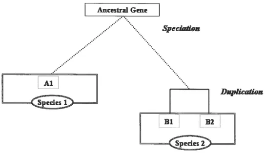
Studying the homologous relationships of genes within and between various genomes and differentiating between orthologs and paralogs is a central aspect of comparative genomics.figure 4 shows a verysimple explanation of the difference between orthologs and paralogs. The genes Ai, Bi and 32 have evolved from an ancestral gene by speciation followed by a duplication event in species B. Gene Al from species A is an ortholog of gene Bi and gene B2 in species B illustrating that one gene in a particular species may have more than one ortholog in the other. Gene Bi and gene B2 in species B, which were formed by gene duplication, are paralogs to each other.
Speciahon
DRptkWiJrn
Figure 4: Schematic representation of speciation and duplication
Genes Ai, Bi and B2 are formed from an ancestral gene by a speciation and duplication event. Gene Al from species A has two orthologs in species B, genes 31 and B2. Bi and 32 are paralogs.
figure 5 depicts different evolutionary scenarios that one encounters while
studying the evolution of genes in gene families are depicted to explain the relationships between genes in ternis of orthologs, paralogs and gene Ioss. An ancestral gene undergoes duplication in species O to give the genes, A, B and C. This is followed by a speciation event with genes Ai, Bi and Clin Species i and genes A2 and B2 in Species 2. The gene Al is an ortholog of A2 and Bi is an ortholog of 32. The gene Cl does not have a corresponding ortholog, as the Species 2 lost the gene after speciation. The genes AI, Bi and Cl are paralogs within species 1 and, A2 and B2 are paraiogs within species 2. Furthermore, as explicitly pointed out recently by fitch (Fitch 2000) and as often ignored, gene Ai (species i) is also a paralog of gene B2 (species 2), gene Bi (species 1) is a paraiog of gene A2 (species 2) and gene Cl is paralog of gene A2 and B2 (species 2). from these explanations, it is clear that orthologs are homologous genes residing in different species, while paralogs may not only refer to the homoÏogy relationship between genes from the same species but also from different species. It is essential to understand that both orthologs and paralogs are free to diverge and do not necessarily aiways have the same function (Thornton and DeSalle 2000).
Duplk&dsnewm
Geiz, Ie
Figure 5: Schematic representation of different evolutionary processes shaping the gene familles in different species.
Gene duplication, speciation and loss lead to the formation of genes Ai, Bi, Cl in species 1 and A2 and B2 in species 2.
csO
1.4 Inferring gene duplication and gene loss
That two genes are homologous is a hypothesis that needs to be studied and analyzed to be able to derive the relationships between the genes to be either orthologs or paralogs. Studying and analyzing the relationships between gene farnilies i.e. evaluation of orthology or paralogy requires a well formulated approach. In order to be able to postulate theories on how related genes evolved from an ancestral gene i.e. by gene duplication, gene loss or by speciation, one needs to assess homology relationships using a well founded phylogeny.
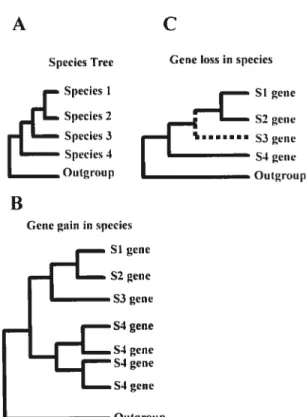
The first step in assessing homology is a sequence alignment of the molecular sequences be it nucleotides or arnino acids. This gives a preliminary measure of possible homology which can then be assessed using a phylogeny. A welI supported phylogeny gives the evolutionaiy relationships bePveen the genes in relation to one another. Comparison of a gene phylogeny between genes with the taxonomic relationships between species, allows gene duplication and loss events to be assessed and roughly dated. As an example, figure 6 shows the different scenarios of gene duplication and gene Ioss. In figure 6A, it can be seen that a duplication event prior to the speciation event resulted in species 2, 3 and 4 created the paralogous gene groups A, B and eventually C. In a hypothetical situation, suppose the genes 2A, 3C and 4B are missing from the gene tree. Assuming that the studied sequences were derived from completely sequenced genomes, the missing genes could either be due to a loss of genes or their possible pseudogenization in the respective species. That the gene duplication occurred prior to speciation can stili be
resolved by superimposing the gene tree with the species tree. Reconciliation between the species and the gene tree will help resolve the absence of genes 2A, 3C and 4B as can be
seen in figure 6B. This kind of srndy can hence be used to infer the evolution of genes
lA 2A 3A 4A 3C 4C 4D 2B 313 4E lA 3A
4A
3C
4C 4D 2E3B
413
Figure 6: Inferring gene duplication and loss events from a gene tree in comparison with the species tree.
(A) A gene tree showing the phylogeny between genes belonging to species 1, 2, 3 and 4. Genes are represented as A, B, C and D. A gene duplication represented as x occurred prior to the speciation event leading to Species 2, 3 and 4.
(B) A species tree showing the relation between species 1, 2, 3 and 4.
(C) A hypothetical situation where the genes 2A, 3C and 4B are missing from the tree as shown in red. Reconciliation of the phylogenetic tree from (A) with the species tree from (B) helps identify the flot only the duplication event but also the missing genes to be able to infer loss. Adapted from (Thornton and De$alÏe 2000)
j
4
B1.5
Previous Studïes addressing zinc finger gene evolution
About 2000 of the 30,000 genes in the human genorne code for transcription factors (Venter, Adams et al. 2001). C2H2-ZNF are the most common of ail the eukaiyotic transcription factors present in the human genome (encoded by 700 genes). Owing to these facts, the C2H2 zinc finger gene famiiy has been considered to be an evoiutionary piayground for genes to develop and differentiate and hence is an interesting famiiy to study (Looman, Abrink et al. 2002).
The studies pertaining to C2H2-ZNF have mostly been restricted to those associated with a KRAB domain and more specifically to the human genome. A recent study identified 423 KRAB C2H2-ZNF ioci organized into 65 ciusters on the human genome (Huntley, Baggott et al. 2006). Evolutionary studies involving these KRAB C2H2-ZNF genes indicated that the evolutionaiy reiatedness within and among ciusters was flot only associated with physical proximity evoiving through tandem duplications but aiso through distributed duplication and postduplication rearrangement events, which have lcd to the drarnatic increase in the gene numbers of this famiiy in hurnans (Hamiiton, Huntley et ai. 2003; Hamiiton, Huntley et al. 2006; Huntley, Baggott et ai. 2006). Though present in clusters, the KRAB C2H2-ZNF are not co-regulated and they show different pattems of expression (Huntley, Baggott et al. 2006).
A study of one KRAB C2H2-ZNF gene cluster on hurnan chromosome 19, suggested an evolutionary model showing the presence of certain beta-satellite repeat structures symrnetrically ordered with the zinc finger genes in the cluster which have coevolved with the cluster accommodating the expansion of the genes within this cluster (Eichler, Hoffman et al. 1998).
A statistical analysis using phylogenetic models on four hurnan C2H2-ZNF clusters on chromosome 19 indicated that positive selection is the driving force involved in the diversification of the KRAB C2H2 zinc linger genes (Schmidt and Durrett 2004).
Not much is known about the evolutionary histories of these genes in different mammalian genomes and very few studies have been carried out to comparatively analyze their evolution. A preliminaiy report on species-specific expansion of these genes, resulted from one study on a C2H2-ZNF cluster on human chromosome 19 and its syntenically homologous cluster on mouse chromosome 7 (Shannon, Hamilton et al. 2003) . A study on
the evolution of members of the primate-specific ZNF9 1 KRAB subfamily, which are mainly found in a chromosome 19 cluster, revealed that this gene subfamily evolved before the spiit of humans and apes. But afier the split, these genes have continued to evolve differentially be it through tandem duplications or segmental duplications, leading to species-specific genes (Dehal, Predki et al. 2001; Hamilton, Huntley et al. 2006).
Inspite of several studies dealing with these genes, there has neyer been a comprehensive study on the C2H2-ZNF genes and their evolution or their functions. In
order to systematically define and analyze the extent of species-specffic duplication and the role of gene loss in the evolution of these genes, it is important to conduct a comprehensive study of these gene clusters in mammalian genomes to obtain dues on their evolution and their possible implicationson ffinctions specific to each species.
1.6 Hypothesis and Objective
Previous studies on zinc finger genes have provided evidence that zinc finger genes have undergone a huge expansion in vertebrate genomes, with a specific increase in humans. Studies have shown that these genes have been subjected to expansion through tandem duplication and also of the existence of species-specific duplication events (Shannon, Kim et al. 199$; Shannon, Hamilton et al. 2003; Harnilton, Huntley et al. 2006; Huntley, Baggott et al. 2006). A contribution of gene loss in the evolution of C2H2 zinc finger genes has been suggested but neyer tested rigorously.
The main objective of this thesis is to systematically determine to what extent zinc finger genes are subrnitted to species-specific expansion and to assess the potential contribution of gene loss in the evolution of this gene famiiy in mammals. To this end, we have:
1. Assembled a curated database of ail C2H2-ZNF genes in the hurnan genome and identify ail the C2H2-ZNF clusters in the human genome
2. Searched for syntenically homologous clusters in other cornpletely sequenced mammalian genomes, narnely chimpanzee, mouse, rat and dog genomes.
3. Perforrned a phylogenetic analysis of C2H2-ZNF genes from the syntenically homologous clusters.
4. Perfornied a reconciliation of both phylogenetic analyses and physical maps of the clusters with the species tree accounting for the evolutionary history of the species in order to infer gene loss and gain.
These studies should allow us to determine the nature of evolutionary events that shaped this large gene farnily in mammals. In particular, this study wiIl help us to better infer orthology in the various mammals and better understand the evolution and relationships between the different C2H2-ZNF subfarnilies.
Evolution of C2H2-zinc finger genes in mammals:
Species-specific duplication and loss at the level of
clusters, genes and their functional domains.
Hamsa Dhwani Tadepalfy, Gertraud Burger and Muriel Aubry*
Department of Biochemistry, Université de Montreal, C.P.612$,
Succ.Centre-Ville, Montreal, QC, H3C 3J7, Canada
To whom correspondence and reprints should be addressed:
MurielAubry, Ph.D. Departrnent of Biochemistiy Université de Montréal C.P. 6128, Succ. Centre-Ville Montréal, H3C 3J7 Canada
Key words: C2H2/Kruppel, zinc finger, gene family, tandem repeats, gene duplication, gene loss, evolution.
ABSTRACT
C2H2 zinc finger genes (C2H2-ZNF) constitute the iargest class of transcription factors in hurnans and one of the largest gene families in mammals. Often arranged in clusters in the genome, these genes are thought to have undergone a massive expansion invertebrates by a process involving tandem duplication. However, this view is based on lirnited datasets restricted to single chromosome or a specific subfamiiy of C2H2-ZNF genes. Here, we present the first comprehensive study of the dynamic evoiution of the C2H2-ZNF family in mammals. We assernbled the complete repertoire of hurnan C2H2-ZNF genes (718 in total), about 70 % of which are organized into 81 ciusters across ail chromosomes. Based
on an analysis of their N-terminal effector domains, we identified
SET- and HOMEO dornain-encoding ZNF genes as members of two new C2H2-ZNf subfamiiies. We searched for the syntenic counterparts of human clusters in other mammals for which compiete gene data are avaiiable: chirnpanzee, mouse, rat and dog. Cross-species comparisons show a large variation in the numbers of C2H2-ZNF genes within homologous mammalian clusters stiggesting differential pattems of evolution. Phylogenetic anaiysis of selected C2H2-ZNF clusters reveals that differences in C2H2-ZNF gene repertoires across mammais not only originate from differentiai gene duplication but also gene loss. Further, we find variations among orthologs in the number of zinc finger motifs and association of the effector dornains, the later often undergoing sequence degeneration. Based on these resuits and an anaiysis of the exon-intron organization of genes from the large SCAN and KRAB domains-containing subfamilies, we propose a new model for the evolution ofthese subfamilies.
This manuscript includes two supplementaiy Figures and four supplementary Tables INTRODUCTION
The human genome sequence uncovered a large number of gene families oflen arranged in a clustered organization (Ohta 2000; Thornton and DeSalle 2000; Venter, Adams et ai. 2001). C2H2 zinc finger (C2H2-ZNf) genes make tip 2 % of ail the human genes and represent the second largest gene family in humans after the odorant receptor farnily (Lander, Linton et al. 2001) (Schuh, Aicher et al. 1986; Bellefroid, Lecocq et al. 1989; Messina, Glasscock et al. 2004). The first identified members of the C2H2-ZNF family areXen opus TFIIIA and Drosophila Kruppel and thus genes of this family are often called zinc finger genes of the TFIIIA or Kruppel type (Miller, McLachlan et ai. 1985; Schuh, Aicher et al. 1986).
Most of the characterized C2H2-ZNF genes code for transcription factors which bind DNA through their zinc finger region; others bind RNA and their exact function is yet unknown (Theunissen, Rudt et al. 1992; Grondin, Bazinet et al. 1996). The zinc finger region is cornposed of a basic structural unit of 28 amino acids (CX21CX3FX5LX2HX3 4HTGEKPYX, where X is any arnino acid), called the zinc finger motif, that is often repeated in tandem. The two cysteines and two histidines in this motif interact with a zinc ion, stabilizing the proper folding of this motif (Klug and Rhodes 1987; Lee, Gippert et al. 1989; Rhodes and Klug 1993). C2H2-ZNF proteins often contain an effector domain aiways located N-terminal to the zinc finger region, such as the KRAB (Kntppel Associated-Box), SCAN (SRE-ZBP, CTfin5l, AW-1 and Numberl8 cDNA) and BTB (Broad-Complex, Tramtrack and Bric-a-bric) domains. The first two domains are
vertebrate-specific (BelÎefroid, Poncelet et al. 199f; Rosati, Marino et al. 1991; Collins, Stone et al. 2001), while BTB is also present in insects. The KRAB domain includes the box KRAB A (—38 amino acids) involved in transcriptional repression and often a second box, usually KRAB B (—32 amino acids) or in few cases KRAB b or KRAB C (—21 amino acids) box (Witzgall, O’Leary et al. 1994; Looman, Abrink et al. 2002; Urrutia 2003; Looman, Heilman et al. 2004). The KRAB A box and the second KRAB B, b or C box are encoded by separate exons, which are alternatively spliced. The SCAN, also called the leucine-rich (LeR) domain
(—
84 amino acids) (Stone, Maki et al. 2002) mediates protein protein interactions through dimerization (Sander, Haas et al. 2000; Schumacher, Wang et al. 2000). The BTB dornainQ—-
120 amino acids) is a dimerization domain that also acts as a repression dornain in some cases (Melnick, Carlile et al. 2002). In contrast to the SCAN and KRAB domains which are only present in C2H2-ZNf proteins, the BTB domain is also found as a part of actin-binding proteins (Coïlins, Stone et al. 2001). C2H2-ZNF proteins are grouped into different subfamilies based on the type of N-terminal effector dornain present.Initial studies on the C2H2-ZNF gene farnily focused on hurnan chromosome 19, which is particularly enriched in clusters of these genes (Bellefroid, Marine et al. 1993; Eichler, Hoffman et al. 1998). More recent studies deait more specifically with the KRAB subfarnily (Mark, Abrink et aI. 1999; Looman, Abrink et al. 2002; Shannon, Harnilton et al. 2003; Huntley, Baggott et al. 2006). The current view is that C2H2-ZNf genes have undergone a massive expansion during vertebrate evolution by a process involving tandem duplication (Dehal, Predki et al. 2001; Looman, Abrink et al. 2002; Hamilton, Huntley et
al. 2003; Shannon, Harnilton et al. 2003; Harnilton, Huntley et ai. 2006; Huntley, Baggott et al. 2006). Yet, this view may be biased because it is extrapolated from smali subsets of C2H2-ZNF genes.
In this report, we reconstructed a global picture of the evolution of the C2H2-ZNF gene repertoires during mammalian speciation, based on a comprehensive catalogue of ail human C2H2-ZNf genes and their syntenic counterparts present in clusters in other mammals. Our study clearly dernonstrates that this gene farnily expanded and contracted flot only in hurnan but across mammals and in a lineage-specific fashion. In addition, we discovered evolutionary change of individual C2H2-ZNF orthologs invoiving both differential duplication of zinc finger motifs and loss of N-terminai effector dornains. $peciation of mammals is characterized by divergent evolutionary trends at the level of individual C2H2-ZNF genes as well as the entire farnily. This led us to propose a model for the evolution of SCAN, SCAN-KRAB and KRAB subfamilies and points to the importance of comparing complete repertoires rather than C2H2-ZNF genes from specific subfarnilies for gaining insights into the possible orthologous relationships between genes from varions genornes.
METHODS
Collection of human C2H2 zinc finger genes
We conducted an extensive sirnilarity search to identify the compiete repertoire of C2H2-ZNF genes in the hurnan genome (assembly NCBI 36). First, we identified ail the genes annotated as C2H2 and/or Kmppei zinc finger genes by performing an initiai text term search via Entrez (www.ncbi.nlm.nih.gov). Second, we used PROSITE (http://www.expasy.com) to identify ah the proteins which had a zinc finger motif of the C2H2 type as weil as the N-terminal effector domain, if present.
from these searches, the genomic coordinates, chromosome number, position on the chromosome, number of fingers and identified domains were collected for each of the gene and protein sequences (initial dataset). A TBLASN (e-vaiue ctttoff le-3) (Gertz, Yu et ai. 2006) search was done against the genome using each of the gene sequences from the initial dataset as a query. The blast hits were used to generate the final dataset of ah the identified C2H2-ZNF genes (Suppiementary Table Si).
Identification of C2H2-ZNF gene clusters in the human genome
We anaiyzed the relative positions of C2H2-ZNF genes in the human genorne in order to identify the C2H2-ZNF clusters. A distribution of the distances between neighboring C2H2-ZNF genes in the human genome is presented in Supplernentaiy Figure Si. Two consecutive C2H2-ZNF genes are said to beiong to a ciuster if the distance between them is 500 kb regardiess of the presence of other genes within the ciuster , a
threshold classically used in gene farnily studies (Niimura and Nei 2003). Clusters were determined for each hurnan chromosome.
Identification of mammalian C2H2-ZNF ciusters syntenically homologous to human clusters
We searched for clusters hornologous to the human C2H2-ZNf clusters (i.e. syntenically homologous clusters) in other mammals for which complete genorne sequences are available. The assemblies used for Pan troglodytes), Mus muscutus, Rattus norvegicus and Canis famitiaris were chimpanzee Pan Tro- 2.1, mouse NCBI m36, rat RGSC 3.4 and dog Can fam 2.0. We used the linkage maps of Ensembi (http://www.ensembl.org); assignment of syntenic clusters is based on the genes flanking each human cluster and which were mapped in ail the species. Four flanking genes at each extremity were mapped in most instances. Then, we conducted TBLASTN analysis of the syntenic regions comprised between the flanking genes, using as queries the amino acid sequence of the zinc finger region from ail the known human C2H2-ZNF genes from the corresponding region. A hit with e-value le-4 confirrned the respective hornologous clusters in the five mammalian genornes. A comprehensive catalogue of the hurnan C2H2-ZNf clusters and their syntenic counterparts in other mammals is cornpiled in Supplementary Table S4.
Phylogenetic analysis
Phylogenetic analysis was conducted using the amino acid sequences of the zinc
finger region (identified using PROSITE) of C2H2-ZNF genes from selected human
clusters and their syntenically homologous clusters in chimpanzee, mouse, rat and dog. A
multiple sequence alignment of the zinc finger regions of the C2H2-ZNF genes was
generated using the program MUSCLE (Edgar 2004). The alignments were edited to
remove gaps using the program GBLOCKS (Castresana 2000). Maximum Likelihood (ML)
and Bayesian Inference (BI) methods were used to infer the phylogenetic trees and estimate
the clade support. for ML analysis, the program RAxML (RAxML-VI-HPC Version 2.2.1)
(Stamatakis, Ludwig et al. 2005) employing the WAG model of amino acid substitution was used to reconstruct the best tree. Bootstrapping of 100 datasets was irnplemented. The
posterior probabilities were deterrnined by a Bayesian MCMC method implemented in the
program Mr.Bayes v.3. 1 (Huelsenbeck and Ronquist 2001) to test the robustness of the
topology of the tree infened through ML. One million generations were rcin and the trees
were sampled after every 10 generations.
To determine appropriate outgroups for our analysis, we searched the nr database to look
for close homologs in non-mammals using TBLASTN (e-value eut off le-4). In addition to
the Xfin sequence from Xen opus Ïaevis, we obtained a set of zinc finger genes from Chicken (Gallus gaïlus, Assembly WASHUC2) specifically selected for each human C2H2-ZNF cluster based on an extensive similarity search. To select the chicken outgroup,
a TBLASTN (e-value cutoff le-4) search was done against the chicken genome using each
The top 10 hits for each query sequence were ail analysed using a CD-HIT anaiysis
(Identity threshold = 100%, 95% and 90%) (Li, Jaroszewski et aI. 2001) to produce a final
set of non-redundant representative chicken sequences, ail used as a partofthe outgroups.
Sequence analysis to confirm the Ioss of domains
In the case where loss of a dornain was suspected, we conducted an extensive
sequence analysis to mie out the possibility that these domains would have been rnissed
either due to a frame-shift or inadequate exon-intron spiicing of the gene and thus
inappropriate amino acid translation, preventing recognition by PROSITE
(http://www.expasy.com). Firstiy, for each particular C2H2-ZNF genes where loss of an
N-tennina1 dornain was suggested, we systematically collected the nucleotide sequence of
the region ranging from the stop of translation of the previous gene to the start of
translation of the next gene. We conducted a TBLASTN search of this region using die
amino acid sequence of the dornain of interest (present in the colTesponding orthoiogs and
the consensus of the domains selected from randomly seiected sequences) as a query to
confirm the absence of the domain in the C2H2-ZNF gene of interest. Secondly, we
obtained the exon-intron structure of these genes using the Ensernbl Genorne Browser
(http://www.ensembi.org). In order to search for exonic 01. intronic sequences which may
exhibit significant identity with the nucleotide sequence of the domain of interest. For this
purpose we conducted a BLAST anaiysis of the individual exon and intron sequences with
the nucleotide sequence of the various domains that are present in the coiresponding
Flowchart of the study
figure 1 summarizes the flowchart of our analysis procedure of C2H2-ZNF genes and clusters in mammals.
RESULTS
Compilation of a comprehensive catalogue of human C2H2-ZNF genes
Previous studies reported the existence of at least 564 C2H2-ZNF genes in the
human genome and suggested that this family may include approximately 700-800 genes
(Bellefroid, Lecocq et al. 1989; Bellefroid, Poncelet et al. 1991). As a first step to study the
evolution of ZNF genes, we established a comprehensive catalogue of the
C2H2-ZNf genes in the hurnan genome. By conducting an extensive simiiarity search (see
Methods), we identified 71$ C2H2- ZNF genes (compiied in Supplementary Table Si). 0f the 718 genes, 66 are annotated as pseudogenes in Genflank. For ail genes, we determined their exact position onthe chromosomes, their orientation, the number of finger motifs and the effector domains.
These genes are distributed across ail chromosomes of the human genorne
(Supplementaiy Table $2). As reported earlier, chromosome 19 has the highest number
(Venter, Adams et al. 2001) and density ofC2H2-ZNF genes, inciuding 40% (289) ofthe
71$ human C2H2-ZNF genes, whereas this chromosome corresponds to only 2.1 % of the
human genome. More than haÏf (58%) of the C2H2-ZNf genes encode conserved N tenuinal domains, the KRAB, SCAN and BTB dornains (figure 2A). typically involved in transcriptional regulation (Kim, Chen et al. 1996; Collins, Stone et al. 2001) and form
different C2H2-ZNF subfamilies. Further, we discovered two additïonal dornains typical of transcription regulators, the SET and HOMEO domains that are also encoded by
C2H2-ZNf genes. While the KRAB subfarnily represents almost haif of the C2H2-ZNF genes
subfamilies account for oniy a small percentage (—12%) of the C2H2-ZNF genes (figure 2A).
Clustered organization of human C2112-ZNf genes
It was reported earlier that chromosome 19 is particularly rich in tandemly duplicated C2H2-ZNf genes and that KRAB C2H2-ZNF genes. are clustered on several other chromosomes (Dehal, Predki et al. 2001; Rousseau-Merck, Koczan et al. 2002). In order to trace the duplication history of the entire C2H2-ZNF repertoire, we studied the distribution of these genes across the whole human genome. Two consecutive C2H2-ZNf genes were considered to belong to a cluster if the distance between them is 500 Kb, regardless of the presence of other genes or pseudogenes within the cluster (see Methods). Using this definition, we identified 81 human C2H2-ZNF clusters accounting for 72 % of the total numberofC2H2-ZNF genes (518 of the 718) (Supplementary Tables S2 and S3). The rernaining genes are dispersed as singletons. Among these clusters, 3 1 ¾ include exclusively tandemly organized C2H2-ZNF genes with no other intervening genes (figure 2B, Supplementary Table S3). The number of C2H2-ZNF genes per cluster ranges from 2 to 76 with an average of 6. As illustrated in the Figure 2B, about 75 % ofthe total number of C2H2-ZNf clusters has between two to six genes. Consistent with previous reports, chromosome 19 flot only has the Iargest number of C2H2-ZNF clusters (Supplernentaiy Table S2) but also hosts the largest clusters (>12 genes) (see figure 2B and Supplementary Table S3).
We find that the large majority of KRAB (89 %) and SCAN (90 %) types of C2H2-ZNF genes are arranged in clusters (Figttre 2A and Supplementary Table S2). This contrasts with the BTB subfarnily of C2H2-ZNF genes or those lacking regulatory domains which occur more offen as singletons in the genome. An analysis of the composition of individual clusters revealed that two-third of the clusters contains a mixture of various C2H2-ZNF subfamilies (‘mixed clusters’, Supplementaiy Table S3). The few ciusters made up of a single C2H2-ZNF gene subfamily (‘pure ciusters’) are ofsmall size (<4 genes).
Identification and comparison of syntenic C2H2-ZNF clusters across mammals
With the ultimate goal to study the evolution of zinc finger genes, we identified and compiied clusters in completely sequenced mammalian genomes (i.e. chimpanzee, mouse, rat and dog) that are syntenically homologous to those ofhurnan. SyntenicaÏly homologous clusters were identified by the genes flanking each ciuster. Then, ail the C2H2-ZNF genes found within the delimited syntenic regions were identified using a TBLASTN search (sec Methods). The $1 human C2H2-ZNF clusters and their syntenic counterparts in other mammais are listed in Supplementaiy Table 54, which also inciudes information on the orientation ofthe genes in the clusters, their associated domains, the number of zinc finger motifs and the flanking genes.
Primates (Homo sapiens and Pan troglodytes) stood out for their large number of both C2H2-ZNF clusters and genes within them, as compared to rodents (Mts musculus