HAL Id: inserm-00718204
https://www.hal.inserm.fr/inserm-00718204
Submitted on 16 Jul 2012
HAL is a multi-disciplinary open access
archive for the deposit and dissemination of
sci-entific research documents, whether they are
pub-lished or not. The documents may come from
teaching and research institutions in France or
abroad, or from public or private research centers.
L’archive ouverte pluridisciplinaire HAL, est
destinée au dépôt et à la diffusion de documents
scientifiques de niveau recherche, publiés ou non,
émanant des établissements d’enseignement et de
recherche français ou étrangers, des laboratoires
publics ou privés.
Transfer RNA(Ala) recognizes transfer-messenger RNA
with specificity; a functional complex prior to entering
the ribosome?
Reynald Gillet, Brice Felden
To cite this version:
Reynald Gillet, Brice Felden. Transfer RNA(Ala) recognizes transfer-messenger RNA with specificity;
a functional complex prior to entering the ribosome?. EMBO Journal, EMBO Press, 2001, 20 (11),
pp.2966-76. �10.1093/emboj/20.11.2966�. �inserm-00718204�
Reynald Gillet and Brice Felden
1Laboratoire de Biochimie Pharmaceutique, Faculte de Pharmacie, Universite de Rennes I, UPRES Jeune Equipe 2311, IFR 97, 2 avenue du Pr LeÂon Bernard, 35043 Rennes, France 1
Corresponding author e-mail: bfelden@univ-rennes1.fr
tmRNA (SsrA or 10Sa RNA) functions as both a transfer RNA and a messenger RNA, rescues stalled ribosomes and clears the cell of incomplete polypep-tides. We report that native Escherichia coli tmRNA interacts speci®cally with native or synthetic E.coli tRNA alanine (tRNAAla) in vitro, alanine being the
®rst codon of the tmRNA internal open reading frame. Aminoacylatable RNA microhelices also bind tmRNA. Complex formation was monitored by gel retardation assays combined with structural probes. Nucleotides from the acceptor stem of tRNAAla are
essential for complex formation with tmRNA. tRNAAla
isoacceptors recognize tmRNA with different af®ni-ties, with an important contribution from tRNAAla
post-transcriptional modi®cations. The most abundant tRNAAla isoacceptor in vivo binds tmRNA with the
highest af®nity. A complex between tRNAAla and
tmRNA might involve up to 140 tmRNA molecules out of 500 present per E.coli cell. Our data suggest that tmRNA interacts with the tRNA that decodes the resume codon prior to entering the ribosome. Biological implications of promoting speci®c com-plexes between tmRNA and aminoacylatable RNAs are discussed, with emphasis on primitive versions of the translation apparatus.
Keywords: evolution/protein synthesis/tRNAAla/tmRNA/
trans-translation
Introduction
In bacteria, transfer-messenger RNA (tmRNA), known alternatively as SsrA RNA or 10Sa RNA, rescues stalled ribosomes and contributes to the degradation of incom-pletely synthesized peptides. As an ancillary role (Huang et al., 2000), this RNA encodes a peptide tag that is incorporated at the end of the aberrant polypeptide and targets it for proteolysis. This process, referred to as trans-translation, is frequent when Escherichia coli cells grow, but is not essential. The gene encoding tmRNA is, however, considered essential for Mycoplasma genitalium and Mycoplasma pneumoniae (Hutchison et al., 1999), and without doubt for Neisseria gonorrhoeae survival (Huang et al., 2000). Foreign and arti®cial mRNAs are substrates for trans-translation both in vitro and in vivo. In E.coli cells, however, the only known endogenous target for trans-translation is the mRNA encoding the Lac repressor
involved in cellular adaptation to lactose availability (Abo et al., 2000). tmRNA function is also required for the ef®cient growth of Bacillus subtilis under various stresses (Muto et al., 2000). Altogether, evidence suggests that tmRNA expression becomes crucial when bacteria have to adapt to environmental changes.
tmRNA acts initially as tRNA, being aminoacylated at its 3¢ end with alanine by alanyl-tRNA synthetase (Komine et al., 1994; Ushida et al., 1994), to add alanine to the stalled polypeptide chain. Resumption of translation ensues not on the mRNA upon which the ribosomes were stalled, but at an internal position in tmRNA. Termination soon occurs and permits ribosome recycling. The current model is that aminoacylated tmRNA is ®rst recruited to the ribosomal A site. Subsequently, the nascent polypeptide chain is transferred to the tRNA portion of aminoacylated tmRNA. The ribosome trans-locates and the incomplete mRNA is replaced with the open reading frame (ORF) of tmRNA possessing a termination codon (for a review see Karzai et al., 2000). Thus, tmRNA has a dual function in bacteria. First, as a tRNA, thanks to a partial structural analogy with canonical tRNAs, and then as an mRNA, with an internal coding sequence that begins, in the vast majority of known tmRNA sequences, with an alanine (resume) codon.
Primitive versions of the translation apparatus were proposed to be made solely of RNAs (Noller et al., 1992; Piccirilli et al., 1992). To study how protein synthesis may have looked some 3.8 billion years ago, before protein-based life emerged, one might contemplate designing an `all RNA' system that can form a peptide bond. Recent structural (Nissen et al., 2000) and functional (Muth et al., 2000) evidence suggests that the ribosome is a catalytic RNA (ribozyme), and also that an aminoacyl-tRNA synthetase ribozyme can aminoacylate a tRNA (Lee et al., 2000). Also, iterative RNA selection previously identi®ed ribozymes that form amide bonds between RNA and an amino acid, or between two amino acids (Zhang and Cech, 1997).
In today's cellular protein factory, the ribosome orchestrates the process of protein synthesis, bringing tRNAs and mRNAs into proximity (Figure 1A). There are intrinsic assets in selecting tmRNA as a model to form a peptide bond without the help of ribosomal proteins (Figure 1B). First, tmRNA consists of a single poly-ribonucleotide chain that sustains two main functions in protein synthesis: (i) the adapter between the genetically encoded message and the newly synthesized polypeptide (tRNA function), and (ii) the encoded message itself (mRNA function). Secondly, tmRNA occurs in vivo in all bacteria with a precise biological function, in contrast to in vitroselected RNAs. Thirdly, tmRNAs are long enough (between 260 nucleotides for the shortest sequences and ~430 nucleotides for the longer ones) to form an RNA core
Transfer RNA
Ala
recognizes transfer-messenger RNA
with speci®city; a functional complex prior to
entering the ribosome?
with an intricate tertiary structure, a prerequisite for an RNA that stands as a candidate for mimicking primitive versions of the translation apparatus. Fourthly, tmRNA is not a ribosomal RNA. Thus, it could have arisen before the RNA±protein-based polypeptides synthesis machinery.
As a step toward this task, we investigated whether tmRNA could interact speci®cally with either native or synthetic canonical tRNA(s), as well as with minimalist RNA structures for aminoacylation, as microhelices, proposed to be present when the genetic code was shaped from an operational RNA code that related RNA sequences and/or structures to speci®c amino acids (Schimmel et al., 1993). Our initial hypothesis was that canonical tRNAs or RNA microhelices might recognize tmRNA with speci®city in vitro. An initial recognition between tRNA and tmRNA could involve speci®c inter-actions outside of the tmRNA internal ORF. Subsequently, nucleotides from the anticodon of the tRNA could trigger speci®c pairings with matching codons of the tmRNA ORF, mimicking a `codon±anticodon' interaction. Minimalist structures for aminoacylation might also be capable of complex formation with tmRNA. Here, we report that native E.coli tmRNA interacts either with native or synthetic tRNAAlafrom E.coli in vitro, as well as
with RNA microhelices whose sequences are derived from tRNAAlaisoacceptors. The structural basis of these speci®c
interactions between tmRNA and aminoacylatable RNAs was de®ned further by chemical and enzymatic probes in
solution. Our results suggest that the tRNA that decodes the resume codon of E.coli tmRNA is recruited prior to its interaction with the ribosome.
Results
tRNAAla, but not tRNAAspor tRNAGln, binds tmRNA
with speci®city in vitro
Among all tRNAs, we reason that native tRNAAla from
E.colihas a relatively higher advantage in binding tmRNA via a codon±anticodon interaction, since there are four alanine codons in the tmRNA ORF, including the resume codon. Monitoring the appearance of a slow-migrating band that is largely pulled apart from a 76-nucleotide labeled tRNA (size difference of 76 + 363 nucleotides) should provide an unambiguous answer as to whether or not a speci®c tmRNA±tRNAAlacomplex forms in vitro. A
speci®c gel-retarded band is observed with labeled native E.colitRNAAla, but not with native tRNAAsp(Figure 2A).
For tRNAAla, the gel-retarded band migrates slower than
labeled tmRNA alone (Figure 2A, left), suggesting that the complex contains full-length tmRNA. The complex between tmRNA and tRNAAla involves non-covalent
interactions, since the gel-retarded band disappears in the presence of 8 M urea (data not shown). With native tRNAAsp, (one matching codon in the tmRNA ORF), there
are no detectable gel-retarded bands. An ~320 molar
Fig. 2. (A) Native gel retardation assays between native canonical tRNAs and tmRNA from E.coli. Labeled native tRNAAla3, but not
native tRNAAsp, interacts with unlabeled tmRNA. (B) Labeled tRNAAla3
(0.5 pmol) binds tmRNA in the presence of all tRNAs from E.coli. Fig. 1. Schematic illustration of (A) the usual `ribosome-driven'
protein synthesis compared with (B) an hypothetical `tmRNA-driven' peptide bond formation. Tripartite lines are the triplets of the messenger RNA (1). RNAs that receive the amino acid during transpeptidation are either (A) a canonical tRNA or (B) tmRNA (2). A canonical tRNA (3) donates the amino acid prior to transpeptidation. The ribosome (4) is either (A) present or (B) absent. Notice that in (B), tmRNA is the mRNA and also the tRNA (1 and 2).
excess of tmRNA is still unable to gel-shift tRNAAsp
(Figure 2A, right), demonstrating that not all tRNAs are able to bind tmRNA. tRNAGln was also assayed, since it
has no matching codon within the tmRNA ORF. Upon the addition of up to a 600 molar excess of tmRNA to in vitro transcribed tRNAGln, there is no detectable complex
formation (not shown). Interestingly, labeled tRNAAla
binds unlabeled tmRNA even in the presence of an ~200 molar excess of all tRNA isoacceptors from E.coli (0.5 pmol of tRNAAla for 100 pmol of total tRNAs;
Figure 2B).
Native E.coli tRNAAlaisoacceptors bind E.coli
tmRNA with different af®nities
In the E.coli tmRNA ORF, there are four alanine codons: two, including the resume codon, are `GCA' and two are `GCU'. Native E.coli tRNAAlaisoacceptors 1 (tRNAAla1)
and 3 (tRNAAla3) differ in their anticodon triplet sequence,
GGC and UGC, respectively, with only tRNAAla3 being
able to form three canonical pairs with a GCA codon. Also, these two native tRNAAlahave identical nucleotide
sequences, except at seven positions including position 34, and except for two modi®ed nucleotides at positions 8 and 34 (Sprinzl et al., 1998). Gel retardation assays were performed between these two native tRNAAlaisoacceptors
and native tmRNA from E.coli (Figure 3). For labeled native tRNAAla1 (~0.5 pmol), 10 pmol of unlabeled
tmRNA are required to visualize a gel-retarded band,
which becomes darker upon increasing tmRNA concen-tration and migrates slower than labeled tmRNA alone (black arrow in Figure 3, left). For labeled native tRNAAla3, however, 0.6 pmol of unlabeled tmRNA are
suf®cient to observe, at a similar location to that for tRNAAla1, a gel-retarded band, which also becomes darker
when tmRNA concentration increases (black arrow in Figure 3, right). Native tRNAAla1forms dimers in solution,
as indicated by an additional band (white arrow in Figure 3, left), which migrate at a slower pace than tRNAAla1alone.
tRNAAla1 from E.coli is known to be a `dimer-forming'
tRNA (Kholod, 1999). Native tRNAAla3, however, does
not dimerize in solution (Figure 3, right).
For both native tRNAAla1and tRNAAla3, the
concentra-tion of the E.coli tmRNA±tRNAAlacomplex was plotted
versus tmRNA concentration (Figure 3, bottom). For tRNAAla1, plateau levels are up to 2% with a 3.5 mM
dissociation constant, whereas plateau levels of tRNAAla3
are up to 4% with a 0.5mM dissociation constant (the data are from six independent experiments for each tRNA; Table I). Compared with native tRNAAla3, the reduced
ability of native tRNAAla1 to bind tmRNA can be
explained because of either: (i) its ability to form dimers in solution; (ii) its sequence differences at seven positions; (iii) its content in modi®ed nucleosides differing at two positions; or (iv) the involvement of the correct anticodon to bind the resume codon, which is present in tRNAAla3but
not in tRNAAla1. To address these issues one by one, four
tRNAAlaconstructs named tRNAAla1, tRNAAla2, tRNAAla3
and tRNAAla1±2 were designed, cloned, and their
corres-ponding RNAs produced in vitro (Figure 4). Between all of the synthetic tRNAAlaconstructs inspired by tRNAAla
isoacceptors, the sequence varies only at 12 positions (Figure 4, black circles), the remaining 64 nucleotides being identical. These 12 variable positions are clustered into two sets: one that gathers seven nucleotides within the acceptor branch and another that includes ®ve nucleotides from the anticodon stem±loop.
Fig. 3. Native gel retardation assays between tmRNA and native tRNAAlaisoacceptors 1 and 3 from E.coli. For native tRNAAla3, the
binding plateaus show all the experimental values collected from four independent experiments. For native tRNAAla1, only the experimental
values from the upper panel are shown, and six independent experiments were performed, with binding plateaus ¯uctuating from 1 to 2%. The black and white arrows point to tRNAAla±tmRNA
complexes and tRNA dimers, respectively.
Table I. Kinetic parameters of complex formation between E.coli tmRNA and native or synthetic aminoacylatable RNAs
RNAs Plateau levelsa
(%)
Kda (mM) Escherichia colitRNAAla b
native tRNAs
1 1.5 3.5
3 4 0.5
in vitrotranscribed tRNAs
1 0.8 1.2
3 0.8 1.2
2 4.5 2.5
1±2 5 2.5
Escherichia colitRNAAlamicrohelices
1±3 0.5 0.7
2 2 0.7
Escherichia colitRNAAsp 0 n.d.
Yeast tRNAAsp 0 n.d.
Escherichia colitRNAGln 0 n.d. aPlateau levels and K
dare 60.5% and 60.5 mM, respectively.
bNomenclature and numbering of all tRNA sequences are from Sprinzl
et al. (1998). n.d., not determined.
tRNAAlapost-transcriptional modi®cations are
important for binding tmRNA
Compared with their corresponding native counterparts, synthetic tRNAAla1 and tRNAAla3 bind tmRNA poorly,
only up to 0.8% at the plateau (compare Figures 3 and 5). For both synthetic tRNAsAladeprived of modi®ed
nucleo-sides, a gel-retarded band that corresponds to a tRNA±tmRNA complex is only detectable when 25 pmol of unlabeled tmRNA are present (Figure 5). Nevertheless, for both RNAs the binding is speci®c, increases in a concentration-dependent manner and reaches a plateau that is 3- to 4-fold reduced compared with their native counterparts (Figure 5; Table I). This result demonstrates the importance of the modi®ed bases in forming a stable complex between tmRNA and tRNAAlain solution.
In vitrotranscribed tRNAAla1also dimerizes in solution
(white arrow, Figure 5), demonstrating that post-transcrip-tional modi®cations are not required for dimer formation.
With the exception of the A49-U65 pair, which is ¯ipped
over (U49-A65), tRNAAla3 has the acceptor branch of
tRNAAla1 (Figure 4). Interestingly and in contrast to
tRNAAla1, native or in vitro transcribed tRNAAla3does not
form any detectable dimers in solution. It demonstrates that minor sequence variations in the anticodon stem±loop can convert a forming tRNA to a non dimer-forming tRNA, independently of the presence or absence of modi®ed nucleosides.
The sequence of the tRNAAlaacceptor stem is
essential for binding tmRNA
tRNAAla2differs from tRNAAla1at 12 positions (Figure 4).
Also, this tRNA has six variable nucleotides compared with tRNAAla3, but an identical anticodon stem±loop
(Figure 4). This tRNA alanine was originally sequenced by Williams et al. (1974) and has a G3´C70pair instead of the
G3´U70 pair that is required as a major determinant for
aminoacylation with alanine (for a review see Varani and McClain, 2000). Thus, we purposefully changed nucleo-tides at positions 69 and 70 into C69 and U70in order to
design a tRNA construct that is chargeable with alanine. Considering also the in vivo data from two-dimensional polyacrylamide gel electrophoresis fractionation of E.coli tRNAs, which has characterized only two tRNAAla
isoacceptors (Dong et al., 1996), the existence of this third tRNAAlaisoacceptor in vivo is highly questionable.
In vitro transcribed tRNAAla2 does not form any
detectable dimers in solution. With tRNAAla2, up to 4.5%
of the complex with tmRNA is formed, with a dissociation constant of 2.5mM (Table I; Figure 5). In a concentration-dependent manner, three gel-retarded bands appear
Fig. 4. Sequences and secondary structures of the puri®ed native tRNAAla, as well as the synthetic tRNAAlaconstructs designed,
produced and tested for binding E.coli tmRNA. A sequence consensus for E.coli tRNAAlasecondary structure (top panel), with the black dots
and stars corresponding to the location of the variable nucleotides and the post-transcriptional modi®cations, respectively. Native or synthetic tRNAAlaconstructs are numbered from 1 to 3 (nomenclature from
Sprinzl et al., 1998), with 1-2 being a chimera between 1 and 2. In all four tRNAs, invariant nucleotides are in white dots and variable nucleotides are indicated.
Fig. 5. Native gel retardation assays between E.coli tmRNA and labeled synthetic tRNAAlaconstructs 1, 2 and 3. The binding curves are
derived from the experiments shown above. The experimental values were reproduced from at least three independent experiments for each synthetic tRNA. The white arrow points to tRNA dimers. For a direct comparison between natives and synthetic tRNAAla1and tRNAAla3, the
binding plateaus of their natives counterparts are indicated by dotted lines. For tRNAAla2, the binding curve does not include complex
(occasionally a smear, probably containing several con-formers), together with a gel-accelerated one (asterisk, Figure 5). The bands were excised from the gel, eluted passively and reverse transcribed with a DNA oligo-nucleotide complementary to 13 oligo-nucleotides at the tmRNA 3¢ end. Only the gel-retarded ones contain full-length tmRNA in complex with tRNAAla2, whereas the
fast-migrating band contains a tmRNA fragment. The speci®c cleavage of tmRNA is a direct consequence of tRNAAla2
binding, since it only appears when tRNAAla2is added. The
tmRNA conformation is very ¯exible and contains several sequence stretches that are particularly unstable in solution (Felden et al., 1997).
A tRNAAla chimera was also designed and produced
in vitrothat recapitulates the anticodon branch of tRNAAla1
with the accepting branch of tRNAAla2, and was named
tRNAAla1±2 (Figure 4). With tRNAAla1±2, up to 5% of the
complex with tmRNA is also formed, with a dissociation constant of 2.5 mM (Table I) and a migration pattern similar to that of tRNAAla2(data not shown). tRNAAla2and
tRNAAla1±2gel-shift tmRNA with similar (if not identical)
ef®ciencies, both for their plateau levels and dissociation constants (Table I). This demonstrates that the nucleotide sequence at the ®ve variable positions within the anticodon stem±loop (positions 28, 32, 34, 38 and 42) has no effect on binding, and that the sequence and/or structural elements in tRNA allowing its ef®cient interaction with tmRNA are elsewhere. Also, it demonstrates that sequences within tRNAAla2/Ala1±2 acceptor branches are
responsible for a speci®c tmRNA fragmentation.
In vitro transcribed tRNAAla1 binds tmRNA very
weakly, whereas tRNAAla1±2 is a good binder, with only
seven nucleotide changes all in the acceptor branch between the two RNAs. This demonstrates that the sequence and/or structure of tRNAAla1±2 at these very
few positions are essential for interacting with tmRNA. In vitrotranscribed tRNAAla1and tRNAAla3have identical
sequences in their acceptor branches, except for a base pair involving nucleotides 49 and 65, i.e. A49´U65in tRNAAla1
and U49´A65 in tRNAAla3. We have already demonstrated
that ®ve nucleotide differences in their anticodon branch have no effect on binding tmRNA. Since both tRNAAla1
and tRNAAla3 bind tmRNA poorly, any nucleotide
com-bination at positions 49 and 65 that maintains pairing is of no consequence to complex formation. Out of 76 nucleotides from tRNAAla, the ones that are required to
interact with tmRNA are within a set of ®ve positions, all in the acceptor stem, including base pairs 5±68 and 6±67 and position 68, according to the numbering of canonical tRNAs.
Structural evidences reinforcing the implication of tRNAAlaacceptor stem in binding tmRNA
To characterize further the interaction, we monitored the conformation in solution of synthetic tRNAAla2and native
tRNAAla3 in the presence and absence of tmRNA, using
structural probes (Figure 6). Out of the restricted set of nucleotides from synthetic tRNAAla2, including those
required to interact with tmRNA, direct evidence for those directly involved in binding tmRNA is still missing. Also, additional structural domains other than the acceptor stem might also be involved, but not essential for complex formation. Finally, whether nucleotides from the acceptor
stem of native tRNAAlaare important for binding tmRNA
remains to be established. Lead acetate cleaves RNA single strands and its speci®c requirements for cleavage depend on very subtle conformational changes in RNAs. Thus, it might help in deciphering discrete conformational changes in tRNA structure when in complex with tmRNA. Ribonuclease V1, from cobra venom, cleaves RNA double
strands or stacked nucleotides, and was used to monitor whether the overall architecture of synthetic or native tRNAAla was altered upon binding to tmRNA. For
structural probing, the experimental conditions correspond to the binding plateau derived from the gel retardation assays: 0.5 pmol of both native tRNAAla3 and synthetic Fig. 6. Nuclease mapping and lead acetate probing data collected on native tRNAAla3(right) and synthetic tRNAAla2(left), in the presence
or absence of tmRNA. Autoradiograms of 16% denaturing PAGE of cleavage products of 3¢-labeled tRNAs, with similar migration times. Lanes C, incubation controls; lanes V1, RNase V1mapping; lanes Pb, lead acetate-induced hydrolysis; lanes T1, RNase T1hydrolysis ladder; lanes U2, RNase U2hydrolysis ladder; lane AH, alkaline hydrolysis ladder. For clarity, all of the ®ve structural domains of canonical tRNAs are indicated. Mapping data differences in the presence and absence of tmRNA are indicated on tRNAAlasecondary structures.
When tmRNA was present, only a reactivity enhancement towards structural probes was observed for speci®c nucleotides from both native and synthetic tRNAAla. These nucleotides are marked with black arrows
for lead-induced cuts and with black arrowheads for RNase V1. For native tRNAAla3, the modi®ed nucleotides are circled and nomenclature
is as follows: m7G, 7-methylguanosine; Ucmo5, uridine 5-oxyacetic
tRNAAla2 with 20 or 50 pmol of tmRNA, respectively
(Figures 3 and 5).
Except for the acceptor stem, the overall conformation of synthetic tRNAAla2 is not perturbed in the absence or
presence of tmRNA, as shown by identical chemical and enzymatic probing patterns (Figure 6, left). Strikingly, when tmRNA is present, the pattern of lead-induced cleavages is modi®ed at two positions within the acceptor stem of synthetic tRNAAla2. Two nucleotides, C6and A7,
become reactive to lead acetate when tmRNA is present (Figure 6, left). Reactivity differences at these two positions were observed with both a 5¢- and a 3¢-labeled tRNA (data not shown). Of the two nucleotides, C6
belongs to the limited set established previously as being required for complex formation with tmRNA. Also, C6is
paired with G67in the tRNAAla2secondary structure and A7
is at the 3¢ side of C6, facing C68(Figure 4). Probing data
indicate that when tRNAAla2 binds tmRNA, two
nucleo-tides within the acceptor stem that are not reactive towards single-stranded speci®c probes in the absence of tmRNA are reactive when tmRNA is present, suggesting that the C6-G67base pair is disrupted. According to these structural
probes, no differences in reactivity are observed for nucleotides 5, 67 and 68 when synthetic tRNAAla2 binds
tmRNA.
Strikingly, for native tRNAAla3 in the presence and
absence of tmRNA, the very few differences in the reactivity of nucleotides towards structural probes are also concentrated within the acceptor stem, with one exception within the D stem (Figure 6, right). An increase of RNase V1-induced cuts at positions 68, 71 and 72 has been
consistently observed in the presence of tmRNA. This suggests that part of the acceptor stem is stabilized further in the presence of tmRNA. Unlike synthetic tRNAAla2,
positions 6 and 7 within native tRNAAla3 are already
cleaved by lead in the absence of tmRNA. tRNAAlamicrohelices bind tmRNA
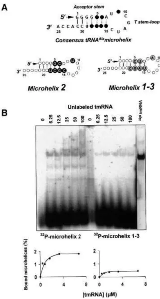
We anticipated that a minimalist RNA construct recapitu-lating a 7-base-pair tRNAAla acceptor stem capped by a
7-nucleotide loop might be suf®cient to interact with tmRNA. For the alanine system, aminoacylation is deter-mined by a single G3´U70pair (Francklyn and Schimmel,
1989), implying that this RNA microhelix can be amino-acylated with alanine, and, if it interacts with tmRNA, will simplify further our working system for peptide bond formation in an `all RNA' environment.
Two RNA microhelices were designed and produced in vitro(Figure 7A). The sequence of microhelix 1±3 is derived from tRNAAla1 and tRNAAla3. The sequence of
microhelix 2 is derived from tRNAAla2, which binds
tmRNA with af®nity. Microhelix 1±3 forms seven base pairs, whereas microhelix 2 has only six base pairs with an A7´C15 mismatch. Both microhelices bind tmRNA with
speci®city, demonstrating that the tRNAAlaacceptor stem
is necessary and suf®cient to promote binding with tmRNA (Figure 7B; Table I). Also, binding of a micro-helix does not require an A7´C15 mismatch within the
acceptor stem. Compared with full-length tRNAAla, the
complex between tmRNA and RNA microhelices is gel-retarded to a lower extent, accounting for a microhelix being 3-fold smaller than a tRNA (compare the migration pattern of the gel-retarded complex in Figures 3 and 7).
Microhelix 1±3, as for in vitro transcribed tRNAAla1and
tRNAAla3, binds tmRNA weakly (~0.5%), but microhelix 2,
as for tRNAAla2, binds tmRNA with plateau levels 4-fold
higher (2% of complex formation at the plateau; Table I). Thus, both microhelices and full-length tRNAs are recognized by tmRNA by similar rules. Also, as for tRNAAla2, there are speci®c cleavages of tmRNA when
microhelix 2 binds tmRNA (lower bands in Figure 7B, left), but not for microhelix 1±3, as for tRNAAla1 and
tRNAAla3. These tmRNA fragments are still able to bind
microhelix 2, as for tRNAAla2. Both microhelices bind
tmRNA with similar dissociation constants (0.7 mM; Table I). For both synthetic tRNAAla1 and tRNAAla3,
dissociation constants are also equivalent but higher than their corresponding microhelices (1.2mM; Table I). This suggests that these two tRNAs possess sequences and/or structural features outside of their acceptor stem that act as negative elements (anti-determinants) when binding tmRNA.
Fig. 7. Native gel retardation assays between RNA microhelices and tmRNA. (A) A sequence consensus (top) depicted on secondary structure models of the minimalist RNAs, with the black circles corresponding to the variable nucleotides. (B) The binding curves are derived from the experiment shown above. For each RNA microhelix, the experimental values were reproduced in three independent experiments.
Upon binding tmRNA, the loop of the RNA microhelix unfolds
The conformations of RNA microhelices were monitored with or without tmRNA in solution, using lead acetate and RNase V1. Nuclease S1, speci®c for RNA single strands,
was also used (Figure 8). The probing data will be compared with that collected with full-length tRNAAla
(Figure 6). In the absence of tmRNA, microhelix 2 is cut by RNase V1 at position 19, cleaved by RNase S1 at
positions 9±12 and also by lead at position 14 (Figure 8), three arguments supporting the existence in solution of a stem capped by a loop. Strikingly, when 50 pmol of unlabeled tmRNA (corresponding to the binding plateau; Figure 7B) are added to microhelix 2, there is a signi®cant enhancement of RNase S1 cleavages at positions 8±10.
Also, lead-induced cleavages appear at positions 13 and 14. Positions 8±14 correspond to the seven nucleotides of the loop. RNase V1-induced cleavage at position 19 is still
visible, suggesting that the stem is still folded when RNA microhelix 2 binds tmRNA. To test whether the A7´C15
mismatch is what allows this large unfolding of the loop to occur, the conformation of microhelix 1±3, with or without tmRNA, was also monitored in solution. In the presence of tmRNA, a similar probing pattern was observed for microhelix 1±3, suggesting that the reactivity of its 7-nucleotide loop towards single-stranded speci®c probes is signi®cantly increased when in complex with tmRNA (data not shown). All of the sequence differences between the two RNA microhelices affect binding ef®ciency, but probably not the recognition process (two microhelices give similar footprints). Nucleotides in common between the two microhelices encompass the ones with an increased reactivity towards structural probes when in complex with tmRNA. Surprisingly, microhelix 1±3 is less stable in solution compared with microhelix 2, as shown by the absence of an RNase V1-induced cleavage at
position 19 (not shown).
Blocking the tmRNA ORF with antisense oligonucleotides does not impair its binding to either native or synthetic tRNAAla
Antisense DNAs targeting the internal ORF of tmRNA might affect binding of either native or in vitro transcribed tRNAAla. Antisense oligonucleotides with complementary
regions including either one, two, three or all four alanine codons within the tmRNA internal ORF were designed (DNAs a±d, with DNA d blocking all 10 codons of the tmRNA ORF; Figure 9A), and their putative interference with tRNAAlabinding assayed. All four antisense
oligo-nucleotides bind a complex between tmRNA and native tRNAAla1 or tRNAAla3, substantiated by RNase
H-medi-ated cleavages of tmRNA when in complex with the antisense DNAs. In the presence of RNase H and each of the four antisense DNAs, a 5¢-labeled tmRNA is entirely cleaved into an ~100-nucleotide fragment (Figure 9A). Since the mRNA module of E.coli tmRNA starts at position 90 and ends at position 122, this demonstrates that all four antisense DNAs bind within the tmRNA internal ORF. Notice that in the presence of DNA antisense c, two distinct RNase H-mediated cleavages are observed, whereas a single cut within the tmRNA ORF is induced by either DNA antisense a, b or d. This result probably re¯ects a dynamic equilibrium of binding of DNA c to tmRNA. DNA c, but not the other three DNAs, has to compete against an 11-base-pair stem (helix H4; Felden et al., 1997) to bind the 3¢ side of the tmRNA ORF. In the presence of each antisense DNA and compared with tRNA and tmRNA alone, the intensity of the gel-retarded band is not decreased (Figure 9B). Quantitation of the gel-retarded bands shows no difference with or without antisense DNAs. Similar results were obtained with synthetic tRNAAla2(data not shown). These results suggest that the
primary binding sites between tRNAAlaand tmRNA do not
involve the tmRNA internal ORF. No detectable differ-ence in the reactivity towards structural probes of the tRNAAlaanticodon loop with or without tmRNA (Figure 6)
suggests that the tRNAAla anticodon loop is also not
involved.
Fig. 8. Nuclease mapping and lead acetate probing data collected on RNA microhelix 2, in the presence or absence of puri®ed tmRNA. Autoradiograms of 20% denaturing polyacrylamide gels of cleavage products of 5¢-labeled RNA microhelices. Lanes S1, RNase S1 mapping; all other lanes are as in Figure 6. Sequencing tracks are numbered every four to ®ve nucleotides. Structural mapping data in the presence or absence of tmRNA are indicated. When tmRNA is present, only reactivity enhancement towards structural probes are observed, all located in the loop. Straight and circled arrows correspond to lead and nuclease S1cleavages, respectively, with the thickness and darker color referring to the intensity of cleavage (weak, medium and strong). Black arrowheads correspond to RNase V1cleavages. The molar ratio between microhelices and tmRNA is 100:1.
Active conformers of tmRNA are able to participate in complex formation with tRNAAla
After aminoacylation of tmRNA with [3H]alanine by
puri®ed AlaRS and subsequent binding with tRNAAla3,
74 6 26 d.p.m. of [3H]alanine are in the complex lane
between alanylated-tmRNA and tRNAAla, whereas there is
no radioactivity in the complex lane between uncharged tmRNA and tRNAAla (Figure 10; three independent
experiments). Consequently, complex formation between tmRNA and tRNAAlaas a result of an artifact solely seen
with inactive tmRNA conformers is excluded.
Discussion
General considerations
Native or synthetic tRNAAla isoacceptors from E.coli
interact speci®cally with puri®ed tmRNA from E.coli in vitro. tRNAAlapost-transcriptional modi®cations
are important, but not essential, for binding tmRNA. Interestingly, native tRNAAla3 binds tmRNA much more
tightly compared with native tRNAAla1, whereas their
synthetic counterparts deprived of modi®ed nucleosides bind tmRNA equally (Table I). Compared with native tRNAAla1, native tRNAAla3 has an additional
post-tran-scriptional modi®cation at position 34 (a uridine 5-oxyacetic acid) but lacks a modi®ed uridine at position 8. Synthetic tRNAAla and tRNAAlamicrohelices
bind tmRNA on a different basis compared with native tRNAAla. Engineering microhelices by chemical synthesis
to introduce the two modi®ed nucleosides at positions analogous to those present in the T-loops of canonical tRNAs might reveal their putative involvement in binding tmRNA. Binding plateaus between native-synthetic tRNAAlaand tmRNA do not exceed several percent, with
dissociation constants ranging from 0.5 (native tRNAAla3)
to 3.5 mM (native tRNAAla1). Other native or synthetic
tRNAs, such as tRNAAspor tRNAGln, do not bind tmRNA,
suggesting that speci®c features within the tRNAAla
structure allow the interaction to proceed. Four indepen-dent pieces of experimental evidence suggest that nucleotides within the acceptor stem of either native or synthetic tRNAAlaare responsible for a speci®c interaction
with tmRNA. First, using a variety of synthetic constructs derived from the sequence of tRNAAla, ®ve nucleotides
were identi®ed, all in the acceptor stem, as being among those required to bind tmRNA. Secondly, structural probing of a synthetic tRNAAla indicates that when in
the presence of tmRNA, nucleotides at positions 6 and 7 within the acceptor stem of tRNAAla become single
stranded. Thirdly, structural probing of a native tRNAAla
indicates that when in the presence of tmRNA, the ®rst base pairs of the acceptor stem are signi®cantly stabilized. Fourthly, RNA microhelices recapitulating a tRNAAla
acceptor stem are able to bind tmRNA with speci®city. Kinetics of complex formation between tRNAAla
and tmRNA
Plateau levels and dissociation constants (Kd) between
tmRNA and various RNA constructs were measured and compared (Table I). As negative controls, E.coli or yeast tRNAAsp, as well as E.coli tRNAGln, do not bind tmRNA. Fig. 9. (A) RNase H-mediated cleavages of the tmRNA internal ORF
with four antisense DNA oligonucleotides. Five percent denaturing PAGE, with the upper arrow pointing to the Xylene cyanol dye (corresponds to the migration of an ~130mer) and the lower arrow pointing to the Bromophenol blue dye (corresponds to the migration of an ~30mer), respectively. a, b, c and d are the four antisense DNAs, and their respective targets within and around the tmRNA internal ORF are indicated. (B) Native gel retardation assays with antisense DNA oligonucleotides targeting the tmRNA internal ORF, in the presence of natives tRNAAla1or tRNAAla3.
Fig. 10. Active conformers of tmRNA (aminoacylated form) are able to participate in complex formation with tRNAAla.
Plateau levels and Kdbetween various tRNAAlaconstructs
and tmRNA vary from 1 to 5% and from 0.5 to 3.5mM, respectively. Full-length native and synthetic tRNAAla
possess the higher binding plateaus, whereas one native tRNAAla isoacceptor and the RNA microhelices have
higher dissociation constants (Table I). Direct comparison of both the binding plateaus and the Kdbetween synthetic
tRNAAla 1, 2 and 3 and their corresponding RNA
microhelices 1±3 and 2 is achievable. Compared with full-length tRNAs, the binding plateaus of the corres-ponding RNA microhelices are reduced 2-fold with a 2- to 4-fold decrease in their dissociation constants (Table I). These results suggest that compared with full-length tRNAAla, RNA microhelices bind tmRNA with more
ease, accounting for a lower Kd, probably because they are
three times smaller than full-length tRNAs. Their inter-action with tmRNA, however, is probably not as stable as with full-length tRNAAla, as suggested by lower binding
plateaus.
Structural basis of the interaction between tRNAAlaand tmRNA
When tmRNA interacts with synthetic or native tRNAAla,
only minor differences in the reactivity of nucleotides of both RNAs towards structural probes were detected. This could have been anticipated from only a few percent of complex formation out of total RNAs. Other subtle structural perturbations are probably not detected when the two RNAs interact with each other, with the approach described here, and await further structural mapping to be delineated. Footprints between tRNAAlaand tmRNA were
performed with native tRNAAla3and synthetic tRNAAla2,
since gel retardation assays have shown that they bind tmRNA with the lower Kd(Table I). Within the tRNAAla
structure, nucleotides with increased reactivity towards chemical and enzymatic probes are mostly clustered in the acceptor stem. For synthetic tRNAAla2, the acceptor stem
partially unfolds when tmRNA binds. For RNA micro-helices, the TyC loop unfolds. For native tRNAAla3, both
the acceptor and the D stems are stabilized when tmRNA binds. Antisense oligonucleotides targeting the anticodon stem±loop of either native or synthetic tRNAAlaincrease
its binding 2-fold (data not shown). This suggests a negative contribution of the anticodon stem±loop in complex formation, a result that is in agreement with a higher af®nity of the RNA microhelices for tmRNA compared with their full-length tRNA counterparts. Speci®c interactions between tmRNA and either synthetic, native or minimalist tRNAAlastructures are predicted to be
different. Recognition between two RNAs might be very adaptable.
What are the sequences and/or structural domains within tmRNA involved in binding tRNAAla or the
RNAAlamicrohelices? Within the ribose-phosphate
back-bone of tmRNA, speci®c cleavages appear when either tRNAAla2, tRNAAla1±2or RNA microhelix 2 interact with
tmRNA (this is not observed with either native or synthetic tRNAAla1 and tRNAAla3, or with microhelix 1±3). Thus,
speci®c nucleotides from the acceptor stem of tRNAAla2,
including G5, C6, C66, G67 and C68, trigger speci®c cuts
within the tmRNA sequence. Interestingly, these tmRNA fragments still bind tRNAAla (or the microhelix) with
speci®city. Mapping these tmRNA fragments will identify
the structural domains that are dispensable for binding synthetic tRNAAla. The band containing the tmRNA
fragment in complex with synthetic tRNAAla2was excised
from the gel, eluted passively and reverse transcribed with a DNA oligonucleotide complementary to the tmRNA 3¢ end. A speci®c band with a length <280 nucleotides was obtained (data not shown). This demonstrates that these tmRNA fragments are missing at least the ®rst 80 nucleotides from the 5¢ end and are still capable of speci®c binding with tRNAAla2.
Biological signi®cance of the interaction between tRNAAlaand tmRNA
We report that native E.coli tmRNA interacts with two native tRNAAlaisoacceptors from E.coli with af®nity and
speci®city in vitro. Compared with tRNAAlaisoacceptor 1,
tRNAAlaisoacceptor 3 binds tmRNA with a dissociation
constant 7-fold lower and an ~2-fold higher binding plateau (Table I). Out of 46 tRNAs in E.coli, tRNAAla
isoacceptor 3 is the seventh most abundant tRNA in E.coli. However, the intracellular concentration of each tRNA varies as a function of the growth rate. tmRNA is present in E.coli cells in low abundance, at ~500 copies per cell, at a growth rate for which ~5000 ribosomes are present (Lee et al., 1978). Interestingly, at a similar condition of growth, native tRNAAlaisoacceptor 3 represents 5% of the
total tRNA population in E.coli, that is 3250 6 220 molecules per cell (Ala3 is equivalent to Ala1B; Dong et al., 1996). Thus, in vivo, there is a 6-fold excess of tRNAAla3 compared with tmRNA. In these conditions, a
complex can form in vitro between tRNAAla3and tmRNA.
Native tRNAAla isoacceptor 1, however, represents only
0.95% of the total tRNA population in E.coli (620 6 60 molecules per cell) and is one of the eight least abundant tRNAs in E.coli (Ala1 is equivalent to Ala2; Dong et al., 1996).
Binding of native tmRNA to either native tRNAAla1or
native tRNAAla3in vitrois stable at pHs varying between
5.0 and 8.0. All the in vitro binding assays were carried out at 37°C in the presence of monovalent (200 mM NH4Cl),
divalent (3 mM MgCl2) and multivalent (10 mM
spermi-dine) ions, at concentrations compatible with those in E.colicells. Depending on the growth rate of E.coli cells, there is 5±6% of tRNAAla3(Dong et al., 1996). Figure 2B
demonstrates that tmRNA binds native tRNAAla
iso-acceptor 3 in the presence of all tRNAs from E.coli, even when there is only 5% of labeled tRNAAla3compared
with total tRNAs. At a 200 molar excess of all tRNAs from E.coli, the binding between labeled tRNAAla3and tmRNA
decreases slightly (~10%), which is likely to account for the competition of unlabeled tRNAAla isoacceptors
from the mixture of all tRNAs with labeled tRNAAla3
(Figure 2B). Moreover, active conformers of tmRNA (aminoacylated form) are able to participate in complex formation with tRNAAla3(Figure 10). However, this does
not preclude an initial recognition between both uncharged tRNAAlaand tmRNA, which are subsequently
aminoacyl-ated by a common aminoacyl-tRNA synthetase, AlaRS. Altogether, our results suggest that a speci®c complex between native tRNAAlaisoacceptors and native tmRNA is
likely to form in vivo. Considering the binding plateaus measured in vitro, and also that the stoichiometry between tRNAAla and tmRNA might be one-to-one in vivo, this
complex between tmRNA and tRNAAlacould involve up
to 140 tmRNA molecules out of the 500 per cell (1.5% of the 620 molecules of tRNAAla1 and 4% of the 3250
molecules of tRNAAla3, in complex with tmRNA in vitro).
During trans-translation, the current model is that alanine-charged tmRNA recognizes stalled ribosomes, binds as a tRNA to the ribosomal A site, and donates the charged alanine to the nascent polypeptide chain via transpeptidation (for a review see Karzai et al., 2000). The stalled mRNA is then replaced by tmRNA and resumption of translation ensues at an internal alanine (resume) codon in tmRNA. Out of 140 known tmRNA sequences from 118 species (tmRNA website; Williams, 2000), alanine is the resume codon for >80% of all tmRNA sequences, the remaining ones possessing either a glycine, an aspartic acid or a valine resume codon. This suggests that an alanine codon is preferred for resuming translation within the tmRNA internal ORF. For the other few species that use either glycine, aspartic acid or valine as resume codons in their respective tmRNA-mediated, protein-tagging systems, their tmRNA structures might allow speci®c recruitment of either tRNAGly, tRNAAsp or tRNAVal, but
not tRNAAla. In E.coli tmRNA, mutating the resume codon
from alanine (GCA) to either serine (UCA) or valine (GUA) still allows tagging of a truncated protein in vivo (Williams et al., 1999), albeit to lower levels. However, tmRNA variants disallowing the proper utilization of the resume codon are not able to transfer the uncoded alanine attached to the 3¢ end of tmRNA to the nascent polypeptide (Williams et al., 1999). These data are in agreement with our results and suggest that tmRNA interacts with the tRNA that decodes the resume codon prior to entering the ribosome. E.coli tRNAAla3is the only isoacceptor with a
5¢-UGC-3¢ anticodon that can form three canonical pairs with the resume codon (5¢-GCA-3¢) from E.coli tmRNA. Here, we show that native tRNAAla3binds tmRNA with the
highest af®nity in vitro (Table I). Local recruitment and enrichment around tmRNA of the tRNA species that has to pair with the resume codon might help re-registration of the tag reading frame during trans-translation. Speci®c recruitment of tRNAAla by tmRNA involves structural
domains outside of its internal ORF, but this does not exclude the possibility that the acceptor stem of tRNAAla
has to be recognized ®rst, with a subsequent recruitment of its anticodon loop at the resume codon. De®ning the recognition elements within tmRNA that are required for binding tRNAAla with accuracy might reveal further
biological insights.
Towards peptide bond formation between Ala-RNAAlaand Ala-tmRNAAla
Alternatively, speci®c complex formation between tmRNA and tRNAAla might re¯ect only an ancient
interaction between two aminoacylatable RNAs, when the translational apparatus might have required a covalent linkage between aminoacylated tRNAs and mRNAs, as for tmRNA, to recruit a second aminoacylated tRNA for peptide bond formation. If true, a small percentage of binding is only observed today, as putative remnants of these early events in the history of the genetic code. Aminoacylatable RNA microhelices were proposed to be present during these initial stages of the genetic code establishment (Tamura and Schimmel, 2001). Strikingly,
RNA microhelices also bind tmRNA, even with a 2- to 4-fold higher af®nity compared with their full-length tRNA counterparts. Thus, our initial model for peptide bond formation will now be simpli®ed further, a step forward towards de®ning the smallest machinery entirely made of RNAs capable of peptide bond formation, inspired from a molecule that is still functional in the 21st century.
Materials and methods
DNA oligonucleotides and enzymes
All the synthetic DNA oligonucleotides were synthesized by Cybergene (Saint-Malo, France). Four DNAs: 5¢-TATTAAGCTGCTAAAGCG-TAGTTTTCGTCGTTTGCGACTA-3¢, 5¢-TTAAGCTGCTAAAGC-GTAG-3¢, 5¢-CAGGTTATTAAGCTGCTAA-3¢ and 5¢-TCGTCGTTT-GCGACTATTT-3¢ were used as antisense targeting either tmRNA internal ORF. Thirteen DNA primers: 5¢-GGGGATCCTGGTGGA-GGCGCGCGGG-3¢, 5¢-GTATGTTGTGTGGAATTGT-3¢, 5¢-GGA-AGCTTAATACGACTCACTATAGGGGGCATAGCTCAG-3¢, 5¢-GGG-GATCCTGGTGGAGCTATGCGG-3¢, 5¢-TAATACGACTCACTA-TA-3¢, 5¢-GGAAGCTTAATACGACTCACTATAGGGGCTATAGCT-CAG-3¢, 5¢-GGGGATCCTGGCGGAACGGACGGGAC-3¢, 5¢-TTA-AGTTGGGTAACGCCAG-3¢, 5¢-GGAAGCTTAATACGACTCACT-ATAGGAGCGGTAGTTCAG-3¢, 5¢-TGGTGGAGCTAGATCGAATAG-CCCCTATAGTGAGTCGTATTA-3¢, 5¢-TGGTGGAAGCGGATC-GAATGCCCCCTATAGTGAGTCGTATTA-3¢, 5¢-TGGTGGAGC-TGGCGGGA-3¢ and 5¢-TGGTGGAGCTGGC-3¢ were used for cloning all of the synthetic tRNAs and for direct transcription of the RNA microhelices. T7 RNA polymerase was prepared according to Wyatt et al. (1991). Restriction enzymes BamHI, HindIII, BstN1, alkaline phospha-tase and T4 polynucleotide kinase were from New England Biolabs (Berverly, MA). AMV reverse transcriptase, Taq DNA polymerase, T4 DNA ligase and T4 RNA ligase were from Gibco-BRL Life Technologies (Cergy-Pontoise, France). RNases S1, V1, U2, and T1 were from Amersham-Pharmacia-Biotech (Orsay, France). RNase H was from Sigma-Aldrich (Saint-Quentin, France). [g-32P]ATP
(3000 mCi/mmol), [a-32P]pCp (3000 mCi/mmol) andL-[3-3H]alanine
(74 Ci/mmol) were from NEN (Paris, France).
Preparation of RNAs and aminoacylation reaction
Escherichia colitmRNA was overexpressed in E.coli cells and puri®ed as previously described (Felden et al., 1997). Synthetic RNAs were cloned downstream of a T7 RNA polymerase promoter as described (Perret et al., 1990). Plasmids were linearized with BstN1 restriction nuclease before transcription, so that in vitro transcribed RNAs will end with the 3¢-terminal CCA triplet. In vitro transcription of synthetic tRNAs and RNA microhelices was performed as described (Felden et al., 1994). Electrophoresis on denaturing gels separates the transcribed RNAs from non-incorporated nucleotides and DNA fragments. Appropriate bands were electroeluted, and pure in vitro transcribed RNAs were recovered by ethanol precipitation. Puri®ed native tRNAs were from Subriden (Rolling Bay, WA). Aminoacylation reactions were performed in a medium containing 25 mM Tris±HCl pH 7.5, 7.5 mM MgCl2, 2 mM ATP, 5 mM b-mercaptoethanol, 10 mM KCl, 200 pmol of tmRNA, 50 mM3H-labeled
alanine and 700 nM puri®ed E.coli AlaRS. Incubations were at 37°C for 30 min, then 200 mM cold potassium acetate pH 5.0 was added, followed by a phenol extraction of the enzyme at pH 4.3.
Gel retardation assays and structural mapping procedures Labeling at the 5¢ end of the RNAs was performed with [g-32P]ATP and
phage T4 polynucleotide kinase after dephosphorylation with alkaline phosphatase (Silberklang et al., 1977). Labeling at the 3¢ end was carried out by ligation of [g-32P]pCp using T4 RNA ligase. After labeling, the
RNAs were gel puri®ed at a nucleotide resolution for tRNAs and RNA microhelices, eluted passively, and ethanol precipitated. RNAs were denatured for 2 min at 80°C in a folding buffer (5 mM MgCl2, 20 mM NH4Cl, 10 mM HEPES±KOH pH 6.9) and then slowly cooled down to room temperature for 30 min. Standard assays contained 0.5 pmol of labeled RNA in the presence of the appropriate concentration of aminoacylated or uncharged tmRNA in a binding buffer (10 mM spermidine, 3 mM MgCl2, 200 mM NH4Cl, 80 mM HEPES±KOH pH 6.9) to a ®nal volume of 15 ml. A 30 min incubation at 37°C was either followed by enzymatic and chemical footprints between tRNAAlaand
tmRNA, or subjected directly to electrophoresis in a 5% non-denaturing polyacrylamide gel, in 45 mM Tris±HCl pH 8.3, 43 mM boric acid, 0.1 mM MgCl2at 4°C and 10 V/cm (Ramos and MartõÂnez-Salas, 1999). For the aminoacylated tmRNA, electrophoresis was in a 5% non-denaturing polyacrylamide gel overnight in 0.1 M Na acetate pH 5.0. Bands corresponding to aminoacylated tmRNA, either free or in complex with tRNAAla3, were excised, passively eluted in water, and3H was
counted on a Wallac 1409 (Perkin-Elmer). Binding assays, gel excision, passive elution and counting were performed with uncharged tmRNA in identical conditions, as negative controls.
Digestions with the various ribonucleases (V1 at 0.075 units, S1 at 40 units, U2at 0.4 units and T1at 0.2 units) and probing with lead acetate (a ®nal concentration of 2.5 mM) were performed as described (Felden et al., 1997). Quantitation of selected bands was as described (Felden et al., 1998). Relative amounts of RNA±RNA complexes were analyzed on a PhosphorImager with ImageQuant (Molecular Dynamics, Sunnyvale, CA). Data are represented as the percentage of the RNA complex of interest relative to the input probe, calculated as the sum of the intensity of all bands in the corresponding lane.
Inhibition assays with antisense DNA oligonucleotides Four synthetic DNA oligonucleotides complementary to various portions of the tmRNA ORF were used. For annealing, 100 pmol of tmRNA or tRNA and 1000 pmol of an oligonucleotide were incubated in folding buffer for 2 min at 80°C. After annealing, gel retardation assays were performed as described. RNase H digestion assays of antisense DNA±tmRNA duplexes were adapted from Matveeva et al. (1997); both the molar ratio between the antisense DNAs and tmRNA as well as the annealing step were as for the binding assays between tmRNA, labeled tRNAs and the antisense DNAs shown in Figure 9B.
Acknowledgements
Prof. C.Florentz and Dr G.Eriani (IBMC, Strasbourg) kindly provided us with bacterial strains encoding either tRNAAla1/tRNAGlnor tRNAAspfrom
E.coli, respectively. This work was funded by a Human Frontier Science Program Research Grant (RG0291/2000-M 100), a Research Grant entitled `Recherche Fondamentale en Microbiologie et maladies infectieuses' and an `Action ConcerteÂe Incitative Jeunes Chercheurs 2000' from the French Ministry of Research, to B.F.
References
Abo,T., Inada,T., Ogawa,K. and Aiba,H. (2000) SsrA-mediated tagging and proteolysis of LacI and its role in the regulation of lac operon. EMBO J., 19, 3762±3769.
Dong,H., Nilsson,L. and Kurland,C.G. (1996) Co-variation of tRNA abundance and codon usage in Escherichia coli at different growth rates. J. Mol. Biol., 260, 649±663.
Felden,B., Florentz,C., GiegeÂ,R. and Westhof,E. (1994) Solution structure of the 3¢-end of brome mosaic virus genomic RNAs. Conformational mimicry with canonical tRNAs. J. Mol. Biol., 235, 508±531.
Felden,B., Himeno,H., Muto,A., McCutcheon,J.P., Atkins,J.F. and Gesteland,R.F. (1997) Probing the structure of the Escherichia coli 10Sa RNA (tmRNA). RNA, 3, 89±103.
Felden,B., Hanawa,K., Atkins,J.F., Himeno,H., Muto,A., Gesteland,R.F., McCloskey,J.A. and Crain,P.F. (1998) Presence and location of modi®ed nucleotides in Escherichia coli tmRNA: structural mimicry with tRNA acceptor branches. EMBO J., 17, 3188±3196.
Francklyn,C. and Schimmel,P. (1989) Aminoacylation of RNA minihelices with alanine. Nature, 337, 478±481.
Huang,C., Wolfgang,M.C., Withey,J., Koomey,M. and Friedman,D.I. (2000) Charged tmRNA but not tmRNA-mediated proteolysis is essential for Neisseria gonorrhoeae viability. EMBO J., 19, 1098±1107.
Hutchison,C.A., Peterson,S.N., Gill,S.R., Cline,R.T., White,O., Fraser, C.M., Smith,H.O. and Venter,J.C. (1999) Global transposon mutagenesis and a minimal Mycoplasma genome. Science, 286, 2165±2169.
Karzai,A.W., Roche,E.D. and Sauer,R.T. (2000) The SsrA-SmpB system for protein tagging, directed degradation and ribosome rescue. Nature Struct. Biol., 7, 449±455.
Kholod,N.S. (1999) Dimer formation by tRNAs. Biochemistry (Mosc.), 64, 298±306.
Komine,Y., Kitabatake,M., Yokogawa,T., Nishikawa,K. and Inokuchi,H. (1994) A tRNA-like structure is present in 10Sa RNA, a small stable RNA from Escherichia coli. Proc. Natl Acad. Sci. USA, 91, 9223±9227.
Lee,N., Bessho,W., Weil,K., Szostak,J.W. and Suga,H. (2000) Ribozyme-catalyzed tRNA aminoacylation Nature Struct. Biol., 7, 28±33.
Lee,S.Y., Bailey,S.C. and Apirion,D. (1978) Small stable RNAs from Escherichia coli: evidence for the existence of new molecules and for a new ribonucleoprotein particle containing 6S RNA. J. Bacteriol., 133, 1015±1023.
Matveeva,O., Felden,B., Audlin,S., Gesteland,R.F. and Atkins,J.F. (1997) A rapid in vitro method for obtaining RNA accessibility patterns for complementary DNA probes: correlation with an intracellular pattern and known RNA structures. Nucleic Acids Res., 25, 5010±5016.
Muth,G.W., Ortoleva-Donnelly,L. and Strobel,S.A. (2000) A single adenosine with a neutral pKain the ribosomal peptidyl transferase center. Science, 289, 947±950.
Muto,A., Fujihara,A., Ito,K.I., Matsuno,J., Ushida,C. and Himeno,H. (2000) Requirement of transfer-messenger RNA for the growth of Bacillus subtilisunder stresses. Genes Cells, 5, 627±635.
Nissen,P., Hansen,J., Ban,N., Moore,P.B. and Steitz,T.A. (2000) The structural basis of ribosome activity in peptide bond synthesis. Science, 289, 920±930.
Noller,H.F., Hoffarth,V. and Zimniak,L. (1992) Unusual resistance of peptidyl transferase to protein extraction procedures. Science, 256, 1416±1419.
Perret,V., Garcia,A., Puglisi,J., Grosjean,H., Ebel,J.-P., Florentz,C. and GiegeÂ,R. (1990) Conformation in solution of yeast tRNAAsptranscripts
deprived of modi®ed nucleotides. Biochimie, 72, 735±743.
Piccirilli,J.A., McConnell,T.S., Zaug,A.J., Noller,H.F. and Cech,T.R. (1992) Aminoacyl esterase activity of the Tetrahymena ribozyme. Science, 256, 1420±1424.
Ramos,R. and MartineÂz-Salas,E. (1999) Long-range RNA interactions between structural domains of the aphthovirus internal ribosome entry site (IRES). RNA, 5, 1374±1383.
Schimmel,P., GiegeÂ,R., Moras,D. and Yokoyama,S. (1993) An operational RNA code for amino acids and possible relationship to genetic code. Proc. Natl Acad. Sci. USA, 90, 8763±8768.
Silberklang,M., Prochiantz,A., Haenni,A.L. and Rajbhandary,U.L. (1977) Studies on the sequence of the 3¢-terminal region of turnip-yellow-mosaic-virus RNA. Eur. J. Biochem., 72, 465±478.
Sprinzl,M., Horn,C., Brown,M., Ioudovitch,A. and Steinberg,S. (1998) Compilation of tRNA sequences and sequences of tRNA genes. Nucleic Acids Res., 26, 148±153.
Tamura,K. and Schimmel,P. (2001) Oligonucleotide-directed peptide synthesis in a ribosome- and ribozyme-free system. Proc. Natl Acad. Sci. USA, 98, 1393±1397.
Ushida,C., Himeno,H., Watanabe,T. and Muto A. (1994) tRNA-like structures in 10Sa RNAs of Mycoplasma capricolum and Bacillus subtilis. Nucleic Acids Res., 22, 3392±3396.
Varani,V. and McClain,W.H. (2000) The G´U wobble base pair: a fundamental building block of RNA structure crucial to RNA function in diverse biological systems. EMBO rep., 1, 18±23.
Williams,K.P. (2000) The tmRNA website. Nucleic Acids Res., 28, 168. Williams,K.P., Martindale,K.A. and Bartel,D.P. (1999) Resuming translation on tmRNA: a unique mode of determining a reading frame. EMBO J., 18, 5423±5433.
Williams,R.J., Nagel,W., Roe,B. and Dudock,B. (1974) Primary structure of E.coli alanine transfer RNA: relation to the yeast phenylalanyl tRNA synthetase recognition site. Biochem. Biophys. Res. Commun., 60, 1215±1221.
Wyatt,J.R., Chastain,M. and Puglisi,J.D. (1991) Synthesis and puri®cation of large amounts of RNA oligonucleotides. Biotechniques, 11, 764±769.
Zhang,B. and Cech,T.R. (1997) Peptide bond formation by in vitro selected ribozymes. Nature, 390, 96±100.
Received December 21, 2000; revised April 4, 2001; accepted April 5, 2001