HAL Id: inserm-00187321
https://www.hal.inserm.fr/inserm-00187321
Submitted on 14 Nov 2007
HAL is a multi-disciplinary open access archive for the deposit and dissemination of sci-entific research documents, whether they are pub-lished or not. The documents may come from teaching and research institutions in France or abroad, or from public or private research centers.
L’archive ouverte pluridisciplinaire HAL, est destinée au dépôt et à la diffusion de documents scientifiques de niveau recherche, publiés ou non, émanant des établissements d’enseignement et de recherche français ou étrangers, des laboratoires publics ou privés.
Tool/tissues interaction modeling for transluminal
angioplasty simulation
Tanguy Le Fol, Pascal Haigron, Antoine Lucas
To cite this version:
Tanguy Le Fol, Pascal Haigron, Antoine Lucas. Tool/tissues interaction modeling for
translu-minal angioplasty simulation. 29th Annual International Conference of the IEEE Engineering
in Medicine and Biology Society, 2007. EMBS 2007., Aug 2007, Lyon, France. pp.1734-1737,
Abstract— In this paper, a simulation environment is describe for balloon dilation during transluminal angioplasty. It means simulating geometrical and physical data from the inflation of a balloon while integrating data from tissues analysis and parameters from virtual tool-tissues interactions. In this context, three main behaviors has been identified: soft tissues, crush completely under the effect of the balloon, calcified plaques, do not admit any deformation but could move in deformable structures and blood vessel wall and organs, try to find their original forms.
A deformable soft tissue model is proposed, based on the Enhanced ChainMail method to take into account tissues deformation during dilatation. We improved the original ChainMail method with a “forbidden zone” step to facilitate tool/tissues interactions. The simulation was implemented using five key steps: 1) initialization of balloon parameters; 2) definition of the data structure; 3) dilatation of the balloon and displacement approximation; 4) final position estimation by an elastic relaxation; and 5) interpolation step for visualization. Preliminary results obtain from patient CT data are reported.
I. INTRODUCTION
HISpaper is aimed at studying the minimally invasive procedure called percutaneous transluminal angioplasty (PTA) on narrowed arteries by means of simulation. PTA consists in introducing a balloon-catheter in the stenosis area and inflating a balloon to expand the blood vessel lumen. Even though angioplasty is a quite common practice, it’s important to simulate and predict mechanical behaviors of interaction between balloon, plaque and artery.
There are a number of algorithms developed for modeling and simulation for various computer graphics applications (surgery modeling, flight simulator...). Depending on the constraints (nature of the objects, time, realism...), researchers may use different techniques that could be classified by their geometrical or physical formalism. Non-physically (or geometrically) based methods are used as a simplified physical principle to achieve a reality-looking effect. They allow a user to deform the space containing the objects or to manipulate operators like bend and twist onto primitives or sampling data [1]. This type of method is very fast but do not use physical properties. Those methods are not realistic enough, that is why they are often used in combination with other methods [2,3] or used for specific topologies [4,5]. For applications which demand realistic simulation of deformable physical bodies, physical modeling is more convenient. The two main physical approaches for soft-tissue modeling over the past decade have been the mass-spring approach, and the
*
T. Le Fol, P. Haigron, A. Lucas are with INSERM, U642, Rennes, F-35000, France; Université de Rennes 1, LTSI, F-35000, France.
{tanguy.lefol, pascal.haigron, antoine.lucas}@univ-rennes1.fr
Finite Element Method (FEM). FEM, firstly developed in 1950 by M. Turner, is the most realistic method. However, their computational costs make them hard to be used in real-time application, since the true elastic behavior of soft tissue is nonlinear, and for large meshes, a large system of equations has to be solved. To counter this, Bro-Nielsen & al [6] proposed a simplified approach with a condensation procedure. This condensation step effectively reduces the size of the linear system of equations. Though, interactive topology changes, such as cutting of soft tissues, are not possible since the pre-processing assumes a certain topology. Holzapfel & al. [7] and Di Puccio & al. [8] applied FEM optimized for angioplasty in real-time, but only for few millimeters around blood vessel’s lumen, preventing to take into account the constraint imposed by external tissues. Mass-spring models [9] comprise a set of nodes connected by springs, with point masses attached at each node. Real time performance can be achieved with a limited number of nodes, but the behavior is often unrealistic and can be unstable. Moreover, mass-spring approach requires computation time similar to the FEM optimized model, while giving worse results [10]. Therefore, whilst much research effort has been spent on improving such techniques in the area of physical accuracy and performance, other methods, such as the ChainMail algorithm [11, 12, 13], have been proposed aiming at interactive frame rates. This method, combines mechanical behaviors and geometric laws. Its performances make a good compromise between computation time and realism.
In this context, the overall objective of this first work is to develop a fast, realistic and patient specific simulation based on Chain-mail approach. This paper is organized as follows: Section II introduces ChainMail principle and presents the proposed method for angioplasty simulation. Section III illustrates experimental results for 2D CT images. Finally, in Section IV, conclusion of the presented work and the direction of our future research are described.
II. SIMULATION METHOD
The general method (Figure 1), based on Le Fol & al. [14], initialize the balloon in the blood vessel lumen. The meshing is initialized according to the Enhanced ChainMail formalism. The balloon (described in two different ways: a disc composed with linked elements, and a “forbidden area” for tissues) inflates and an approximation of the tissues’ reaction is obtained from the propagation of the displacement. A minimal energy level is then calculated, called elastic relaxation. Finally, a classical interpolation allows displaying result.
Tool/tissues interaction modeling for transluminal angioplasty simulation
T. Le Fol* , P. Haigron*, A. Lucas*
T
This material is presented to ensure timely dissemination of scholarly and technical work. Copyright and all rights therein are retained by authors or by other copyright holders.
All persons copying this information are expected to adhere to the terms and constraints invoked by each author's copyright. In most cases, these works may not be reposted without the explicit permission of the copyright holder.
HAL author manuscript inserm-00187321, version 1
Fig. 1. Inflation simulation diagram.
In the ChainMail algorithm [11], volume elements were linked to their nearest neighbors (4 or 8 in 2D and 6 or 18 in 3D). When an element of the data’s structure is moving, it communicates the information concerning its new state to its neighbors to actualize their positions and to update the whole structure. This approach requires two initial processes, the creation of the data structure and the definition of the geometrical constraints. The structure is composed of elements, called « linked elements », which represent the volume data. An element holds the information about the deformation, the elasticity parameters and its number of neighbors. Then, a set of geometric constrains that consists of two pairs of translations and 2 single shears (Min, Max and MaxShear, assuming different in x and y) in the 2D case (Figure 2) are used to denote material properties.
Fig. 2. Valid region for an element defined by its neighbors [11].
To take into account heterogeneous nature of tissues, Schill & al. [12] proposed an « Enhanced ChainMail » method which combines the parameters of each linked element with those of its neighbors. For example, if A and B are two linked elements neighbors, the parameter for each of them are . A B A B M in M in M in M ax M ax M ax = + = +
A. Initialization of the balloon parameters
In this work, we consider 2D angioscan images, where the tool/tissues interactions zone is defined by the vessel lumen (pre-segmented by a region growing process). This zone determines the place where the balloon, considered as
non-compliant one, is placed and inflated. This balloon is described as a circle which radius is fixed by a ray casting process (the smallest is conserved) and located at the mass center of the vessel lumen (Figure 3).
Fig. 3. Initialization of the balloon. Lumen segmentation (a), mass-center calculation (b), minimal radius detection (c), initial balloon (d).
B. Definition of the data structure
The meshing initialization process defines some simulation’s parameters and builds the structure: a 2D grid, the number of linked neighbors and the geometrical constraints. Each pixel corresponds to a node/linked element of the grid (Figure 4). A regular Cartesian grid has been chosen for the simulation because it takes advantage of the high resolution data produced by CT-scanners and provides a direct access to the data.
Fig. 4. Meshing initialization with ChainMail formalism.
The elements density in the Cartesian grid is taken as parameter to define the geometrical constraints according to the curves of the Figure 5 and [14]. On these curves, three main classes are considered where constraints are defined in percentage from the initial distance.
H(Air)
90%
50%
10%
Soft Elastic Rigid
Distance Rigid threshold Density (Hounsfield) H(Bone) Max Min MaxShearX MaxShearY MaxY MinY MinX MaxX a) b) c) d) Calcification Vessel wall Blood Link (1) (2)
Fig. 5. Geometric constraints definition curves according to density.
C. dilation of the balloon and Displacement approximation
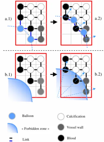
Two methods of tool/tissues interaction are considered. The first one, called here the classical method, describes the balloon as a group of linked elements (regarded as rigid tissues thanks to the non-compliance of the balloon) arranged in circle with a radius calculated in part A (Figure 6 (a.1)) and connected to the linked elements belonging to tissues. In an iterative way, the step of dilation is incremented (increasing the radius of the balloon) and neighbors from the balloon, belonging to the tissues, move following their constraints and propagate the displacement (Figure 6 a.2)).When it comes to the second method, the balloon is considered as a “forbidden area”. In this zone, linked elements cannot stay (Figure 6 b.1)). When the balloon inflates, the “forbidden area” increases and if elements belonging to the tissues are in this zone, they are moved towards the radial direction established between the center of the balloon and the old position of the element. Then, displacement is propagated (Figure 6 b.2)).
The dilation of the balloon generates a displacement of the tissues. The propagation of the disturbance is made by the Enhanced ChainMail algorithm and gives an approximation of the deformation. The process of propagation uses the geometrical constraints to check that each element remains between the minimum and maximum distance to its neighbors. A disturbance is produced when an element is moved and violates these constraints.
The rules which control the propagation of the disturbance are established thanks to the links and the geometrical constraints. The elements concerned must move in order to find a new configuration which satisfies the constraints. In order to manage the multiple collisions and displacements (caused by dilation), each element is moved only once according to the greatest displacement generated by the neighbors’ elements initiator of the disturbance.
D. Elastic relaxation
ChainMail step does not guaranty a global minimal energy configuration, notably for elastic tissues which try to recover the initial structures. Relaxation makes it possible to find such a configuration. The approach employs a function based on the distances between the neighbor’s elements, from Gibson & al. [13].
E. Interpolation
The last stage of the treatment consists of a simple interpolation. In the classical case, the goal of the interpolation is to reposition the elements in a Cartesian grid. This enables, at the following iteration, to link easily the balloon with the tissues. Indeed, if the position of the elements belonging to tissues is unchanged and if a larger balloon is created (with a greater number of elements defining it to avoid the holes between 2 elements of the balloon when it is inflated), there is not enough link, on tissues side, compared to the number of link, on balloon class. So, it is necessary to create heavy rules to establish
which element of the balloon links with which element of the tissues. In the “forbidden area” formalism, the interpolation is used only for the visualization of the result and is not taken into account in the treatment for the following iteration. There is not new link to establish, it is only necessary to increase the “forbidden area”. This last stage achieved, the process is repeated until satisfaction.
Fig. 6. Balloon/tissues interaction. a.1) Balloon describes by linked elements. a.2) When these elements move, they transmit their displacement. b.1) Balloon describes like a “forbidden area”. b.2) When the balloon dilates, the points lately introduced into the zone are moved.
III. RESULTS
To illustrate results, 2 examples of stenosis vessel are employed. These images are 100 × 100 Pixels from angioscan. This system was implemented in C++ and uses VTK. The examples and the results reported here were obtained with a processor of 3.20 GHz with 2Go of RAM.
For both methods, it can be seen that the physical behavior for hard plaques (rigidity) and for soft plaques (compression) moves apart from classical elastic laws, and is essentially well reproduced in the Chain-Mail approach, considering geometric constraints.
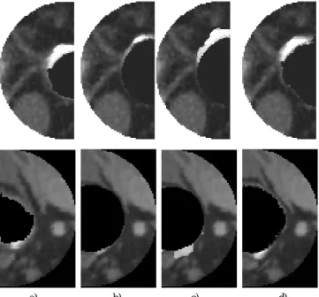
However, the classical method for balloon/tissues interaction by its successive interpolations, degrade the image. Indeed, the interpolation means the close pixels to create a new Cartesian grid and when many pixels, belonging to blood, are pressed against elastic tissues and/or
a.1) b.1) Balloon « Forbidden zone » Link Calcification Vessel wall Blood a.2) b.2)
calcification, the new calculated intensity of the pixels is low. The elastic tissues and calcifications disappear (Figure 7 (b), grey and white densities). An idea to avoid this is to keep the pixels of strong intensity: if in the calculation of the new intensity, elements of elastic tissue or calcification are detected, the elements of blood will not be taken into account in the calculation. But this new interpolation spreads out the tissues with strong intensity (Figure 7 (c), white density). Finally, in the two preceding cases, interpolation creates a new grid of element where tension and compression are at zero. That leads to a loss of crucial information for elastic relaxation.
Fig. 7. Qualitative results from dilation simulation. a) Original image. b) Result with classical balloon/tissues interaction and classical interpolation. c) Result with classical balloon/tissues interaction and high density priority interpolation. d) Result after dilation simulation with “forbidden area” tool/tissues interaction.
The “forbidden area” method preserves each class of density and allows the tissues to express elastic behaviors (Figure 7 (d)). Because the elements involved are the same since the beginning of the simulation, tensions and compressions are kept and updated each iteration. Nevertheless, with this method, the number of elements that has to be moved increase with the radius of the balloon, spending more and more computing time (beginning at 0.2 second per iteration until 1.5 to 2 seconds). The classical method, on the contrary, has to move a quasi-constant and fewer numbers of elements (0.2 sec per iteration).
IV. CONCLUSION
The objective of this work was to study a model of dilation in order to carry out a specific patient simulation method. The methodology suggested, applied in 2D, makes it possible to analyze the potential of such an approach which combines the geometrical and physical considerations to model the interactions during an angioplasty. The data structure initialized as a Cartesian grid gives direct access to the patient data. The Enhanced ChainMail method, by its low
time consuming and its modularity, permits to develop a fast simulation which could manage interactions between the balloon and a large surrounding of the vessel wall. The definition of the geometrical constraints seems well adapted for hard and soft plaques. The proposed method of balloon/tissues interaction based on “forbidden area” makes it possible to observe better reaction for elastic tissues. The formalism suggested brings up to new perspectives for a 3D simulation in endovascular intervention. Future work will relate to the connection between real and virtual parameters as well as an evaluation of reflected forces from tissues to find the new position of the balloon.
ACKNOWLEDGMENT
The project was supported by regional council of Brittany and INSERM (Institut National de la Santé et de la Recherche Médicale).
REFERENCES
[1] A. Barr: “Global and local deformations of solid primitives” in
Computer Graphics 18(3):21-29,1984.
[2] D.C Tseng, J.Y. Lin, C.H. Lee: “A Hybrid Physical Deformation Modeling for Laparoscopic Surgery Simulation.” in Proc. World
Congress on Medical Physics and Biomedical Engineering, Chicago,
July 23-28, 2000.
[3] G. Sela, S. Schein, G. Elber: “Real-Time Incision Simulation Using Discontinuous Free Form Deformation.” in ISMS: 114-123, 2004. [4] J. Lenoir, P. Meseure and C. Chaillou: “ Surgical Thread Simulation”
in Conference for Computer-aided Medecine and Surgery (MS4CMS), 2002.
[5] L. France, J. Lenoir, A. Angelidis, P. Meseure, M. Cani, F. Faure, C. Chaillou: “A Layered Model of a Virtual Human Intestine for Surgery Simulation” in Medical Images Analysis, Elsevier Sciences, 2004.
[6] M. Bro-Nielsen and S. Cotin: “Real-time Volumetric Deformable Models for Surgery Simulation using Finite Elements and Condensation” in Computer Graphics Forum, 15(3) 57-66
Eurographics'96, 1996.
[7] G. A. Holzapfel, C. A. J. Schulze-Bauer and M. Stadler, “Mechanics of angioplasty: Wall, balloon and stent.” appeared as Proceedings in
Mechanics in Biology, J. Casey and G. Bao, eds, AMD-Vol. 242,
BED-Vol. 46, New York, 141-156, 2000.
[8] F. Di Puccio, P. Forte, G. Guarneri: “Finite element modelling of balloon angioplasty.” in IASTED International Conference
BIOMECHANICS, 97-102, 2003.
[9] U. Kühnapfel, H. K. Çakmak and H. Maass: “Endoscopic Surgery Training using Virtual Reality and deformable Tissue Simulation” in
Computers & Graphics 24(2000) 671-682, Elsevier (2000)
[10] M. Harders, R. Hutter, A. Rutz, P. Niederer, G. Székely: “Comparing a simplified FEM approach with the mass-spring model for surgery simulation.” In Stud Health Technol Inform, 2003.
[11] S. F. Frisken Gibson: “3D ChainMail: A Fast Algorithm for Deforming Volumetric Objects.” in Proceeding of 1997 Symposium
on interactive 3D Graphics: 149-154, 1997.
[12] M. A. Schill, S. F. Frisken Gibson, H. J. Bender, R. Männer: “Biomechanical Simulation of the Vitreous Humor in the Eye Using and Enhanced ChainMail Algorithm.” volume 1496 of LNCS. Springer-Verlag, 1998.
[13] S. F. Frisken Gibson: “Using Linked Volumes to Model Object Collisions, Deformation, Cutting, Carving, and Joining.” in IEEE
Trans. Vis. Comput. Graph. 5(4): 333-348, 1999.
[14] T. Le Fol, O. Acosta-Tamayo, A. Lucas, P. Haigron: “angioplasty simulation using ChainMail method.” in Proc. SPIE 6509, 2007. a) b) c) d)
![Fig. 2. Valid region for an element defined by its neighbors [11].](https://thumb-eu.123doks.com/thumbv2/123doknet/14365155.503383/3.892.510.785.168.378/fig-valid-region-element-defined-neighbors.webp)