CD5L/AIM Regulates Lipid Biosynthesis
and Restrains Th17 Cell Pathogenicity
The MIT Faculty has made this article openly available.
Please share
how this access benefits you. Your story matters.
Citation
Wang, Chao et al. “CD5L/AIM Regulates Lipid Biosynthesis and
Restrains Th17 Cell Pathogenicity.” Cell 163.6 (2015): 1413–1427.
As Published
http://dx.doi.org/10.1016/j.cell.2015.10.068
Publisher
Elsevier
Version
Author's final manuscript
Citable link
http://hdl.handle.net/1721.1/105772
Terms of Use
Creative Commons Attribution-NonCommercial-NoDerivs License
CD5L/AIM regulates lipid biosythesis and restrains Th17 cell
pathogenicity
Chao Wang1, Nir Yosef4,8, Jellert Gaublomme2,3, Chuan Wu1, Youjin Lee1, Clary B. Clish2,
Jim Kaminski8, Sheng Xiao1, Gerd Meyer Zu Horste1, Mathias Pawlak1, Yasuhiro Kishi1,9,
Nicole Joller1, Katarzyna Karwacz1, Chen Zhu1, Maria Ordovas-Montanes1, Asaf Madi1,2,
Ivo Wortman2, Toru Miyazaki6, Raymond A. Sobel5, Hongkun Park2,3, Aviv Regev2,7, and
Vijay K. Kuchroo1,2
1Evergrande Center for Immunologic Diseases, Harvard Medical School and Brigham and Women’s Hospital, Boston, MA 02115, USA
2Broad Institute of MIT and Harvard, 415 Main Street, Cambridge, MA 02142, USA
3Department of Chemistry and Chemical Biology & Department of Physics, Harvard University, 12 Oxford Street, Cambridge, Massachusetts 02138, USA
4Department of Electrical Engineering and Computer Science, University of California, Berkeley, CA 94720, USA
5Palo Alto Veteran’s Administration Health Care System and Department of Pathology, Stanford University School of Medicine, Stanford, California, USA
6Laboratory of Molecular Biomedicine for Pathogenesis, Center for Disease Biology and Integrative Medicine, Faculty of Medicine, The University of Tokyo, Tokyo 113-0033, Japan 7Howard Hughes Medical Institute, Department of Biology, Massachusetts Institute of Technology, Cambridge, MA 20140
8Center for Computational Biology, University of California, Berkeley, CA 94720, USA
9Mitsubishi Tanabe Pharma Corporation, Kamoshida-cho 1000, Yokohama, 225-0002, Japan
Corresponding authors: Vijay K. Kuchroo ([email protected]) or Aviv Regev ([email protected]).
Author Contributions
C.W. (C. Wang) and V.K.K. conceived the project. C.W. designed and performed most biological experiments. J. G., N.Y, Y.L., H.P., V.K.K and A.R. conceived and performed the original single cell analysis and ranking to identify putative pathogenicity regulators. N.Y. and A.R. devised the computational analyses. C.W. J.G. Y.L. and C.C. performed the lipidomics experiments, and C.W., J.K. and N.Y. analyzed the data. C. Wu contributed to experiments and design. I.W. processed RNA-seq samples and A.M. analyzed RNAseq data. S.X., G.M.ZH. M.P. K.K. M.O. and Z.C. contributed to vector constructs used for experiments. Y.K. and N.J. contributed to setting up the project and acquisition of the CD5L−/− mice. R.S. performed histology analysis for EAE. T.M. provided CD5L−/− mice.
Conflict of Interest
During the course of the research, VKK had an ownership interest in Tempero Pharmaceuticals, a company that was working in the area of Treg-Th17 biology and developing treatments for autoimmune diseases in areas related to this research. VK’s ownership ended in October 2014. VK’s interests were reviewed and managed by the Brigham and Women’s Hospital and Partners HealthCare in accordance with their conflict of interest policies.
Publisher's Disclaimer: This is a PDF file of an unedited manuscript that has been accepted for publication. As a service to our
customers we are providing this early version of the manuscript. The manuscript will undergo copyediting, typesetting, and review of
HHS Public Access
Author manuscript
Cell. Author manuscript; available in PMC 2016 December 03.
Published in final edited form as:
Cell. 2015 December 3; 163(6): 1413–1427. doi:10.1016/j.cell.2015.10.068.
Author Manuscript
Author Manuscript
Author Manuscript
Summary
Th17 cells play a critical role in host defense against extracellular pathogens and tissue
homeostasis, but can induce autoimmunity. The mechanisms implicated in balancing ‘pathogenic’ and ‘non-pathogenic’ Th17 cell states remain largely unknown. We used single-cell RNA-seq to identify CD5L/AIM as a regulator expressed in ‘non-pathogenic’ but not in ‘pathogenic’ Th17 cells. Although CD5L does not affect Th17 differentiation, it is a functional switch that regulates the pathogenicity of Th17 cells. Loss of CD5L converts ‘non-pathogenic’ Th17 cells into ‘pathogenic’ cells that induce autoimmunity. CD5L mediates this effect by modulating the intracellular lipidome, altering fatty acid composition, and restricting cholesterol biosynthesis, and thus ligand availability for Rorγt, the master transcription factor of Th17 cells. Our study identifies CD5L as a critical regulator of the Th17 cell functional state and highlights the importance of lipid metabolism in balancing immune protection and disease induced by T cells.
Introduction
IL-17-producing Th17 cells are present at tissue inflammation sites and contribute to the pathogenesis of human autoimmune diseases and relevant murine models (Kleinewietfeld and Hafler, 2013; Lee et al., 2014). However, not all Th17 cells induce tissue inflammation and disease (i.e. are ‘pathogenic’). Th17 cells that line the normal gut mucosa regulate tissue homeostasis by preventing invasion of gut microflora and promoting epithelial barrier functions (Guglani and Khader, 2010). In addition, Th17 cells play a crucial role in host defence against pathogens such as fungi (Candida albicans) and extracellular bacteria (Staphalococcus aureus) (Gaffen et al., 2011; Romani, 2011). Therefore, Th17 cells exhibit great diversity in their function. The extracellular signals and intracellular mechanisms that control these distinct functions of Th17 cells in vivo have not been identified.
Th17 cells with distinct effector functions can also be generated in vitro by different cytokine combinations. We (Bettelli et al., 2006) and others (Mangan et al., 2006; Veldhoen et al., 2006) found that two cytokines, IL-6+TGF-β1, can differentiate naïve T cells into Th17 cells in vitro, although these cells are poor inducers of experimental autoimmune encephalomyelitis (EAE), an autoimmune disease model for human multiple sclerosis. Exposure to the pro-inflammatory cytokine IL-23 can transform these cells into disease-inducing pathogenic Th17 cells (Awasthi et al., 2009; Cua et al., 2003; Jager et al., 2009; Langrish et al., 2005; McGeachy et al., 2007; McGeachy et al., 2009). Other cytokine combinations such as IL-1β+IL-6+IL-23 (Chung et al., 2009; Ghoreschi et al., 2010) or TGF-β3+IL-6+IL-23 (Lee et al., 2012) can also induce Th17 cells that elicit EAE with severe tissue inflammation. Comparing gene expression profiles of Th17 cells generated with distinct in vitro differentiation protocols led to the identification of a signature that distinguishes pathogenic from non-pathogenic Th17 cells (Lee et al., 2012), consisting of 16 pro-inflammatory genes expressed in pathogenic Th17 cells (e.g., T-bet, GMCSF and IL-23R) and 7 regulatory genes in pathogenic cells (e.g., IL-10). Exposure of non-pathogenic Th17 cells to IL-23 converts them into a non-pathogenic phenotype, with diminished expression of the regulatory signature and with acquisition of the pro-inflammatory
signature genes, suggesting IL-23 is a master cytokine that dictates the functional phenotype of Th17 cells.
Author Manuscript
Author Manuscript
Author Manuscript
In humans, two subtypes of Th17 cells were described with specificity for different pathogens. Th17 cells that co-produce IL-17 with IFNγ were generated in response to
Candida albicans, whereas Th17 cells that co-produce IL-17 with IL-10 have specificity for Staphylococcus aureus infection (Zielinski et al., 2012). Both IL-1 and IL-23 can
differentially affect the development of distinct Th17 subtypes in humans. Comparison of the human Th17 subsets with Th17 cells in mice suggests the C. albicans-specific Th17 cells may mirror the pathogenic Th17 cells we and others have described in mouse models of autoimmunity with expression of proinflammatory signature genes, whereas S. aureus-specific Th17 cells are more similar to the non-pathogenic Th17 cells.
Identifying specific molecular switches that drive pathogenic vs. non-pathogenic Th17 cells will allow selective inhibition of pathogenic Th17 cells, while sparing non-pathogenic, potentially tissue-protective Th17 cells. To date, the intracellular mechanisms by which IL-23 evokes the pathogenic phenotype in differentiating Th17 cells are not well understood. Genomic approaches provide a compelling unbiased method to find such mechanisms (Wu et al., 2013; Yosef et al., 2013), but it is likely that pathogenic and non-pathogenic cells co-exist in vivo, and co-differentiate in vitro, limiting our power to detect subtler signals on a population level. Indeed, our previous study comparing populations of in vitro-derived pathogenic and non-pathogenic cells (Lee et al., 2012) did not find strong candidate regulators, but rather effector molecules. The advent of single-cell RNA-seq (Ramskold et al., 2012) opens the way to identify such subtler, yet physiologically important, regulators. Here, we used single-cell RNA-seq profiles of Th17 cells from in vivo autoimmune lesions and from in vitro differentiation (Gaublomme et al., cosubmitted) and identified a novel regulator of Th17 pathogenicity, CD5-like (CD5L). CD5L is predominantly expressed in non-pathogenic Th17 cells and is down-regulated upon exposure to IL-23. Loss of CD5L converts non-pathogenic Th17 cells into disease-inducing Th17 cells by regulating the Th17-cell lipidome. CD5L decreases the level of polyunsaturated fatty acyls (PUFA), affecting the expression of key cholesterol biosynthesis enzymes, and in turn affecting the binding and activity of Rorγt, the master transcription factor of Th17-cell differentiation. Thus, we discovered CD5L as a critical regulator that distinguishes Th17 functional states, and identified T-cell lipid metabolism as an integral component of the pathways regulating Th17 cell pathogenicity.
Results
Single-cell RNA-seq identifies CD5L as a candidate regulator of pathogenicity
To identify regulators of Th17 cell function, we analyzed single-cell RNA-seq profiles of Th17 cells isolated from the central nervous system (CNS) during EAE in vivo or differentiated in vitro under non-pathogenic (TGF-β1+IL-6) and pathogenic (IL-1β
+IL-6+IL-23) conditions (Gaublomme et al., cosubmitted). We used three lines of evidence to rank genes for their potential association with pathogenicity: (1) transcript’s correlation with the first principal component (PC) of single Th17 cells differentiated in vitro
(TGF-β1+IL-6), which showed the presence of two anti-correlated modules: a “pro-inflammatory
module” (positively correlated with Il17a expression) and a “regulatory module” (positively correlated with Il10 expression); (2) co-variation of transcripts in vitro (TGF-β1+IL-6) with
Author Manuscript
Author Manuscript
Author Manuscript
a cell pathogenicity score, defined as the difference in the average expression of genes from our previously defined pro-inflammatory and regulatory signatures (Lee et al., 2012); and (3) transcript’s correlation with the first two PCs of single Th17 cells isolated from the CNS and lymph node during EAE in vivo. These showed that Th17 cells span a functional spectrum along the first PC (from effector, memory to exhausted state) and the second PC (from a naïve-like to terminally differentiated state).
Cd5l is one of the high-ranking genes by single-cell analysis of potential regulators,
exhibiting two surprising features: although Cd5l is expressed in Th17 cells derived under non-pathogenic conditions (Figure 1A), in these non-pathogenic cells, Cd5l positively correlates with the first PC of in-vitro derived cells and co-varies with other genes in the pro-inflammatory module (Figure S1A, B, C). In addition, Cd5l positively correlates with the cell pathogenicity score (Figure 1B, C). Comparing Cd5l expression at the single-cell level in Th17 cells (sorted IL-17.GFP+) derived in vitro showed ~80% of Th17 cells derived with IL-1β+IL-6+IL-23 lacked Cd5l expression, whereas Th17 cells differentiated with TGF-β1+IL-6 predominantly expressed Cd5l (Figure 1A). Neither Th17 cells differentiated under an alternative pathogenic condition (TGF-β3+IL-6) nor encephalitogenic Th17 cells sorted from the CNS of mice undergoing active EAE expressed Cd5l at the single-cell level (Figure 1A). However, Cd5l expressed in non-pathogenic Th17 cells (unsorted single-cell analysis, Figure S1A) correlates with the first PC and co-varies with the pro-inflammatory module (Figure S1B) that is indicative of the pathogenic signature (Figure S1C) as previously defined (Lee et al., 2012). Furthermore, Cd5l correlates with the defining signature of the pro-inflammatory module, and negatively correlates with that of the regulatory module (Figure 1C). Finally, it is among the top 8 genes in the single-cell based pro-inflammatory module whose expression most strongly correlates with our previously defined pathogenic gene signature (Figure 1B, p = 2.63 × 10^−5).
CD5L is a member of the scavenger receptor cysteine rich superfamily (Sarrias et al., 2004). It is expressed in macrophages and can bind cytosolic fatty acid synthase in adipocytes following endocytosis (Miyazaki et al., 1999). CD5L is also a receptor for pathogen associated molecular patterns (PAMPs), and may regulate innate immune responses (Martinez et al., 2014). However, its expression has not been reported in T cells, and its role in T-cell function has not been identified.
CD5L expression is associated with non-pathogenic Th17 cells in vitro and in vivo
We hypothesized that the preferential expression of CD5L in non-pathogenic Th17 cells, but in association with the pro-inflammatory module, may reflect a unique role for CD5L in regulating the transition between a non-pathogenic and pathogenic state. While co-expression with the pro-inflammatory module (Figure 1C) and correlation with a
pathogenicity signature (Figure 1B) per se could suggest a function as a positive regulator of pathogenicity, the apparent absence of CD5L from Th17 cells differentiated in vitro under the pathogenic conditions or isolated from lesions in the CNS (Figure 1A) suggested a more nuanced role. We hypothesized that CD5L is a negative regulator of pathogenicity,
explaining its absence from truly pathogenic cells. In fact, mRNAs encoding negative
Author Manuscript
Author Manuscript
Author Manuscript
regulators of cell states are often positively co-regulated with the modules they suppress in eukaryotes from yeast (Pe’er et al., 2002; Segal et al., 2003) to human (Amit et al., 2007). We first validated and extended our initial finding that CD5L is uniquely expressed in non-pathogenic Th17 cells by analyzing naïve CD4 T cells cultured under various differentiation conditions using qPCR and flow cytometry (Figure 1D, E, F). At the mRNA level, we found little Cd5l expression in Th0, Th1 or Th2 helper T cells, high expression in Th17 cells differentiated with TGF-β1+IL-6, but low expression in Th17 cells differentiated with IL-1β +IL-6+IL-23 or in iTregs (Figure 1D). Protein measurements confirmed the presence of CD5L in a large proportion of non-pathogenic Th17 cells (Figure 1F).
Next, we explored whether CD5L expression is associated with less pathogenic Th17 cells
in vivo. We analyzed Th17 cells isolated from mice induced with EAE. Th17 cells
(CD3+CD4+IL-17.GFP+) sorted from the spleen expressed Cd5l but IL-17− T cells did not (Figure 1G). In contrast, Cd5l was not expressed in Th17 cells from the CNS despite significant expression of Il17 (Figure 1H), consistent with the single-cell RNA-seq data (Figure 1A). Next, we analyzed Th17 cells from mesenteric lymph nodes (mLN) and lamina propria (LP) of naïve mice, where Th17 cells contribute to tissue homeostasis and mucosal barrier function. IL-17+ but not IL-17− T cells harvested from mLN and LP expressed high levels of Cd5l (Figure 1I and data not shown). Thus, CD5L is a gene expressed in non-pathogenic but not non-pathogenic Th17 cells in vivo.
We asked if IL-23, known to make Th17 cells more pathogenic, can regulate Cd5l expression. We hypothesized that if CD5L is a positive regulator of IL-23-dependent pathogenicity, its expression will be increased by IL-23, whereas if it is a negative regulator, its expression will be suppressed. As IL-23R is induced after T-cell activation, we
differentiated naïve T cells with TGF-β1+IL-6 for 48h and expanded them in IL-23 in fresh media. IL-23 suppressed Cd5l (Figure 1E), consistent with these cells acquiring a pro-inflammatory module and becoming pathogenic Th17 cells, and with our hypothetical assignment of CD5L as a negative regulator of pathogenicity. CD5L expression can be promoted by STAT3 but not RORγt (Figure S1D, E), as IL-23 can enhance STAT3 function further studies are required to elucidate the pathways involved in regulating CD5L
expression.
CD5L represses effector functions without affecting Th17 differentiation
To analyze the functional role of CD5L in vivo, we immunized mice with MOG35–55/CFA to induce EAE. CD5L−/− mice exhibited more severe clinical EAE that persisted for at least 28 days, whereas wildtype (WT) mice began recovering 12 days post immunization (Figure 2A). Similar frequencies of FoxP3+ CD4+ Treg cells were found in WT and CD5L−/− mice, suggesting that the increased severity of the disease was not due to changes in the number of Tregs in CD5L−/− mice (Figure S2A). In contrast, more CD4 T cells produced IL-17 and fewer cells produced IFNγ in the CNS of CD5L−/− mice (Figure 2A, S2B). In response to MOG reactivation in vitro, cells from the draining lymph nodes of CD5L−/− mice showed higher proliferative responses and produced more IL-17 (Figure S2C, S2D). These observations are consistent with either a direct or indirect role for CD5L in defining Th17 cell function.
Author Manuscript
Author Manuscript
Author Manuscript
We studied the impact of CD5L on Th17 cells differentiated from naïve WT and CD5L−/− T cells by analyzing signature gene expression. CD5L deficiency did not affect Th17
differentiation as measured by IL-17 expression (Figure 2B, C), nor did it affect other Th17 signature genes including Il17f, Il21, Il23r, Rorc or Rorα (Figure 2D). Of note, under the
non-pathogenic differentiation condition, CD5L−/− Th17 cells made less IL-10 (Figure 2C, D). These observations suggest that changes in differentiation alone cannot explain the increased susceptibility to EAE in CD5L−/− mice, but that CD5L may indeed affect the internal state of differentiated Th17 cells.
We determined if CD5L regulates effector/memory Th17 cells by differentiation of non-pathogenic Th17 cells from naïve cells. Upon restimulation, more CD5L−/− Th17 cells produced IL-17 and expressed IL-23R without affecting viability (Figure 2E and data not shown), suggesting that CD5L deficiency leads to more stable expansion of Th17 cells. Consistently, CD5L−/− Th17 cells expressed more Il17 and Il23r, less Il10 and similar levels of Rorc or Rorα (Figure 2F). Thus, CD5L does not regulate Th17 cell differentiation, but affects Th17 cell expansion and/or effector functions over time. Similarly, effector memory cells (CD4+CD62L−CD44+) isolated ex vivo from CD5L−/− mice have higher frequencies of IL-17+ and lower frequencies of IL-10+ cells (Figure 2G, S2E), possibly reflecting the greater stability of Th17 cells that persist in the repertoire of CD5L−/− mice. To address if Th17 cells isolated in vivo also produced more IL-17 per-cell, we sorted RORγt+ (GFP+) effector/memory T cells from WT and CD5L−/− mice and found more IL-17+ and fewer IL-10+ cells in CD5L−/− cells, suggesting RORγt+ cells are better IL-17 producers in the absence of CD5L (Figure 2H, S2F).
CD5L is a major switch that regulates Th17 cells pathogenicity
To determine if loss of CD5L can convert non-pathogenic Th17 cells into disease-inducing Th17 cells, we crossed CD5L−/− mice to 2D2 transgenic mice expressing a T-cell receptor specific for MOG35–55/IAb (Bettelli et al., 2003). Naïve CD5L−/− 2D2 T cells were differentiated with the non-pathogenic (TGF-β1+IL-6) Th17 condition and transferred into WT recipients. A similar frequency of IL-17+ T cells was generated from WT and CD5L−/− 2D2 naïve cells (Figure 3A), consistent with the observation CD5L does not affect Th17 differentiation.
We compared clinical and histological disease progression in the recipients of WT and CD5L−/− 2D2 cells. As expected, recipients (6/13) of WT 2D2 Th17 cells showed little sign of EAE. Strikingly, all (12/12) CD5L−/− 2D2 recipients developed severe EAE with optic neuritis, had significant weight loss and developed more ectopic lymphoid follicle-like structures in the CNS (Figure 3B, C), a hallmark of disease induced by pathogenic IL-23-treated Th17 cells (Peters et al., 2011). Thus, T cell intrinsic expression of CD5L plays a pivotal role in restraining Th17 cell pathogenicity. We analyzed the phenotype of T cells from the CNS of mice undergoing EAE. The 2D2 CD5L−/− Th17 cells retained more IL-17+ and fewer IL-10+ cells (Figure 3D, S3A). A considerable proportion of endogenous T cells produced IL-10 compared to transferred 2D2 T cells (Figure S3A), suggesting that
extracellular IL-10 is not sufficient to restrain the pathogenicity of CD5L−/− Th17 cells. WT 2D2 T cells also acquired IFNγ expression in vivo, whereas CD5L−/− 2D2 T cells produced
Author Manuscript
Author Manuscript
Author Manuscript
little IFNγ, suggesting CD5L may also regulate Th17 cell stability. Consistently, naïve CD5L−/− 2D2 T cells transferred into WT hosts immunized with MOG35–55/CFA without inducing EAE made more IL-17 and little IL-10 in contrast to WT 2D2 T cells (Figure 3E, S3B).
As IL-23 suppresses CD5L (Figure 1E) and CD5L restrains Th17 cell pathogenicity, we reasoned that sustained CD5L expression should antagonize IL-23-driven pathogenicity. To test this hypothesis, we generated a retroviral vector for ectopic expression of CD5L. Naive 2D2 T cells were differentiated with IL-1β+IL-6+IL-23, transduced with CD5L, transferred into WT recipients, and followed for weight loss and the development of clinical EAE (Experimental Procedures). 2D2 T cells transduced with CD5L (CD5L-RV 2D2) had a small reduction in IL-17 and higher IL-10 levels (Figure S3C). Ectopic expression of CD5L in pathogenic Th17 cells reduced their pathogenicity as CD5L-RV 2D2 recipients had reduced weight loss and a significant decrease in the incidence and peak severity of EAE (Figure S3D, E). Furthermore, CD5L-RV 2D2 Th17 cells transferred in vivo lost IL-17 production and began producing IFNγ (Figure S3F). Therefore, sustained expression of Cd5l in pathogenic Th17 cells converts them to a less pathogenic and less stable phenotype in that these cells lose the expression of IL-17 and acquire an IFNγ-producing phenotype in vivo. This observation, combined with the observation that the loss of CD5L converts non-pathogenic Th17 cells into non-pathogenic Th17 cells in vivo, unequivocally supports the role of CD5L as a negative regulator of the functional pathogenic state of Th17 cells.
Consistent with our functional findings, CD5L regulates expression of several pathogenicity signature genes. We differentiated naïve WT and CD5L−/− T cells with TGF-β1+IL-6. CD5L−/− Th17 cells upregulated several effector molecules of the pathogenic signature, including Il23r, Il3, Ccl4, Gzmb, Lrmp, Lag3 and Sgk1, and down-regulated several non-pathogenic signature genes, including Il10, Il9 and Maf as compared to WT Th17 cells (Figure 3F). Several other signature genes were not affected by the loss of CD5L, suggesting a more nuanced mechanism.
CD5L shifts the fatty acid composition in Th17 cell lipidome and restricts cholesterol synthesis, an endogenous source of Rorγt ligand
As CD5L regulates lipid metabolism by binding to fatty acid synthase in the cytoplasm of adipocytes (Kurokawa et al., 2010), we hypothesized that CD5L could regulate Th17-cell function by regulating fatty acid (FA) profiles in T cells. We asked if lipid metabolites are regulated by CD5L and if any such changes are associated with the increased pathogenicity of CD5L−/− Th17 cells. We profiled the lipidome of WT and CD5L−/− Th17 cells
differentiated under the non-pathogenic β1+IL-6) and pathogenic
(TGF-β1+IL-6+IL-23) conditions using a non-targeted approach. We detected 178 lipid
metabolites from Th17 cells, 39 of which showed differences among various Th17 polarizing conditions (Figure 4A, S4A, p < 0.05, fold change > 1.5; Table S1). Strikingly, non-pathogenic WT Th17 cells had a unique lipidome profile that was distinct from those of CD5L−/− Th17 cells and WT Th17 cells differentiated with TGF-β1+IL-6+IL-23 (Figure 4A, S4A).
Author Manuscript
Author Manuscript
Author Manuscript
We analyzed the FA profile and lipid class in the Th17 cell lipidome. As we did not detect free FA except myristic acid, we analyzed the FA content (side-chain) of the lipids in Figure 4A. WT non-pathogenic Th17 cells (compared to CD5L−/− Th17 cells of the same
conditions) have increased polyunsaturated fatty acid (PUFA), accompanied by a decrease in lipids containing saturated (SFA) and monounsaturated fatty acids (MUFA) (Figure 4B). We then extended our analysis to the 178 lipids detected. Not all PUFA are different in WT
vs. CD5L−/− Th17 cells: linoleic acid (C18:2) and linolenic acid (C18:3) are equally distributed in the lipidome, whereas downstream PUFA, in particular arachidonic acid (C20:4), are elevated in WT non-pathogenic Th17 cells (Figure S4B). In contrast, MUFA is equivalently distributed and the corresponding SFA is decreased in WT non-pathogenic Th17 cells (Figure S4C). The PUFA increase in WT non-pathogenic Th17 is equivalently distributed among the phospholipid and neutral lipid compartments (Figure 4C), whereas the relative decrease of SFA is only significant in phospholipid (Figure S4C). Finally,
comparing the difference in specific lipid species (Figure S4D), we found a higher level of cholesterol ester (CE), lysophosphatidylcholine (LPC) and phosphatidylcholine (PC), as well as decreased triacylglyceride (TAG) in both the CD5L−/− and more pathogenic cells (Figure S4D). Taken together, these findings suggest CD5L predominantly regulates FA composition in Th17 cells, resulting in elevation of PUFA and changes in specific lipid species, including cholesterol metabolites. Similar changes are also observed in WT Th17 cells differentiated under the pathogenic condition.
Cholesterol metabolites, such as oxysterols, can function as agonists of Rorγt (Jin et al., 2010; Soroosh et al., 2014), and the cholesterol synthesis pathway has been linked to the production of endogenous Rorγt ligand. While we did not detect any oxysterols or intermediates of cholesterol synthesis, the higher level of cholesterol esters (Figure S4D) prompted us to further investigate the cholesterol pathway.
We confirmed the higher intensity of free cholesterol in CD5L−/− Th17 cells using microscopy (Figure S4E). Next, we analyzed the expression of cyp51 and sc4mol, two enzymes of the cholesterol synthesis pathway responsible for generating endogenous Rorγt ligands (Santori et al., 2015), and found both increased in CD5L−/− Th17 cells or in pathogenic WT Th17 cells (Figure 4D), suggesting this may be a common mechanism by which Th17 cells regulate their function.
We asked if the change in FA profile in CD5L−/− Th17 cells is responsible for the regulation of cyp51 and sc4mol. Indeed, while SFA had a modest effect, PUFA abolished the increased expression of the enzymes in CD5L−/− Th17 cells (Figure 4D). Thus CD5L can regulate fatty acid composition in Th17 cells and alter the cholesterol synthesis pathway, a source of Rorγt ligand.
CD5L and PUFA/SFA profile regulate Rorγt function in a ligand-dependent manner
We analyzed if CD5L and the PUFA/SFA profile can alter Rorγt binding and function. Our previous chromatin immunoprecipitation (ChIP)-Seq analysis (Xiao et al., 2014) suggested Rorγt binds at several sites in the promoter and intronic regions of Il23r and Il17 and near CNS-9 of Il10 (Figure S5A) where other Il10-regulating transcription factors, such as cMaf, also bind (Xiao et al., 2014). As CD5L restrains IL-17 and promotes IL-10 in Rorγt+ Th17
Author Manuscript
Author Manuscript
Author Manuscript
cells (Figure 2H) and CD5L−/− Th17 cells have more cholesterol metabolites and lower PUFA (Figure 4A, 4B, S4D, S4E), we hypothesized that CD5L regulates the expression of IL-23R, IL-17, IL-10 and, in turn, pathogenicity by affecting the binding of Rorγt to these targets by changing the SFA/PUFA profile and cholesterol biosynthesis.
We assessed if CD5L regulates Rorγt binding and transcription using ChIP-PCR and luciferase reporter assays. ChIP of Rorγt showed higher binding in the Il17 and Il23r region and reduced binding to the Il10 region in CD5L−/− Th17 cells despite similar Rorγt
expression compared to WT (Figure 5A, B, S5B). Further, CD5L overexpression was sufficient to suppress Rorγt-dependent transcription of Il17 and Il23r luciferase reporters (Figure 5C, S5C) and to enhance the transcription of the Il10 reporter (Figure S5D). This effect of CD5L is not observed with PPARγ, another regulator of Il10, further supporting the hypothesis that the effect of CD5L depends on Rorγt (Figure S5E).
We then examined whether changing the lipidome of WT Th17 cells with exogenous SFA or PUFA can regulate Rorγt binding to genomic regions (Figure 5A, B and S5B). SFA enriched binding of Rorγt at Il17 and Il23r loci and PUFA decreased such binding (Figure 5A, S5B). Instead, PUFA increased Rorγt binding to the Il10 CNS-9 locus (Figure 5B), suggesting that manipulation of the lipid content of Th17 cells can indeed modulate Rorγt binding to DNA.
We reasoned that if CD5L regulates Rorγt transcriptional activity by limiting Rorγt ligand, adding exogenous agonists of Rorγt would rescue CD5L-induced suppression. Indeed, 7β, 27-dihydroxycholesterol, previously shown as an endogenous ligand of Rorγt (Soroosh et al., 2014), rescued the CD5L-driven suppression of Il17 reporter transcription, suggesting ligand availability partly contributes to the regulation of Rorγt function by CD5L (Figure 5D). Consistently, CD5L inhibited IL-17 expression in unpolarized Th0 cells with ectopic Rorγt expression and this inhibition could be partially rescued by the addition of a Rorγt ligand (Figure 5E). Addition of Rorγt ligand also increased IL-17 production from non-pathogenic Th17 cells (Figure 5F), suggesting that ligand restriction may be one of the mechanisms by which CD5L regulates Th17 cell pathogenicity. We then determined if SFA/ PUFA regulate Rorγt activity through Rorγt ligand. While Rorγt strongly transactivates the
Il23r enhancer in the presence of an agonistic ligand, the addition of PUFA to the agonist
ligand inhibited Rorγt-mediated Il23r transactivation and enhanced Il10 transactivation (Figure S5F, G). Similarly, adding SFA alone had little impact on Rorγt-dependent transcription, but it modified the transcriptional effect of oxysterol (Figure S5F, G). Thus, PUFA/SFA can modulate Rorγt transcriptional activity via a Rorγt-ligand dependent mechanism, although the precise mechanism of exogenous PUFA and SFA require further studies. Taken together, these observations suggest that CD5L shifts the FA composition in the lipidome, changes Rorγt ligand availability and Rorγt genomic binding, and regulates
Il23r and Il10, members of the proinflammatory vs. regulatory modules.
PUFA/SFA regulate Th17 cell and contribute to CD5L function
As CD5L−/− Th17 cells have an altered balance in lipid saturation, and PUFA/SFA modulate Rorγt binding and function, we analyzed the relevance of FA moieties to Th17 cell function and their contribution to CD5L-driven Th17 cell pathogenicity. We first tested the effect of
Author Manuscript
Author Manuscript
Author Manuscript
PUFA/SFA on the generation of Th17 cells. WT Th17 cells were differentiated with
TGF-β1+IL-6 and expanded using IL-23 in fresh media with either PUFA or SFA. PUFA
suppressed IL-17 and IL-23R expression consistent with reduced transactivation in WT but not in Rorγt−/− Th17 cells, suggesting PUFA can limit pathogenic Th17 cell function in a Rorγt dependent manner (Figure 6A, B). CD5L−/− Th17 cells differentiated with
TGF-β1+IL-6 were also sensitive to PUFA treatment, resulting in reduced percentage of IL-17+ CD4+ T cells (Figure 6C). In contrast, addition of SFA only slightly increased the
expression of both IL-17 and IL-23R expression, and this effect was not significant, possibly because pathogenic Th17 cells had already very high levels of SFA.
We studied the contribution of lipid saturation to Th17 cell pathogenicity. We speculated that if the balance of lipid saturation distinguishes non-pathogenic WT Th17 cells and pathogenic CD5L−/− Th17 cells, the addition of SFA to WT and PUFA to CD5L−/− Th17 cells can result in reciprocal changes in the transcriptional signature relevant to Th17 cell pathogenicity. We analyzed the expression of a 312 gene signature of Th17 cell
differentiation and function (Yosef et al., 2013) in SFA- or control-treated WT Th17 cells and in PUFA- or control-treated CD5L−/− Th17 cells differentiated with TGF-β1+IL-6. Of those genes that are differentially expressed (Table S2, > 1.5 fold), PUFA-treated CD5L−/− Th17 cells resemble WT non-pathogenic Th17 cells, and SFA-treated WT non-pathogenic Th17 cells are more similar to CD5L−/− Th17 cells (Figure 6D, Table S2). qPCR analysis confirmed that PUFA and SFA reciprocally regulated effector molecule expression of the pathogenicity signature (Lee et al., 2012), including Il10, Il23r, Ccl5, Csf2 and Lag3 (Figure 6E). Notably, in some cases PUFA and SFA have the same effects; for example, Il22 expression is increased following either FA treatment. Taken together, these observations suggest that the balance of lipid saturation contributes to CD5L-dependent regulation of Th17 cells by regulating the Th17-cell transcriptome.
DISCUSSION
Th17 cells are a helper cell lineage capable of diverse functions ranging from maintaining gut homeostasis, mounting host defense against pathogens, to inducing autoimmune
diseases. How Th17 cells can mediate such diverse and opposing functions remains a critical open question. Addressing this is especially important since anti-IL-17 and Th17-based therapies have been highly efficacious in some autoimmune diseases, but had no impact on others (Baeten and Kuchroo, 2013; Genovese et al., 2010; Hueber et al., 2012; Leonardi et al., 2012; Papp et al., 2012; Patel et al., 2013), even when Th17 cells have been genetically linked to the disease process (Cho, 2008; Lees et al., 2011). Using single-cell genomics we have addressed this issue and have identified novel functional regulators of pathogenicity in Th17 cells.
Here, we highlight and investigate CD5L as one of the novel regulators that affect the pathogenicity of Th17 cells. We show that: (1) Among CD4 T cells, CD5L is highly expressed only in non-pathogenic Th17 cells, but in them positively co-varies with a pro-inflammatory module, a pattern consistent with being a negative regulator of pathogenicity; (2) CD5L does not affect Th17 differentiation but affects their long-term expansion and function; (3) CD5L-deficiency converts non-pathogenic Th17 cells into pathogenic Th17
Author Manuscript
Author Manuscript
Author Manuscript
cells; (4) CD5L regulates lipid metabolism in Th17 cells and alters their fatty acid composition; and (5) change in the lipidome in CD5L−/− Th17 cells affects the ligand availability and binding of Rorγt to its target genes.
In a seemingly paradoxical way, CD5L is expressed only in non-pathogenic Th17 cells, but in co-variance with the pro-inflammatory module. This observation led us to hypothesize that CD5L is a negative regulator of a non-pathogenic to pathogenic transition, since negative regulators are often known to co-vary in regulatory networks with the targets they repress in organisms from yeast (Segal et al., 2003) to mammals (Amit et al., 2007; Amit et al., 2009). Our functional analysis bears out this hypothesis, suggesting that CD5L might indeed be expressed to restrain the pro-inflammatory module in the non-pathogenic Th17 cells. Similarly, other genes with this specific pattern, i.e. exclusive expression in non-pathogenic cells but in co-variance with the pro-inflammatory module, may also be repressors that quench pro-inflammatory effector functions and make Th17 cells non-pathogenic. Thus, depending on the environmental context or trigger, non-pathogenic Th17 cells can be readily converted into pathogenic Th17 cells by inhibiting a single gene like CD5L. This is supported by our data showing IL-23R signalling can suppress CD5L and persistent CD5L expression inhibits the pro-inflammatory function of Th17 cells. In addition to suppressing the pro-inflammatory module, CD5L also promotes the regulatory module, acting as a switch to allow rapid responses to environmental triggers such that Th17 cells can change their functional phenotype without intermediary pathways.
Both pathogenic and non-pathogenic Th17 cells are present in peripheral lymphoid organs, but pathogenic Th17 cells appear at sites of tissue inflammation (CNS) and non-pathogenic Th17 cells appear in the gut or other mucosal surfaces. This is mirrored in the expression of CD5L. IL-23, which is present in the CNS during EAE, can suppress CD5L and convert non-pathogenic Th17 cells into pathogenic Th17 cells. At steady state, it is unknown what promotes CD5L expression and non-pathogenicity in the gut. TGF-β could be a candidate given its abundance in the intestine and its role in both differentiation of IL-10-producing CD4 T cells in vivo (Konkel and Chen, 2011; Maynard et al., 2007) and Th17 cell differentiation (Bettelli et al., 2006; Veldhoen et al., 2006). Specific commensal bacteria (Ivanov et al., 2009; Yang et al., 2014) and metabolites from microbiota (Arpaia et al., 2013) can also regulate T cell differentiation. Notably, CD5L is reported as a secreted protein and can recognize PAMPs (Martinez et al., 2014). It is possible CD5L expressed by non-pathogenic Th17 cells in the gut can interact with the immune cells interacting with gut microbiota and maintain gut tolerance and a non-pathogenic Th17 phenotype. Other CD5L-expressing cells in the intestine may also contribute to such a function. Therefore, the two functional states of Th17 cells may be highly plastic, in that either pathogenic or non-pathogenic Th17 cells can be generated by sensing changes in the tissue micro-environment. CD5L is critical for maintaining the non-pathogenic functional state of Th17 cells, and IL-23 rapidly suppresses CD5L rendering the cells pathogenic. This hypothesis also predicts that non-pathogenic Th17 cells can be easily converted into pathogenic Th17 cells by production of IL-23 locally in the gut during inflammatory bowel disease.
How does CD5L regulate Th17 cell pathogenicity? We provide evidence CD5L can regulate Th17 cell function by regulating intracellular lipid metabolism and limiting Rorγt ligand.
Author Manuscript
Author Manuscript
Author Manuscript
CD5L inhibits the de novo synthesis of fatty acid through direct binding to fatty acid synthase. We discovered that in Th17 cells CD5L is more than a general inhibitor, as it regulates the fatty acid composition of PUFA vs. SFA and MUFA. We showed CD5L suppresses the cholesterol synthesis pathway by regulating critical enzymes sc4mol and cyp51 and the addition of PUFA could reverse this phenotype. Importantly, exogenous Rorγt ligand can rescue the suppressive effect of CD5L on IL-17 expression. PUFA metabolites can function as ligands of several transcription factors and the exact mode of function for PUFA requires further investigation. We showed that PUFA limits ligand-dependent function for Rorγt, such that in the presence of CD5L or PUFA, Rorγt binding to the Il17a and Il23r loci is decreased, along with reduced transactivation of both genes, whereas binding at and expression from the Il10 locus is enhanced.
Notably, Rorγt’s ability to regulate Il10 expression was not reported previously. As CD5L does not impact overall Th17 cell differentiation, this suggests a nuanced effect of CD5L and lipid balance on Rorγt function, enhancing its binding to and transactivation at some loci, while reducing it in others. In Th17 cells, Stat3 and c-Maf can promote Il10 (Stumhofer et al., 2007; Xu et al., 2009). As Stat3, C-Maf and Rorγt can all bind to the same Il10 enhancer element, it is possible that, depending on the quality and quantity of the available ligands, Rorγt may interact with other transcription factors and regulate Il10 transcription. This supports a hypothesis in which the spectrum of Rorγt ligands depends, at least in part, on the CD5L-regulated PUFA vs. SFA lipid balance in the cell, and these resulting ligands can impact the specificity of Rorγt, allowing it to assume a spectrum of functional states. Several metabolic pathways are associated with Th17 cell differentiation. HIF1α regulates Th17 cells through direct transactivation of Rorγt (Dang et al., 2011; Shi et al., 2011) and acetyl-coA carboxylase influences the Th17/Treg balance through the glycolytic and lipogenic pathways (Berod et al., 2014). Mice harbouring mutations in genes that regulate Th17 cell differentiation and function acquire an obese phenotype, associating Th17 cell development with obesity (Ahmed and Gaffen, 2010; Jhun et al., 2012; Mathews et al., 2014; Winer et al., 2009). A hallmark of obesity is the accumulation of saturated fat and cholesterol and mice fed with a diet rich in PUFA were reported to have reduced severity of chronic colitis and Th17 cell polarization (Monk et al., 2013; Monk et al., 2012). In this study, we provided evidence that at the cellular level, lipidome saturation can promote Th17 cell function by regulating Rorγt function.
In conclusion, by using single-cell genomics and computational analysis, we identified CD5L as a novel repressor of Th17 cell pathogenicity, highlighting the power of single-cell genomics to identify molecular switches that are otherwise obscured by population-level genomic profiles. CD5L appears to be a molecular switch that does not affect Th17 differentiation per se but one that impacts the function (pathogenic vs. non-pathogenic phenotype) of Th17 cells, potentially by regulating the quality and/or quantity of available Rorγt ligands, allowing a single master regulator to possibly assume multiple functional states. Our results connect the lipidome to essential functions of immune cells, opening new avenues for sensitive and specific therapeutic intervention.
Author Manuscript
Author Manuscript
Author Manuscript
Experimental Procedures
For additional procedures, please refer to Supp. Experimental Procedures.
Single-cell RNAseq data acquisition and analysis
We profiled the transcriptome of 806 Th17 cells, either harvested in vivo or differentiated in
vitro. For in vivo experiments, CD3+CD4+IL-17A.GFP+ cells were isolated from draining LNs and CNS of mice at peak of EAE. For in vitro experiments, cells were sorted at 48h post induction of differentiation of naïve CD4+ T cells under different conditions. We had at least two independent biological replicates for each in vivo and in vitro condition (except for TGF-β3+IL-6 for which we only had one replicate), as well as two technical replicates for two in vivo conditions. For analysis, refer to Supp. Experimental Procedures.
Lipidomics
Th17 cells were differentiated from naïve WT and CD5L−/− T cells. Culture media were snap frozen. Cells were harvested at 96h. 10×106 cells per sample were snap frozen and extracted in either 80% methanol (for fatty acids and oxylipids) or isopropanol (for polar and nonpolar lipids). Two liquid chromatography tandem mass spectrometry (LC-MS) methods were used to measure fatty acids and lipids in cell extracts. Further details are provided in Supp. Experimental Procedures.
Supplementary Material
Refer to Web version on PubMed Central for supplementary material.
Acknowledgments
We are grateful for Mary Collins, Patrick Burkett and Alan Saghatelian for critical reading of the manuscript. We thank Leslie Gaffney for assistance with preparation of figures. We thank the Broad Metabolomics Platform for the metabolite profiling experiments. This project was supported by the Klarman Cell Observatory at the Broad Institute (AR and HP), HHMI (AR), in part by the Koch Institute Support (core) Grant P30-CA14051 from the National Cancer Institute, the Evergrande Center for Immunologic diseases (VKK), Grant # NS030843, AI039671, NS076410, AI056299 and the Crohn’s and Colitis Foundation of America (VKK). C.Wu was supported by a Career Transition Award from the National Multiple Sclerosis Society TA-3059-A-2) and US National Institutes of Health (4R00AI110649). C.Wang was supported by an endMS Postdoctoral Fellowship from the Multiple Sclerosis Society of Canada.
References
Ahmed M, Gaffen SL. IL-17 in obesity and adipogenesis. Cytokine & growth factor reviews. 2010; 21:449–453. [PubMed: 21084215]
Amit I, Citri A, Shay T, Lu Y, Katz M, Zhang F, Tarcic G, Siwak D, Lahad J, Jacob-Hirsch J, et al. A module of negative feedback regulators defines growth factor signaling. Nature genetics. 2007; 39:503–512. [PubMed: 17322878]
Amit I, Garber M, Chevrier N, Leite AP, Donner Y, Eisenhaure T, Guttman M, Grenier JK, Li W, Zuk O, et al. Unbiased reconstruction of a mammalian transcriptional network mediating pathogen responses. Science. 2009; 326:257–263. [PubMed: 19729616]
Arpaia N, Campbell C, Fan X, Dikiy S, van der Veeken J, deRoos P, Liu H, Cross JR, Pfeffer K, Coffer PJ, et al. Metabolites produced by commensal bacteria promote peripheral regulatory T-cell generation. Nature. 2013; 504:451–455. [PubMed: 24226773]
Author Manuscript
Author Manuscript
Author Manuscript
Awasthi A, Riol-Blanco L, Jager A, Korn T, Pot C, Galileos G, Bettelli E, Kuchroo VK, Oukka M. Cutting edge: IL-23 receptor gfp reporter mice reveal distinct populations of IL-17-producing cells. Journal of immunology. 2009; 182:5904–5908.
Baeten DL, Kuchroo VK. How Cytokine networks fuel inflammation: Interleukin-17 and a tale of two autoimmune diseases. Nature medicine. 2013; 19:824–825.
Berod L, Friedrich C, Nandan A, Freitag J, Hagemann S, Harmrolfs K, Sandouk A, Hesse C, Castro CN, Bahre H, et al. De novo fatty acid synthesis controls the fate between regulatory T and T helper 17 cells. Nature medicine. 2014; 20:1327–1333.
Bettelli E, Carrier Y, Gao W, Korn T, Strom TB, Oukka M, Weiner HL, Kuchroo VK. Reciprocal developmental pathways for the generation of pathogenic effector TH17 and regulatory T cells. Nature. 2006; 441:235–238. [PubMed: 16648838]
Bettelli E, Pagany M, Weiner HL, Linington C, Sobel RA, Kuchroo VK. Myelin oligodendrocyte glycoprotein-specific T cell receptor transgenic mice develop spontaneous autoimmune optic neuritis. The Journal of experimental medicine. 2003; 197:1073–1081. [PubMed: 12732654] Cho JH. The genetics and immunopathogenesis of inflammatory bowel disease. Nature reviews
Immunology. 2008; 8:458–466.
Chung Y, Chang SH, Martinez GJ, Yang XO, Nurieva R, Kang HS, Ma L, Watowich SS, Jetten AM, Tian Q, et al. Critical regulation of early Th17 cell differentiation by interleukin-1 signaling. Immunity. 2009; 30:576–587. [PubMed: 19362022]
Cua DJ, Sherlock J, Chen Y, Murphy CA, Joyce B, Seymour B, Lucian L, To W, Kwan S, Churakova T, et al. Interleukin-23 rather than interleukin-12 is the critical cytokine for autoimmune
inflammation of the brain. Nature. 2003; 421:744–748. [PubMed: 12610626]
Dang EV, Barbi J, Yang HY, Jinasena D, Yu H, Zheng Y, Bordman Z, Fu J, Kim Y, Yen HR, et al. Control of T(H)17/T(reg) balance by hypoxia-inducible factor 1. Cell. 2011; 146:772–784. [PubMed: 21871655]
Gaffen SL, Hernandez-Santos N, Peterson AC. IL-17 signaling in host defense against Candida albicans. Immunologic research. 2011; 50:181–187. [PubMed: 21717069]
Genovese MC, Van den Bosch F, Roberson SA, Bojin S, Biagini IM, Ryan P, Sloan-Lancaster J. LY2439821, a humanized anti-interleukin-17 monoclonal antibody, in the treatment of patients with rheumatoid arthritis: A phase I randomized, double-blind, placebo-controlled, proof-of-concept study. Arthritis and rheumatism. 2010; 62:929–939. [PubMed: 20131262]
Ghoreschi K, Laurence A, Yang XP, Tato CM, McGeachy MJ, Konkel JE, Ramos HL, Wei L, Davidson TS, Bouladoux N, et al. Generation of pathogenic T(H)17 cells in the absence of TGF-beta signalling. Nature. 2010; 467:967–971. [PubMed: 20962846]
Guglani L, Khader SA. Th17 cytokines in mucosal immunity and inflammation. Current opinion in HIV and AIDS. 2010; 5:120–127. [PubMed: 20543588]
Hueber W, Sands BE, Lewitzky S, Vandemeulebroecke M, Reinisch W, Higgins PD, Wehkamp J, Feagan BG, Yao MD, Karczewski M, et al. Secukinumab, a human anti-IL-17A monoclonal antibody, for moderate to severe Crohn’s disease: unexpected results of a randomised, double-blind placebo-controlled trial. Gut. 2012; 61:1693–1700. [PubMed: 22595313]
Ivanov II, Atarashi K, Manel N, Brodie EL, Shima T, Karaoz U, Wei D, Goldfarb KC, Santee CA, Lynch SV, et al. Induction of intestinal Th17 cells by segmented filamentous bacteria. Cell. 2009; 139:485–498. [PubMed: 19836068]
Jager A, Dardalhon V, Sobel RA, Bettelli E, Kuchroo VK. Th1, Th17, and Th9 effector cells induce experimental autoimmune encephalomyelitis with different pathological phenotypes. Journal of immunology. 2009; 183:7169–7177.
Jhun JY, Yoon BY, Park MK, Oh HJ, Byun JK, Lee SY, Min JK, Park SH, Kim HY, Cho ML. Obesity aggravates the joint inflammation in a collagen-induced arthritis model through deviation to Th17 differentiation. Experimental & molecular medicine. 2012; 44:424–431. [PubMed: 22513335] Jin L, Martynowski D, Zheng S, Wada T, Xie W, Li Y. Structural basis for hydroxycholesterols as
natural ligands of orphan nuclear receptor RORgamma. Molecular endocrinology. 2010; 24:923– 929. [PubMed: 20203100]
Kleinewietfeld M, Hafler DA. The plasticity of human Treg and Th17 cells and its role in autoimmunity. Seminars in immunology. 2013; 25:305–312. [PubMed: 24211039]
Author Manuscript
Author Manuscript
Author Manuscript
Konkel JE, Chen W. Balancing acts: the role of TGF-beta in the mucosal immune system. Trends in molecular medicine. 2011; 17:668–676. [PubMed: 21890412]
Kurokawa J, Arai S, Nakashima K, Nagano H, Nishijima A, Miyata K, Ose R, Mori M, Kubota N, Kadowaki T, et al. Macrophage-derived AIM is endocytosed into adipocytes and decreases lipid droplets via inhibition of fatty acid synthase activity. Cell metabolism. 2010; 11:479–492. [PubMed: 20519120]
Langrish CL, Chen Y, Blumenschein WM, Mattson J, Basham B, Sedgwick JD, McClanahan T, Kastelein RA, Cua DJ. IL-23 drives a pathogenic T cell population that induces autoimmune inflammation. The Journal of experimental medicine. 2005; 201:233–240. [PubMed: 15657292] Lee Y, Awasthi A, Yosef N, Quintana FJ, Xiao S, Peters A, Wu C, Kleinewietfeld M, Kunder S,
Hafler DA, et al. Induction and molecular signature of pathogenic TH17 cells. Nature immunology. 2012; 13:991–999. [PubMed: 22961052]
Lee Y, Collins M, Kuchroo VK. Unexpected targets and triggers of autoimmunity. Journal of clinical immunology. 2014; 34(Suppl 1):S56–60. [PubMed: 24789684]
Lees CW, Barrett JC, Parkes M, Satsangi J. New IBD genetics: common pathways with other diseases. Gut. 2011; 60:1739–1753. [PubMed: 21300624]
Leonardi C, Matheson R, Zachariae C, Cameron G, Li L, Edson-Heredia E, Braun D, Banerjee S. Anti-interleukin-17 monoclonal antibody ixekizumab in chronic plaque psoriasis. The New England journal of medicine. 2012; 366:1190–1199. [PubMed: 22455413]
Mangan PR, Harrington LE, O’Quinn DB, Helms WS, Bullard DC, Elson CO, Hatton RD, Wahl SM, Schoeb TR, Weaver CT. Transforming growth factor-beta induces development of the T(H)17 lineage. Nature. 2006; 441:231–234. [PubMed: 16648837]
Martinez VG, Escoda-Ferran C, Tadeu Simoes I, Arai S, Orta Mascaro M, Carreras E, Martinez-Florensa M, Yelamos J, Miyazaki T, Lozano F. The macrophage soluble receptor AIM/Api6/ CD5L displays a broad pathogen recognition spectrum and is involved in early response to microbial aggression. Cellular & molecular immunology. 2014; 11:343–354. [PubMed: 24583716] Mathews JA, Wurmbrand AP, Ribeiro L, Neto FL, Shore SA. Induction of IL-17A Precedes
Development of Airway Hyperresponsiveness during Diet-Induced Obesity and Correlates with Complement Factor D. Frontiers in immunology. 2014; 5:440. [PubMed: 25309539]
Maynard CL, Harrington LE, Janowski KM, Oliver JR, Zindl CL, Rudensky AY, Weaver CT. Regulatory T cells expressing interleukin 10 develop from Foxp3+ and Foxp3− precursor cells in the absence of interleukin 10. Nature immunology. 2007; 8:931–941. [PubMed: 17694059] McGeachy MJ, Bak-Jensen KS, Chen Y, Tato CM, Blumenschein W, McClanahan T, Cua DJ.
TGF-beta and IL-6 drive the production of IL-17 and IL-10 by T cells and restrain T(H)-17 cell-mediated pathology. Nature immunology. 2007; 8:1390–1397. [PubMed: 17994024]
McGeachy MJ, Chen Y, Tato CM, Laurence A, Joyce-Shaikh B, Blumenschein WM, McClanahan TK, O’Shea JJ, Cua DJ. The interleukin 23 receptor is essential for the terminal differentiation of interleukin 17-producing effector T helper cells in vivo. Nature immunology. 2009; 10:314–324. [PubMed: 19182808]
Miyazaki T, Hirokami Y, Matsuhashi N, Takatsuka H, Naito M. Increased susceptibility of thymocytes to apoptosis in mice lacking AIM, a novel murine macrophage-derived soluble factor belonging to the scavenger receptor cysteine-rich domain superfamily. The Journal of experimental medicine. 1999; 189:413–422. [PubMed: 9892623]
Monk JM, Hou TY, Turk HF, McMurray DN, Chapkin RS. n3 PUFAs reduce mouse CD4+ T-cell ex vivo polarization into Th17 cells. J Nutr. 2013; 143:1501–1508. [PubMed: 23864512]
Monk JM, Jia Q, Callaway E, Weeks B, Alaniz RC, McMurray DN, Chapkin RS. Th17 cell accumulation is decreased during chronic experimental colitis by (n-3) PUFA in Fat-1 mice. J Nutr. 2012; 142:117–124. [PubMed: 22131549]
Papp KA, Leonardi C, Menter A, Ortonne JP, Krueger JG, Kricorian G, Aras G, Li J, Russell CB, Thompson EH, et al. Brodalumab, an anti-interleukin-17-receptor antibody for psoriasis. The New England journal of medicine. 2012; 366:1181–1189. [PubMed: 22455412]
Patel DD, Lee DM, Kolbinger F, Antoni C. Effect of IL-17A blockade with secukinumab in autoimmune diseases. Annals of the rheumatic diseases. 2013; 72(Suppl 2):ii116–123. [PubMed: 23253932]
Author Manuscript
Author Manuscript
Author Manuscript
Pe’er D, Regev A, Tanay A. Minreg: inferring an active regulator set. Bioinformatics. 2002; 18(Suppl 1):S258–267. [PubMed: 12169555]
Peters A, Pitcher LA, Sullivan JM, Mitsdoerffer M, Acton SE, Franz B, Wucherpfennig K, Turley S, Carroll MC, Sobel RA, et al. Th17 cells induce ectopic lymphoid follicles in central nervous system tissue inflammation. Immunity. 2011; 35:986–996. [PubMed: 22177922]
Ramskold D, Luo S, Wang YC, Li R, Deng Q, Faridani OR, Daniels GA, Khrebtukova I, Loring JF, Laurent LC, et al. Full-length mRNA-Seq from single-cell levels of RNA and individual circulating tumor cells. Nature biotechnology. 2012; 30:777–782.
Romani L. Immunity to fungal infections. Nature reviews Immunology. 2011; 11:275–288.
Santori FR, Huang P, van de Pavert SA, Douglass EF Jr, Leaver DJ, Haubrich BA, Keber R, Lorbek G, Konijn T, Rosales BN, et al. Identification of natural RORgamma ligands that regulate the development of lymphoid cells. Cell metabolism. 2015; 21:286–297. [PubMed: 25651181] Sarrias MR, Gronlund J, Padilla O, Madsen J, Holmskov U, Lozano F. The Scavenger Receptor
Cysteine-Rich (SRCR) domain: an ancient and highly conserved protein module of the innate immune system. Critical reviews in immunology. 2004; 24:1–37. [PubMed: 14995912] Segal E, Shapira M, Regev A, Pe’er D, Botstein D, Koller D, Friedman N. Module networks:
identifying regulatory modules and their condition-specific regulators from gene expression data. Nature genetics. 2003; 34:166–176. [PubMed: 12740579]
Shi LZ, Wang R, Huang G, Vogel P, Neale G, Green DR, Chi H. HIF1alpha-dependent glycolytic pathway orchestrates a metabolic checkpoint for the differentiation of TH17 and Treg cells. The Journal of experimental medicine. 2011; 208:1367–1376. [PubMed: 21708926]
Soroosh P, Wu J, Xue X, Song J, Sutton SW, Sablad M, Yu J, Nelen MI, Liu X, Castro G, et al. Oxysterols are agonist ligands of RORgammat and drive Th17 cell differentiation. Proceedings of the National Academy of Sciences of the United States of America. 2014; 111:12163–12168. [PubMed: 25092323]
Stumhofer JS, Silver JS, Laurence A, Porrett PM, Harris TH, Turka LA, Ernst M, Saris CJ, O’Shea JJ, Hunter CA. Interleukins 27 and 6 induce STAT3-mediated T cell production of interleukin 10. Nature immunology. 2007; 8:1363–1371. [PubMed: 17994025]
Veldhoen M, Hocking RJ, Atkins CJ, Locksley RM, Stockinger B. TGFbeta in the context of an inflammatory cytokine milieu supports de novo differentiation of IL-17-producing T cells. Immunity. 2006; 24:179–189. [PubMed: 16473830]
Winer S, Paltser G, Chan Y, Tsui H, Engleman E, Winer D, Dosch HM. Obesity predisposes to Th17 bias. European journal of immunology. 2009; 39:2629–2635. [PubMed: 19662632]
Wu C, Yosef N, Thalhamer T, Zhu C, Xiao S, Kishi Y, Regev A, Kuchroo VK. Induction of pathogenic TH17 cells by inducible salt-sensing kinase SGK1. Nature. 2013; 496:513–517. [PubMed: 23467085]
Xiao S, Yosef N, Yang J, Wang Y, Zhou L, Zhu C, Wu C, Baloglu E, Schmidt D, Ramesh R, et al. Small-molecule RORgammat antagonists inhibit T helper 17 cell transcriptional network by divergent mechanisms. Immunity. 2014; 40:477–489. [PubMed: 24745332]
Xu J, Yang Y, Qiu G, Lal G, Wu Z, Levy DE, Ochando JC, Bromberg JS, Ding Y. c-Maf regulates IL-10 expression during Th17 polarization. Journal of immunology. 2009; 182:6226–6236. Yang Y, Torchinsky MB, Gobert M, Xiong H, Xu M, Linehan JL, Alonzo F, Ng C, Chen A, Lin X, et
al. Focused specificity of intestinal TH17 cells towards commensal bacterial antigens. Nature. 2014; 510:152–156. [PubMed: 24739972]
Yosef N, Shalek AK, Gaublomme JT, Jin H, Lee Y, Awasthi A, Wu C, Karwacz K, Xiao S, Jorgolli M, et al. Dynamic regulatory network controlling TH17 cell differentiation. Nature. 2013; 496:461–468. [PubMed: 23467089]
Zielinski CE, Mele F, Aschenbrenner D, Jarrossay D, Ronchi F, Gattorno M, Monticelli S,
Lanzavecchia A, Sallusto F. Pathogen-induced human TH17 cells produce IFN-gamma or IL-10 and are regulated by IL-1beta. Nature. 2012; 484:514–518. [PubMed: 22466287]
Author Manuscript
Author Manuscript
Author Manuscript
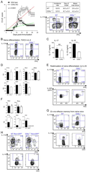
Figure 1. CD5L is a candidate regulator of Th17 cell functional states
(A–C) Single-cell RNA-seq analysis. (A) Cd5l expression of single-cells from in-vitro generated and in-vivo sorted Th17 cells (IL-17.GFP+) from mice at the peak of EAE. (B,C) Correlation of Cd5l expression in non-pathogenic Th17 cells (TGF-β1+IL-6) with (B) the
cell pathogenicity score (based on the pathogenic signature of (Lee et al., 2012)). p = 2.63
×10−5 (Wilcoxon ranksum test, comparing signature scores of Cd5l expressing vs. non-expressing cells); (C) the founding signature genes of the single-cell based proinflammatory (red) and regulatory (green) modules (Solid bars, significant correlation (p < 0.05); striked bars, none significant correlation).
(D–F) Validation of CD5L expression in vitro. Naïve T cells (CD4+CD62L+CD44−CD25−) were sorted and differentiated as indicated and analyzed by qPCR for CD5L expression at 48h (D) and 72h (E) and by flow cytometry at 48h (F); (E) IL-23 or control was added at 48h in fresh media. (G–I) Validation of Cd5l expression in vivo. (G,H) IL-17A.GFP reporter mice were immunized to induce EAE. Cells were sorted from spleen (G) and CNS (H) at the peak of disease. Cd5l and Il17a expression are measured by qPCR. Figure shown is representative data of three technical replicates from two independent experiments. (I) Cells were sorted from the gut of naïve mice and the number of RNA transcripts measured by nanostring nCounter platform (supp. Experimental Procedures). See also Figure S1.
Author Manuscript
Author Manuscript
Author Manuscript
Figure 2. CD5L represses effector functions without affecting Th17 cell differentiation
(A) EAE was induced by MOG/CFA (40μg) immunization. Left panel is pooled results from 3 independent experiments. Right panel: cytokine profile of CD4 T cells isolated from CNS at day 15 post immunization with summary data in Figure S2B. (B–D) Naïve splenic T cells were sorted and differentiated with TGF-β1+IL-6 for 48h. Th17 cell signature genes were measured by flow cytometry (B), ELISA (C) and qPCR (D). (E–F) Effector Th17 cells were differentiated as in B and resuspended in fresh media with no cytokines for 72h followed by restimulation. Gene profile was measured by flow cytometry (E) and qPCR (F). (G–H) Effector memory T cells (CD4+CD62L−CD44+) (G) or Effector memory Th17 cells (CD4+CD62L−CD44+RorγtGFP+) (H) were sorted from spleen of naïve mice and activated with TCR stimulation. See also Fig S2.
Author Manuscript
Author Manuscript
Author Manuscript
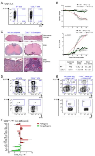
Figure 3. CD5L is a switch that regulates Th17 cell pathogenicity
(A–D) Passive EAE is induced as previously described (Jager et al., 2009). Naïve WT or CD5L−/− 2D2 splenic T cells were differentiated with TGF-β1+IL-6 and transferred into syngeneic WT hosts. (A) Cytokine profile of 2D2 T cells after differentiation at day 4. (B) Weight and clinical score of recipient mice undergoing EAE. (C) Representative histology of optic nerve (upper 2 panels, Hematoxylin and eosin stain) and spinal cord (lower 4 panels, Luxol fast blue-hematoxylin and eosin stains). Demyelination is indicated by loss of normal blue staining of myelin in lower panels. (D) Cytokine profile of WT and CD5L−/− 2D2 cells isolated from CNS at day 27 post transfer. Cells were gated on Va3.2+CD4+. (E) CD45.1 WT recipients received 100,000 naïve WT or CD5L−/− 2D2 T cells and were immunized the following day with MOG/CFA without pertussis toxin. Cytokine profile of 2D2 T cells was examined on day 10 in draining LN. (F) Expression profile of pathogenicity signature genes in WT and CD5L−/− Th17 cells differentiated with TGF-β1+IL-6 as in Figure 2E. Data are summary of at least three independent experiments. See also Figure S3.
Author Manuscript
Author Manuscript
Author Manuscript
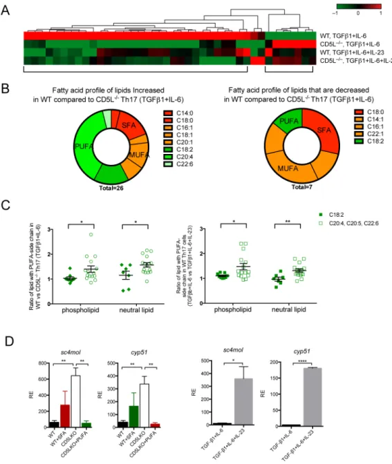
Figure 4. CD5L shifts the fatty acid composition of Th17 cell lipidome and modulates Rorγt ligand availability
(A–C) Lipidome analysis of Th17 cells showing altered SFA/MUFA and PUFA composition between WT and CD5L−/− Th17 cells (Exprimental Procedures). (A) Heatmap of 39 metabolites (rows) with significantly different levels among any Th17 cell conditions (columns) and with a fold change of at least 1.5. Median Intensity of each metabolite is shown (color bar is normalized per row). (B) Lipids from the two clusters in (A) are partitioned based on the length and saturation of their fatty acyl (FA) side chains. Those carrying more than one FA are further grouped by their FAs with the least saturation or longest carbon chain (in that order). Complete FA profile is shown in Figure S4BC. (C) Ratio of specific lipids in WT vs. CD5L−/− Th17 cells carrying various PUFA side chains. Phospholipids included in this analysis: phosphatidylcholine, phosphatidylethanolamine,
Author Manuscript
Author Manuscript
Author Manuscript
phosphatidylserine and their respective lyso-metabolites. Neutral lipid included in this analysis: Triacylglyceride, diacylglyceride and monoacylglyceride. Asterisk (*) denotes to p < 0.05 in Student’s t-test. (D) Expression of cyp51 and sc4mol mRNA in WT or CD5L−/− Th17 cells (TGF-β1+IL-6, left panels) or WT Th17 cells (TGF-β1+IL-6 with control or IL-23, right panels). SFA (palmitic acid, 25uM) or PUFA (arachidonic acid, 25uM) was added at 48h and cells analyzed at 96h. See also Figure S4 and Table S1.
Author Manuscript
Author Manuscript
Author Manuscript
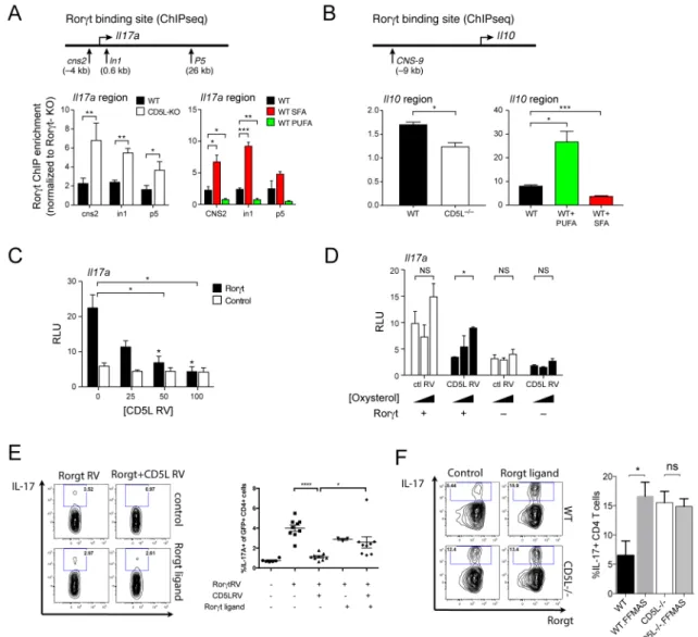
Figure 5. CD5L and PUFA/SFA profile regulate Rorγt function in a ligand-dependent manner
(A, B) Rorγt ChIP-PCR analyses in WT and CD5L−/− Th17 cells. WT, CD5L−/− and Rorγt−/− Th17 cells were differentiated with TGF-β1+IL-6 for 96h. Enrichment of Rorγt binding to genomic regions of Il17 (A) and Il10 (B) is measured using qPCR. For fatty acid experiments, 10μM of either SFA (palmitic acid) or PUFA (arachidonic acid or
docosahexaenoic acid showed similar results) was added to WT Th17 cell culture at day 0. Three independent experiments were performed. (C, D) Rorγt transcriptional activity was measured by luciferase reporter of Il17 promoter in EL4 cells transfected with CD5L-RV at 0, 25, 50, 100ng (C) or 100ng with 7, 27 dihydroxycholsterol (5, 0.5 or 0.05uM) (D). (E) Naïve WT T cells were activated without polarizing cytokines (Th0) and infected with retrovirus expressing Rorγt in the presence of control-RV or CD5L-RV with or without FF-MAS (5uM) as a source of Rorγt ligand. Each dot represents an independent infection. (F) WT or CD5L−/− naïve cells were differentiated with TGF-β1+IL-6. At 48h, cells were replated in fresh media with either control or FF-MAS (5uM) as a source of Rorγt ligand. Cells were harvested for FACS analysis 72h later. See also Figure S5.
Author Manuscript
Author Manuscript
Author Manuscript
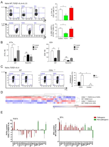
Figure 6. PUFA/SFA regulate Th17 cell function and contribute to CD5L-dependent regulation of Th17 cells
(A, B) Naïve T cells were sorted from WT or IL-23RGFP reporter mice (A); or WT or Rorc−/− mice (B) and differentiated with TGF-β1+IL-6 followed by addition of IL-23 at 48h in fresh media with either PUFA (arachidonic acid, 10uM) or SFA (palmitic acid, 20uM). Cells were analyzed by flow cytometry (A) or qPCR (B) at 96h. The concentration of free fatty acids was predetermined in titration experiments (data not shown). (C) Naïve WT and CD5L−/− T cells were differentiated with TGF-β1+IL-6 and replated in fresh media with control or 5uM of PUFA at 48h. Cells were analyzed at 96h. (D, E) Same as in C. PUFA (5uM) or SFA (25uM) were added 48h. Cells were analyzed by nanostring analysis (D) or qPCR (E) at 96h. (D) Genes that are differentially expressed between any of the four samples and are with a fold change of at least 1.5 are plotted in heatmap. Color scale is normalized per row (subtracted by mean and divided by standard deviation). All data are summary of 3 independent experiments. See also Table S2.